PRE-EXPOSURE PROPHYLAXIS FOR HIV: LINKING ANTIRETROVIRAL
PHARMACOKINETICS AND PHARMACODYNAMICS TO IDENTIFY OPTIMAL
DOSING STRATEGIES
Melanie R. Nicol
A dissertation submitted to the faculty of the University of North Carolina at Chapel Hill in
partial fulfillment of the requirements for the degree of Doctor of Philosophy in Pharmaceutical
Sciences in the Eshelman School of Pharmacy (Pharmacotherapy and Experimental
Therapeutics).
Chapel Hill
2014
Approved by:
Angela D.M. Kashuba
James Heyward Hull III
Julie A.E. Nelson
iii
ABSTRACT
Melanie R. Nicol: Pre-Exposure Prophylaxis for HIV: Linking Antiretroviral Pharmacokinetics and Pharmacodynamics to Identify Optimal Dosing Strategies
Under the direction of: Angela D.M. Kashuba
Despite utilization of potent combination antiretroviral therapy, the incidence of HIV in the U.S. has not declined over recent years. Several clinical trials recently demonstrated that antiretrovirals can protect vulnerable mucosal surfaces against infection. However, the protection conferred ranged from 0% to 73%, underscoring the importance of identifying drug concentrations needed at mucosal surfaces. Additionally, antiretrovirals have complex pharmacology in mucosal tissues with unpredictable drug penetration within and between drug classes. The ultimate goal of this work was to identify factors influencing variability in drug penetration and to develop an explant tissue culture model that could be used to evaluate the efficacy of tenofovir and maraviroc, two antiretroviral candidates for oral pre-exposure prophylaxis strategies.
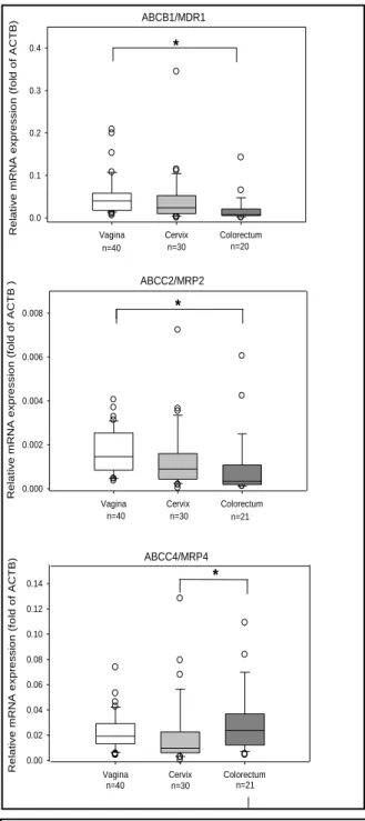
Using real-time PCR and immunochemistry, mRNA and protein expression of three efflux (ABCB1/MDR1, ABCC2/MRP2, ABCC4/MRP4) and three uptake (SLC22A6/OAT1, SLC22A8/OAT3, SLCO1B1/OATP1B1) transporters was described in female genital (FGT) and colorectal tissues (CRT) from 99 donors. Epithelial expression of efflux transporters was found to be two- to four-fold greater in FGT than CRT. These data support clinical findings of higher maraviroc and tenofovir concentrations in CRT compared to FGT after oral dosing. Quantifying mucosal transporter expression and localization was a useful technique to understand drug tissue distribution and facilitate antiretroviral selection to target these tissues.
iv
developed using the R5, T-tropic strain HIV-1JR-CSF as the inoculum, and was coupled with a novel real-time PCR assay to quantify spliced RNA that detected replication within 24-72 hours after HIV exposure. In this model, concentrations required to protect vaginal explants were 10-1000 fold higher than in a TZM-bl cell monolayer. A single oral dose of tenofovir and maraviroc at 200% treatment doses protected cervical and vaginal biopsies in 3/6 healthy volunteer women. TZM-bl cells over predicted efficacy by
49%, and tissue explants under predicted by 36%. This translational approach, comparing
v
To my wonderful and loving family, Mom, Dad, Derek, Ethan, Frances, Stephanie, and Truman
vi
ACKNOWLEDGEMENTS
Completion of this doctoral dissertation would not have been possible without the meaningful
contributions of many individuals in both my personal and professional life.
First, I would like to thank my advisor Angela Kashuba. Dr. Kashuba has been a phenomenal
mentor over the past five years and I suspect I will be coming to her for guidance for the rest of my career. Her door was always open for anything and everything I needed. She was always pushing me to a level of achievement that I wouldn’t have thought myself capable of. I would also like to thank Dr. Hull
for stepping in as my committee chair. Dr. Hull has been a dedicated educator and I appreciate his ability to teach applied statistics in a way relevant to drug research. Dr. Corbett has been a wonderful role model in the clinic and in the classroom. I appreciate her sharing with me not only her knowledge on
antiretroviral therapy, but more importantly her approach to solving clinical problems. Dr. Kroetz’s expertise in transporter pharmacology was instrumental to the development of my dissertation. Lastly, Dr. Nelson, always more than generous with her time, proved to be an invaluable advisor on the molecular and virologic portions of my project. Her expertise contributed significantly to the multidisciplinary nature of my project and my development as a translational researcher.
Outside of my committee, there have been many others who have mentored me in my path to obtaining a PhD. Dr. Talbot was the first to introduce me to scientific research and without his
vii
the amount I have learned from her about performing clinical research is astounding. I also want to thank Dr. Fedoriw for serving as a collaborator on these studies. His expertise in tissue pathology proved instrumental in moving both the explant and transporter projects along. Katie Molan, CFAR statistician also provided helpful input on the statistical analysis for many of these studies. I would also like to extend a special thanks to Drs. Myron Cohen and Ron Swanstrom for providing valuable feedback at various time points throughout the development of my dissertation.
I want to thank all the members of the Kashuba laboratory group for their support, both personal and professional. Cindi Emerson, Kuo Yang, Julie Dumond, Craig Sykes, Nicole White, Stephanie Malone, Ruili Wang, Michelle Brosnan-Cook, and Steve Jennings not only provided excellent analytical tools for the completion of my studies, but also contributed to the fantastic working group that made it a pleasure to come into the laboratory every day. I was fortunate to be trained alongside several amazing graduate students and fellows during my time in the Kashuba lab. I am grateful to consider Racheal Kendrick, Jessica Adams, Corbin Thompson, Mackenzie Cottrell and Christine Trezza as both friends and professional colleagues.
I need to acknowledge the remarkable core facilities at UNC whose resources allowed for experiment completion and whose personnel provided valuable insight into my projects. Tissue
Procurement Facility provided invaluable services in the steady supply of tissues without which none of the research here would have been possible. The Urogynecology Department at UNC, in particular Dr. Elizabeth Geller, deserves thanks for assisting with recruitment of tissue donors. Tissue processing and immunohistochemical staining was performed by the Translational Pathology Laboratory. Michele Mathews in particular was instrumental in the work performed. The nurses and staff at the Clinical and Translational Research Center provided excellent service and assistance in the performance of my clinical study. I would like to acknowledge the subjects who participated in the clinical study. It is because of volunteers like them that drug development is able to move forward.
I would like to thank Kathy Maboll, and Arlo Brown, and the administrative staff of
viii
thank the DPET faculty. I feel incredibly fortunate to have been taught by such an esteemed group of professors. I also acknowledge the DPET fellows and graduate students that I have interacted with over my time here. They were a pleasure to work and learn with.
ix
TABLE OF CONTENTS
ABSTRACT ... iii
ACKNOWLEDGEMENTS ... vi
LIST OF FIGURES ...xiv
LIST OF TABLES ... xv
LIST OF ABBREVIATIONS ...xvi
Chapter I: Background-Pharmacologic Opportunities for HIV Prevention ...1
Summary ... 1
Introduction ... 1
Current Approaches to Pre-Exposure Prophylaxis of HIV Infection ... 3
Current Approaches to Post-Exposure Prophylaxis ... 15
Current Approaches to Treatment of the Index Case ... 17
Developmental Obstacles in HIV Prevention Studies ... 22
Future Role of Clinical Pharmacology to Bridge the Gaps ... 25
Conclusion ... 28
Specific Aims ... 29
x
Summary ... 31
Introduction ... 32
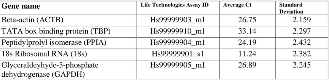
Materials and Methods ... 33
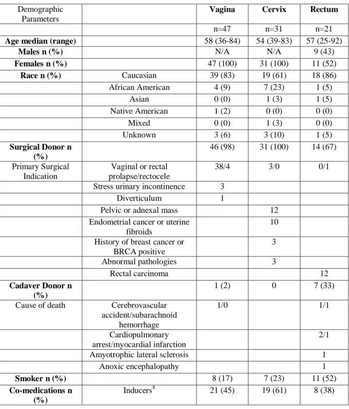
Tissue Procurement ... 33
mRNA Expression ... 34
Protein Expression ... 35
Statistical Analysis ... 36
Results ... 37
ABC Efflux Transporters: ... 37
SLC Uptake Transporters:... 45
Role of Menopause Status in Efflux Transporter mRNAExpression ... 46
Discussion ... 46
Chapter III: A Human Tissue Explant Model as a Preclinical Tool to Evaluate HIV Pre-Exposure Prophylaxis Candidates ... 52
Summary ... 52
Introduction ... 53
Materials and Methods ... 54
Tissue Procurement ... 54
Immunohistochemistry and Immune Cell Quantification ... 54
Ex-vivo Explant Cultures ... 55
MTT Viability Assay ... 56
xi
Viral Propagation and Explant Infections ... 57
Immunohistochemistry to Measure Intracellular p24 ... 57
Real-time PCR to Measure Proviral DNA ... 58
Spliced Viral RNA Assay ... 59
Statistical Analysis ... 60
Results ... 61
Expression and Variability of HIV Target and Immune Cells in Explant Tissues ... 61
Changes in Architecture, Immune Cell Composition, and Viability over Time ... 62
Uptake, Elimination, and Metabolism of Antiretroviral Drugs in Explant Tissues ... 64
Identification and Quantification of HIV Infection in Explant Culture ... 66
Discussion ... 68
Chapter IV: Concentration-Response Relationship of Tenofovir and Maraviroc in Human Explant Tissue ... 73
Summary ... 73
Introduction ... 74
Methods ... 75
Tissue Procurement ... 75
Concentration-Response Relationship in Vaginal Explant Tissue ... 75
Concentration-Response in TZM-bl Cells ... 76
Data analysis ... 76
Results ... 77
xii
MVC Efficacy in Vaginal Explants is Short Lived ... 78
Additive Efficacy of Tenofovir and Maraviroc in Cell Culture ... 79
Prediction of Efficacy from Oral Tenofovir Disoproxil Fumarate and Maraviroc ... 79
Discussion ... 80
Chapter V: Efficacy of Oral Maraviroc and Tenofovir for HIV Prevention: Proof of Concept Study to Evaluate Explant Model Predictions... 84
Summary ... 84
Introduction ... 84
Methods ... 85
Study Design ... 85
Peripheral Blood Mononuclear Cell Stimulation and Infection ... 86
Cervical and Vaginal Biopsy Ex-vivo HIV Challenge ... 87
Results ... 88
Study Participants and Adverse Events ... 88
Oral Maraviroc and Tenofovir Protection of Cervical and Vaginal Biopsies ... 89
PBMC as a Surrogate for Donor Susceptibility to HIV Infection ... 89
Discussion ... 90
Chapter VI: Perspectives and Future Directions ... 94
Clinical Pharmacology for HIV Prevention: Advances and Limitations ... 94
The Future of PrEP ... 98
Applications beyond Prevention ... 99
xiii
APPENDIX : Transporter Gene Expression Statistical Tables ... 101
xiv
LIST OF FIGURES
Figure 1: Opportunities for prevention ... 2
Figure 2: Timeline of mucosal infection... 15
Figure 3 Relationship of total genital tract exposure to blood plasma protein binding ... 22
Figure 4: What should be measured for accurate PK-PD evaluations? * ... 26
Figure 5 mRNA expression of three ABC transporters in vaginal, cervical, and colorectal tissues. ... 40
Figure 6 Immunohistochemical staining of vaginal, cervical, and colorectal tissues. . ... 41
Figure 7 Positive and Negative Controls for Transporter Immunohistochemistry. ... 42
Figure 8 Lower Magnification Images (original objective magnification 20 or 40X. ... ….44
Figure 9: Hypothesized influence of drug transporters on drug exposure in colorectal and vaginal tissues. . ... ……….47
Figure 10: HIV target cells and relevant immune cells are expressed in explant tissue sources………...60
Figure 11: Explant tissue composition is variable………..……….61
Figure 12: Sloughing of the epithelial layer and tissue architecture changes………..62
Figure 13: Rapid changes occur in explant tissue over time in culture……….………..63
Figure 14: Antiretrovirals are rapidly taken up by tissue and rapidly eliminated………64
Figure 15: Exlpant tissues can be infected with a clinically relevant strain HIV-1JR-CSF/hPBMC……..….65
Figure 16: Spliced viral RNA and replication in explant tissue………..68
Figure 17: Inhibition of HIV is correlated with tissue concentrations of TFVdp in human vaginal explant tissue………...77
Figure 18: Maraviroc inhibits HIV infection in vaginal explants for 24 hours. ……….…78
xv
LIST OF TABLES
Table 1: Summary of completed clinical trials involving pharmacotherapy for prevention... 11 Table 2: Exposures of antiretrovirals in relevant tissues compared to blood plasma……….………...…..21 Table 3: Expression and variability in mRNA expression of five endogenous control genes.. ... 35 Table 4 Demographics of tissue donors.. ... 38 Table 5 The proportion of samples for each transporter with quantifiable mRNA expression... ... 45 Table 6: Demographic information for vaginal explant donors used in spliced RNA assay time course.. 67 Table 7 Emax model predictions of oral MVC and TDF efficacy at 50, 100 and 200% dosing.. ... 79
xvi
LIST OF ABBREVIATIONS
3TC Lamivudine
3TC-tp Lamivudine triphosphate
ABC Abacavir
AIDS Acquired Immunodeficiency Syndrome
ARV(s) Antiretroviral(s) AUC Area under the curve
BP Blood plasma
CVF Cervicovaginal fluid
dATP deoxyadenosine triphosphate dCTP deoxycytidine triphosphate FGT Female genital tract FTC Emtricitabine
FTC-tp Emtricitabine triphosphate HIV Human Immunodeficiency Virus IHC Immunohistochemistry
LC Liquid chromatography MDR1 Multidrug resistant protein1
MRP2 Multidrug resistance-associated protein 2 MRP4 Multidrug resistance-associated protein 4 MS Mass spectrometry
xvii
MTT methylthiazolydiphenyl-tetrazolium MVC MaravirocnPEP nonoccupational Post-exposure prophyalxis NRTI Nucleotide reverse transcriptase inhibitor NNRTI Non-nucleotide reverse transcriptase inhibitor OAT1 Organic anion transporter 1
OAT3 Organic anion transporter 3
OATP1B1 Organic anion transporting polypeptide 1B1 PBMC Peripheral blood mononuclear cell
PCR Polymerase chain reaction PEP Post-exposure prophyalxis
PD Pharmacodynamics
PHA Phytohemagglutinin PK Pharmacokinetics
PrEP Pre-exposure prophylaxis
SHIV Simian-human (chimeric) immunodeficiency virus SIV Simian immunodeficiency virus
TCID50 Tissue culture infectious dose (50%) TDF Tenofovir disoproxil fumarate
TFV Tenofovir
TFVdp Tenofovir diphosphate TPF Tissue Procurement Facility
TPL Translational Pathology Laboratory
1
Chapter I: Background-Pharmacologic Opportunities for HIV Prevention
1Summary
Innovations in antiretroviral treatment strategies have resulted in treated, HIV-infected patients having similar life expectancies as their uninfected counterparts. Yet, the number of
individuals capable of HIV transmission is increasing: for every person initiating antiretroviral (ARV) treatment, four more become infected with HIV. The limited progress with microbicides and
vaccines for HIV prevention reinforce the need for a concentrated exploration of the utility of antiretrovirals. Preliminary animal studies with topical and systemic antiretrovirals show promise. However, current clinical trials were designed without a comprehensive understanding of
antiretroviral pharmacokinetic-pharmacodynamic relationships in HIV prevention. This review focuses on current strategies for prevention of HIV infection, and how the tools of pharmacology can be a valuable resource for determining pharmacodynamic targets, providing interspecies scaling of exposures, identifying the optimal drugs/drug combinations, doses, and dosing regimens, and
designing efficient clinical trials.
Introduction
Over the past 10 years, innovations in antiretroviral treatment strategies have resulted in
treated, HIV-infected patients having similar life expectancies as their uninfected counterparts [1].
1
2
Yet the number of individuals capable of HIV transmission is increasing. According to the UNAIDS, the number of newly infected individuals worldwide in 2012 was approximately 2.3 million [2]. Since the early 1990s, transmission rates have been relatively stable in the United States [3]. This is due, in part, to increased efforts in HIV screening, placing patients into care, and
behavioral interventions [4]. However, countries which lack access to resources have not had similar success. For example, between 2001 and 2008 in the Middle East and North Africa, the number of newly infected individuals increased by 17% and the number of adult and child deaths due to AIDS increased by 80% [5]. The World Health Organization estimates that in resource limited settings, for every person initiating antiretroviral (ARV) treatment, four more become infected with HIV [6]. Since current treatment interventions will not likely curb the HIV epidemic, other prevention efforts are desperately needed.
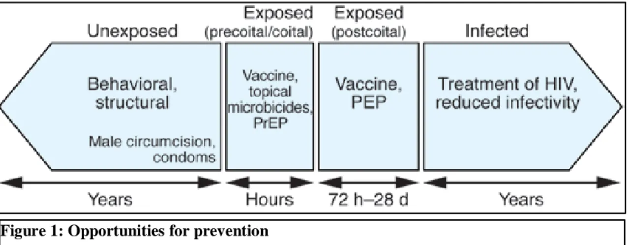
As illustrated in Figure 1, interventions for HIV prevention can be divided into four main strategies based on the time course of exposure. In an individual who has yet to be exposed to HIV, behavioral and structural interventions may be effective [4]. In an exposed individual at the time of coital exposure, a vaccine that provides neutralizing antibodies or a pre-exposure prophylaxis (PrEP) intervention with topical microbicides/antiretrovirals, or systemic antiretrovirals may be effective. In an exposed individual shortly after the time of exposure, a vaccine or a post-exposure prophylaxis (PEP) intervention with antiretrovirals may be effective. Once infected, an individual may be rendered less infectious if placed on potent combination antiretroviral therapy [7, 8].
3
The efficacy for each of the aforementioned interventions depends on the type and timing of the intervention. Structural barriers such as male circumcision and condom use, along with other behavioral interventions, have shown 50-80% efficacy in clinical trials [9]. However, consistent implementation of these practices remains challenging, particularly due to social and cultural
conditions that can preclude women from being empowered to utilize them [10]. Recent advances in vaccine development are encouraging [11], but worldwide implementation of an effective vaccine is years away. Therefore, the potential of pharmacologic interventions to curtail the HIV epidemic quickly is significant.
These strategies include microbicides and traditional antiretroviral drugs used systemically or topically. Typically, microbicides act in the vaginal lumen to either destroy the virus before it can traverse mucosal surfaces, or prevent viral attachment to mucosal surfaces. This is in contrast to antiretrovirals which are distributed into tissues following oral or topical application, and act at the mononuclear cellular level to prevent viral entry or replication. This article provides a brief review of these pharmacologic opportunities and their roles in the exposure continuum (Figure 1). We also summarize current obstacles facing the clinical pharmacology of HIV prevention, and possible opportunities to overcome them.
Current Approaches to Pre-Exposure Prophylaxis of HIV Infection
4
(12%) in a Phase III study involving 892 female sex workers in four countries [13]. Four nonspecific microbicides (Cellulose Sulfate, Carraguard, Buffer Gel, Pro 2000) have failed to demonstrate efficacy in clinical trials [12]. VivaGel ® (SPL7013 3%; a membrane binding inhibitor) was found in a Phase I study to be well tolerated but adherence was reduced in the active arm compared to placebo and no further studies are currently planned [14].
With the disappointing results of microbicide trials, topical formulations of antiretrovirals are actively being investigated. Nucleoside/tide analogue (N(t)RTIs) and nonnucleoside analogue (NNRTI) reverse transcriptase inhibitors are of greatest interest due to their pre-integration activity, long (intracellular) half-life, safety profile, and success in preventing infection in animal models [15, 16]. Some leading candidates as of 2010 are summarized below.
Tenofovir (TFV) is a nucleotide reverse transcriptase inhibitor that has advanced the farthest in PrEP clinical trials. It has been FDA approved as Viread ® for HIV treatment since 2001 as the salt form tenofovir disoproxil fumarate (TDF), and is often used with the NRTI, Emtriva ®
emtricitabine (FTC) as the fixed dose combination Truvada ®. CAPRISA 004 was a Phase II safety and efficacy study looking at coital-dependent use of 1% TFV gel in 900 high-risk South African women [17]. Results of this randomized, double-blinded trial which followed the women for 30 months provided both encouraging and informative data in regards to the future of pharmacology for prevention. When applied vaginally within 12 hours before and within 12 hours after sex, the gel demonstrated a 39% reduction in HIV infections in the study population with 60 seroconversions in the placebo group (n=444) compared to 38 infections in the treatment group (n=445). Importantly, the ability of the gel to protect against HIV infection closely followed the pharmacology. In the group of women who seroconverted, 45% had detectable concentrations in the cervicovaginal fluid (CVF) (samples drawn average of 4.5 days after use) and a median concentration of 1 ng/mL. This is in contrast to the group of women who remained protected from HIV of whom 96% had detectable
5
In contrast to the success of CAPRISA 004, in 2011 a Phase II study in African women which included an arm to evaluate daily use of 1% TFV gel, halted enrollment after external review by the Data Safety Monitoring Board determined futility [18]. The reasons for the failure to find a drug effect in this study are still being explored. FACTS001 was launched in October and is a Phase III regulatory study which plans to enroll 2900 women to confirm the findings of CAPRISA 004 regarding BAT24 dosing (www.facts-consortium.co.za).
The efficacy of vaginally applied TFV 1% gel has also been investigated in a macaque model of repeated low-dose exposure to a chimeric simian –human immunodeficiency virus (SHIVSF162P3 10 50% tissue culture infectious doses or 1.5 x 10⁶ copies RNA per exposure]) [19]. Three milliliters of TFV 1% gel, TFV 1% + FTC 5% combination gel or placebo gel was administered vaginally 30 minutes before viral challenge to groups of six macaques. All animals in the TFV or TFV+FTC gel groups (6/6) remained protected from infection after 20 weekly viral challenges, whereas only 1/6 animals in the placebo group remained uninfected (p<0.001). The average plasma TVF
concentration from these protected animals 30 minutes after dosing was 16 ng/mL for TFV gel, and 39 ng/mL for TFV+FTC gel. Comparatively, preliminary pharmacokinetic data in humans following a four gram intravaginal dose of 1% TFV gel demonstrates that blood plasma concentrations above 5 ng/mL are seldom achieved [20].
Using male rhesus macaques, Cranage et al demonstrated that rectal application of three mL of TFV 1% gel up to two hours before exposure to simian immunodeficiency virus (SIVmac251/32H, 20 median rectal infectious doses) prevented infection in 6/9 animals, whereas only 1/4 animals in the placebo group remained uninfected [21]. Analysis of plasma concentrations 15 minutes after dosing suggested that protection was associated with higher TFV exposures; the lowest plasma TFV concentration conferring protection was 120 ng/mL. Assuming the average male rhesus monkey weighs 8 kg to a human’s 70 kg, in order to achieve the same mg/kg dose, nearly 30 mL of gel would
6
Tenofovir has also been shown safe and efficacious in cervical and colorectal tissue explants. In a polarized cervical explant culture (where only the epithelial layer is exposed to the air interface), the application of a 1mg/mL TFV gel reduced p24 antigen concentrations from an HIVBaL inoculation by 10-fold, compared to untreated explants [22]. Following a one hour drug incubation, colorectal tissue demonstrated 100% protection from infection (two hour exposure to 30X the tissue culture ID50 (TCID50) HIVBaL) at concentrations of 10 and 100 μg/mL and >90 % inhibition at concentrations of 0.1 and 1 μg/mL [23]. In colorectal biopsies collected from HIV-negative volunteers before and after
rectal dosing with vaginally formulated 1% TFV gel, a reduction in p24 antigen was observed that was found to be correlated (r2 =0.23; p=0.002) with measured intracellular tenofovir diphosphate (TFVdp) concentrations [24]. However, a significant increase in the adverse events reported after seven days of gel use determined that the vaginally-formulated TFV gel was not appropriate for rectal use.
UC781 is a nonnucleoside reverse transcriptase inhibitor with an intracellular half-life of 5.5 days [25]. This long half-life confers protection against HIV infection in explant culture models up to six days after initial drug exposure [26]. In colorectal explants, complete protection from HIVBaL infection was seen at UC781 concentrations ranging from 10-100 μM [23]. A Phase I study showed no significant adverse events after single or seven days of 0.1% or 0.25% UC781 gel use and also demonstrated efficacy in ex vivo biopsy challenge [27]. In cervical explant tissue, 100 μM UC781 was able to confer complete protection from a two hour exposure to HIVBaL. At concentrations as low as 1 nM, greater than 99% protection was seen [26]. Using 20 pig-tailed macaques, Patton et al concluded that UC781 0.1% and 1% gel formulations were safe after four daily doses of rectal (2.5 mL) or vaginal (1.5 mL) applications based on in-vitro toxicity assays, colposcopy examination, vaginal pH, rectal lavage examinations, rectal pH, and rectal micro flora. [28] Even upon repeated dosing, UC781 was not detected in the animals’ plasma, suggesting minimal systemic absorption. Up
to six hours following administration of 1% gel, vaginal lavage samples contained UC781
7
sponsor for UC781 halted the development of UC781 as a microbicide and as of 2014, no additional clinical studies are planned.
Dapivirine (TMC120) is a 2nd generation nonnucleoside reverse transcriptase inhibitor, which maintains efficacy against many NNRTI-resistant strains of HIV [29]. While dapivirine’s poor bioavailability precludes its utility for oral dosing, its potential to achieve high local concentrations in a topical formulation may prove advantageous. In a cervical explant model, lack of detectable
proviral DNA with dapivirine exposure 0.001 μM demonstrated its potential efficacy in prevention
of HIV acquisition [30]. At concentrations of 1-10μM, dapivirine demonstrated a protective effect against HIV-1 infection for up to 6 days after drug exposure. Pharmacokinetic evaluations performed after seven days of vaginally-administered [14C]dapivirine 0.009% gel (0.5 mL per day in rabbits and 1 mL per day in macaques) demonstrated low to undetectable plasma concentrations, with cervical and vaginal tissue concentrations being greater than the in vitro EC90 (0.9 ng/mL) up to 48 hours after dosing [31, 32]. Histopathologic analysis of tissues from both species demonstrates that most drug remains on the mucosal surface, within the keratinized layer of the epithelium, or in compromised
8
matrix IVR produced plasma concentrations 50-fold that of the reservoir IVR (Cmax 1194 vs. 51.87 pg/mL), both rings produced vaginal fluid concentrations above dapivirine’s EC50 of 0.3 ng/mL for the entire 28 days of the study and for at least 5 days following removal (Cmax 7.5-14.37 μg/g for reservoir and 850-1900 μg/g for matrix IVR). As of 2014, two Phase III studies (ASPIRE and The Ring Study) are currently underway to evaluate efficacy of a 25 mg DPV ring inserted once monthly in African women[34, 35].
Although topical formulations of ARVs allow high local exposure while limiting systemic exposure, restrictions to their use may include a difficult application technique, adherence, user and partner acceptance, local irritation, and multiple applications for one encounter if multiple mucosal surfaces require protection. Systemic dosing of ARVs provides an alternate approach for protection and may overcome some of these challenges. Finding a pill that a woman can take in a coitally-independent manner can circumvent disclosure to their partners and assist with empowerment.
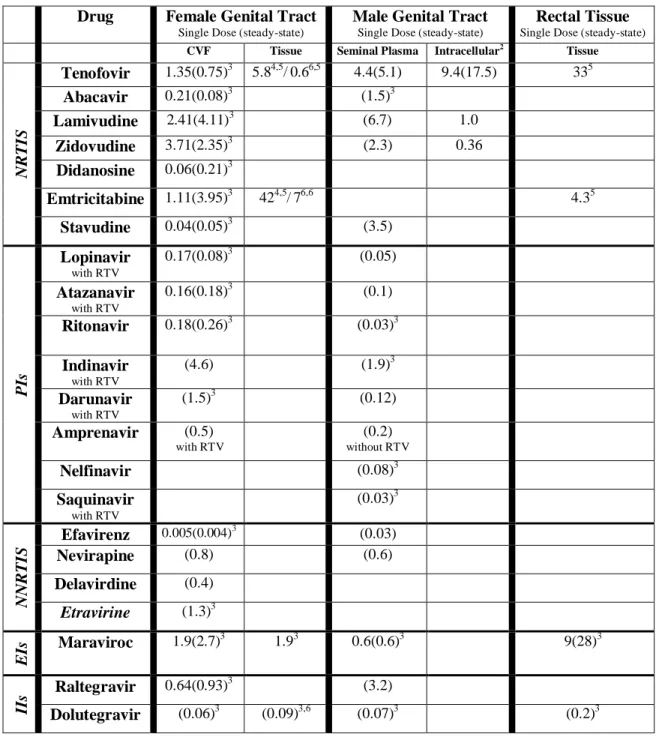
Currently, the target drug exposure required to prevent HIV infection at mucosal surfaces is unknown. Therefore, the present assumption is that higher drug exposures are better for conferring protection. However, ARVs differ greatly in their ability to penetrate mucosal tissues or secretions [36-39]. Generally, highly protein-bound compounds do not gain access to these secondary compartments due to their affinity for plasma proteins such as albumin and a1-acid glycoprotein. Drugs such as the protease inhibitors, which are 95-99% bound to plasma proteins, achieve female genital tract concentrations < 50% that of blood plasma [38]. In contrast, most N(t)RTIs have a low degree of protein-binding (<0.7-49%) and achieve concentrations 2- to 6-fold higher than in blood plasma [38]. However, protein binding is not the only predictor of ARV exposure. For example, maraviroc, a cellular entry inhibitor that demonstrates 85% plasma protein binding, has high
penetration into CVF and vaginal tissue. After seven days of 300 mg twice daily dosing, the AUCs of CVF and vaginal tissue are 2.7 and 1.9 times higher than blood plasma [37]. Raltegravir, an integrase
9
Currently there are no published data regarding intrasubject pharmacokinetic variability in the female genital tract; no one has sampled the same individuals under steady state conditions over multiple days. However, the intersubject variability of drug exposure in the female genital tract is generally greater than in blood plasma (the coefficients of variation range from approximately 50-200% in CVF and 2-100% in blood plasma).
An additional consideration of antiretroviral pharmacokinetics and pharmacodynamics at mucosal surfaces is their protein binding within the mucosal secretions. CVF concentrations of albumin and a1-acid glycoprotein are both <1% of their concentration in plasma [40]. Although the protein binding of drugs in genital secretions has not been extensively evaluated, maraviroc has recently shown 10-fold less protein binding in CVF than blood plasma (7.5% vs 75%) [37]. This phenomenon must be considered in pharmacokinetic-pharmacodynamic analysis of antiretroviral prevention strategies.
Penetration of drugs into rectal tissues also has implications for HIV transmission. Exposures (AUC12 hour) of maraviroc are ~30 times higher in rectal tissues than blood plasma [41]. Some drugs, such as the nucleoside/tide analogues require cellular uptake and phosphorylation in order to be active against the RT enzyme. Intracellular and extracellular concentrations of tenofovir and emtricitabine have been recently evaluated in blood plasma as well as cervical, vaginal, and rectal tissues after a single dose [42]. Sampling was performed from 24 hours to 14 days post-dose. TFV exposures (as measured by AUCDay 1-14) were 5.8, 0.6, and 34 times higher in cervical, vaginal and rectal tissues, respectively. FTC exposures were 41, 7, and 4.4 times higher in cervical, vaginal and rectal tissues, respectively. Intracellular concentrations of TFVdp were detected in all tissues for at least seven days. FTCtp (emtricitabine triphosphate) was only detected in tissues for two days post-dosing, despite PBMC concentrations persisting for 10 days. These initial tissue data suggest that these ARVs may be promising candidates for HIV prevention. However, more comprehensive pharmacokinetic data
10
Several systemically-dosed ARVs have been tested for PrEP efficacy in nonhuman primates and humanized mice. Oral dosing of CMPD167, a CCR5 inhibitor, was able to prevent infection from vaginal exposures to SHIV162P3 in 50% (10/20) of macaques when drug was continued for 10 days following exposure and in 25% of macaques (1/4) if drug was given only for four days prior to exposure [43]. Subbarao et al demonstrated that, with weekly rectal exposure to low dose SHIV (3.8 x 10⁵ viral particles per exposure) , oral daily (n=4) or weekly (n=4) doses of 22 mg/kg TDF can delay time to infection by 4.5-5.5 weeks and provide a 60% decrease in the per-exposure probability of infection [44]. In this model, viral inoculation was performed two hours after tenofovir
administration: this was based on PK sampling in a single macaque in which a concentration of 633 ng/mL was observed at two hours post-dose. Single blood plasma sampling in these macaques at two hours post dose revealed lower TFV exposures in 80% (4/5) of the macaques that were infected (10-137 ng/mL) compared to those that were protected (mean 2000 ng/mL). In 2008, Garcia-Lerma et al evaluated five different routes, combinations, and/or schedules of TFV and FTC in a rectal, low dose SHIV (7.5 x 10⁵ viral particles per exposure) repeat-exposure macaque model [45]. Oral doses were selected as a result of preliminary PK dose ranging analyses in 11 animals (5 for TDF and 6 for FTC) demonstrating similar AUCs to humans at doses of TDF 20-24mg/kg and FTC 20mg/kg (AUC for TFV were 3.2-3.4 μg * h/mL and within human range of 1.03-3.56 μg * h/mL; AUC for FTC was 13.2 μg * h/mL in macaques and ~10 μg * h/mL in humans[38]). In this investigation, complete
protection from HIV infection was achieved with daily subcutaneous dosing of TVF 22 mg/kg and FTC 20 mg/kg. Other daily regimens (subcutaneous FTC 20 mg/kg alone or oral TDF 22mg/kg + FTC 20 mg/kg) achieved only partial protection (33% and 67% protected, respectively).
Daily intraperitoneal (IP) doses of TDF+FTC (5.2 + 3.5mg) was also protective in a
humanized BLT mouse model. All mice (5/5) given FTC+TDF daily beginning two days before, and continuing for seven days after, vaginal inoculation with approximately 9 x 10⁴ tissue culture
11
Following daily IP injections for three days, animals were exposed to HIV-1 JR-CSF in the rectal mucosa or by intravenous injection while continuing to receive daily TDF+FTC for an additional four days [47] Using this regimen, no infections were noted with rectal challenge (n=9) and only one was infected with intravenous challenge (n=9). Similar to macaques, pharmacologic data using these doses in mice are lacking. Using weight-based scaling these mice received IP doses of ~30 mg/kg, which is likely to exceed the ~ 4 mg/kg being used in current PrEP clinical trials.
Table 1: Summary of completed clinical trials involving pharmacotherapy for prevention
Trial Prevention Area
Exposure Intervention Results Sample Size Reference
Caprisa 004 Phase 2; PrEP vaginal Topical tenofovir gel Results published 2010 HIV incidence reduced by 39%
900 women Abdool Karim Q, et al. Science. Sep 3;329(5996):11 68-74. CDC 4323 Phase 2; PrEP penile or rectal Once daily oral TDF Results published 2011. No difference in risk behavior. No major safety concerns.
400 MSM Liu, A.Y., et al. 2011. PLoS One 6:e23688.
iPrEx Phase 3; PrEP penile or rectal Once daily oral TDF/FTC Results published 2010. HIV incidence reduced by 44%.
2,499 MSM Grant, R.M., et al. 2010. N Engl J Med 363:2587-2599. CDC 4370 (Bangko k Study) Phase 2/3; PrEP
parenteral Once daily oral TDF Results published 2013. HIV incidence reduced by 49%. 2,400 IV drug users Choopanya, K., et al. 2013. Lancet
12
CDC 4940 (TDF2) Phase 2; PrEP penile or vaginal Once daily oral TDF/FTC Results published 2012. HIV incidence reduced by 62% 1,200 heterosexual men and women Thigpen, M.C., et al. 2012. N Engl J Med 367:423-434. Partners PrEP Phase 3; PrEP penile or vaginal Once daily oral TDF or TDF/FTC Results published 2012. HIV incidence reduced by 67% in the TDF alone arm and 75% in the TDF/FTC arm 4,700 sero-discordant couplesBaeten, J.M., et al. 2012. N Engl J Med 367:399-410. FEM-PrEP Phase 3; PrEP in women
vaginal Once daily oral TDF/FTC Stopped early for futility in 2011. 3,900 women Van Damme, L., et al. 2012. N Engl J Med 367:411-422. Voice (MTN 003) Phase 2;PrEP
vaginal Once daily oral TDF and TDF/FTC and topical TDF Stopped early for futility (oral TDF and topical TFV in 2011, TDF/FTC in 2012) 5,000 women http://www.mtn stopshiv.org/no de/70
13
HPTN 052 Phase 3; treatment for preventio n vaginal or rectal Delayed or immediate initiation of ARV therapy Published in 2011. Immediate initiation reduced transmissi on by 96%1,750 sero-discordant couples
Cohen, M.S.,et al. 2011. N Engl J Med 365:493-505.
Despite the sparse human pharmacokinetic data and the pharmacologic limitations of the animal data, a number of clinical studies were initiated in multiple at-risk populations with standard dosing of tenofovir with or without emtricitabine. Table 1 summarizes the results of these PrEP investigations. The promising results from these trials led to the 2012 FDA approval for pre-exposure prophylaxis as an added indication for Truvada (TDF+FTC) [11]. Although many of these studies investigated daily dosing, this regimen may not be practical for all individuals. Daily dosing may result in decreased adherence rates (prophylaxis fatigue) and unnecessary systemic toxicity. Adherence appears to be higher when individuals perceive themselves at high risk of HIV acquisition
[48, 49]. Therefore, intermittent PrEP (iPrEP), using coitally-dependent or independent dosing, is also being investigated.
There are several ARVs with a pharmacokinetic profile that is favorable for iPrEP. The pharmacokinetic profiles of maraviroc, TFV, and FTC have been discussed in detail above. Maraviroc has a long residence time in the genital tract, remaining above the protein-free IC50 in cervicovaginal fluid for at least 72 hours after a dose [37]. TFV and FTC also have extended pharmacokinetic profiles in mucosal surfaces after a single dose [36, 38]. An analysis of blood and rectal cells in macaques following oral TDF+FTC (20 + 22 mg/kg) revealed long intracellular half-lives for TFVdp (78-170 hours) and FTCtp (15-49 hours) [50]. Interestingly, despite the presence of TFVdp in rectal mononuclear cells at two hours post-dose, FTCtp was not detected until 24 hours post-dose. The reason for this finding is still under investigation.
14
and 24 hours after repeated low dose SHIV rectal exposures were protected from infection over 14 weeks [45]. A recent investigation has demonstrated maximum protection from 14 weekly SHIV exposures when a 22 hour subcutaneous pre-exposure dose is followed by a two hour post-exposure dose (Hazard Ratio (HR) 16.7) [50]. This protection decreases when the pre-exposure dose is extended to three and seven days prior to infection (HR 15.4 and 9.3) or if the post-exposure dose is delayed by 22 or 26 hours post-infection (HR 4.1 and 4.)
Before a clinical trial for iPrEP can be performed, thorough extracellular and intracellular pharmacokinetic analysis must be performed with these agents to allow optimal interspecies scaling from humanized mice, to macaques to humans. Although it’s not known if the PK-PD relationship
with antiretrovirals and HIV prevention is similar between these species, it would be useful to assure at least similar ARV exposures. HPTN 066, a multisite Phase I study evaluated intracellular and extracellular pharmacokinetics of four different treatment regimens in HIV-negative men and women (Arm 1: 300mg TDF/ 200mg FTC weekly; Arm 2: 300 mg TDF/ 200 mg FTC twice weekly; Arm 3: 600 mg TDF/400 mg FTC twice weekly; Arm 4 300 mg TDF/ 200 mg FTC daily) [51]. Sampling of plasma, rectal, seminal, and vaginal fluid, cells, and tissues suggest that intracellular phosphorylated moieties may not be dose proportional across different dosing regimens and inter-donor variability in these tissue compartments ranges from 33-63%. Additional studies with sensitive analytical
15
Current Approaches to Post-Exposure Prophylaxis
Post-exposure prophylaxis (PEP) involves prevention of HIV infection after a known or
suspected exposure in a health care worker (occupational PEP), or a risky behavioral encounter (non-occupational PEP; nPEP). Guidelines and recommendations for (non-occupational and non-(non-occupational PEP come from small
studies, case reports, and animal data. Adequately powered interventional clinical trials are cost
prohibitive due to low HIV infection rates and the need for an active control arm. Current CDC guidelines for
occupational post exposure prophylaxis use an algorithm based on type of exposure and the infection status of the source [52]. Guidelines emphasize that medications should be started as soon as possible following exposure and, due largely to data from animals, four weeks of therapy are recommended [53]. As of 2013, it is recommended that at least three antiretrovirals be used for PEP. Due to its generally high tolerability, the preferred regimen consists of the integrase inhibitor raltegravir in combination with Truvada (TFV/FTC).
One case-control study investigating seroconversions of healthcare workers following needlestick injury demonstrated that, after controlling for other factors (eg. deep injury, visible blood on the needle terminal illness of the source) the use of ZDV reduced the risk of infection by 81%
16
(adjusted OR: 0.21 p=0.002) [54]. This provides important proof-of-concept for utilizing antiretrovirals in a post-exposure prophylaxis setting.
Nonoccupational exposures include those that involve the male and female genital tracts, the rectal mucosa, and exposure from IV-drug use. Historically, the use of nPEP has been less consistent than with occupational PEP, as the benefit of its routine use has been debated. One major concern is behavioral disinhibition. The potential for increased risky behaviors with increased availability of nPEP has been both supported and refuted in observational studies [36]. However, studies and case reports have supported the feasibility of nPEP [36], and in 2005, the CDC issued recommendations for its use (http://www.cdc.gov/hiv/resources/guidelines/index.htm). For similar reasons as PEP, an adequately powered interventional study for nPEP is unlikely to be performed. Since there is no evidence that one particular group of drugs is more effective than others, selection is based on patient specific issues and prescriber experience. Preferred regimens include two nucleoside analogues (3TC or FTC with ZDV or TDF) with
lopinavir/ritonavir or efavirenz.
Critical to nPEP is the timing with which prophylaxis is initiated. Based on the timeline of mucosal infection developed from animal and in-vitro models (Figure 2) a 24 hour window of protection may be optimal for intervention. The CDC recommendation of 28 days of treatment initiated within 72 hours of exposure is supported by animal studies. Maximum protection from infection in macaques given high-dose subcutaneous TFV (30 mg/kg/day) is obtained when treatment is initiated within 24 hours of exposure and continued for 28 days [53]. Delaying treatment by 48 or 72 hours after exposure and continuing treatment for 28 days was < 20% effective at protection. If treatment was started within 24 hours but stopped after 10 days, treatment was only 50% effective. If treatment was started within 24 hours and stopped after three days, treatment was completely
17
Current Approaches to Treatment of the Index Case
The model for sustaining an epidemic has been applied to HIV infection [55]. The
requirements to sustain an epidemic are summarized in the mathematical equation R₀=β*Δ*C. R₀ is
defined as the number of secondary individuals infected by a single infected person. β is the transmission probability per exposure (including the size/concentration of the inoculum), Δ is the
length of time an individual remains infected, and C is the rate at which the infected individual acquires new partners. According to this model, R₀ must be >1 for the HIV epidemic to be
maintained. In the absence of a cure for HIV, there is little that can affect Δ. While behavioral interventions and education have impacted C to a degree, β remains the main target that can be
manipulated to control the HIV epidemic. Chakraborty et al developed a model of sexual
transmission in which a logarithmic increase in the probability of male-to-female HIV transmission occurs with increasing seminal HIV RNA concentrations and numbers of receptors in cervical tissue
[56]. These mathematical formulas illustrate the potential public health benefit in suppressing HIV transmission through treatment of HIV infection. Although local suppression of viral replication is
paramount to reducing infectivity [7], there is no known HIV RNA threshold below which a person can be rendered noninfectious. Observational and prospective studies have suggested this is a blood HIV-RNA concentration below 3500 copies/mL [7]. However, similar data for the genital tract are lacking, largely due to limitations in sample collection and insufficiently sensitive HIV RNA detection methods. Assays able to detect low viral copy numbers require large sample volumes beyond what is often feasible to collect for genital secretions.
Observational studies have demonstrated the ability of ART to reduce HIV transmission. An Ugandan study demonstrated that initiation of antiretroviral therapy (drugs not specified) with
18
infections in the partners of those receiving ART, and 171 infections in couples that were not treated (OR: 0.87, 95% CI: 0.79 - 0.96) [58].
Mathematical models have also been developed to predict the benefit of ARV therapy on reducing transmission [7]. These models vary in their predictions based on assumptions regarding the number needed to be treated, the infectivity of the HIV positive individuals, and the ability of ARVs to suppress HIV RNA. While some of these models suggest that widespread use of preventative ART would curb the epidemic, others indicate that due to sexual disinhibition and increase in HIV
resistance, any benefit would be negated [7]. Acutely infected individuals have high viral shedding from the genital tract often before HIV RNA is detected in blood. Since these individuals may be responsible for a significant proportion of new HIV infections, targeting this cohort for early testing and treatment may make an important contribution to public health.
The choice of ARVs for “sterilization” of the genital tract is currently unknown. Drug
exposure in the female genital tract has been reviewed above (see section “Current Approaches to
Pre-Exposure Prophylaxis of HIV Infection“). Similar to the female genital tract, antiretrovirals display differential penetration into the male genital tract [36, 41, 59-62]. Nucleoside analogues (abacavir: ABC, ZDV, TDF, and 3TC) achieve seminal exposures ranging from 150-600% that of blood plasma [36]. Following oral administration of TDF, intracellular seminal mononuclear TVF-DP concentrations are at least 8 times higher than in PBMCs [59]. However, this increased extracellular-intracellular concentration relationship in semen does not hold for ZDV or 3TC. Despite 4-6 fold higher seminal plasma concentrations, intracellular seminal mononuclear cell concentrations of ZDV-TP are 40% of PBMCs, and seminal mononuclear cell concentrations of 3TC-ZDV-TP are approximately equal to PBMCs [62]. As in the female genital tract, there are no data regarding intrasubject pharmacokinetic variability in the male genital tract. However, intersubject variability in drug exposure is generally greater in the male genital tract (the coefficients of variation range from
19
The pharmacokinetics of newer classes of antiretrovirals in semen has recently been
evaluated. Maraviroc AUC in semen is 40% lower than blood plasma. However, similar to findings in the female genital tract, low protein binding in semen results in free drug concentrations 28-fold higher than the protein-free IC90 [41]. Raltegravir concentrates 3-6 times higher in semen than blood plasma [60], although the protein binding in seminal plasma has not been determined. A summary of antiretroviral exposure in relevant mucosal fluids and tissues is presented in Table 2.
An important consideration in antiretroviral choices for HIV prevention is genital tract compartmentalization. Genotypic differences in HIV isolated from blood plasma and the male and female genital tract have been found, and is evidence for viral compartmentalization [63, 64]. These resistance patterns coincide with observed antiretroviral drug exposure in genital secretions. For example, protease inhibitors have the poorest penetration into the genital tract. This may explain the presence of protease-resistant viral isolates found in seminal plasma and vaginal fluid [36]. Likewise, an increased number of mutations conferring NNRTI resistance have been detected in the genital tract of treatment experienced women: <1% of efavirenz (an NNRTI) blood plasma exposure is achieved in cervicovaginal fluid [38]. Although it has not been adequately tested, it is assumed that
antiretrovirals with a higher exposure to the genital tract will be more effective at reducing viral loads in the genital compartments. Understanding drug penetration into the genital tract will allow for educated choices for therapy to “sterilize” this compartment [8].
In January of 2008, an international debate followed the release of a consensus statement by Swiss HIV experts declaring that an HIV-infected person on effective antiretroviral therapy (defined
as suppressed viremia for 6 months) with no other sexually transmitted infections, may be
20
21
Table 2 Exposures of Antiretrovirals in Relevant Tissues Compared to Blood Plasma.
Comparisons are tissue/BP paired samples unless otherwise noted. NRTI=Nucleotide reverse
transcriptase inhibitor, PI= Protease inhibitor, NNRTI=Non-nucleotide reverse transcriptase inhibitor, EI=Entry inhibitor, II=Integrase inhibitor
2
Seminal mononuclear cells compared to PBMCs
3
AUC CVF, SP, or tissue: AUC BP ratio
4 cervical
5
AUC tissue1-14 days: AUC BP1-14 day
6
vaginal
Drug Female Genital Tract
Single Dose (steady-state)
Male Genital Tract
Single Dose (steady-state)
Rectal Tissue
Single Dose (steady-state)
CVF Tissue Seminal Plasma Intracellular2 Tissue
N
R
TIS
Tenofovir 1.35(0.75)3 5.84,5/0.66,5 4.4(5.1) 9.4(17.5) 335
Abacavir 0.21(0.08)3 (1.5)3
Lamivudine 2.41(4.11)3 (6.7) 1.0
Zidovudine 3.71(2.35)3 (2.3) 0.36
Didanosine 0.06(0.21)3
Emtricitabine 1.11(3.95)3 424,5/76,6 4.35
Stavudine 0.04(0.05)3 (3.5)
PIs
Lopinavir
with RTV
0.17(0.08)3 (0.05)
Atazanavir
with RTV
0.16(0.18)3 (0.1)
Ritonavir 0.18(0.26)3 (0.03)3
Indinavir
with RTV
(4.6) (1.9)3
Darunavir
with RTV
(1.5)3 (0.12)
Amprenavir (0.5)
with RTV
(0.2)
without RTV
Nelfinavir (0.08)3
Saquinavir with RTV (0.03)3 N N R TIS
Efavirenz 0.005(0.004)3 (0.03)
Nevirapine (0.8) (0.6)
Delavirdine (0.4)
Etravirine (1.3)3
E
Is Maraviroc 1.9(2.7)
3
1.93 0.6(0.6)3 9(28)3
IIs
Raltegravir 0.64(0.93)3 (3.2)
22
Developmental Obstacles in HIV Prevention Studies
All current completed clinical trials for HIV prevention have evaluated TDF alone or in combination with FTC. These were chosen based on safety, availability, and preliminary nonhuman primate data. Since preclinical pharmacology was not prospectively or concomitantly performed for the effective macaque subcutaneous doses ranging from 22-30 mg/kg, standard labeled treatment
dosing strategies were pursued for the clinical studies. This approach has a number of limitations in HIV prevention research, and more data are required to make rational choices for drugs, doses, and
dosing strategies.
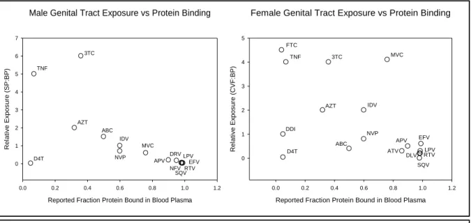
Quantifying the pharmacokinetics of antiretrovirals in mucosal tissues is complex. Blood plasma concentrations do not always correlate with genital or rectal concentrations, and accumulation ratios vary between and within therapeutic classes [36, 38, 41]. The mechanisms behind these differences have yet to be fully elucidated. In the male genital tract, the primary influence appears to be protein binding. However, as illustrated in Figure 3 this is not the case for the female genital tract.
Figure 3 Relationship of total genital tract exposure to blood plasma protein binding 3TC: lamivudine; ABC: abacavir; APV: amprenavir; ATV: atazanavir; AZT: zidovudine; D4T: stavudine; DDI:didanosine; DLV: delavirdine; EFV: efavirenz; FTC: emtricitabine; TNF: tenofovir; IDV: indinavir; LPV: lopinavir; MVC: maraviroc; NVP: nevirapine; RTV: ritonavir; SQV: saquinavir
Male Genital Tract Exposure vs Protein Binding
Reported Fraction Protein Bound in Blood Plasma
0.0 0.2 0.4 0.6 0.8 1.0 1.2
R el at iv e E x po s ur e (S P :B P ) 0 1 2 3 4 5 6 7 D4T TNF 3TC AZT ABC IDV NVP MVC APV DRV NFV SQVRTV LPV EFV
Female Genital Tract Exposure vs Protein Binding
Reported Fraction Protein Bound in Blood Plasma
0.0 0.2 0.4 0.6 0.8 1.0 1.2
R el at iv e E x po s ur e (C V F :B P ) 0 1 2 3 4 5 FTC
TNF 3TC MVC
23
Further investigations on the physicochemical properties of antiretrovirals that confer increased mucosal penetration are required.
Although the majority of antiretrovirals act intracellularly, only the nucleoside/tide analogue reverse transcriptase inhibitors require further phosphorylation in order to exert antiviral activity. Since an extracellular-intracellular disconnect between ZDV, 3TC, and TFV has been found between semen and blood [59, 62], additional studies quantifying intracellular moieties in the female genital tract will be important for identifying appropriate concentration targets for HIV prevention. Quantification of the intersubject variability in subpopulations of mononuclear cells susceptible to HIV infection may also provide insight into the probability of protection in a targeted population. Finally, additional insight into what portions of the female genital tract are most susceptible to HIV infection (vagina vs cervix vs uterus), or which portion can be a pharmacologic and virologic
surrogate for the others, is required. Advanced physiologic-based modeling may be able to assist in this effort.
Developing a reliable animal model for prevention studies has been challenging. Human immunodeficiency virus infection is a species-specific disease which limits animal model flexibility. For decades, research has been limited to macaque models, due to the permissibility of SIV: a virus similar, but not identical, to HIV. SIV’s distinct co-receptor usage can confound results of prevention
studies [68]. SIVmac and SIVmc most closely resemble HIV-1 in progression of plasma RNA and CD4 T cell response [8]. The chimeric SHIV, while containing a portion of the HIV-1 genome, does not mimic the progression of infection as closely as SIV. Some models utilize progesterone therapy to thin the epithelial layer of the vaginal tract, increase the probability of infection, and increase the likelihood of documenting an antiretroviral effect. However, the impact this intervention has on antiretroviral concentrations or efficacy in these tissues is unknown. Finally, exposure models also differ. The low-dose repeat exposure model was developed to mimic the human condition. The single
24
with the high dose model (75%) [69]. The variability between these models is a strong consideration when assessing outcomes.
Interspecies scaling of antiretroviral exposures between macaques and humans has been difficult due to the limited amount of pharmacokinetic information generated in these studies. The only systemic antiretroviral regimens that have demonstrated full protection against rectal, vaginal, and oral exposure to SHIV in macaques is daily or intermittent subcutaneous administration of 22 mg/kg TFV or 22 mg/kg TFV+ 20 mg/kg FTC [15, 45]. Although no Cmax or AUC value was
reported for subcutaneous TFV, the Cmax of the selected human-equivalent oral dose (633 ng/mL) [44] was approximately twice as high as the observed Cmax in humans [38]. Local exposure of
antiretrovirals in mucosal tissues following topical administration remains to be measured
comprehensively in animal models. Given the smaller genital and rectal cavities of macaques, it is possible that animals in the preclinical trials discussed above received supratherapeutic dosing.
The humanized mouse model has recently been developed to overcome some of the logistical challenges with macaques. The first humanized mouse was from a SCID (severe combined
immunodeficiency) variant. The mutation in the prkdc gene leading to loss of function in mature T and B lymphocytes allows these mice to be infected with foreign tissue without rejection [16]. Since only the implanted tissues can be infected with HIV, this has limited applications for HIV prevention research. The SCID-hu PBL model was developed by IP injection of human peripheral blood lymphocytes. Although populations of human B and T cells can be recovered, the repertoire is limited and transient, with no renewal capacity. These mice have been used to study viral pathogenesis and screen vaccine candidates. However, the limited numbers of human cells in mucosal tissues make them poor candidates for prevention research. In the SCID-hu Thy/Liv model, human thymus and liver are grafted into the SCID mouse, with subsequent production of human mononuclear cells (including CD4+ cells). However, this model is not susceptible to mucosal
25
mouse generally have more extensive humanization of mucosal tissues. Rag2-/- IL2Rγc-/- mice are developed by intrahepatic placement of human CD34+ hematopoetic stem cells which then differentiate into human B, T, and dendritic cells. These mice are susceptible to HIV following systemic, vaginal, and rectal exposure. Additionally, appropriate viral suppression and CD4+ T cell recovery can be achieved upon administration of antiretroviral therapy [70]. The NOD/SCID- IL2Rγc
mice are formed upon introduction of CD34+ hematopoetic stem cells, either
intrahepatically or intravenously. This model is currently undergoing investigations as to their susceptibility to mucosal transmission of HIV. In the NOD/SCID BLT mouse, human fetal liver and human fetal thymus are implanted under the kidney capsule of the NOD/SCID mouse with
subsequent transplantation of human fetal liver CD34+ hematopoetic stem cells. This model has shown a robust humanization of the rectal and female genital tracts and susceptibility to both vaginal and rectal HIV transmission [16]. However, no pharmacokinetic investigations in this model have been performed.
An alternative, or complementary, approach to a preclinical animal model for HIV prevention strategies is the tissue explant culture model. This technique has been previously used with human thymus tissue and more recently applied to rectal, vaginal, and cervical tissues: common sites of exposure in HIV transmission [71]. This promising ex vivo approach allows human tissue from healthy volunteers to be used in the study of HIV transmission, in addition to antiretroviral safety, efficacy, and pharmacology. Insights into successful prevention strategies may be obtained by varying drug doses, viral isolates and titers in this model. This technique has been utilized to evaluate several candidate microbicides [23, 26, 30], as well as tenofovir [22, 24].
Future Role of Clinical Pharmacology to Bridge the Gaps
As more predictive preclinical models are developed for prevention, the tools of26
scaling of exposures, identifying the optimal drugs/drug combinations, doses, and dosing regimens, and designing efficient clinical trials.
Characterization of the pharmacokinetics occurring at mucosal surfaces prone to infection (rectal, vaginal, cervical) has provided valuable information on which antiretrovirals should be pursued in prevention trials. Those drugs that accumulate in tissues (eg TFV, FTC, 3TC, maraviroc) as well as those that achieve concentrations above their respective IC90 (eg raltegravir, atazanavir, darunavir) are candidates for further investigation. However, the specific pharmacokinetic behavior of these drugs in tissues remains unknown, including residence times, cellular uptake, and intracellular phosphorylation. While it is known that certain physiochemical properties such as protein binding, lipophilicity, and molecule size have an impact on penetration into genital tract secretions, there remain undefined mechanisms of uptake, particularly for the female genital tract. Even without a complete mechanistic understanding of drug movement between these compartments,
pharmacokinetic modeling may still allow for accurate estimation of concentrations in the genital tract.
Identifying the concentration of
antiretroviral(s) needed at the site of exposure to prevent HIV infection is critical to this field. Preclinical models have shown that antiretrovirals can protect against HIV infection. However, in the absence of rigorous pharmacokinetic data,
it is difficult to determine how these results can be extrapolated to humans. More deliberate, well designed sampling strategies in preclinical models can provide a great deal of information with only
27
minimal increase in cost and effort. The tissue explant culture model may also assist in defining PK-PD relationships in cervical, vaginal, and rectal mucosal tissues. Physiological based modeling may then assist in selecting the most appropriate drugs or combinations, doses, and dose strategies to move forward in clinical investigations.
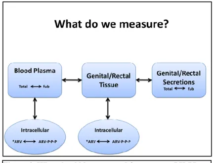
Once drug targets for prevention are identified, determining the appropriate surrogate
sampling schemes to generate data in large groups of individuals will be required. Figure 4 represents the compartments from which pharmacokinetic data can be obtained. It is possible that only
intracellular ARV concentrations from a specific cellular subset in tissues predict efficacy. However, to expediently perform larger population PK-PD modeling analysis from registration clinical trials, surrogates of this exposure will need to be developed. However, optimal use of these surrogates requires accurate information on when the interventions are used. Without proper dose-timing information, any drug exposure measure will have little value as a tool for adherence.
Likewise, when comparing local exposures in the mucosal tissues to blood plasma
concentrations, single paired concentrations will not yield as comprehensive an understanding of drug exposure as AUC measures. In order to calculate an AUC however, either extensive intra-individual sampling is needed, or pharmacokinetic models with population pharmacokinetic approaches are required. Therefore, many of the clinical trials conducted thus far have not prospectively
incorporated this degree of pharmacokinetic sampling. Future studies which include a thoughtful approach to obtaining pharmacokinetic data will provide additional insight into the pharmacologic relationships between dose, plasma concentrations, mucosal concentrations, and efficacy.
Finally, an important consideration in analyzing clinical trial data is the confounder of adherence to therapy. In the case of prevention research, reliance is placed on the individual subjects to ensure adherence. To adequately interpret clinical trial outcomes, investigators require a thorough understanding of the number of subjects in the active treatment arm not taking their medications, and
28
the time and resources invested in the clinical trial. Real-time pharmacokinetic sampling with subsequent adherence interventions could be built into these investigations for pharmacologic quality assurance.
Conclusion
The recent failures with microbicide and vaccine candidates for HIV prevention necessitate a focus on topical and systemic antiretroviral therapy. Implementation of an acceptable and efficacious antiretroviral prevention strategy will dramatically impact the global HIV epidemic and significantly reduce the burden of disease. Both topical and systemic animal model data are promising. From these data, a number of prevention studies were created to investigate a limited range of
antiretrovirals in a variety of populations and geographic settings (as of 2010, nearly 20,000 individuals were enrolled in PrEP trials). As these clinical trials were not designed to provide definitive information to correlate drug exposure with HIV protection, many pharmacologic opportunities still exist. The target antiretroviral exposure for prevention of infection in animals or tissues is unknown, as is the effect of combining antiretrovirals from different drug classes. New analytical capabilities and novel sampling strategies allow cellular pharmacokinetics and dynamics to be more diligently investigated and to identify surrogate markers for protection. Assessing
29
Specific Aims
AIM 1: Quantify gene and protein expression, variability, and localization of drug transporters
ABCB1 (MDR1), ABCC2 (MRP2), ABCC4 (MRP4), SLC22A6 (OAT1), SLC22A8 (OAT3), and
SLC01B1 (OATP1B1) in vaginal, cervical, and colorectal tissues. Drug transporters expressed on
relevant cells in mucosal tissues have the potential to impact the efficacy of antiretrovirals. To date, there has been little exploration of transporter distribution in these tissues. If identified as a
significant predictor of tissue drug exposure, screening for transporter affinity could increase the efficiency in identifying next generation compounds for HIV prevention. Discussed in Chapter II
AIM 2: Develop an ex vivo tissue model that can be used to identify the concentration of TFV
and MVC in vulnerable mucosal tissue that will provide complete protection against HIV.
2a: Measure variability of immune cell populations, localization and viability of vaginal, cervical, and rectal tissue. Unlike cell-based assays, the explant model is not able to control for the number of target cells present in each culture, leading to high variability in infection response. Quantification of specific immune cells in each explant prior to infection, can define a baseline to which infection response can be normalized. Discussed in Chapter III
2b: Determine sensitivity of alternative methods to detect early HIV tissue infection. The widely used measurement of p24 in the culture supernatant in tissue explant experiments is not able to distinguish between inoculum virus and newly infected cells, and the resultant data are highly variable. More sensitive methods are needed to accurately identify and measure the low frequency of infected cells early in one infection. Discussed in Chapter III
30
determine whether the combination of TFV and MVC has additive or synergistic protection against HIV. Discussed in Chapter IV
AIM 3: Validate the findings from the tissue explant model in AIM 2 in a healthy volunteer
study with ex-vivo HIV challenges. Coupled with mucosal exposure data generated in our lab, the dose response model generated in Aim 2a will be used to perform PK/PD modeling that will identify the most promising intermittent oral dosing regimen to protect tissues from HIV infection. This regimen will be administered to healthy female volunteers, with subsequent tissue biopsy harvest for drug concentration analysis and ex-vivo HIV challenge to verify protection. If successful, this will provide a new Proof of Concept model for drug development. Discussed in Chapter V