CYTOSOLIC LPS ACTIVATES CASPASE-11: IMPLICATIONS FOR INNATE IMMUNITY AND MANAGEMENT OF SEPTIC SHOCK
Jon Alan Hagar
A dissertation submitted to the faculty at the University of North Carolina at Chapel Hill in partial fulfillment of the requirements for the degree of Doctor in Philosophy in the Department
of Microbiology and Immunology.
Chapel Hill 2016
Approved by: Nathaniel Moorman Jenny Ting
ABSTRACT
Jon Alan Hagar: Cytosolic LPS activates caspase-11: Implications for innate immunity and management of septic shock
(Under the direction of Edward A. Miao)
Caspases are either apoptotic or inflammatory. Amongst inflammatory caspases, caspase-1 and -caspase-1caspase-1 trigger pyroptosis, a form of programmed cell death. Whereas both can be detrimental in inflammatory disease, only caspase-1 has an established protective role during infection. In Chapter 2, we report that caspase-11 is required for innate immunity to cytosolic, but not vacuolar, Gram-negative bacteria. Burkholderia species that naturally invade the cytosol triggered caspase-11, as did cytosol invading mutants of the normally vacuolar pathogens Salmonella typhimurium and Legionella pneumophila. This pathway protected mice from lethal challenge with B. thailandensis and B. pseudomallei. Thus, caspase-11 is critical for surviving exposure to ubiquitous environmental pathogens.
During endotoxemia, excessive caspase-11 activation causes shock. In Chapter 3, we report that contamination of the cytoplasm by lipopolysaccharide (LPS) is the signal that triggers caspase-11 activation in mice. Priming the caspase-11 pathway in vivo resulted in extreme sensitivity to subsequent LPS challenge in both wild type and Tlr4-deficient mice, whereas caspase 11-deficient mice were relatively resistant. Together, our data reveal a new pathway for detecting cytoplasmic LPS.
Treating hyperglycemia with insulin improves the survival of patients in intensive care units (ICU); however, extreme controversy has surrounded how stringently blood glucose should be controlled. Amongst subgroups of ICU patients, those with sepsis are particularly prone to hypoglycemia during insulin therapy, which is a risk factor for death. Why sepsis predisposes to hypoglycemia remains unknown. In Chapter 4, we show that co-delivery of insulin with
ACKNOWLEDGEMENTS
TABLE OF CONTENTS
LIST OF TABLES ... x
LIST OF FIGURES ... xi
LIST OF ABBREVIATIONS ... xiii
CHAPTER 1: INTRODUCTION ... 1
DETECTION OF CYTOSOLIC BACTERIA BY INFLAMMATORY CASPASES ... 2
Summary ... 2
Introduction ... 3
The inflammatory caspases ... 3
The inflammasomes ... 4
Burkholderia ... 4
Shigella ... 6
Francisella ... 7
Listeria ... 8
Rickettsia ... 9
Mycobacterium ... 10
Conclusions ... 11
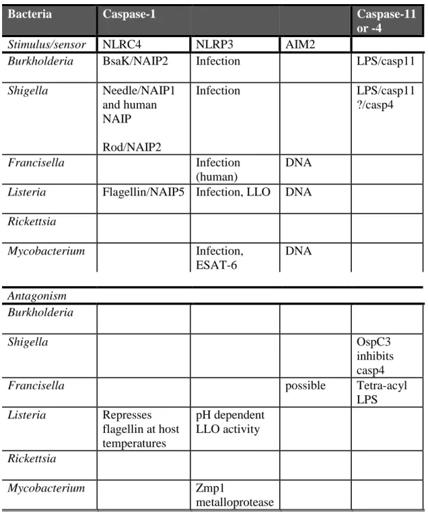
Tables... 12
CHAPTER 2: CASPASE-11 PROTECTS AGAINST BACTERIA THAT
ESCAPE THE VACUOLE ... 19
SUMMARY ... 19
CASPASE-11 PROTECTS AGAINST BACTERIA THAT ESCAPE THE VACUOLE ... 20
MATERIALS AND METHODS ... 26
FIGURES AND TABLES ... 31
REFERENCES ... 42
CHAPTER 3: CYTOPLASMIC LPS ACTIVATES CASPASE-11: IMPLICATIONS IN TLR-4 INDEPENDENT ENDOTOXIC SHOCK ... 46
SUMMARY ... 46
CYTOPLASMIC LPS ACTIVATES CASPASE-11: IMPLICATIONS IN TLR-4 INDEPENDENT ENDOTOXIC SHOCK ... 47
MATERIALS AND METHODS ... 53
FIGURES AND TABLES ... 59
REFERENCES ... 66
CHAPTER 4: LPS POTENTIATES INSULIN-DRIVEN HYPOGLYCEMIC SHOCK ... 69
SUMMARY ... 69
LPS POTENTIATES INSULIN-DRIVEN HYPOGLYCEMIC SHOCK ... 70
MATERIALS AND METHODS ... 75
FIGURES AND TABLES ... 81
REFERENCES ... 91
CHAPTER 2 – CASPASE-11 PROTECTS AGAINST BACTERIA
THAT ESCAPE THE VACUOLE ... 93 CHAPTER 3 – CYTOPLASMIC LPS ACTIVATES CASPASE-11:
LIST OF TABLES
Table 1.1. Cell tropism and vacuolar escape determinants of cytosolic bacteria. ... 12
Table 1.2. Interaction of inflammatory caspases and cytosolic bacteria. ... 13
Table 2.1. Strain and plasmid list... 40
Table 2.2. Numbers of mice used in survival experiments ... 41
Table 3.1. Strain list. ... 65
LIST OF FIGURES
Figure 2.1. Schematic of inflammasome detection pathways... 31
Figure 2.2. Burkholderia detection and protection conferred by Casp1/11 is independent of all known canonical inflammasomes. ... 32
Figure 2.3. Burkholderia protection conferred by Casp1/11 is independent of all known canonical inflammasomes. ... 33
Figure 2.4. Diverse cytosolic bacteria activate pyroptosis independent of NLRC4, NLRP3 and ASC. ... 34
Figure 2.5. Caspase-11 mediates pyroptosis after infection by cytosolic bacteria. ... 35
Figure 2.6. TLR ligands and IFN-γ enhance Casp11 expression and caspase-11- dependent cell death. ... 36
Figure 2.7. Caspase-11 is not required for pyroptosis induced by flagellin expressing wild type L. pneumophila. ... 37
Figure 2.8. Morphology of S. typhimurium ΔsifA-induced pyroptosis. ... 38
Figure 2.9. Caspase-11 protects against cytosolic bacteria in vivo. ... 39
Figure 3.1. Schematic of canonical and non-canonical inflammasome pathways. ... 59
Figure 3.2. Cytoplasmic LPS triggers caspase-11 activation. ... 60
Figure 3.3. Upregulation of caspase-11 potentiates pyroptosis but not proteolytic processing. .. 61
Figure 3.4. Listeria and CTB mediate caspase-11 activation by LPS. ... 62
Figure 3.5. Caspase-11 responds to distinct lipid A structures. ... 63
Figure 4.1. Co-delivery of LPS and insulin drives hypoglycemic shock. ... 81
Figure 4.2. LPS delivered in OptiMEM rapidly induces shock. ... 82
Figure 4.3. LPS impairs insulin clearance and amplifies insulin receptor signaling. ... 83
Figure 4.4. LPS delays insulin clearance but does not affect serum Evan’s blue concentrations. 84 Figure 4.5. LPS enhances insulin signaling via caspase-11, TLR4, and complement. ... 85
Figure 4.6. CVF fails to rescue wild-type mice. ... 86
Figure 4.7. Eicosanoid signaling acts downstream of LPS to amplify insulin activity. ... 87
Figure 4.8. Lethality of LPS and insulin challenge is independent of effects on hematocrit. ... 88
Figure 4.9. Proposed interaction of LPS with insulin signaling and clearance pathways. ... 89
LIST OF ABBREVIATIONS
BMM Bone-marrow derived macrophage
CARD Caspase activation and recruitment domain CIT Conventional insulin therapy
COX Cyclooxygenase CTB Cholera toxin B
DAMP Damage-associated molecular pattern DC Dendritic cell
ICU Intensive care unit
IFN Interferon
IIT Intensive insulin therapy
IL Interleukin
IR Insulin receptor LLO Listeriolysin O LPS Lipopolysaccharide
Mφ Macrophage
NLR NOD-like receptor
PAMP Pathogen-associated molecular pattern PGE2 Prostaglandin E2
PMN Polymorphonuclear
CHAPTER 1: INTRODUCTION
Caspase-11 is a member of the inflammatory caspase family, which includes caspase-1, caspase-12, and the human caspase-11 orthologues caspase-4 and -5(1). Caspases are cysteine-dependent proteases that recognize various conserved aspartate residues in substrate proteins(2). These enzymes are involved in a number of processes, but are perhaps most well known for their roles in programmed cell death and immunity.
Caspase-11 was discovered during a screen of mouse complementary DNA for caspase-1 homologs(3), where it was found to mediate the lethality of endotoxic shock in mice1. While caspase-1 and caspase-11 both have well established roles in inflammatory diseases, only caspase-1 had been shown to protect against certain lethal infections. When we began the work described herein, we were investigating a novel caspase-1 activation pathway triggered by the cytosol-invasive bacterium Burkholderia thailandensis that was critical for mice to resist lethal infection; i.e. mice deficient in all known caspase-1 activators resisted lethal B. thailandensis infection, whereas mice deficient in Casp1 itself uniformly succumbed to infection. Shortly thereafter, Dr. Vishva Dixit’s laboratory made the key observations that all Casp1-deficient mice carry an inactivating passenger mutation in the adjacent Casp11 gene, and that this mutation accounted for several phenotypes previously ascribed to caspase-1 deficiency(4). Hypothesizing that caspase-11 might be involved in murine resistance to B. thailandensis, our story soon
1
evolved into the first description of caspase-11 protecting a host from lethal infection, described in Chapter 2. Subsequent work identified lipopolysaccharide (LPS) as the bacterial ligand to which caspase-11 responds, a story described in Chapter 3. Finally, we serendipitously discovered that LPS signaling via caspase-11 and the other cellular LPS sensor, TLR4, can inhibit clearance of exogenous insulin, which models insulin therapy administered during sepsis treatment; this work is described in Chapter 4.
In the next section of this chapter, we review inflammatory caspase interaction with cytosol invasive bacteria, which is a unifying theme of this dissertation. This review describes the state of the field before and shortly after we published the work described in Chapters 2 and 3, as well as a more detailed description of inflammatory caspase biology.
DETECTION OF CYTOSOLIC BACTERIA BY INFLAMMATORY CASPASES2 Summary
The sanctity of the cytosolic compartment is rigorously maintained by a number of innate immune mechanisms. Inflammasomes detect signatures of microbial infection and trigger
caspase-1 or caspase-11 activation, culminating in cytokine secretion and obliteration of the replicative niche via pyroptosis. Recent studies have examined inflammatory caspase responses to cytosolic bacteria, including Burkholderia, Shigella, Listeria, Francisella, and Mycobacterium species. For example, caspase-11 responds to LPS introduced into the cytosol after
Gram-negative bacteria escape the vacuole. Not surprisingly, bacteria antagonize these responses; for
2
example, Shigella delivers OspC3 to inhibit caspase-4. These findings underscore bacterial coevolution with the innate immune system, which has resulted in few, but highly specialized cytosolic pathogens.
Introduction
The immune defenses of the extracellular environment are severe, as are those of the phagolysosome. The prospect of refuge from these insults therefore makes the cytosolic
compartment a theoretically attractive refuge for potential bacterial pathogens. However, the fact that bona fide cytosolic bacteria can be counted on one’s fingers (see Table 1 for a summary of these pathogens, their cell tropisms, and their mechanisms for invading the cytosol) highlights the successful immune defenses employed to maintain the sterility of the cytosolic niche. A number of cytosolic sensors detect signatures of infection, initiating potent inflammatory responses and/or host cell death. The importance of inflammatory caspases in this regard is underscored by the extreme susceptibility of mice deficient in these enzymes to infection by cytosolic pathogens. Interestingly, the few cytosolic specialist pathogens are among the most virulent known. Herein, we discuss the role of inflammatory caspases in the innate immune response to cytosolic bacteria, focusing on recent advances in our understanding of how cells detect intruders and trigger caspase activation, and how caspases mediate containment of the infection.
The inflammatory caspases
immunologically silent form of programmed cell death. In contrast, the inflammatory caspases, caspase-11 (or the presumed human homologs caspase-4 and caspase-5) and caspase-1, initiate a form of lytic cell death termed pyroptosis following their activation, which releases
inflammatory mediators, removes the replicative niche of cytosolic bacteria, and exposes intruders to extracellular defenses and neutrophils(5) (reviewed in (6)). In addition, caspase-1 mediates the maturation and secretion of pro-IL-1β and pro-IL-18, two pleiotropic inflammatory cytokines best known for inducing fever and interferon (IFN)-γ secretion, respectively(2).
The inflammasomes
The inflammatory caspases are expressed as inactive zymogens3. The canonical
inflammasomes, a class of cytosolic pattern recognition receptors (PRR), activate caspase-1 in response to specific signatures of infection. A theorized non-canonical inflammasome(s) is proposed to activate caspase-11(4). Relevant inflammasomes and their agonists are detailed in Table 2; for in-depth review, see (6) and (2).
Burkholderia
B. pseudomallei and B. thailandensis have served as models for studying the interaction of inflammatory caspases and cytosolic bacteria. These Gram-negative bacteria exist
ubiquitously in the soil of southeast Asia and sporadically elsewhere(10). Although closely related, only B. pseudomallei causes severe human and murine disease; however, B.
3
thailandensis can infect macrophages and epithelial cells both in vitro and in vivo. B.
pseudomallei and B. thailandensis rapidly escape the vacuole via their type III secretion system (T3SS) (11)(12). NLRC4 is positioned to detect signatures of T3SS activity, alerting the immune system to pathogens that reprogram and parasitize host cells. Not surprisingly, we and others found that macrophage infection triggers NLRC4 activation (13)(14). Mediating this activation, we showed that the T3SS rod protein BsaK is detected through NLRC4(15), and Zhao and colleagues demonstrated that NAIP2 is the sensor upstream of NLRC4(16). Later the T3SS needle protein BsaL, as well as needle proteins from a variety of other bacteria, was found to be detected by murine NAIP1 and human NAIP, both signaling through NLRC4
downstream(16)(17)(18). By an ill-defined mechanism, Burkholderia species also activate NLRP3(13)(14). Together, NLRC4 and NLRP3 are critical for mice to resist intranasal B. pseudomallei challenge(13). In this model, IL-18 is central to this resistance, coordinating bacterial clearance, whereas IL-1β secretion mediates immune pathology driven by neutrophil recruitment.
activation(19)(9). Although enhanced by TLR4 signaling, this pathway can proceed
independently of extracellular LPS signaling. Thus, Tlr4–/– mice primed with a TLR3 agonist succumb to secondary LPS challenge in a model of endotoxic shock. Previous studies indicate that during prolonged infections, caspase-11 activates in response to all Gram-negative
bacteria[4](20)(21)(22). We speculate that such activation may reflect vacuole leakage events that accumulate over 16h, which may have relevance in the setting of Gram-negative septic shock. In contrast, caspase-11 rapidly responds to L. pneumophila infection in pre-activated macrophages(23)(24); whether vacuolar integrity is compromised under these conditions remains to be examined. The physiologic role of caspase-11 during infection is to combat cytosolic bacteria. The upstream sensor that detects cytosolic LPS remains unknown.
Shigella
Members of the Gram-negative Shigella genus are exquisitely adapted to cause human gastrointestinal disease. S. flexneri infects a variety of cell types, such as intestinal epithelial cells and macrophages. Following phagocytosis by macrophages or T3SS-mediated uptake by
epithelial cells, S. flexneri rapidly escapes the phagosome. In vitro, S. flexneri is robustly
detected by caspase-1 via NLRC4(25) and, under some conditions, NLRP3(26). As an aflagellate bacterium, S. flexneri does not expressed flagellin. We showed that the MxiI rod protein is detected via NLRC4(15), and Zhao showed this was via NAIP2(16). The S. flexneri needle component MxiH is also detected by murine NAIP1 and human NAIP (17). As with
function in resistance to Shigella infection remains to be determined; however, we have found that both S. flexneri infection and transfection of S. flexneri lysates into macrophages activate caspase-11 in vitro (our unpublished observations), indicating that S. flexneri lipid A can be detected by the caspase-11 pathway.
Recently, work employing a Guinea pig model of Shigella infection, which more faithfully models human infection than mouse models, has implicated caspase-4 in host
resistance to S. flexneri(27). Kobayashi and colleagues found that caspase-4 mediates epithelial cell death in response to several enteric pathogens, and that S. flexneri secretes an inhibitor of caspase-4 activation, OspC3, to counteract this innate immune response in vitro and in vivo. Remarkably, the authors found that OspC3 is specific in antagonizing caspase-4 and does not associate with caspase-11, highlighting the specificity of Shigella species for infecting humans. Future research will determine whether 4 responds to cytoplasmic LPS as does caspase-11, which would situate caspase-4 as a key preserver of cytosolic sterility.
Francisella
The causative agent of tularemia, Gram-negative F. tularensis is among the most infectious and virulent pathogens; thus, it is classified as a category A bioweapon. F. tularensis infects a variety of cell types, with macrophages and neutrophils representing the primary
by F. novicida, results in little detectable caspase-1 activation(36), suggesting virulent strains have evolved to evade AIM2. A better understanding of this difference may have implications for both the treatment of and vaccination against tularemia.
Francisella species express tetra-acylated LPS. Not surprisingly, we have found that macrophages do not activate caspase-11 after infection by F. novicida(9). However, transfection of penta-acylated lipid A from an lpxF mutant, but not wild-type tetra-acylated lipid A, triggers caspase-11 dependent pyroptosis. Therefore, Francisella species appear to have evolved to evade a major host cytosol surveillance pathway, the non-canonical inflammasome.
Listeria
Listeria monocytogenes is a Gram-positive saprophyte and facultative pathogen that causes self-limited gastroenteritis in immunocompetent individuals. Of particular concern for the immunocompromised, L. monocytogenes infections can progress to cause sepsis, encephalitis, and death; in pregnant mothers, it can trigger abortion. L. monocytogenes readily escapes into the cytosol of epithelial cells and macrophages using the pore-forming toxin listeriolysin O (LLO).
In vivo, Casp1–/– Casp11–/– mice may have increased susceptibility to L. monocytogenes infection (45); however, this was not replicated in another publication(44). Furthermore, the contributions of individual inflammasomes during in vivo infection are not defined.
Nevertheless, L. monocytogenes appears to have evolved to limit inflammasome detection: LLO activity is optimal in the acidic environment of the phagosome, thus limiting its potential to trigger NLRP3; flagellin expression is repressed during growth at host temperature; and few bacteria lyse in the cytosol, thus limiting cytosolic DNA exposure. The efficiency of these evasive strategies is demonstrated by the rapid clearance of L. monocytogenes forced to express flagellin in vivo(44)(46).
By virtue of its nature as a Gram-positive bacterium, L. monocytogenes does not contain LPS, and thus is not detected by caspase-11(9)(47).
Rickettsia
Mycobacterium
Among Mycobacterium species, M. marinum is distinct in that it rapidly escapes the phagosome to replicate in the cytosol and spread cell-to-cell. Vacuolar escape requires ESAT-6, a secretion product of the ESX-1 type VII secretion system suggested to have membrane pore forming activity(50). Although M. tuberculosis is traditionally considered a vacuolar pathogen of macrophages, recent studies suggest it may exist in the cytosol for at least part of its intracellular life cycle (reviewed in (51)).
A number of studies have investigated the role of inflammatory caspases in immunity to M. tuberculosis and M. marinum. While the in vivo importance of IL-1 and IL-1 are well
accepted, the role of NLRP3, ASC, and caspase-1 remain controversial both in vivo and in vitro (for a more in-depth review, see (2)). Herein we limit our discussion to the recent studies
examining caspase-1 activation in response to cytosolic bacterial exposure. Several studies implicate ESX-1 and ESAT-6 in caspase-1 activation(52)(53)(54)(55)(56)(57). Abdallah and colleagues suggest that ESX-1 translocation of mycobacteria to the cytosol potentiates
Conclusions
In recent years, our understanding of inflammatory caspase activation has expanded to include several new sensor-stimulus pairs, such as AIM2 and DNA, NAIP1 and the T3SS needle, and LPS and the non-canonical inflammasome. These findings have elucidated how the
inflammatory caspases and, more generally, the innate immune system restrict the ability of pathogens to establish cytosolic growth niches. At the same time, they pose a number of
Tables
Genus Gram +/- Cell tropism Vacuolar escape
determinants, bacterial
Burkholderia - Mφ, PMN,
epithelial cells
T3SSBSA
Shigella - Mφ, DC,
intestinal epithelial cells
Mxi-Spa T3SS, IpaB
Francisella - Mφ, PMN, DC,
epithelial cells, hepatocytes
IglC, MglA, FTT11103
Listeria + Mφ, intestinal
epithelial
LLO,
phospholipase C
Rickettsia - Vascular
endothelial, Mφ
Phospholipases, hemolysin Mycobacterium Acid-fast + Mφ ESX-1 T7SS,
ESAT-6
Bacteria Caspase-1 Caspase-11 or -4
Stimulus/sensor NLRC4 NLRP3 AIM2
Burkholderia BsaK/NAIP2 Infection LPS/casp11 Shigella Needle/NAIP1
and human NAIP Rod/NAIP2
Infection LPS/casp11
?/casp4
Francisella Infection
(human)
DNA Listeria Flagellin/NAIP5 Infection, LLO DNA Rickettsia
Mycobacterium Infection, ESAT-6
DNA
Antagonism Burkholderia
Shigella OspC3
inhibits casp4
Francisella possible Tetra-acyl
LPS Listeria Represses
flagellin at host temperatures
pH dependent LLO activity Rickettsia
Mycobacterium Zmp1
metalloprotease
REFERENCES
1. J. Shi et al., Inflammatory caspases are innate immune receptors for intracellular LPS. Nature (2014).
2. J. von Moltke, J. S. Ayres, E. M. Kofoed, J. Chavarría-Smith, R. E. Vance, Recognition of bacteria by inflammasomes. Annual review of immunology 31, 73–106 (2013).
3. S. Wang et al., Identification and characterization of Ich-3, a member of the interleukin-1beta converting enzyme (ICE)/Ced-3 family and an upstream regulator of ICE. J Biol Chem 271, 20580–20587 (1996).
4. N. Kayagaki et al., Non-canonical inflammasome activation targets caspase-11. Nature 479, 117–21 (2011).
5. E. A. Miao et al., Caspase-1-induced pyroptosis is an innate immune effector mechanism against intracellular bacteria. Nature immunology 11, 1136–42 (2010).
6. Y. Aachoui, V. Sagulenko, E. A. Miao, K. J. Stacey, Inflammasome-mediated pyroptotic and apoptotic cell death, and defense against infection. Current opinion in microbiology 16, 319–26 (2013).
7. K. M. Boatright et al., A unified model for apical caspase activation. Mol Cell 11, 529–541 (2003).
8. P. Broz, J. von Moltke, J. W. Jones, R. E. Vance, D. M. Monack, Differential requirement for Caspase-1 autoproteolysis in pathogen-induced cell death and cytokine processing. Cell host & microbe 8, 471–83 (2010).
9. J. A. Hagar, D. A. Powell, Y. Aachoui, R. K. Ernst, E. A. Miao, Cytoplasmic LPS activates caspase-11: implications in TLR4-independent endotoxic shock. Science (2013).
10. B. J. Currie, D. A. Dance, A. C. Cheng, The global distribution of Burkholderia pseudomallei and melioidosis: an update. Transactions of the Royal Society of Tropical Medicine and Hygiene 102 Suppl 1, S1–4 (2008).
11. N. R. Adler et al., The molecular and cellular basis of pathogenesis in melioidosis: how does Burkholderia pseudomallei cause disease? FEMS microbiology reviews 33, 1079–99 (2009). 12. C. T. French et al., Dissection of the Burkholderia intracellular life cycle using a
photothermal nanoblade. Proceedings of the National Academy of Sciences of the United States of America 108, 12095–100 (2011).
14. Y. Aachoui et al., Caspase-11 protects against bacteria that escape the vacuole. Science (New York, NY) 339, 975–8 (2013).
15. E. A. Miao et al., Innate immune detection of the type III secretion apparatus through the NLRC4 inflammasome. Proceedings of the National Academy of Sciences of the United States of America 107, 3076–80 (2010).
16. Y. Zhao et al., The NLRC4 inflammasome receptors for bacterial flagellin and type III secretion apparatus. Nature 477, 596–600 (2011).
17. J. Yang, Y. Zhao, J. Shi, F. Shao, Human NAIP and mouse NAIP1 recognize bacterial type III secretion needle protein for inflammasome activation. Proceedings of the National Academy of Sciences of the United States of America (2013).
18. S. K. Cassidy et al., D. J. Philpott, Ed. Membrane Damage during Listeria monocytogenes Infection Triggers a Caspase-7 Dependent Cytoprotective Response. PLoS pathogens 8, e1002628 (2012).
19. N. Kayagaki et al., Noncanonical Inflammasome Activation by Intracellular LPS Independent of TLR4. Science (2013).
20. V. A. Rathinam et al., TRIF licenses caspase-11-dependent NLRP3 inflammasome activation by gram-negative bacteria. Cell 150, 606–19 (2012).
21. P. Broz et al., Caspase-11 increases susceptibility to Salmonella infection in the absence of caspase-1. Nature 490, 288–91 (2012).
22. P. Gurung et al., Toll or interleukin-1 receptor (TIR) domain-containing adaptor inducing interferon-β (TRIF)-mediated caspase-11 protease production integrates Toll-like receptor 4 (TLR4) protein- and Nlrp3 inflammasome-mediated host defense against enteropathogens. The Journal of biological chemistry 287, 34474–83 (2012).
23. C. N. Casson et al., Caspase-11 activation in response to bacterial secretion systems that access the host cytosol. PLoS pathogens 9, e1003400 (2013).
24. C. L. Case et al., Caspase-11 stimulates rapid flagellin-independent pyroptosis in response to Legionella pneumophila. Proceedings of the National Academy of Sciences of the United States of America 110, 1851–6 (2013).
25. T. Suzuki et al., Differential regulation of caspase-1 activation, pyroptosis, and autophagy via Ipaf and ASC in Shigella-infected macrophages. PLoS pathogens 3, e111 (2007).
26. B. K. Davis et al., Cutting edge: NLRC5-dependent activation of the inflammasome. Journal of immunology (Baltimore, Md : 1950) 186, 1333–7 (2011).
27. T. Kobayashi et al., The Shigella OspC3 effector inhibits caspase-4, antagonizes
28. J. D. Hall et al., Infected-host-cell repertoire and cellular response in the lung following inhalation of Francisella tularensis Schu S4, LVS, or U112. Infection and immunity 76, 5843–52 (2008).
29. M. T.-H. Huang et al., Deletion of ripA alleviates suppression of the inflammasome and MAPK by Francisella tularensis. Journal of immunology (Baltimore, Md : 1950) 185, 5476–85 (2010).
30. V. A. Rathinam et al., The AIM2 inflammasome is essential for host defense against cytosolic bacteria and DNA viruses. Nature immunology 11, 395–402 (2010).
31. T. Fernandes-Alnemri et al., The AIM2 inflammasome is critical for innate immunity to Francisella tularensis. Nature immunology 11, 385–93 (2010).
32. J. W. Jones et al., Absent in melanoma 2 is required for innate immune recognition of Francisella tularensis. Proceedings of the National Academy of Sciences of the United States of America 107, 9771–6 (2010).
33. K. Peng, P. Broz, J. Jones, L.-M. Joubert, D. Monack, Elevated AIM2-mediated pyroptosis triggered by hypercytotoxic Francisella mutant strains is attributed to increased intracellular bacteriolysis. Cellular microbiology 13, 1586–600 (2011).
34. R. Pierini et al., AIM2/ASC triggers caspase-8-dependent apoptosis in Francisella-infected caspase-1-deficient macrophages. Cell death and differentiation 19, 1709–21 (2012).
35. M. K. Atianand et al., Francisella tularensis reveals a disparity between human and mouse NLRP3 inflammasome activation. The Journal of biological chemistry 286, 39033–42 (2011). 36. J. R. Wickstrum et al., Francisella tularensis induces extensive caspase-3 activation and apoptotic cell death in the tissues of infected mice. Infection and immunity 77, 4827–36 (2009). 37. K. Meixenberger et al., Listeria monocytogenes-infected human peripheral blood
mononuclear cells produce IL-1beta, depending on listeriolysin O and NLRP3. Journal of immunology (Baltimore, Md : 1950) 184, 922–30 (2010).
38. S. E. Warren, D. P. Mao, A. E. Rodriguez, E. A. Miao, A. Aderem, Multiple Nod-like receptors activate caspase 1 during Listeria monocytogenes infection. Journal of immunology (Baltimore, Md : 1950) 180, 7558–64 (2008).
39. S. Kim et al., Listeria monocytogenes is sensed by the NLRP3 and AIM2 inflammasome. European journal of immunology 40, 1545–51 (2010).
40. J. Wu, T. Fernandes-Alnemri, E. S. Alnemri, Involvement of the AIM2, NLRC4, and NLRP3 inflammasomes in caspase-1 activation by Listeria monocytogenes. Journal of clinical
41. K. Tsuchiya et al., Involvement of absent in melanoma 2 in inflammasome activation in macrophages infected with Listeria monocytogenes. Journal of immunology (Baltimore, Md : 1950) 185, 1186–95 (2010).
42. N. Ozören et al., Distinct roles of TLR2 and the adaptor ASC in IL-1beta/IL-18 secretion in response to Listeria monocytogenes. Journal of immunology (Baltimore, Md : 1950) 176, 4337– 42 (2006).
43. J.-D. Sauer et al., Listeria monocytogenes triggers AIM2-mediated pyroptosis upon infrequent bacteriolysis in the macrophage cytosol. Cell host & microbe 7, 412–9 (2010).
44. J.-D. Sauer et al., Listeria monocytogenes engineered to activate the Nlrc4 inflammasome are severely attenuated and are poor inducers of protective immunity. Proceedings of the National Academy of Sciences of the United States of America 108, 12419–24 (2011).
45. N. M. Tsuji et al., Roles of caspase-1 in Listeria infection in mice. International immunology 16, 335–43 (2004).
46. S. E. Warren et al., Generation of a Listeria vaccine strain by enhanced caspase-1 activation. European journal of immunology 41, 1934–40 (2011).
47. N. J. Mueller, R. A. Wilkinson, J. A. Fishman, Listeria monocytogenes infection in caspase-11-deficient mice. Infection and immunity 70, 2657–64 (2002).
48. S. Radulovic et al., Rickettsia-macrophage interactions: host cell responses to Rickettsia akari and Rickettsia typhi. Infection and immunity 70, 2576–82 (2002).
49. J. Turco, H. H. Winkler, Effect of mouse lymphokines and cloned mouse interferon-gamma on the interaction of Rickettsia prowazekii with mouse macrophage-like RAW264.7 cells. Infection and immunity 45, 303–8 (1984).
50. J. Smith et al., Evidence for pore formation in host cell membranes by ESX-1-secreted ESAT-6 and its role in Mycobacterium marinum escape from the vacuole. Infection and immunity 76, 5478–87 (2008).
51. A. Welin, M. Lerm, Inside or outside the phagosome? The controversy of the intracellular localization of Mycobacterium tuberculosis. Tuberculosis (Edinburgh, Scotland) 92, 113–20 (2012).
52. K.-W. Wong, W. R. Jacobs Jr, Critical role for NLRP3 in necrotic death triggered by Mycobacterium tuberculosis. Cellular microbiology 13, 1371–84 (2011).
53. B. B. Mishra et al., Mycobacterium tuberculosis protein ESAT-6 is a potent activator of the NLRP3/ASC inflammasome. Cellular microbiology 12, 1046–63 (2010).
55. I. C. Koo et al., ESX-1-dependent cytolysis in lysosome secretion and inflammasome activation during mycobacterial infection. Cellular microbiology 10, 1866–78 (2008).
56. T. Kurenuma et al., The RD1 locus in the Mycobacterium tuberculosis genome contributes to activation of caspase-1 via induction of potassium ion efflux in infected macrophages. Infection and immunity 77, 3992–4001 (2009).
57. A. Welin, D. Eklund, O. Stendahl, M. Lerm, Human macrophages infected with a high burden of ESAT-6-expressing M. tuberculosis undergo caspase-1- and cathepsin B-independent necrosis. PloS one 6, e20302 (2011).
58. M. A. Abdallah et al., Mycobacterial secretion systems ESX-1 and ESX-5 play distinct roles in host cell death and inflammasome activation. Journal of immunology (Baltimore, Md : 1950) 187, 4744–53 (2011).
59. P. S. Manzanillo, M. U. Shiloh, D. A. Portnoy, J. S. Cox, Mycobacterium tuberculosis activates the DNA-dependent cytosolic surveillance pathway within macrophages. Cell host & microbe 11, 469–80 (2012).
60. H. Saiga et al., Critical role of AIM2 in Mycobacterium tuberculosis infection. International immunology 24, 637–44 (2012).
CHAPTER 2: CASPASE-11 PROTECTS AGAINST BACTERIA THAT ESCAPE THE VACUOLE4
SUMMARY
Caspases are either apoptotic or inflammatory. Amongst inflammatory caspases, caspase-1 and -caspase-1caspase-1 trigger pyroptosis, a form of programmed cell death. Whereas both can be detrimental in inflammatory disease, only caspase-1 has an established protective role during infection. Here, we report that caspase-11 is required for innate immunity to cytosolic, but not vacuolar, bacteria. Although Salmonella typhimurium and Legionella pneumophila normally reside in the vacuole, specific mutants (sifA and sdhA, respectively) aberrantly enter the cytosol. These mutants triggered caspase-11, which enhanced clearance of S. typhimurium sifA in vivo. This response did not require NLRP3, NLRC4, or ASC inflammasome pathways. Burkholderia species that naturally invade the cytosol also triggered caspase-11, which protected mice from lethal
4
This chapter contains a manuscript that previously appeared as an article in Science and reflects the co-first author efforts of Dr. Youssef Aachoui, Dr. Irina Leaf, and me. Specifically, I
contributed directly to the performance of experiments depicted in Fig. 2.5B, C, and E – H, Fig. 2.6C and D, and Fig. 2.9F. Additionally, I helped analyze the data in all figures and co-wrote the manuscript with Dr. Aachoui and Dr. Miao. This is the author's version of the work. It is posted here by permission of the AAAS for personal use, not for redistribution. The definitive version was published in Science 339, 2013, doi: 10.1126/science.1230751. The full citation is: Aachoui, Y.*, Leaf, I.A.*, Hagar, J.A.*, Fontana, M.F., Campos, C.G., Zak, D.E., Tan, M.H., Cotter, P.A., Vance, R.E., Aderem, A., and Miao, E.A. Caspase-11 protects against bacteria that escape the vacuole. Science. 339, pp. 975-978, 2013.
challenge with B. thailandensis and B. pseudomallei. Thus, caspase-11 is critical for surviving exposure to ubiquitous environmental pathogens.
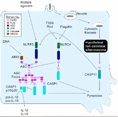
CASPASE-11 PROTECTS AGAINST BACTERIA THAT ESCAPE THE VACUOLE Canonical inflammasomes, such as NLRP3, NLRC4, and AIM2, are cytosolic sensors that detect pathogens or danger signals and activate caspase-1, leading to secretion of the
proinflammatory cytokines interleukin (IL)-1β and IL-18, and pyroptosis, a form of programmed cell death (1). Pyrin domain-containing inflammasomes, including NLRP3, signal through the ASC adaptor protein to recruit caspase-1 (Fig. 2.1). Many diverse agonists cause cytosolic perturbations that are detected through NLRP3; however the underlying mechanisms remain obscure (2). In contrast, the CARD domain-containing inflammasome NLRC4 can signal
directly to caspase-1 resulting in pyroptosis, as well as indirectly through ASC to promote IL-1β and IL-18 secretion (Fig. 2.1) (1, 3). NLRC4 detects bacterial flagellin and type III secretion system (T3SS) rod or needle components within the macrophage cytosol (4–6). Together, NLRC4 and the ASC dependent inflammasomes account for all known canonical caspase-1 activation pathways.
Burkholderia pseudomallei is a Gram-negative bacterium endemic to Southeast Asia that causes mellioidosis and is a potential biologic weapon (7). B. pseudomallei uses a T3SS to escape the phagosome and replicate in the cytosol. NLRC4 and NLRP3 both detect B. pseudomallei, promoting IL-1β secretion from murine bone marrow-derived macrophages (BMM) ((8) and Fig. 2.2A). Despite encoding many of the same virulence factors as B.
NLRP3 and NLRC4 accounted for all IL-1β secretion in response to B. thailandensis (Fig. 2.2B). We next determined whether inflammasome activation is critical to survival following B.
thailandensis challenge using caspase-1 deficient mice. Kayagaki et al. recently showed that all existing caspase-1 deficient mice also lack caspase-11 due to the backcrossing of a mutant Casp11 allele from 129 into C57BL/6 mice (10). Inflammasome detection was critical for resistance to B. thailandensis, as Casp1–/–Casp11–/– animals succumbed to the infection (Fig. 2.2C and Fig. 2.3A). In contrast, wild type C57BL/6 mice survived high dose intraperitoneal or intranasal challenge (Fig. 2.2C and Fig. 2.3A). Surprisingly, Nlrc4–/–Asc–/– mice that are deficient in all known canonical inflammasomes were also resistant (Fig. 2.2D and Fig. 2.3B). This
indicated that an unknown signaling pathway provides protection via either caspase-1 or -11 (see pathway schematic Fig. 2.1). Resistance to B. thailandensis was at least partially independent of IL-1β and IL-18, depending on the route of infection (Fig. 2.2E and Fig. 2.3C), suggesting that both cytokines and pyroptosis can contribute to protection. We therefore examined pyroptosis in vitro, and found that cytotoxicity in response B. thailandensis was impaired in Casp1–/–Casp11–/– BMM (Fig. 2.2F). Consistent with our in vivo data, pyroptosis in vitro did not require Nlrc4 or Asc (Fig. 2.2F). B. pseudomallei similarly triggered pyroptosis in Nlrc4–/–Asc–/– macrophages (Fig. 2.2G). These results indicate that a pyroptosis-inducing pathway distinct from all known canonical inflammasomes detects B. thailandensis and protects against lethal infection.
Inflammasomes discriminate pathogens from non-pathogens by detecting contamination or perturbation of the cytosolic compartment (11). The B. thailandensis T3SS facilitates bacterial access to the cytosol, and was required for induction of pyroptosis, whereas the
In order to establish their intracellular vacuolar growth niche, Salmonella typhimurium and Legionella pneumophila use T3SS and T4SS, respectively, to translocate effector proteins that work in concert to maintain the stability of these altered bacteria-containing vacuoles (12– 14). Loss of the S. typhimurium SifA or L. pneumophila SdhA effectors causes rupture of the vacuole and release of bacteria into the cytosol (15–17). S. typhimurium uses two distinct T3SS encoded by the Salmonella pathogenicity island 1 (SPI1) and SPI2; these two T3SS translocate distinct batteries of effectors, such as SifA by SPI2 (18). While S. typhimurium-expressing SPI1 and flagellin are readily detected by NLRC4 (19, 20), bacteria grown under conditions that mimic the vacuolar environment express SPI2 and repress flagellin, minimizing canonical inflammasome detection (1, 11, 21). Infection of BMMs with S. typhimurium that lacked sifA, however, significantly increased IL-1β secretion and pyroptosis (Fig. 2.4, B and C). IL-1β secretion was dependent on canonical inflammasomes (Fig. 2.4B), whereas pyroptosis was still observed in Nlrc4–/–Asc–/– and Nlrp3–/–Nlrc4–/– macrophages (Fig. 2.4C). Furthermore, the
NLRC4 inflammasome agonist flagellin was not required for these responses (Fig. 2.4, D and E). Thus, macrophages detect S. typhimurium when it aberrantly enters the cytosol, activating
pyroptosis independent of all known canonical inflammasomes.
L. pneumophila also translocates flagellin through its T4SS. Thus, L. pneumophila mutants lacking flagellin (∆flaA) evaded NLRC4 detection (Fig. 2.4F) (2). In contrast, L. pneumophila ∆flaA ∆sdhA mutants induce caspase-1 activation (16, 17), IL-1β secretion (17),
absence of flagellin and ASC (Fig. 2.4G), ruling out all canonical inflammasomes in triggering pyroptosis under these infection conditions. These data demonstrate that diverse bacteria are detected in the cytosol.
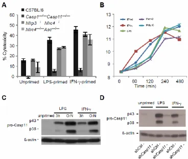
Because IL-1β secretion required the canonical inflammasomes whereas pyroptosis did not, we hypothesized that cell death is triggered by a distinct mechanism mediated by caspase-11. Like caspase-1, caspase-11 is an inflammatory caspase that can directly trigger pyroptosis (Fig. 2.1). Caspase-11 can also promote IL-1β secretion dependent upon NLRP3, ASC, and caspase-1 (10, 22–24). Because caspase-1 is activated by recruitment to an oligomerized platform known as the inflammasome, Kayagaki et al. hypothesized that a similar oligomeric structure would activate caspase-11, which they termed the non-canonical inflammasome (10). Although the cholera toxin B subunit and many different Gram-negative bacteria can trigger caspase-11 activation in vitro (10, 22–24), the nature of the physiologic stimulus that activates caspase-11 during infection remains uncertain.
Caspase-11 activation requires priming through a Toll like receptor 4 (TLR4)-TRIF-STAT1 pathway (10, 22–24). Consistent with this, Tlr4–/– and Trif–/– macrophages did not undergo pyroptosis after S. typhimurium ∆sifA infection, whereas cell death was observed in macrophages deficient in the other TLR4 adaptor, Myd88 (Fig. 2.5A). This dependence could be overcome by priming the macrophages with interferon (IFN)-γ (Fig. 2.5A), which signals
through STAT1. Interestingly, IFN-γ or LPS priming significantly increased the sensitivity of macrophages to S. typhimurium ∆sifA (Fig. 2.5A and Fig. 2.6A). These priming effects
Caspase-11 alone promoted pyroptosis without IL-1β secretion after B. thailandensis infection, whereas caspase-1 enabled both responses (Fig. 2.5B). This is consistent with B. thailandensis detection through NLRC4 and/or NLRP3 activating caspase-1 (8) and an additional pathway activating caspase-11. In contrast, the responses to S. typhimurium ∆sifA or L. pneumophila ∆flaA ∆sdhA acted through caspase-11, and not caspase-1 (Fig. 2.5, C and D). We further confirmed that caspase-11 was responsible for the cell death observed in Nlrc4–/–Asc–/– macrophages using short hairpin (sh)RNAmir (Fig. 2.5, E and F, and Fig. 2.6E). Finally, Casp11–/– BMM revealed that caspase-11 was required for pyroptosis after B. thailandensis, S. typhimurium ∆sifA, and L. pneumophila ∆flaA ∆sdhA (Fig. 2.5, G to I). Although a previous report suggested that NLRC4 signals through caspase-11 to alter phagosomal trafficking (25), we saw no evidence that NLRC4 contributes to caspase-11 dependent cell death (Fig. 2.2F, Fig. 2.4D, and Fig. 2.7). Pyroptosis initiated by caspase-11 was morphologically similar to pyroptosis triggered by caspase-1 (Fig. 2.8, A and B). Therefore, macrophages activate caspase-11 in response to cytosolic B.
thailandensis, S. typhimurium, or L. pneumophila (Fig. 2.1).
clears S. typhimurium ∆sifA in vivo; in contrast, wild type S. typhimurium effectively evades caspase-11 (23) by remaining within the vacuole. The remaining S. typhimurium ∆sifA
attenuation likely reflects the role of sifA as a virulence factor promoting intracellular replication. Moreover, all known canonical inflammasomes were dispensable for S. typhimurium ∆sifA clearance, as were IL-1β and IL-18 (Fig. 2.9D), implicating pyroptosis as the mechanism of clearance. Clearance of bacteria after pyroptosis is mediated by neutrophils through generation of reactive oxygen (21). Consistent with this, NADPH oxidase deficient p47phox–/– mice were also defective for clearance of S. typhimurium ∆sifA (Fig. 2.9D). Interestingly, TLR4 and IFN-γ were not required (Fig. 2.9E), suggesting that there is redundant priming of caspase-11 pathways in vivo. Therefore, caspase-11 protects mice from S. typhimurium ∆sifA, and because IL-1β and IL-18 are not required, pyroptosis is likely to be the mechanism of bacterial clearance in this case.
We next examined the susceptibility of Casp11–/– mice to the naturally cytosolic pathogens B. thailandensis and B. pseudomallei. While C57BL/6 mice are resistant to B.
thailandensis infection, Casp11–/– mice succumbed (Fig. 2.9F). Likewise, Casp11–/– succumbed to B. pseudomallei infection, whereas C57BL/6 mice survived (Fig. 2.9G). Since Nlrc4–/– mice are also susceptible to B. pseudomallei infection (8), we conclude that both caspase-1 and caspase-11 play critical roles in limiting B. pseudomallei infection.
presence of bacteria within the cytosol. Caspase-11 also responds to vacuolar bacteria under delayed kinetics, but such responses have not been shown to provide protection from infection in vivo (10, 22–24). LPS-induced septic shock is mediated by caspase-11 (10), suggesting that caspase-11 can be activated by other mechanisms besides cytosol-localized bacteria. Thus, we propose that caspase-11 provides protection against pathogens, but is dysregulated during overwhelming infection, contributing to septic shock and mortality. It will be interesting to determine if caspase-11 triggers eicosanoid secretion as is seen for caspase-1, and whether these mediators contribute to septic shock (26). The identity of the hypothesized non-canonical inflammasome(s) that activate caspase-11 and the precise nature of the activating signal will shed more light on the mechanisms by which caspase-11 can both promote innate immunity and exacerbate immunopathology. These insights may lead to novel therapies to treat infection and sepsis.
MATERIALS AND METHODS Mice and in vivo infections.
Wild-type C57BL/6 (Jackson Laboratory), Asc–/–(27), Nlrp3−/− (28), Nlrc4−/− (27), Nlrp3– /–
Nlrc4–/–, Nlrc4–/–Asc–/–, Il1b–/–Il18–/– (29, 30), Casp1–/–Casp11129mt/129mt referred to as Casp1–/– Casp11–/– (31), Casp11−/− (10), Ifng−/− (Jackson # 002287) (32), Tlr4lps-del/lps-del referred to as Tlr4–/– (Jackson # 007227), Trif Lps2/Lps2 referred to as Trif–/– (Jackson # 005037), Myd88−/−
(Jackson # 009088), and Ncf1m1J/m1J referred to as p47phox–/– (Jackson # 004742) (33) mice were used in this study. Mice were housed in a specific pathogen–free facility. All protocols were approved by the Institutional Animal Care and Use Committee at the University of North
or The University of California at Berkeley and met guidelines of the US National Institutes of Health for the humane care of animals.
For study of lethal B. thailandensis challenge, mice were infected via intraperitoneal (i.p.) injection with 2 × 10⁷ cfu (except Fig. 2.2D at 2 x 10⁶ cfu) or intranasal (i.n.) inoculation with 1x10⁴ cfu. For Figure 4F, mice were infected with 2 x 10⁷ cfu i.p. of B. thailandensis that was passaged through as Casp1–/–Casp11–/– mouse (strain E264-1); this strain displays more synchronized infection kinetics than the parental E264. For B. pseudomallei infection studies, mice were infected with 100 cfu i.n. For monotypic S. typhimurium and S. typhimurium ∆sifA challenges, C57BL/6 mice were infected via i.p. injection with 1000 cfu. Because Casp1–/– Casp11–/– mice have a mild innate susceptibility to S. typhimurium infection, a lower dose of 250 cfu was used, which yielded more comparable infection kinetics in comparison to C57BL/6 mice. For numbers of mice used in lethal challenges see Table S3. For studies of coinfection with S. typhimurium and S. typhimurium ∆sifA, 4 or 5 mice were infected with 5x10⁴
cfu each of S. typhimurium pWSK29 (ampicillin resistant) and S. typhimurium ∆sifA pWSK129 (kanamycin
resistant). Spleens were harvested 2 days post infection and homogenized in sterile PBS. Viable cfu in homogenates were enumerated by plating serial dilutions on agar containing ampicillin (100μg/mL) or kanamycin (40μg/mL). Bacterial competitive indices were calculated as the log of (S. typhimurium ∆sifA cfu / S. typhimurium cfu).
Bacterial growth conditions
in LB overnight at 37°C. For induction of SPI2 expression, bacteria were cultured as previously described (34). Briefly, freshly streaked bacterial colonies were used to inoculate LB. After 16-20h growth at 37°C, bacteria were pelleted, washed with PBS 3X, and then back-diluted to an OD600 of 0.026 in SPI2 media and grown 16-18h at 37°C. SPI2 media: 0.1% w/v casamino acids, 38mM glycerol, 5mM KCl, 7.5mM (NH4)2SO4, 0.5mM K2SO4, 1mM KH2PO4, 100mM Tris, 100mM BisTris, 200uM MgCl2, 100mM Hepes; pH to 6.5. Legionella strains were grown overnight at 37°C in buffered yeast extract supplemented with FeNO3, thymidine, and cysteine (35).
Macrophage culture, infection, and analysis of inflammasome activation
BMMs were prepared as described (20). For infections, macrophages were seeded into 96-well tissue culture treated plates at a density of 5x10⁴ cells/well (B. thailandensis, B. pseudomallei, S. typhimurium) or 1x10⁵ cells/well (L. pneumophila). When indicated,
macrophages were primed with lipopolysaccharide (50 ng/ml) or IFN-γ (8 ng/ml ) overnight. Bacteria were added to BMMs at MOI 50 (B. thailandensis, B. pseudomallei, S. typhimurium) or MOI 1 (L. pneumophila), centrifuged for 5 min at 200 xg (B. thailandensis, B. pseudomallei, S. typhimurium) or 10 min at 400 xg (L. pneumophila), and then incubated at 37°C for 1hr. After 1 hour extracellular bacterial growth was stopped by addition of 15 μg/ml gentamicin (S.
typhimurium) or 300 μg/ml kanamycin (B. thailandensis). Supernatant samples were collected at
the indicated time points. Cytotoxicity was defined as the percentage of total lactate
dehydrogenase released into the supernatant and was determined as described (36) or using the CytoTox 96 assay kit (Promega). IL-1β secretion was determined by enzyme-linked
Complementation and knockdown of Casp1 and Casp11
Bone marrow derived macrophages were immortalized (iBMM) as described (37). For complementation of Casp1 and Casp11 in Casp1–/–Casp11–/– iBMMs, macrophages were
transduced with pMXsIP (38) derived retrovirus carrying Casp1 or Casp11; for complementation of primary BMMs, macrophages were transduced with MSCV derived retroviruses (39). For knockdown of Casp11 expression in immortalized Nlrc4–/–Asc–/– iBMMs, macrophages were transduced with LMP retrovirus carrying shRNAmir seed sequences targeting Casp11 transcripts or scrambled control sequence (Open Biosystems, UNC Lenti-shRNA Core Facility).
Caspase-11 mRNA and protein expression
Total RNA was extracted using TRIzol solution (Invitrogen) and overall RNA quality was analyzed with an Agilent 2100 Bioanalyzer. Sample mRNA was amplified, labeled and hybridized to GeneChip Mouse Genome 430 2.0 arrays according to the array manufacturer’s instructions (Affymetrix). Probe intensities were measured and then processed with Affymetrix GeneChip operating software into image analysis (.CEL) files. The Affymetrix CEL files were normalized with robust multi-array average expression measure (40) and baseline scaling using the software Bioconductor (41). Plotted are average log2 normalized expression intensities, computed from 2-3 biological replicates/condition.
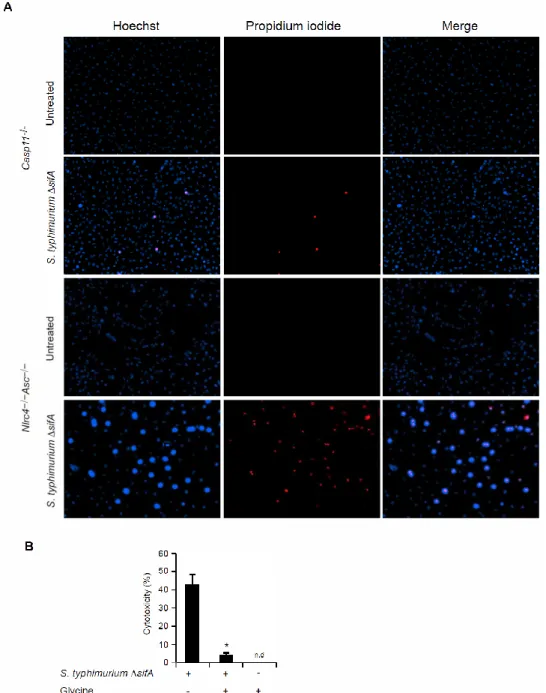
Fluorescence Microscopy
Nlrc4–/–Asc–/– and Casp11–/– BMMs were seeded onto glass cover slips in 24-well plates at a density of 2.78x10⁵ cells/well. Macrophage were primed with IFN-γ (8 ng/ml) overnight then infected with S. typhimurium ∆sifA. At 8 hours post infection, cells were washed with PBS and then incubated for 15 min on ice with Hoechst (0.12 µg/ml) and Propidium Iodide (PI) (1 µg/ml). Images were acquired using an EVOS florescent microscope.
Statistical analysis
FIGURES AND TABLES
Figure 2.1. Schematic of inflammasome detection pathways.
Figure 2.2. Burkholderia detection and protection conferred by Casp1/11 is independent of all known canonical inflammasomes.
Figure 2.3. Burkholderia protection conferred by Casp1/11 is independent of all known canonical inflammasomes.
(A-C) Wild type C57BL/6 or the indicated knockout mice were infected i.n. with B.
Figure 2.4. Diverse cytosolic bacteria activate pyroptosis independent of NLRC4, NLRP3 and ASC.
(A) LPS-primed BMMs were infected for 4h with either B. thailandensis or the indicated mutants and cytotoxicity was determined. BMMs were infected for 8h with (B) S. typhimurium or S. typhimurium ∆sifA, (D) S. typhimurium ∆sifA or S. typhimurium ∆sifA flgB (D) and
Figure 2.5. Caspase-11 mediates pyroptosis after infection by cytosolic bacteria. (A-I) Macrophage cytotoxicity and IL-1β secretion were determined after infection with S. typhimurium ΔsifA (8h), L. pneumophila ΔflaA ΔsdhA (4h), or B. thailandensis (4h). (A) C57BL/6, Casp1–/–Casp11–/–, Tlr4–/–, Trif–/–, and Myd88–/– BMM infected with S. typhimurium ∆sifA with or without IFN-γ priming prior to infection. (B and C) Retroviral transduction was used to complement Casp1 or Casp11 in Casp1–/–Casp11–/– iBMM. Macrophages were primed with LPS (B) or IFN-γ (C) and responses to B. thailandensis (B) or S. typhimurium ΔsifA (C) infection were examined. (D) Control or complemented Casp1–/–Casp11–/– BMM infected with L. pneumophila ΔflaA ΔsdhA. (E and F) Retroviral transduction was used to introduce control or Casp11-targeting shRNAmir into Nlrc4–/–Asc–/– iBMM. Macrophages were primed overnight with LPS (E) or IFN-γ (F) and then infected as indicated. (G to I) C57BL/6, Casp1–/–Casp11–/–, and Casp11–/– BMM infected with B. thailandensis (G), S. typhimurium ΔsifA (H), or L.
Figure 2.6. TLR ligands and IFN-γ enhance Casp11 expression and caspase-11- dependent cell death.
(A) Untreated, LPS-primed, or IFN-!-primed BMMs were infected with S. typhimurium ΔsifA and cytotoxicity was determined. (B) Transcriptional upregulation of Casp11 in C57BL/6 BMMs after priming with the indicated molecules was determined using Affymetrix GeneChip
technology. (C) Caspase-11 expression in untreated, LPS-primed, and IFN-γ-primed C57BL/6 BMMs was determined by immunoblot. Blots were stripped and β-actin expression was
determined as a loading control. (D) Caspase- 11 expression in untreated, LPS-primed, and IFN-γ-primed control or Casp11 shRNAexpressing Nlrc4-Asc iBMMs was determined by
Figure 2.7. Caspase-11 is not required for pyroptosis induced by flagellin expressing wild type L. pneumophila.
Figure 2.8. Morphology of S. typhimurium ΔsifA-induced pyroptosis.
Figure 2.9. Caspase-11 protects against cytosolic bacteria in vivo.
(A and B) S. typhimurium or S. typhimurium ∆sifA were injected i.p. into C57BL/6 (1000 cfu) or Casp1–/–Casp11–/– mice (250 cfu) and survival was monitored. (C to E) The indicated mice were infected with 5x10⁴ cfu of both wild type S. typhimurium and S. typhimurium ∆sifA marked with ampicillin or kanamycin resistance, respectively. Bacterial loads from 3-4 mice per genotype were determined 48h later and competitive index calculated (CI = log (S. typhimurium ∆sifA/ S. typhimurium)). A CI of −1 corresponds to 10 cfu of S. typhimurium for every 1 cfu of S.
Name of Strain Designation Notes Reference S. typhimurium ATCC 14028s wild type www.atcc.org S. typhimurium CS401 14028s strepR,
phoN::Tn10dCm
Samuel I Miller S. typhimurium ∆sifA JAF57 CS401 ∆sifA (42)
S. typhimurium ∆sifA flgB
JAF57 flgB CS401 ∆sifA
flgB44::Tn10
S. Yamaguchi B. thailandensis E264 wild type www.atcc.org B. thailandensis E264-1 mouse passaged wild
type
this study B. thailandensis T3SS
mutant
Joseph Mougous B. thailandensis T6SS
mutant
Joseph Mougous B. pseudomallei Bp340 1026b amrRAB-oprA (43)
B. pseudomallei ∆purM
Bp82 purine auxotroph (44)
L. pneumophila LP02 wild type;
Philad elp hia-1 rpslL hsdR thyA
-(46)
L. pneumophila ∆flaA flagellin mutant (47) L. pneumophila ∆flaA
∆sdhA
flagellin, sdhA mutant (48)
Plasmids Resistance Notes
pWSK29 Amp Low copy vector (45)
pWSK129 Kan Low copy vector (45)
Figure Mice genotype Number of mice
Fig.1C C57Bl/6 21
Nlrc4−/− 15
Casp1−/−Casp11−/− 21
Fig.1d Nlrc4−/−Asc−/− 10
Casp1−/−Casp11−/− 5
Fig.1E C57Bl/6 10
Il1b−/−Il18−/− 10
Casp1−/−Casp11−/− 10
Fig.4A C57Bl/6 – S. typhimurium 10
C57Bl/6 – S. typhimurium ∆sifA 9 Fig.4B Casp1−/−Casp11−/− – S. typhimurium 9 Casp1−/−Casp11−/− – S. typhimurium ∆sifA 9
Fig.4F C57Bl/6 7
Casp1−/−Casp11−/− 16
Casp11−/− 17
Fig.4G C57Bl/6 8
Casp1−/−Casp11−/− 10
Casp11−/− 11
Fig. 2.3A C57Bl/6 5
Casp1−/−Casp11−/− 5
Fig. 2.3B C57Bl/6 5
Casp1−/−Casp11−/− 8
Nlrc4 10
Asc−/− 7
Nlrp3−/− 9
Nlrc4−/−Asc−/− 9
Fig. 2.3C Il1b−/−Il18−/− 19
Casp1−/−Casp11−/− 10
REFERENCES
1. E. A. Miao, J. V. Rajan, A. Aderem, Caspase-1-induced pyroptotic cell death. Immunol. Rev. 243, 206 (2011).
2. L. Franchi, R. Muñoz-Planillo, G. Núñez, Sensing and reacting to microbes through the inflammasomes. Nat. Immunol. 13, 325 (2012).
3. P. Broz, J. von Moltke, J. W. Jones, R. E. Vance, D. M. Monack, Differential requirement for Caspase-1 autoproteolysis in pathogen-induced cell death and cytokine processing. Cell Host Microbe 8, 471 (2010).
4. E. A. Miao et al., Innate immune detection of the type III secretion apparatus through the NLRC4 inflammasome. Proc. Natl. Acad. Sci. U.S.A. 107, 3076 (2010).
5. Y. Zhao et al., The NLRC4 inflammasome receptors for bacterial flagellin and type III secretion apparatus. Nature 477, 596 (2011).
6. E. M. Kofoed, R. E. Vance, Innate immune recognition of bacterial ligands by NAIPs determines inflammasome specificity. Nature 477, 592 (2011).
7. W. J. Wiersinga, B. J. Currie, S. J. Peacock, Melioidosis. N. Engl. J. Med. 367, 1035 (2012). 8. I. Ceballos-Olvera, M. Sahoo, M. A. Miller, L. Del Barrio, F. Re, Inflammasome-dependent pyroptosis and IL-18 protect against Burkholderia pseudomallei lung infection while IL-1β is deleterious. PLoS Pathog. 7, e1002452 (2011).
9. W. J. Wiersinga, T. van der Poll, N. J. White, N. P. Day, S. J. Peacock, Melioidosis: Insights into the pathogenicity of Burkholderia pseudomallei. Nat. Rev. Microbiol. 4, 272 (2006). 10. N. Kayagaki et al., Non-canonical inflammasome activation targets caspase-11. Nature 479, 117 (2011).
11. E. A. Miao, J. V. Rajan, Salmonella and Caspase-1: A complex interplay of detection and evasion. Front Microbiol 2, 85 (2011).
12. N. Schroeder, L. J. Mota, S. Méresse, Salmonella-induced tubular networks. Trends Microbiol. 19, 268 (2011).
13. S. Shoma et al., Critical involvement of pneumolysin in production of interleukin-1alpha and caspase-1-dependent cytokines in infection with Streptococcus pneumoniae in vitro: a novel function of pneumolysin in caspase-1 activation. Infect. Immun. 76, 1547 (2008).
15. C. R. Beuzón et al., Salmonella maintains the integrity of its intracellular vacuole through the action of SifA. EMBO J. 19, 3235 (2000).
16. E. A. Creasey, R. R. Isberg, The protein SdhA maintains the integrity of the Legionella-containing vacuole. Proc. Natl. Acad. Sci. U.S.A. 109, 3481 (2012).
17. J. Ge, Y.-N. Gong, Y. Xu, F. Shao, Preventing bacterial DNA release and absent in melanoma 2 inflammasome activation by a Legionella effector functioning in membrane trafficking. Proc. Natl. Acad. Sci. U.S.A. 109, 6193 (2012).
18. J. van der Heijden, B. B. Finlay, Type III effector-mediated processes in Salmonella infection. Future Microbiol. 7, 685 (2012).
19. L. Franchi et al., Cytosolic flagellin requires Ipaf for activation of caspase-1 and interleukin 1β in salmonella-infected macrophages. Nat. Immunol. 7, 576 (2006).
20. E. A. Miao et al., Cytoplasmic flagellin activates caspase-1 and secretion of interleukin 1β via Ipaf. Nat. Immunol. 7, 569 (2006).
21. E. A. Miao et al., Caspase-1-induced pyroptosis is an innate immune effector mechanism against intracellular bacteria. Nat. Immunol. 11, 1136 (2010).
22. P. Gurung et al., Toll or interleukin-1 receptor (TIR) domain-containing adaptor inducing interferon-β (TRIF)-mediated caspase-11 protease production integrates Toll-like receptor 4 (TLR4) protein- and Nlrp3 inflammasome-mediated host defense against enteropathogens. J. Biol. Chem. 287, 34474 (2012).
23. P. Broz et al., Caspase-11 increases susceptibility to Salmonella infection in the absence of caspase-1. Nature 490, 288 (2012).
24. V. A. K. Rathinam et al., TRIF licenses caspase-11-dependent NLRP3 inflammasome activation by gram-negative bacteria. Cell 150, 606 (2012).
25. A. Akhter et al., Caspase-11 promotes the fusion of phagosomes harboring pathogenic bacteria with lysosomes by modulating actin polymerization. Immunity 37, 35 (2012).
26. J. von Moltke et al., Rapid induction of inflammatory lipid mediators by the inflammasome in vivo. Nature 490, 107 (2012).
27. S. Mariathasan et al., Differential activation of the inflammasome by caspase-1 adaptors ASC and Ipaf. Nature 430, 213 (2004).
28. S. Mariathasan et al., Cryopyrin activates the inflammasome in response to toxins and ATP. Nature 440, 228 (2006).
30. K. Takeda et al., Defective NK cell activity and Th1 response in IL-18-deficient mice. Immunity 8, 383 (1998).
31. K. Kuida et al., Altered cytokine export and apoptosis in mice deficient in interleukin-1 beta converting enzyme. Science 267, 2000 (1995).
32. D. K. Dalton et al., Multiple defects of immune cell function in mice with disrupted interferon-gamma genes. Science 259, 1739 (1993).
33. C. K. Huang, L. Zhan, M. O. Hannigan, Y. Ai, T. L. Leto, P47(phox)-deficient NADPH oxidase defect in neutrophils of diabetic mouse strains, C57BL/6J-m db/db and db/+. J. Leukoc. Biol. 67, 210 (2000).
34. E. A. Miao, J. A. Freeman, S. I. Miller, Transcription of the SsrAB regulon is repressed by alkaline pH and is independent of PhoPQ and magnesium concentration. J. Bacteriol. 184, 1493 (2002).
35. B. Byrne, M. S. Swanson, Expression of Legionella pneumophila virulence traits in response to growth conditions. Infect. Immun. 66, 3029 (1998).
36. T. Decker, M. L. Lohmann-Matthes, A quick and simple method for the quantitation of lactate dehydrogenase release in measurements of cellular cytotoxicity and tumor necrosis factor (TNF) activity. J. Immunol. Methods 115, 61 (1988).
37. S. E. Warren et al., Cutting edge: Cytosolic bacterial DNA activates the inflammasome via Aim2. J. Immunol. 185, 818 (2010).
38. T. Kitamura et al., Retrovirus-mediated gene transfer and expression cloning: Powerful tools in functional genomics. Exp. Hematol. 31, 1007 (2003).
39. R. G. Hawley, F. H. Lieu, A. Z. Fong, T. S. Hawley, Versatile retroviral vectors for potential use in gene therapy. Gene Ther. 1, 136 (1994).
40. R. A. Irizarry et al., Exploration, normalization, and summaries of high density oligonucleotide array probe level data. Biostatistics 4, 249 (2003).
41. R. C. Gentleman et al., Bioconductor: Open software development for computational biology and bioinformatics. Genome Biol. 5, R80 (2004).
42. J. A. Freeman, M. E. Ohl, S. I. Miller, The Salmonella enterica serovar typhimurium translocated effectors SseJ and SifB are targeted to the Salmonella-containing vacuole. Infect. Immun. 71, 418 (2003).
43. T. Mima, H. P. Schweizer, The BpeAB-OprB efflux pump of Burkholderia pseudomallei 1026b does not play a role in quorum sensing, virulence factor production, or extrusion of
44. K. L. Propst, T. Mima, K.-H. Choi, S. W. Dow, H. P. Schweizer, A Burkholderia
pseudomallei deltapurM mutant is avirulent in immunocompetent and immunodeficient animals: Candidate strain for exclusion from select-agent lists. Infect. Immun. 78, 3136 (2010).
45. R. F. Wang, S. R. Kushner, Construction of versatile low-copy-number vectors for cloning, sequencing and gene expression in Escherichia coli. Gene 100, 195 (1991).
46. K. H. Berger, R. R. Isberg, Two distinct defects in intracellular growth complemented by a single genetic locus in Legionella pneumophila. Mol. Microbiol. 7, 7 (1993).
47. T. Ren, D. S. Zamboni, C. R. Roy, W. F. Dietrich, R. E. Vance, Flagellin-deficient
Legionella mutants evade caspase-1- and Naip5-mediated macrophage immunity. PLoS Pathog. 2, e18 (2006).
CHAPTER 3: CYTOPLASMIC LPS ACTIVATES CASPASE-11: IMPLICATIONS IN TLR-4 INDEPENDENT ENDOTOXIC SHOCK5
SUMMARY
Inflammatory caspases, such as caspase-1 and -11, mediate innate immune detection of pathogens. Caspase-11 induces pyroptosis, a form of programmed cell death, and specifically defends against bacterial pathogens that invade the cytosol. During endotoxemia, however, excessive caspase-11 activation causes shock. We report that contamination of the cytoplasm by lipopolysaccharide (LPS) is the signal that triggers caspase-11 activation in mice. Specifically, caspase-11 responds to penta- and hexa-acylated lipid A, whereas tetra-acylated lipid A is not detected, providing a mechanism of evasion for cytosol-invasive Francisella. Priming the caspase-11 pathway in vivo resulted in extreme sensitivity to subsequent LPS challenge in both wild type and Tlr4-deficient mice, whereas caspase 11-deficient mice were relatively resistant. Together, our data reveal a new pathway for detecting cytoplasmic LPS.
5
This chapter contains a manuscript that previously appeared as an article in Science. This is the author's version of the work. It is posted here by permission of the AAAS for personal use, not for redistribution. The definitive version was published in Science 341, 2013, doi:
10.1126/science.1240988. The full citation is: Hagar, J.A., Powell, D.A., Aachoui, Y., Ernst, R.K., Miao, E.A. Cytoplasmic LPS activates caspase-11: implications in TLR4-independent endotoxic shock. Science. 341, pp.1250-1253, 2013.
CYTOPLASMIC LPS ACTIVATES CASPASE-11: IMPLICATIONS IN TLR-4 INDEPENDENT ENDOTOXIC SHOCK
Caspases are evolutionarily ancient proteases that are integral to basic cellular
physiology. Although some caspases mediate apoptosis, the inflammatory caspases-1 and -11 trigger pyroptosis, a distinct form of lytic programmed cell death. In addition, caspase-1 processes IL-1 and IL-18 to their mature secreted forms. Caspase-1 is activated by the
canonical inflammasomes, which signal via the adaptor ASC; NLRC4 and NLRP1a/1b can additionally activate caspase-1 directly (1, 2). In contrast to caspase-1, caspase-11 is activated independently of all known canonical inflammasome pathways; the hypothetical caspase-11 activating platform has been termed the non-canonical inflammasome (3). Casp1–/– mice generated from 129 background stem cells are also deficient in Casp11 due to a passenger mutation backcrossed from the 129 background into C57BL/6. Caspase-11 is responsible for certain phenotypes initially attributed to caspase-1, such as shock following endotoxin challenge (3). The physiologic function of caspase-11 is to discriminate cytosolic from vacuolar bacteria (4). In the absence of caspase-11, mice become acutely susceptible to infection by bacteria that escape the phagosome and replicate in the cytosol (4), such as Burkholderia pseudomallei and B. thailandensis. Caspase-11 also responds to vacuolar Gram-negative bacteria, albeit with delayed kinetics (3, 5-7), which may have relevance to its aberrant activation during sepsis. Although these studies demonstrated both detrimental and protective roles for caspase-11, the precise nature of the caspase-11 activating signal remained unknown.