GESTATIONAL AND LACTATIONAL α-LINOLENIC ACID
AVAILABILITY INDUCES EPIGENETIC CHANGES IN THE BRAIN OF
MOUSE OFFSPRING
Fuli He
A thesis submitted to the faculty of the University of North Carolina at Chapel Hill in partial fulfillment of the requirements for the degree of Master of Science in the Department of
Nutrition, Gillings School of Global Public Health.
Chapel Hill 2013
Approved by:
ii © 2013 Fuli He
iii
Abstract
FULI HE: Gestational and Lactational α-Linolenic Acid Availability Induces Epigenetic Changes in the Brain of Mouse Offspring
(Under the direction of Dr. Mihai Niculescu)
It has been acknowledged that dietary n-3 fatty acids improve memory and learning,
in part by altering gene expression in the brain. However, the exact mechanisms are far from
being clear, especially for α-linolenic acid (ALA). Inadequate perinatal nutrition can induce
persistent changes in offspring phenotype, acting through epigenetic process. We
investigated the effects of maternal ALA intakes on the epigenetic regulation of
memory-associated genes, in the offspring brains, at the end of lactation (postnatal day 19, P19).
Postnatal ALA supplementation altered mRNA expression of Mecp2, Ppp1cc, Reelin and
Dnmt3a, while ALA deficiency during gestation induced changes in methylation of CpG
sites in either promoter or intron1 of these genes. In addition, bivariate analysis indicated
some significant associations between CpG site-specific methylation and the expression of
Dnmt3a (p<0.05). In summary, the interplay between ALA availability during gestation and
lactation can differentially alter methylation and expression of genes involved in
neurogenesis and synaptic plasticity, potentially affecting brain development and
memory-related biological processes. However, further studies are needed to confirm the
iv
Acknowledgement
Foremost, I would like to express my sincere gratitude to my advisor Dr. Mihai
Niculescu for the continuous support throughout the last two years I have been in this
program, for his patience, motivation, enthusiasm, and immense knowledge. His guidance
helped me in all the time of research and writing of this thesis. Besides my advisor, I would
like to thank the rest of my thesis committee: Dr. Steven H. Zeisel and Dr. Andrew Swick,
for their encouragement, insightful comments, and hard questions. In particular, I am grateful
to my lab partner, Dr. Daniel Lupu, for his endless patience with my request for advice, the
stimulating discussions, and his encouragement in the last two years. I had a really good time
working with him at UNC NRI in Kannapolis. Also I want to thank all my friends,
particularly Madalina Lupu, Heather Zhao, Yan Ni, Qian Sun, Xiaomeng You and
Sheauching Chai, for the sincere help and guidance. Last but not least, I would like to thank
Michael Lee and my parents, for their unwavering support and love. I would not have been as
v
Table of Contents
List of Tables ... vii
Table of Figures ... viii
List of Abbreviations and Symbols... ix
Chapter I - Background ... 1
Introduction ... 1
Dietary n-3PUFAs and Health ... 2
Effects of Dietary n-3 PUFAs on Gene Expression in the Brain ... 4
Memory and Epigenetic Regulation ... 5
DNA Methylation in Memory Formation ... 7
DNA Methylation Marks in Memory ... 12
Hypothesis ... 12
Chapter II – Journal manuscript ... 1
Introduction ... 1
Materials and Methods ... 20
Results ... 29
Discussion ... 31
Chapter III- Discussion ... 41
Effects of Maternal ALA Availability on Gene Expression in the Offspring Brains ... 41
Effects of Maternal ALA Availability on DNA Methylation Machinery in the Offspring Brains ... 44
vi
vii
List of Tables
Table 1. Summary of epigenetic processes in memory and memory-associated
transcription ... 16
Table 2. Summary of target gene information ... 16
Table 3. Fatty acid composition of diets ... 27
viii
Table of Figures
Figure 1. Metabolism of n-6 and n-3 PUFAs. ... 14
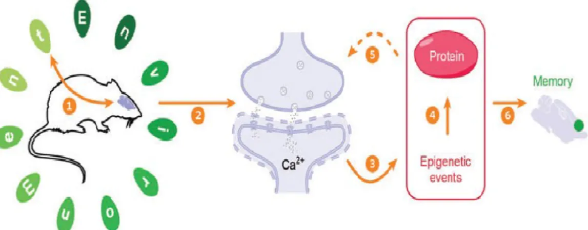
Figure 2. Summary of the basic structural and molecular events that take place during memory formation. ... 15
Figure 3. Study design ... 26
Figure 4. Gene expression assessment ... 35
Figure 5. Bisulfite pyrosequencing for the assement of gene CpG site in promoters or intron1 ... 36
Figure 6. Linear regression for Mecp2 ... 37
Figure 7. Linear regression for Ppp1cc ... 38
Figure 8. Linear regression of Reelin ... 39
ix
List of Abbreviations and Symbols
5-AZA 5-Aza-2-Deoxycytidine
5hmC 5-hydroxymethyl Cytosine
5mC 5-methyl Cytosine
AA Arachidonic Acid
ALA α-Linolenic Acid
ARC Activity-Regulated Cytoskeleton-associated protein
BDNF Brain-Derived Neurotrophic Factor
CFC Contextual Fear Conditioning
CNS Central Nervous System
CREB CAMP Responsive Element Binding protein 1
DHA Docosahexaenoic Acid
DNA Deoxyribonucleic Acid
DNMT DNA Methyltransferase
Egr1 Early Growth Response protein 1
EPA Eicosapentaenoic Acid
ERK Extracellular signal-Regulated Kinase
FA Fatty Acid
Fads2 Fatty Acid Desaturase 2
GADD45B Growth Arrest and DNA-Damage-inducible, beta
HDAC Histone Deacetylase
x
LTP Long-term Potentiation
MBD methyl binding domains
MeCP2 Methyl CpG Binding Protein 2
MS Mass Spectrometry
NMDA N Methyl D Aspartate
PP1 Protein Phosphatase 1
PPAR Peroxisome Proliferator Activated Receptor
PUFA Polyunsaturated Fatty Acid
RT-qPCR Quantitative reverse transcriptase Polymerase Chain Reaction
RNA Ribonucleic Acid
SREBP Sterol Regulatory Element Binding Protein
Chapter I - Background
Introduction
The mechanisms underlying long-term memory formation have been one of the topics
of interest recently. Adequate gene expression patterns are needed for this process, beginning
in fetal life. Most studies focused on the role of DNA binding transcription factors involved
in gene expression regulation. However, the epigenetic markings of chromatin, such as
histone modifications and DNA methylation in the brain, have been recently recognized as an
essential mechanism for brain functions, especially in learning and memory formation. The
epigenetic changes allow nerve cells not only to respond to environmental stimuli and
modulate their profile of gene expression, but also to establish and maintain their own
identity [1-3].
Adequate nutritional intakes during gestation and lactation are thought to be
important for offspring’s growth, especially the brain development. During this period, the
neonate’s brain is experiencing a substantial acceleration in growth, cellular proliferation,
and neuronal and glial differentiation [4]. Several studies have indicated that dietary omega-3
polyunsaturated fatty acids (n-3 PUFAs) are important nutrients for early life.
Supplementation of the lactating mothers with n-3 PUFAs was proved to increase children’s
2
It has been well documented that n-3 PUFAs improve memory and learning process.
However, the exact mechanisms are still far from being clear. Several lines of evidence
suggest that n-3 PUFAs affect gene expression in the brain, which can occur through
interactions with specific or nonspecific ligands that bind to response factors acting on
cis-regulatory elements of the genes. More recently, a clear understanding is developing
concerning the importance of epigenetic-related molecular mechanisms in
transcription-dependent long-term memory formation. Thus, in current study, we aimed to clarify whether
epigenetic mechanisms, specifically DNA methylation, are involved in the n-3 PUFAs
induced gene expression changes.
Dietary n-3PUFAs and Health
PUFAs and the their related complex lipids are important constituents of biological
membranes and contribute to maintaining the structural and functional integrity of cells and
cellular components [6, 7], especially docosahexaenoic acid (DHA; 22:6 n-3) and
arachidonic acid (AA; 20:4 n-6), as the fundamental components of membrane phospholipids
in neural cells. DHA and AA can be obtained directly from the diet or be converted for their
precursors α-linolenic acid (ALA; 18:3 n-3) or linoleic acid (LA; 18:2 n-6), respectively,
which must be obtained from dietary sources because most mammals lack the enzymes to
synthesize them (Figure 1).
ALA is the parent precursor for the n-3 PUFAs elongation and desaturation pathways.
This 18-carbon fatty acid possesses three double bonds and is commonly found in
plant-derived dietary oils such as flaxseed, canola, and soybean oils. All the other common dietary
n-3 PUFAs have longer chains, and are found mainly in fish and fish oils, including the
3
note that human and most other mammals express the enzymes necessary for the conversion
of dietary ALA to any of the other members of the n-3 PUFAs family. One human clinical
trial showed that consumption of ALA-enriched supplements for a 12-wk period was
sufficient to increase EPA and DHA [8].
It has been acknowledged that dietary n-3 PUFAs improve memory and learning, and
also alter the mRNA expression levels of several genes in the brain [9]. Short-term and
long-term feeding of ALA enriched diet to rats significantly improved spatial learning memory,
and altered proteome profiles in the hippocampus, including cytoskeleton and energy
metabolism protein, thereby affecting neurogenesis and synaptic plasticity [10]. Although the
beneficial health effects of n-3 PUFAs have been well documented, modern western diets
enriched in saturated fatty acids and simple carbohydrates are often deficient in n-3 PUFAs.
Thus, n-3 PUFAs are gaining ground in the supplement and functional food market. Most of
the food industry uses vegetable oils, such as flaxseed oil, due to low cost. Thus, it is of more
significance and practical to identify the role of ALA in brain development and cognition.
Flaxseed is a rich source of n-3 PUFAs, which includes a >50% ALA of total FAs
content in the seed. It has been used in human studies aimed at mitigating neurologic and
visual disturbances, folliculitis and growth retardation, and hemorrhagic dermatitis [11]. In
sows and their piglets, dietary supplementation with different forms of flax, such as seed or
oil, during late gestation and lactation has shown significant effects on immune resistance,
and effects on fatty acids (FAs) profiles as an increased n-3 FAs in maternal milk, carcasses,
and brain tissues in their newborn offspring [12]. Notably, a flaxseed diet during the perinatal
4
cognition. Thus, there must be caution in encouraging the maternal intake of flaxseed during
pregnancy and lactation [13].
Effects of Dietary n-3 PUFAs on Gene Expression in the Brain
As we mentioned, dietary n-3 PUFAs can modulate gene expression in the brain or
certain brain regions such as cerebrum and hippocampus [14, 15]. Some gene products, alone
or in combination with the membrane effects of these PUFAs, exert their beneficial effect on
neural functions such as learning and memory. Animal made deficient in n-3 PUFAs showed
a significant decrease in the levels of DHA in their neural tissue, and this loss of DHA was
accompanied by changes in neural function, including membrane-related events, metabolic
events and cellular events [14, 16].
The final FA composition of brain is determined during embryogenesis, particularly
during gestation and early postnatal life [4]. Thus, it is important to ensure the brain supplied
with adequate PUFAs for its functions during this time, since permanent metabolic or genetic
alterations can be initiated during perinatal period in the body and influence the individual’s
health later in life. Previous work from our lab found that maternal ALA availability during
lactation enhanced neurogenesis in the dentate gyrus (DG) of the offspring at postnatal day
19 (P19), and its beneficial effects were offset by maternal ALA deficiency during gestation
[17]. Kitajka, K., et al. also suggested that the supply of n-3 PUFAs during the perinatal
period influenced brain gene expression later in life. DNA microarray analysis showed the
expression of several genes being altered in the rat offspring brains fed n-3 PUFAs deficient
diets in the perinatal period, regardless of the n-3 PUFA-sufficient diets later in life. These
5
The fact that ALA and DHA activate several genes in other tissues, like liver or
adipose tissue, is well known [18-20], but the underlying molecular mechanisms of the direct
effects of n-3 PUFAs diet-induced gene-expression changes in the brain have been addressed
by very few studies [14, 21, 22]. Some studies suggest that PUFAs can directly interact with
transcription factors, like Peroxisome Proliferator Activated Receptor (PPARs), Sterol
Regulatory Element Binding Protein (SREBPs), which further induce the alteration in the
expression of target genes [9]. Interestingly, among all tested FAs, ALA has the highest
binding constant [23]. In current study, we investigated whether ALA could be related to
some other indirect mechanisms which regulate gene expression, such as DNA methylation.
Previously, we have found that maternal exposure to ALA during gestation and lactation
altered fatty acid desaturase 2 (Fads2) DNA methylation in both maternal and offspring
livers [24]. Groups receiving postnatal ALA supplement showed an increase in both Fads2
promoter and intron1 DNA methylation, which was negatively correlated with Fads2 gene
expression. We speculated that ALA supplementation could induce epigenetic changes in
other tissues, such as the brain.
Memory and Epigenetic Regulation
Memory is an abstract term that is not adequately or systematically defined. The most
frequently used definition of memory is that it is the process by which information is
encoded, stored, and retrieved. Specifically, a newly formed memory must first be acquired
(encoding) and then be converted to a more persistent state in a process called consolidation
(storing). Finally, stored memories are subject to retrieval when re-exposure to the initial
environmental stimulus (retrieving) [25]. It has been known that alterations in protein
6
all three stages. Within the neuron, synaptic depolarization activates complex molecular
signaling cascades that coalesce at specific gene loci, and causes acute modulation of
transcriptional efficacy. The resulting protein products are thought to produce stable
alterations in cellular phenotype by affecting the structure and physiology of postsynaptic
dendritic spines [26].
Epigenetic mechanisms have appeared as a central process in learning and memory
(Figure 2). Traditionally, epigenetic mechanisms have been defined as a set of stable and
heritable molecular phenomena that modify gene expression and do not involve alterations of
the DNA sequence of a cell [27]. However, since epigenetic marks can be transient and are
not necessarily heritable, especially when applied to lasting behavioral and cellular memory,
a less comprehensive, but more appropriate definition has been brought up: “the structural
adaptation of chromosomal regions so as to register, signal or perpetuate altered activity
states” [28]. Due to their critical involvement in gene regulation, epigenetic mechanisms
represent an attractive and reversible means for the brain to respond and adapt to specific
environmental changes.
Epigenetic mechanisms are essential to normal development, as they provide the
cellular memory necessary for perpetuating the correct cellular phenotype during mitosis.
Epigenetic mechanisms participate in brain formation and regulate cell physiology and
behavior, including learning and memory, drug addiction, depression and long-term
responses to maternal care [3, 29, 30]. It is important to mention that certain memory tasks
are associated with multiple regions, which may reflect temporally and spatially distinct
7
occur in the hippocampus, after which many are subsequently incorporated into and
maintained in cortical areas [31].
Epigenetic mechanisms include DNA methylation, histone modifications and RNA
interference, all of which are inter-related [32]. Numerous experiments have been carried out
to investigate the activity- and experience-dependent epigenetic mechanisms in the brain,
which are outlined in Table 1. Most research targets the regulation of memory by histone
modifications. For instance, artificially elevating levels of histone acetylation using histone
deacetylase (HDAC) inhibitors enhances induction of long-term potentiation (LTP) in vitro
and formation of long-term memory in vivo. Moreover, acetylation and phosphorylation of
histone H3 are increased in vitro and vivo in the hippocampus following activation of
NMDA receptors and extracellular signal-regulated kinase (ERK) [33, 34].
In this study, we were interested in another epigenetic mechanism, DNA methylation,
that might be involved in perinatal brain development related to the process of memory
formation, since a more recent and burgeoning field is discovering evidence implicating
DNA methylation in mammalian performance (learning and memory) in behavioral tasks.
DNA Methylation in Memory Formation
DNA methylation is one of the primary epigenetic mechanisms for heritable gene
silencing and regulation of gene expression. It has been show to play essential roles in
genomic imprinting, X chromosome inactivation and maintenance of genome stability [2,
35]. This covalent modification of DNA involves conversion of cytosines at CpG
dinucleotides to 5-methylcytosine (5mC). Approximately 60-70% of CpGs in the mammalian
8
of promoters), which are called CpG islands. Predominantly, CpG islands are kept in a
demethylated state and function in part to regulate local gene expression [36]. The
hypermethylation in CpG islands in promoter is usually associated with repression of the
gene transcription. There are two models most acceptable in gene suppression effect:
a)methyl cytosine prevents transcriptional activators from binding to the promoter region, or
b)attracts transcriptional repressors that have methyl binding domains (MBD), such as
methyl CpG binding protein 2 (Mecp2). These either directly influence transcriptional
efficiency or indirectly affect chromatin structure by recruiting HDACs [37]. These two
models have been complemented by recent studies which described active de-methylation
via5-hydroxymethylcytosine (5hmC) [38, 39].
Dynamic DNA methylation events have been reported in the adult brain, particularly
in the hippocampus. During learning, synaptic activity at hippocampal neurons initiates
several signaling cascades, with changes occurring locally at the synapse in addition to the
nucleus where transcriptional changes are made to plasticity-related proteins. These changes
are supported, at least in part, by epigenetic modifications, including DNA methylation [40].
There are three DNA methyltransferases (DNMTs), the “maintenance
methyltransferase” Dnmt1 and the “de novo methyltransferases” DNMT3a and DNMT3b,
that have functional enzymatic activity in mammals. DNMT1 has a substrate preference for
hemi-methylated DNA over unmethylated and interacts with the DNA replication machinery
during the S phase of dividing cells. DNMT3a and DNMT3b can methylate the unmethylated
cytosine of CpG dinucleotides on both strands, altering the epigenetic information content.
These de novo methyltransferases play vital roles in establishing new genomic methylation
9
neurons. Importantly, the three DNMT genes display differential expression profiles in the
central nervous system (CNS). DNMT1 is highly expressed in neurons from embryogenesis
through adulthood [42, 43]. DNMT3b expression is observed in neural progenitor tissue only
during early embryogenesis. DNMT3a is expressed from late embryogenesis to adulthood
with a peak during the early postnatal period, then its level declines but remains detectable in
the post-mitotic neurons of the adult brain [44]. These findings raised the possibility that
DNA methylation patterns might be involved in dynamic changes in the brain for yet
unspecified roles.
Alterations of neuronal gene expression patterns are required for long-term memory
formation or for synaptic plasticity – the ability to change the strength of synaptic
connections – in the CNS [45]. Since DNA methylation is required in neural activity-induced
transcriptional changes, methylation might be of importance in the process of memory
formation. However, whether the subsequent alteration of memory-associated gene
expressions occur within the hippocampus in response to direct changes in DNA methylation
of genes has yet to be determined.
In vitro, methylation inhibitor, zebularine and 5-aza-2-deoxycytidine (5-AZA) were
used to inhibit DNA methyltransferase activity in the hippocampal slices of sacrificed mice.
After inhibition, the promoter regions of the memory enhancer genes, Reelin and
brain-derived neurotrophic factor (Bdnf), induced rapid and dramatic decreases in DNA
methylation, which was speculated to contribute to the failure of LTP in the hippocampal
10
Further, Milier and Sweatt carried out an in vivo study to better illustrate that DNA
methylation was crucial and necessary in memory formation [47].The researchers induced a
group of adult male Spragure-Dawley rats with a contextual fear conditioning (CFC) test.
After sacrificing, the DNA methylation pattern of the memory suppressor gene (protein
phosphatase 1 (Pp1) and the memory enhancer gene Reelin were analyzed in the
hippocampus. They found a rapid methylation and down-regulation of Pp1, along with the
demethylation and activation of Reelin after CFC. However, 5-AZA and zebularine infusion
into the hippocampus immediately reversed the methylation patterns of these two genes,
indicating a potential role in the dysfunction of long-term memory formation. In addition,
with a similar methodology and the same type of rats, alterations were found in chromatin
structure and in methylation, which affected differential regulation of the Bdnf gene
expression, again suggesting the critical role of DNA methylation in the formation of
memory [48].
More recently, Dnmt1 and Dnmt3a double conditional knockout mice were
engineered to illustrate that DNA methylation is required for long-term memory [2]. Results
showed that the complete loss of gene expression of these two proteins in forebrain neurons
led to an abnormal methylation pattern and deregulated expression of some other genes in the
same area, causing deficits in the ability to learn and memorize. The neurons appeared
smaller than those seen in the wild-type mice, though a neuronal loss was not identified.
It is important to note that DNA methylation and histone acetylation may work
together to regulate long-term memory formation and synaptic plasticity. MeCP2 has been
suggested to function as the media that connects DNA methylation and histone acetylation
11
reported [50, 51]. The dominant understanding is that the MeCP2 protein binds to DNA that
has been methylated. Once bound, MeCP2 can interact directly with HDAC1 and 2 through
its transcriptional repressor domain, suggesting that DNA methylation at certain loci
indirectly silences gene expression through chromatin modification. However, a recent
epigenomic analysis suggested this might not be the case [39].
MeCP2 is found in high concentrations in neurons and is associated with maturation
of the CNS and in forming synaptic contact. Its mutations can cause Rett Syndrome, a
debilitating neuro developmental disorder associated with learning and memory deficits from
a young age. It is likely that the inhibition of MeCP2 would mimic the effect of DNMT
blockade. Actually, deletion of Mecp2 resulted in deficits in paired-pulse facilitation, a form
of short-term plasticity [52], while over-expression of Mecp2 led to enhanced short-term
plasticity [51]. It is important to note that the gain-of-function genetic variation in MeCP2
can also lead to MeCP2 duplication syndrome, which indicates that precise regulation of
MeCP2 is a key requirement for the neuronal homeostasis.
The key players involved in dynamic DNA demethylation changes in neurons in the
context of memory formation and storage remain unknown at present. Interestingly, Dnmt3a
and Dnmt3b have been also implicated in active demethylation of DNA [53, 54]. In addition,
another study found that the growth arrest and DNA damage-inducible protein 45 (Gadd45)
family contributed to active DNA demethylation utilizing a mammalian cDNA expression
library and methylated reported constructs [55]. Later, the isoform Gadd45b was reported to
be robustly up-regulated in response to cell depolarization, indicating its function associated
12
DNA Methylation Marks in Memory
DNA methylation has primarily been considered as a transcriptional regulator that
mediates gene expression. However, its regulation and function are likely to be far more
complex in the brain. As mentioned above, DNMT inhibitors have significant effects on
memory formation and storage and behavioral responses to drugs of abuse as well. The
ability of DNA methylation to affect these behaviors raises the question whether those genes,
which are important regulators of these experiences, contain active sites for methylation
and/or demethylation of DNA. In fact, a large number of genes that have previously been
shown to positively and negatively modulate behavioral memory contain dense CpG islands
surrounding their promoter regions. Thus, these genes are potential targets for changes in the
machinery that underlies DNA methylation in neurons.
In the current study, we selected 12genes that were previously reported to be
associated with consolidation of synaptic plasticity or/and long-term memory formation,
including 4 DNA methylation regulatory/machinery genes, Dnmt1, Dnmt3a, Gadd45b and
Mecp2 (Table 2)
Hypothesis
Several studies found that short-term and long-term feeding of ALA-enriched diets
significantly improved memory and learning processes, and also altered mRNA expression
levels of several genes in brain, thereby affecting neurogenesis and synaptic plasticity.
However, most of the studies were conducted in the adult animal models. Importantly, the
neonate’s brain experiences growth spurt during gestation and lactation, thus the perinatal
13
Our previous work revealed that the maternal ALA during gestation and lactation can
alter the hippocampal development in the male offspring at the end of lactation at P19. As a
continuation of this study, we aimed to determine the potential influence of ALA availability
during gestation and lactation on brain development of male offspring, specifically on the
14
Figure 1.Metabolism of n-6 and n-3 PUFAs. (adapted from [57] )
15
Figure 2.Summary of the basic structural and molecular events that take place during memory formation. (adapted from [31] )
16
Table 1. Summary of epigenetic processes in memory and memory-associated transcription
Mechanism Findings Histone
acetylation HDAC activity was negatively associated with hippocampus (especially HDAC2) and cortex-dependent memory in mammals via CREB: CBP-dependent transcriptional activation, and may operate through induced of Nr4a1 and Nr4a2 transcription factors [58]
HDAC inhibition promoted associative memory consolidation and H3 acetylation in Chasmagnathus [59]
Contextual fear conditioning promoted H3 acetylation while latent inhibition training induces H4 acetylation [60]
Over-expression HDACs in the nucleus accumbens impaired conditioned place preference for cocaine, while HAD inhibitors increase cocaine place preference [61]
Histone
phosphorylation
H3S10 phosphorylation was associated with impaired spatial and contextual memory by deletion of msk2, and 2 impaired forced swim memories with deletion of Msk1 [62]
Activation of ERK/MAPK in vitro and contextual fear conditioning in vivo led to an increased histone H3 phosphorylation in hippocampal area [33]
Histone methylation
Over-expression of the histone methyltransferase G9a in the nucleus accumbens impaired conditioned place preference, and opposite result was found in knockdown of G9a [63]
Contextual fear conditioning enhanced H3K4me3 and H3K9me2 marks, regulating genes including Egr1 and Bdnf [64]
DNA methylation Deletion of Mbd1 and Mecp2 impaired long-term memory in rodents [52]
DNMT inhibition impaired long-term memory consolidation in the hippocampus and remote contextual fear memory storage in the cortex
Fear conditioning induced dynamic alterations in DNA methylation at loci including Reelin, Ppp1cb, Bdnf and Egr1 in the hippocampus during memory consolidation [47, 48]
DNMT inhibition or knockout in the nucleus accubens increased development of conditioned place preference for cocaine, whereas over-expression of Dnmt3a reduced cocaine place preference [65]
17
Table 2. Summary of target gene information
Gene Findings
DNA methylation modifiers
Dnmt1 DNMT inhibition and genetic deletion of dnmt1 impaired LTP
Dnmt3a DNMT inhibition and genetic deletion of dnmt3a impaired LTP
Over-expression of dnmt3a increased spine density in nucleus accumbens
Gadd45b Associated with activity-regulated DNA demethylation and postnatal neurogenesis
Mecp2 A transcriptional factor important for controlling gene expression through the interpretation and regulation of epigenetic markers
Mutation in Rett Syndrome
Deletion or truncation impaired LTP expression (reduction in paired-pulse ratio, increase in vesicle release probability, reduction in spontaneous neurotransmission, and enhanced inhibitory tone)
Over-expression led to enhanced short-term plasticity
Memory-associated genes
Arc Required for consolidation of synaptic plasticity and formation of long-term memory
Regulate endocytosis of AMPA receptors in response to synaptic activity[67]
Bdnf Major regulator of synaptic transmission and plasticity at adult synapses in many regions of the CNS[48]
Creb1 Enhanced expression in consolidation of long-term synaptic
facilitation and long-term excitability in sensory neurons of Aplysia.
Reeln Regulate postnatal neurogenesis and enhances spine hypertrophy and LTP
Egr1 Deficient mice have impairments in late phase hippocampal LTP and consolidation of some forms of long-term hippocampus- and
amygdala-dependent memory
Pp1 complex: Ppp1cb/Pp1β & Ppp1cc/Pp1γ
Inhibition of active mouse Protein Phosphatase 1 [Pp1] complex(es) increases LTP of mouse hippocampal tissue that is decreased by oligomeric AMYLOID β-42protein
A rapid methylation and down-regulation of Pp1 was found in mouse hippocampal region along with the demethylation and activation of
Reelin after fear conditioning
Chapter II – Journal manuscript
Introduction
Memory is the process that new information is first acquired (encoding), then
converted to a more persistent state in a process called consolidation (storing) and finally,
stored memories are subject to retrieval when re-exposure to the initial environmental
stimulus (retrieving) [25]. It has been known that alterations in protein synthesis, gene
expression and structural properties of neurons and synapses are required in all three stages
[26].
Several studies have indicated that dietary n-3 PUFAs can improve memory and
learning processes, and also alter the mRNA expression levels of several genes in the brain
[9]. It is important to ensure the brain supplied with adequate PUFAs during gestation and
early postnatal life, since the final FA composition of brain is determined and brain
experiences a growth spurt at this time [4]. Permanent metabolic or genetic alterations can
happen during perinatal period in the body that influence the individual’s health later in life.
Recent research has focused on the association between n-3 PUFAs and better cognitive
development in infancy or slower cognition decrease in the elderly [68]. One study reported
that rats with n-3 FA deficient diet achieved nearly the same level of brain DHA and spatial
19
they were given DHA supplements from birth or weaning. Another study from our lab
showed that in the mouse offspring on postnatal day 19, its brain development is
differentially affected by maternal availability of ALA during gestation and lactation [17].
n-3 PUFAs are gaining ground as dietary supplements due to their potential benefits
on health. There are many sources of n-3 PUFAs used for supplementation. However, most
of the food industry uses vegetable oils containing ALA, instead of fish oils which have
DHA and preformed EPA, because of low cost. Thus, it is of more practical significance to
investigate the effects and mechanisms of ALA on physical health.
Flaxseed is a great source of n-3 PUFAs, which includes a >50% ALA of total FA
present in the seed. It has been used in human studies aimed at mitigating neurologic and
visual disturbances, folliculitis and growth retardation, and hemorrhagic dermatitis [11]. In
sows and their piglets, dietary supplementation with different forms of flax, such as seed or
oil, during late gestation and lactation has shown significant effects on immune resistance,
and effects on fatty acids (FAs) profiles as an increased n-3 FAs in maternal milk, carcasses,
and brain tissues in their newborn offspring [12].
The underlying molecular mechanisms of the direct effects of PUFA diet-induced
gene-expression changes in the brain have been addressed by very few studies. Most studies
suggest that PUFA can directly interact with transcription factors, like PPAR, SREBPs, by
which further induce the alteration in the expression of target genes [9]. However, an
accumulation of recent studies has emphasized the importance of epigenetic mechanisms,
such as DNA methylation, playing a role in alteration of neuronal gene expression pattern,
20
DNA methylation is the covalent modification of DNA, involving conversion of
cytosines at CpG dinucleotides to 5-methylcytosine. Approximately 60-70% of CpGs in the
mammalian genome are highly methylated, except for the CpG-dense area in the vicinity of a
promoter, which is called CpG island. Predominantly, CpG islands are kept in a
demethylated state and function in part to regulate local gene expression [36]. A large
number of genes that have previously been shown to positively and negatively modulate
behavioral memory contain dense CpG islands surrounding their promoter regions. Thus,
these genes are potential targets for changes in the machinery that underlies DNA
methylation in neurons.
In the study, we hypothesized that maternal ALA availability during gestation and
lactation could induce the alterations in the expression of several memory-associated genes in
offspring brains, and these changes are related to epigenetic modifications, specifically DNA
methylation, in the CpG islands spanning the promoter and the intron1 of these genes
Materials and Methods
In order to investigate the effects of perinatal maternal ALA exposure on the
offspring’s brain, female C57BL/6J mice were mated with C57BL/6J males and allowed the
access to an ALA control diet containing soybean oil as fat, or an ALA deficient diet
containing corn oil as the only source of fat before and during gestation, which was followed
by either the same diet in gestation or an ALA sufficient diet containing flaxseed oil in the
lactation period. Male pup brains were snap-frozen in liquid nitrogen and stored at -80 °C on
postnatal day 19. DNA, RNA and protein were extracted from the frozen whole brain
samples for DNA methylation, gene expression and protein expression analysis, respectively.
21 Animals, Diets, and Tissue Collection
This study was approved by the University of North Carolina Institutional Animal
Care and Use Committee. This work is a continuation of our previously published, and a
detailed description can be found in previous published literature [17]. Briefly, mouse
C57BL/6J females (10 wk old; Jackson Laboratory, Bar Harbor, ME, USA) were randomly
assigned into two initial feeding groups (intervention 1, Figure 3.) for 30 days prior, and
during gestation. One group was fed with a defined control diet (AIN-93G, DYETS,
Bethlehem, PA) containing soybean oil as fat source (ALA control, C, n=12), while the other
group was given an AIG-93G modified diet (DYETS) with corn oil as the only source of
fatty acids (ALA deficient, D, n=12). After 30 days, the females were bred overnight with
males that were maintained at all times on the C diet. One day prior to delivery date, the two
groups of females were randomly split into two subgroups (intervention 2). Half from each
group remained on the same diet (n=6), and the other half were switched to a modified
AIN-93G diet containing flaxseed oil (ALA supplemented, S, n=6), until postnatal day 20 (P19,
considering the date of delivery as P0). On P19, all mothers and their pups were sacrificed.
The pups were decapitated, the brains were extracted, snap frozen in liquid nitrogen and
stored at -80 °C. In this study, the brain samples from the male pups were used for further
analysis. However, due to the lack of male pups, only n=5 male brains were included here in
the C-C, C-S and D-S group each.
The fatty acid composition of each diet is indicated in Table 3. Particularly, the diets
contained 18:2 n-6 (LA) and 18:3n-3 (ALA) with the following concentrations: D diet,
87.237 and 1.554 nmol/mg; C diet, 86.938 and 10.827 nmol/mg; S diet, 23.133 and 75.367
22 Real-time RT-PCR
RNA was extracted from the male offspring brains (all at P19) with QIAcube
instrument (Qiagen, Valencia, CA, USA) using AllPrep® DNA/RNA mini kit (Qiagen),
according to manufacturer’s protocol. After the concentration and quality were determined
with NanoPhotometer (Implen GmbH, Munich, Germany), Quantitative reverse transcriptase
PCR (qRT-PCR) method was used for the assessment of gene expression. First, cDNA
synthesis was performed using a QuantiTect reverse transcription kit (Qiagen), on an
Eppendorf Mastercycle ProS (Eppendorf, Hamburg, Germany). The second part was
performed on an Eppendorf Mastercycler Realplex2epGradient S real-time PCR cycler, using
the QuantiTect SYBR Green PCR kit (Qiagen). QuantiTect primer assays (Qiagen) were
purchased for the following genes: Dnmt1, Dnmt3a, Mecp2, Gadd45b, Ppp1cb, Ppp1cc,
Ppp3ca, Arc, Bdnf, Creb1, Reelin, Egr1 and 18s RNA (used as internal reference for each
sample). The real-time PCR reactions were run in triplicate, and data were retrieved as CT
values normalized to 18S and log2 transformed for subsequent statistical analysis. Final data
were expressed as ratios between sample, and the average of the C-C group for each gene.
Bisulfite Pyrosequencing
DNA from male offspring brains (all at P19) was extracted with QIAcube instrument
(Qiagen, Valencia, CA, USA) using AllPrep® DNA/RNA mini kit (Qiagen), following the
specific protocol. Up to 1μg DNA from each sample was treated with sodium bisulfate using
the same instrument and the corresponding protocol (EpiTect kit; Qiagen).
One Pyrosequencing DNA methylation assay was designed for Mecp2 using
23
directly purchased from Qiagen. These assays information, including the assay location,
sequence to analyze, PCR primers, sequencing primers, and PCR conditions are indicated in
Table 4. The amplification of templates was performed on an Eppendorf Mastercycler ProS
using PyroMark PCR kit following the recommended protocol (Qiagen). The PCR condition
was 95°C 15 min; 45 cycles of 94°C 30 s, 56°C 30 s, 72°C 30s; 72°C 10 min and 4°C hold.
The biotinylated strand of the amplified DNA was subjected to pyrosequencing on a
PyroMark MD machine (Qiagen), as described previously [69]. Each sample and sequence
were run in triplicate, and only pyrosequencing reactions that passed the quality test were
included in the analysis. For each sample and sequence, percentage methylated cytosine for
each CpG site was expressed as the average of triplicates, and the values across all sites were
averaged to express the average methylation across each sequence.
Western Blot
Male pup brains were homogenized in lysis buffer (1% sodium dodecil sulfate,
10mM sodium ortovanadate, and 10mM Tris-HCl). Total protein concentration for all
samples was determined using the Lowry protein assay [70]. Proteins were separated by
electrophoresis using polyacrylamide Tris-HCl gels (4-15%; Bio-rad Laboratories, Hercules,
CA, USA) and then blotted on nitrocellulose membranes (0.2micron; Thermo Scientific,
Rockford, IL, USA).
Immunodetection was carried out using SNAP i.d. Protein Detection System
(Millipore, Billerica, MA, USA) according to the dedicated protocol. This system uses
vacuum assisted fluid distribution to rapidly and thoroughly permeate the blot with
24
procedure. The membrane was blocked with 10x blocking buffer (Sigma) diluted with 0.1%
Tween-TBS (TBST) prior to incubation with primary antibody Mecp2 (sc-20700; Santa Cruz
Biotechnology) and Ppp1cc (#orb6773; Biorbyt Ltd) at a 1:200 concentration for ten
minutes. Following primary incubation, membranes were washed three times in TBST, and
incubated with the respective secondary at a 1:5000 concentration for another ten minutes.
Membranes were then washed three times in TBST, incubated in SuperSignal® West Pico
Chemiluminescent Substrate (Thermo Scientific) for five minutes and exposed to
Autoradiography Film (MIDSCI, Missouri, USA) for 40 seconds.
The second immunoblotting with β-Actin (Rabbit mAb HRP conjugate #5125, Cell
Signaling) for internal reference was performed with normal western blot procedure, after
determining that the membrane was properly stripped with Restore Plus Western Blot
Stripping Buffer (Thermo Scientific). Briefly, the membrane was blocked overnight at 4 °C
with gentle shaking and then incubated with β-Actin antibody at 1:1000 dilution in blocking
buffer for 2 hours at room temperature. The same luminal reagent and detection was
performed as we mentioned before. Semi-quantitative analysis was carried out using Image J
and normalized to β-Actin.
Statistical Analysis
All statistical analysis was carried out using SPSS 15.0 software (IBM). For DNA
methylaton, gene expression, protein expression data, results were analyzed for equal
variances, followed by 1-way ANOVA. For each variable, statistical significance between
25
addition, bivariate linear fit analysis was performed for the correlation between two
26
Figure 3. Study design
As described in Materials and methods, there were two internventions in the study. Intervention 1 consisted of the administration of specified diets for 30 days prior to and during pregnancy. Intervention 2 consisted of the administration of specified diets during lactation until P19, when dams and pups were sacrificed.
Table 3.Fatty acid composition of diets
D diet (nmol/mg diet) C diet (nmol/mg diet) S diet (nmol/mg diet)
14:0 Myristic 0.124 0.251 0.151
14:1 Myristoleic 0.011 0.020 0.021
16:0 Palmitic 19.651 19.613 9.628
18:0 Stearic 2.910 2.519 2.365
18:1 n9 Oleic 34.435 33.229 29.756
18:2 n6 LA 87.237 86.938 23.133
18:3 n3 ALA 1.554 10.827 75.367
20:0 Eicosanoic 0.638 0.795 0.222
20:1 n9 11-Eicosenoic 0.352 0.328 0.214
20:2 n6 11,14-Eicosadienoic 0.030 0.067 0.049
20:3 n6 Dihomo-gamma-linolenic ND ND 0.037
20:4 n6 AA ND 0.009 0.078
20:3 n3 11,14,17-Eicosatrienoic 0.187 0.559 0.200
22:0 Behenic 0.012 0.008 ND
22:1 n9 Erucic ND 0.007 ND
20:5 n3 EPA 0.012 0.024 0.013
24:0 Lignoceric 0.228 0.169 0.139
24:1 Nervonic ND ND 0.010
22:5 n3 7,10,13,16,19-Docosapentaenoic ND ND ND
22:6 n3 DHA ND ND ND
ND, not detected
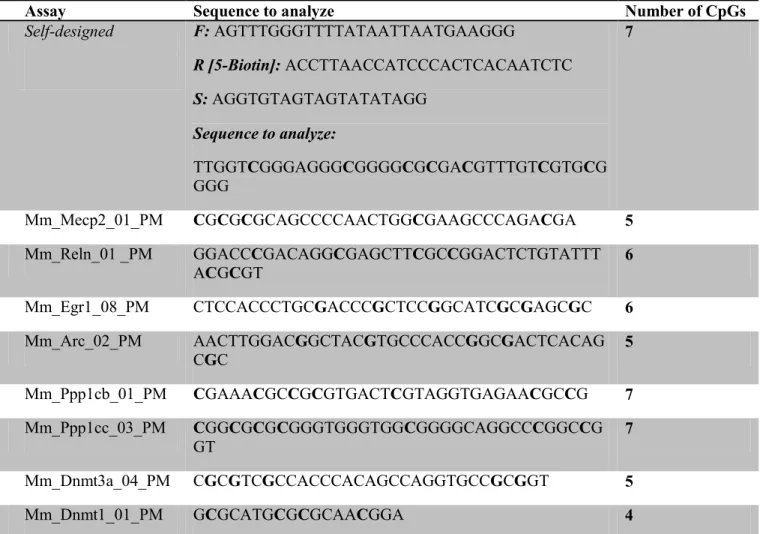
Table 4. Assay information for bisulfite pyrosequencing
Gene position Assay Sequence to analyze Number of CpGs
Mecp2 promoter Self-designed F: AGTTTGGGTTTTATAATTAATGAAGGG
R [5-Biotin]: ACCTTAACCATCCCACTCACAATCTC
S: AGGTGTAGTAGTATATAGG
Sequence to analyze:
TTGGTCGGGAGGGCGGGGCGCGACGTTTGTCGTGCG GGG
7
Mecp2intron1 Mm_Mecp2_01_PM CGCGCGCAGCCCCAACTGGCGAAGCCCAGACGA 5
Reelin promoter Mm_Reln_01 _PM GGACCCGACAGGCGAGCTTCGCCGGACTCTGTATTT ACGCGT
6
Egr1 promoter Mm_Egr1_08_PM CTCCACCCTGCGACCCGCTCCGGCATCGCGAGCGC 6
Arc exon1 Mm_Arc_02_PM AACTTGGACGGCTACGTGCCCACCGGCGACTCACAG CGC
5
Ppp1cb promoter Mm_Ppp1cb_01_PM CGAAACGCCGCGTGACTCGTAGGTGAGAACGCCG 7
Ppp1cc intron1 Mm_Ppp1cc_03_PM CGGCGCGCGGGTGGGTGGCGGGGCAGGCCCGGCCG GT
7
Dnmt3a promoter Mm_Dnmt3a_04_PM CGCGTCGCCACCCACAGCCAGGTGCCGCGGT 5
Dnmt1 promoter Mm_Dnmt1_01_PM GCGCATGCGCGCAACGGA 4
F: forward primer; R, reverse primer; S, sequencing Primer
29
Results
Maternal Exposure to ALA in Lactation Alters Expression of Memory Associated Genes and DNA Methylation Regulatory in Offspring Brains
qRT-PCR assays were performed to ascertain if memory-associated genes exhibited
altered expression in the brain of offspring of male mice with different dietary treatment
during gestation or lactation. Eight genes that have previously been shown to positively and
negatively modulate behavioral memory were chosen for analysis. All of them contain dense
CpG islands surrounding their promoter. Thus, these genes are potential targets for changes
in the machinery that underlies DNA methylation in memory formation. Significant changes
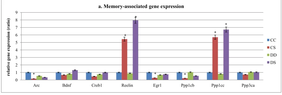
in transcript levels were detected for Arc, Reelin, Egr1, Ppp1cb, and Ppp1cc (Figure 4).ALA
supplementation during lactation induced Arc under-expression in CS group (80% decrease),
Reelin over-expression in CS (44% increase) and DS group (69% increase), Egr1 and
Ppp1cb under-expression in CS group (both around 75% decrease), and Ppp1cc
over-expression in CS (46% increase) and DS group (56% increase), as compared to CC group.
The mRNA expression was also determined in Dnmt1, Dnmt3a, Mecp2 and Gadd45b
(Figure 4b). Dnmt1 expression was suppressed in CS group (73% decrease), while Dnmt3a
was suppressed in both CS (97% decrease) and DS group (93% decrease) and Mecp2 was
over-expressed in CS (33% increase) and DS group (53% increase), when compared to CC
group. Those genes that did not exhibit significant differences between any two groups were
not further investigated.
30
Because mRNA expression differences were identified in several genes, as well as
DNA methylation machinery genes, we further determined whether the DNA methylation of
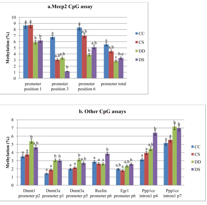
these genes in the promoter or/and intron1 was altered by any dietary treatment. ALA
deficiency during gestation induced a hypomethylation of Mecp2 promoter in the DD (45%
decrease) and DS (40% decrease) group, as compared to CC group (Figure 5a). Interestingly,
one CpG site (position 3) methylation was suppressed by postnatal ALA supplementation
(55% and 80% decrease in CS and DS groups, respectively). We also identified alterations in
several CpG sites in Dnmt1, Dnmt3a, Egr1, Reelin and Ppp1cc (Figure 5b).
We also performed bivariate fit analysis between Dnmt3a/Dnmt1 expression and the
methylation percentage of these CpG assays, so as to determine the potential role of Dnmt1
and Dnmt3a expression in the alterations in DNA methylation induced by maternal ALA
exposure. A significant positive correlation was found between the Dnmt3a gene expression
and position 3 (p3) CpG sites of Mecp2 promoter assay (R² = 0.243, p=0.022; Figure 6a), and
this CpG site was correlated moderately with Mecp2 gene expression negatively (R² = 0.136,
p=0.1; Figure 6b). Meanwhile, there was a negative correlation between Mecp2 and Dnmt3a
gene expression (R² = 0.375, p=0.004; Figure 6c). In addition, the methylation of another
CpG site in Mecp2 intron1 was negatively correlated with Dnmt1 mRNA expression (R² =
0.116, P=0.03; Figure 6d). Mecp2 was also reported to regulate gene expression of epigenetic
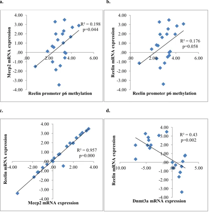
marks. The expression of Reelin was marginally correlated with its CpG site methylation in
promoter (R² = 0.176, p=0.058; Figure 8b) and Mecp2 expression (R² = 0.957, p=0.000;
Figure 8c), while negatively related with Dnmt3a expression (R² = 0.43, p=0.002; Figure 8d)
Similar to what we found between Mecp2 and Dnmt3a, there was a significant
31
0.331, p=0.006; Figure 7a), and the mRNA expression of Ppp1cc was negatively associated
with Dnmt3a expression (R² = 0.431, p=0.002; Figure 7c).
Mecp2 and Ppp1cc Protein levels do not correlate with Transcriptional Changes
We investigated whether the transcriptional changes would induce protein alteration
in the offspring brains. Western blot results showed that there was no significant difference
in Mecp2 and Ppp1cc protein levels between dietary treatments at the end of lactation (Figure
9).
Discussion
The findings of this study support the hypothesis that maternal ALA intake during
pregnancy and lactation alters the expression of memory-associated genes, specifically in the
offspring brain at P19, and that this is in part related with altered epigenetic regulation of the
gene transcription.
Our previous work has shown that postnatal maternal ALA supplementation
promoted neurogenesis in the hippocampus of the offspring [17]. The present findings
indicate that this effect could be a result of increased expression in Ppp1cc, Reelin and
Mecp2 in CS and DS group. Reelin is essential for neuronal lamination and synaptic
plasticity and a decreased transcript level of Reelin has been found as a result of
ALA-deficiency [71]. The increased mRNA level of Ppp1cc is also in agreement with a previous
report that its protein product, Pp1γ promotes neurogenesis by decreasing the
phosphorylation and ubiquitination of Creb in hyperbaric oxygen therapy (HBOT) [72].
32
the maintenance and development of the CNS. Studies suggest that loss- or gain-of Mecp2
function exert bidirectional control (decreased vs. increased) in neurotransmission in cortical
and hippocampal regions of the brain, probably through the regulation of synaptic gene
expression [73]. Our results showed a significant correlation between Mecp2 expression and
Ppp1cc, Reelin mRNA level, indicating that Ppp1cc and Reelin may be potential target genes
for Mecp2.
In contrast to the robust changes in expression, only few changes in CpG methylation
were detected. Different from mRNA expression, the DNA methylation patterns were
associated mostly with gestational ALA exposure, which suggested a particular important
role of DNA methylation during early stages of embryonic and fetal development. It was also
possible that other epigenetic mechanisms, such as histone modification, play a more
important role during lactation period. Also, the Dnmt3a and Dnmt1 expression did not
necessarily predicate DNA methylation, suggesting the speed methylation of response to
Dnmts may vary among different CpG sites. One limitation of our study is that we cannot
differentiate the exact roles of ALA supplementation during gestation vs. lactation, for the
lack of multiple time points, and a CD group (a control or supplemented diet during gestation,
followed by a deficient diet in lactation).
The less significant changes in methylation may also be caused by a small sample
size, and some limitations of the technique, especially the failure to separate 5hmC from
5mC by our current bisulfite-treated pyrosequencing. 5hmC is a marker for DNA
demethylation. The presence of 5hmC in promoter regions was associated with high levels of
transcription. More recently, Mecp2 was identified as the major 5hmC-binding protein in the
33
could speculate, for instance, that Mecp2 binds to 5hmC in Reelin promoter, and in turn
regulates its gene expression, since one CpG site was positively correlated with Reelin and
Mecp2 expression, while being negatively associated with Dnmt3a levels. This should be
further validated by a complementary technique and be applied to a larger sample size, such
MS-based approaches and Tet-assisted bisulfite sequencing (TAB-seq) that can detected
5hmC [74].
Although western blot results did not detect differences in Mecp2 and Ppp1cc protein
level, it does not necessarily mean that their bioactivity has not been altered. More
quantitative and specific assays should be performed. It is also important to note another
limitation in the study: all the results were obtained using the whole brain of offspring.
Evidence has indicated that gene expression differs across brain regions [75]. Studies using
whole brains could result in false–negative results, owing to a dilution effect. Different brain
areas are thought to be involved in specific types of memory. For example, the hippocampus
is thought to be responsible for the spatial learning, while the amygdale is believed to
participate in emotional memory [76]. Thus, protein measurement in these specific areas may
present significant results, especially for Mecp2, Reelin and Ppp1cc, since they have been
suggested to promote neurogenesis.
In summary, our results indicated that maternal ALA supplementation induced
alterations in transcript levels and DNA methylation of several memory-associated genes.
More study and experiments are required to determine the exact timing and mechanism of
these events. A follow-up study should also be performed, in order to clarify the
34
sources, during gestation and lactation on brain function, especially the memory and learning
* * * * * * * 0 1 2 3 4 5 6 7 8 9
Arc Bdnf Creb1 Reelin Egr1 Ppp1cb Ppp1cc Ppp3ca
re la tiv e gene ex press io n (ra tio )
a. Memory-associated gene expression
CC CS DD DS * * * * * 0 1 2 3 4 5 6 7
Dnmt1 Dnmt3a Gadd45b` Mecp2
re la tiv e gene ex press io n (ra tio )
b. Gene expression of epigenetic modifiers
CC CS DD DS
Figure 4. Gene expression assessment
At the end of lactation (P19), gene expression for DNA methylation regulatory genes and memory-associate genes was measured in male pup’s brain, as described in Materials and Methods. a) The expression of Reelin, and Ppp1cc was increased with the ALA supplementation during lactation, while Arc, Egr1 and Ppp1cb gene expression was suppressed only in CS group. No difference between groups was found in the other genes. b) Postnatal ALA supplementation during lactation decreased Dnmt1, Dnmt3aexpression, while increase
Mecp2 expression. No difference between groups was detected for Gadd45b expression.
36
Figure 5.Bisulfite pyrosequencing for the assessment of genes CpG site in promoters or intron1
At the end of lactation period, offspring were euthanized, and brain DNA was used for bisulfite pyrosequencing of gene promoter or intron1. a) Gestation ALA deficiency decreased total DNA methylation in Mecp2 promoter, regardless of ALA availability during lactation. However, one CpG site was hypomethylation groups with postnatal ALA supplementation (CS and DS). b) Two CpGs in
Dnmt3a promoter and one CpG in Ppp1cc intron1 was hypermethylated in DD and DS group. The same trend was found in Dnmt1 promoter p2 and Egr1 promoter p6 CpG site. Hypermethylation was also induced in Reelin promoter p6 and Ppp1cc intron1 p4 CpGs. No difference was found in other CpG site between groups (data not show). Results for bars that do not share a letter differed significantly between the respective groups (p<0.05)
a a a a a a,b a,b a,b b
a,b b
c b b a,b b,c 0 1 2 3 4 5 6 7 8 9 10 promoter
position 1 position 3 promoter position 6 promoter promoter total
M et hy la tio n (%)
a.Mecp2 CpG assay
CC CS DD DS
a
a a
a
a,b
a
a a
a a
a
a
a
a b
b b
a a,b
a,b
b
a,b
b a,b
b
b
b b
0 1 2 3 4 5 6 7 8 Dnmt1
promoter p2 promoter p1 Dnmt3a promoter p5 Dnmt3a promoter p6 Reelin promoter p6 Egr1 intron1 p4 Ppp1cc intron1 p7 Ppp1cc
M et hy la tio n (%)
b. Other CpG assays
37
a. b.
c. d.
Figure 6. Linear regression for Mecp2
Bivariate fit analysis using a linear model was performed on paired variables. a) Mecp2 p3 CpG site methylation in promoter was positively correlated with Dnmt3a mRNA expression. b) This CpG site was also associated with its gene expression in off springs, though not significantly at α=0.05. c) There was also a negative correlation between Mecp2 mRNA expression and Dnmt3a mRNA expression. d) Another CpG site methylation in Mecp2 intron1 was negatively related with Dnmt1 gene expression.
R² = 0.243 P=0.022 .00 2.00 4.00 6.00 8.00 10.00 12.00
-10.00 -5.00 .00 5.00
M ec p2 pro m ot er P 3 m et hy la tio n
Dnmt3a mRNA expression
R² = 0.136 p=0.1 -4.00 -3.00 -2.00 -1.00 .00 1.00 2.00 3.00 4.00
.00 5.00 10.00 15.00
M ec p2 m RNA ex press io n
Mecp2 promoter P3 methylation (%)
R² = 0.375 p=0.004 -4.00 -3.00 -2.00 -1.00 .00 1.00 2.00 3.00 4.00
-10.00 -5.00 .00 5.00
M ec p2 m RNA ex press io n
Dnmt3a mRNA expression
R² = 0.116 P=0.03 .00 2.00 4.00 6.00 8.00 10.00 12.00
-3.00 -2.00 -1.00 .00 1.00 2.00
M ec p2 intr on P 1 m et hy la tio n
38
a. b.
c. d.
Figure 7. Linear regression for Ppp1cc
Same linear model was performed for Ppp1cc and Dnmt3a. a) Regression analysis indicated a significant and negative correlation between Dnmt3a mRNA level and methylation percentage of one CpG site in Ppp1cc intron1. b) The higher methylation of this CpG site was associated with its gene expression negatively only when significance level α was 0.1. c) However, there was an extreme significance in the negative correlation between Dnmt3a and Ppp1cc mRNA expression. d) Ppp1cc mRNA expression was positively associated with Reelin mRNA level
R² = 0.331 p=0.006 .00 .50 1.00 1.50 2.00 2.50 3.00 3.50 4.00 4.50 5.00
-10.00 -5.00 .00 5.00
P pp 1cc intr on1 p3 m et hy la tio
Dnmt3a mRNA expression
R² = 0.142 p=0.09 -3.00 -2.00 -1.00 .00 1.00 2.00 3.00 4.00 5.00
.00 2.00 4.00 6.00
P pp 1cc m RNA ex press io n
Ppp1cc intron p3 methylation
R² = 0.431 p=0.002 -3.00 -2.00 -1.00 .00 1.00 2.00 3.00 4.00 5.00
-10.00 -5.00 .00 5.00
P pp 1cc m RNA ex press io n
Dnmt3a mRNA expression
R² = 0.959 p=0.000 -4.00 -3.00 -2.00 -1.00 .00 1.00 2.00 3.00 4.00
-4.00 -2.00 .00 2.00 4.00 6.00
Ree lin m RNA ex press io n
39
a. b.
c. d.
Figure 8.Linear regression of Reelin
a) There was a positive correlation between the methylation level of Reelin promoter position 6 CpG site and Mecp2 gene expression. b) The higher methylation of this CpG site was correlated with higher Reelin expression. c) Reelin mRNA expression was associated with Mecp2 mRNA expression positively, d) and Dnmt3a mRNA expression negatively.
R² = 0.198 p=0.044 -4.00 -3.00 -2.00 -1.00 .00 1.00 2.00 3.00 4.00
.00 2.00 4.00 6.00
M ec p2 m RNA ex press io n
Reelin promoter p6 methylation
R² = 0.176 p=0.058 -4.00 -3.00 -2.00 -1.00 .00 1.00 2.00 3.00 4.00
.00 2.00 4.00 6.00
Ree lin m RNA ex press io n
Reelin promoter p6 methylation
R² = 0.957 p=0.000 -4.00 -3.00 -2.00 -1.00 .00 1.00 2.00 3.00 4.00
-4.00 -2.00 .00 2.00 4.00
Ree lin m RNA ex press io n
Mecp2 mRNA expression
R² = 0.43 p=0.002 -4.00 -3.00 -2.00 -1.00 .00 1.00 2.00 3.00 4.00
-10.00 -5.00 .00 5.00
Ree lin m RNA ex press io n
40
Figure 9. Protein expression of Mecp2 and Ppp1cc
Western blot analysis was performed to measure the protein level of Mecp2 and Ppp1cc. No difference was detected for this two protein expression in the offspring brains between the four dietary groups.
0 0.2 0.4 0.6 0.8 1 1.2 1.4
Mecp2 Ppp1cc
Rela
tiv
e
pro
tein e
xpress
io
n
(ra
tio
)
b. Protein expression
cc cs dd ds a.
Chapter III- Discussion
n-3 PUFAs gained their reputation and are popular as a supplement in the last few
years, especially in milk and infant food, because of potential health benefits, such as on
brain development. Research performed with long chain PUFAs normally use fish oils that
have DHA, because it is the most abundant n-3 PUFA in the brain. However, the food
industries prefer to use vegetable oils containing ALA as a source of n-3 PUFA due to cost.
Though ALA is the precursor for DHA formation in ALA, it can also function as a cells
signal. Thus the effects of ALA itself on individual health should be further investigated.
This study focused on the role of ALA in brain development, specifically in synaptic
plastic and memory process, during gestation and lactation period. Maternal ALA
supplement induced significant alterations in transcriptional level of several
memory-associated genes, which was in part memory-associated with epigenetic changes. In this chapter, we
would discuss the pertinent issues and research questions, as well as the limitations in the
field of study.
Effects of Maternal ALA Availability on Gene Expression in the Offspring
Brains
We investigated mRNA expression of 8 genes that have been reported to positively
42
genes were found to have a substantial alteration in mRNA levels in the brain of male
offspring (Figure 4). Interestingly, for Reelin, and Ppp1cc, the effects were not offset by
maternal ALA deficiency. According to our previous finding, there was an increased n-3 FAs
levels in the offspring brains at the end of lactation in CS and DS group, and no differences
in DHA content were found between any of the dietary groups of pups [17]. Thus, we can
speculate that maternal ALA supplementation during lactation could have a direct effect on
Reelin and Ppp1cc expression, which may change the memory capacity of the offspring.
Reelin is highly expressed in mammalian brain regions, including the hippocampus,
the developing cerebellum, as the extracellular signaling molecule for the correct positioning
of migrating neuroblasts. Reelin is essential for neuronal lamination and synaptic plasticity.
In our current study, maternal ALA exposure during lactation significantly increased the
Reelin transcript levels in the brain of the pups, which supports evidence that the amount of
Reelin in the cortex and hippocampus of the newborn is reduced as a result of
ALA-deficiency. Reduced concentrations of Reelin may cause a disruption in the cytoarchitecture
of the laminar cortex [71].
Ppp1cc was another gene found to be up-regulated in both pup brains from CS and
DS group. It encodes one of the catalytic subunit of Pp1. Interestingly, Ppp1cb, the gene
encoding another catalytic subunits of Pp1c was significantly down-regulated in the CS
group. The opposite response to ALA supplementation of the two subunits suggests that they
have different interactomes that regulate specific cellular pathways. Pp1 has been implicated
to be involved in protein phosphorylation process, generally as a suppressor of learning and
memory and a potential mediator of cognitive decline during aging [77]. However, few
![Figure 1.Metabolism of n-6 and n-3 PUFAs. (adapted from [57] )](https://thumb-us.123doks.com/thumbv2/123dok_us/8257259.2187698/24.918.139.643.166.773/figure-metabolism-of-n-and-pufas-adapted-from.webp)