Neural Correlates of Emotion Regulation in Schizophrenia
Kelly Duffy
The University of North Carolina at Chapel Hill
Advisor: Dr. Aysenil Belger
Committee:
Dr. Aysenil Belger Dr. Kathleen Gates Dr. Alana Campbell
Acknowledgements
Above all, I would like to thank Elizabeth Andersen, the wonderful leader of this project, for her support, guidance, and endless wealth of knowledge throughout this entire project. I would also like to thank Dr. Ayse Belger and Dr. Alana Campbell for their endlessly valuable and always wonderful mentorship and support. Finally, I would like to thank
Neural Correlates of Emotion Regulation in Schizophrenia
Schizophrenia is a complex neuropsychiatric disorder that is believed to affect around 1.1% of the adult population in the United States (Regier et al. 1993), resulting in
a decreased quality of life for those affected. Patients with schizophrenia have deficits in social (Wiersma et al. 2000), occupational (Ho, Andreasen, & Flaum,1997), and even
residential (Auslander et al. 2001) domains, regardless of if symptoms are in remission (Leung, Bowie, & Harvey, 2008). Further, due to medical complications, patients have an average reduced life expectancy of 20 years (Tiihonen et al., 2009).
Despite its severity and urgency, however, the disorder generally remains poorly understood. Patients can exhibit positive symptoms, which are symptoms that are not
present in those who do not have the disorder, such as hallucinations, delusions, or thought disorders (Regier, 1993). Additionally, patients can have deficits in particular domains, known as negative symptoms, such as avolition, lack of motivation for social
relationships, blunted affect, and—crucially—deficits in emotional domains (Regier, 1993).
Schizophrenia has long been characterized as a disorder with highly diminished emotional processing, particularly in regards to anhedonia (Rado, 1953). Though diminished emotional expression and engagement in pleasurable activities are common
negative symptoms (Blanchard, Kring, Horan, & Gur, 2011), several components of emotion remain very much intact in schizophrenia. Specifically, patients with
Nevertheless, not all aspects of emotional processing appear normal in patients with schizophrenia, including a process called emotion regulation. Emotion regulation is the process by which a person influences what emotion they experience, when it is
experienced, and how it will be experienced (Gross & Thompson, 2007). According to Gross, and various colleagues, emotion regulation has two main classes:
antecedent-focused and response-antecedent-focused (Gross, 1998). Response-antecedent-focused emotion regulation techniques, such as suppression of emotion, are employed after one’s full emotional response has occurred, and they are generally ineffective. Antecedent-focused techniques,
alternatively, are employed before full emotion response tendencies are activated, and are generally much more effective (Gross, 2002).
One often studied and particularly effective form of antecedent-focused emotion regulation is cognitive reappraisal, which is the ability to frame emotional events in a positive light to make them less impactful. For example, during a stressful job interview,
an applicant can neutralize emotional response by looking at the experience as a measure of how suited they are to a particular work environment, rather than as a negative
reflection on their intrinsic abilities (Horan, Hajcak, Wynn, & Green, 2013). Behavioral surveys from some studies have shown that patients with schizophrenia are less likely to engage in cognitive reappraisal than their control counterparts (Livingstone, Harper, &
Gillanders, 2009). Further, electroencephalography studies of cognitive reappraisal as induced by emotional framing paradigms have shown that patients with schizophrenia
The LPP is an event-related potential (ERP) component thought to reflect the sustained attentional processing of motivated stimuli (Hajcak, MacNamara, & Olvet, 2010). The LPP has been frequently employed to study the processing of emotional
images, as it has consistently demonstrated an increased amplitude in response to
emotionally evocative images, both positive and negative, relative to emotionally neutral
images (Horan et al., 2013; Strauss et al., 2013; Kisley et al., 2011). Source space projection analysis has shown that the LPP activity recorded at the scalp is correlated to neural activity distributed across several cortical regions, but predominantly visual
association areas in the parietal, temporal, and occipital lobes (Sabatinelli, Lang, Keil, & Bradley, 2007; Keil, Bradley, Huak, & Rockstroh, 2002). The idea that the emotionality
of a stimulus and its resultant attention allocation leads to enhanced neural processing within the sensory association areas of the cerebral cortex (Vuilleumier, 2005), then, is consistent with the observation of a larger LPP amplitude in response to emotional
images.
The LPP, and the successful modulation of its amplitude in response to external
cues, is affected by numerous factors, such as age (Kisley et al. 2013), task demands (daSilva, Crager, & Puce, 2015), attention (Thiruschselvam et al. 2011), and attention to the emotionality of the stimulus (Ferrari, Codispoti, Cardinale, & Bradley, 2008). Certain
neuropsychiatric disorders appear to affect LPP response as well, such as autism (Fan et al., 2013; Ribeiro, Valasek, Minati, & Boggio, 2013), PTSD (Fitzgerald et al., 2015;
Lobo et al., 2014), and schizophrenia.
et al., 2013; Hajcak et al., 2010), indicating normal processing of emotional stimuli in this domain. However, under framing paradigms, LPP response in patients with
schizophrenia is abnormal compared to controls. Horan and Hajcak (2013), for example,
used an emotional framing paradigm by Foti and Hajcak (2008) meant to simulate cognitive reappraisal, in which participants were shown an emotionally evocative image
(e.g. a tarantula on a man’s shoulder) that was preceded by either a neutralizing (e.g. “This is a harmless pet tarantula sitting on its owner’s shoulder”) or negative (e.g. “A poisonous tarantula is about to bite this man”) auditory cue. Results showed that control
subjects showed a modulated (i.e. decreased) LPP response when a neutralizing, rather than negative, descriptor preceded the picture, and this modulation was not seen in
patients with schizophrenia (Horan et al., 2013). A study by Strauss et al. using the same paradigm replicated these findings, also demonstrating that patients with schizophrenia did not demonstrate the decreased LPP amplitude in response to negative images with
neutralizing, rather than negative, descriptors seen in controls (2013).
Studies using other, very different, framing strategies have also replicated the
finding that reappraisal strategies can modulate the LPP amplitude of emotionally salient stimuli. In a more personalized approach, Hajcak & Nieuwenhuis (2006) asked subjects to generate their own alternative explanations for stimuli, whereas a very subtle frame
was applied by Kisley et al. (2011) by asking participants to categorize stimuli as
“negative/non-negative” and “positive/non-positive.” However, these paradigms have not
Further, in both clinical and non-clinical populations, the relationship between the LPP and associated neural oscillations remains largely unknown. Increasingly, evidence indicates that neural oscillations and their synchrony are key for interneuronal
communication and the processing of disparate information, and oscillations of different wavelengths differentially contribute to cognitive processes (Roach & Mathalon, 2008;
Uhlhaas & Singer, 2012; Moran & Hong, 2011; Spencer et al., 2004). Further, it has been hypothesized that disruption of the coordination of these neural networks may lead to pathological states that subserve various neuropsychiatric and neurodevelopmental
disorders, including schizophrenia (Uhlhaas & Singer, 2012).
Neurooscillatory activity is primarily assessed via time frequency analysis, which
assesses changes in both the magnitude and phase of EEG signal over time, with respect to task events (Roach & Mathalon, 2008). EEG signal is spectrally decomposed to creased magnitude-normalized complex numbers for each time point and frequency bin
(Roach & Mathalon, 2008), which can then be used to assess neurooscillatory activity. Two of the ways in which time-frequency analysis can assess oscillatory behavior is in
terms of evoked power and inter-trial coherence. Evoked power is a measure of changes in EEG power (magnitude), which is obtained by the real component of the spectral decomposition, whereas inter-trial coherence (ITC) is a measure of the phase locking of
oscillations with respect to event onset within a single electrode across trials, and is obtained by the imaginary component of the spectral decomposition (Roach & Mathalon,
Studies evaluating neural oscillations during cognitive processing in patients with schizophrenia have found deficits in both evoked power and ITC across several
frequency bands. Theta-band activity, which ranges from 4-8 Hz, has been associated
with spatial exploration and memory, including working memory (WM), long-term potentiation (LTP), and long-term depression (LTD) (Moran & Hong, 2011; Guderian,
Scott, Richardson-Klavehn, & Duzel, 2009). Theta oscillations have also been implicated in top-down modulation of cognition, particularly cognitive control. A study by
Schmiedt, Brand, Hildebrandt, and Basar-Eroglu (2005), controls showed increased
frontal theta activity to high working memory load conditions and conditions
manipulated to necessitate high cognitive control. Patients with schizophrenia, however,
did not show increased evoked theta to either the high WM load conditions or the high cognitive control conditions.
Patients with schizophrenia have also been reported to have increased theta
activity both locally and globally in unmedicated, first-episode, and chronic patients (Sponheim, Clementz, Iacono, & Beiser, 1994), as well as decreased theta power during
sleep in unmedicated patients (Keshavan et al., 1998). Additionally, decreased coherence in theta activity between frontal and temporal electrodes has been found in patients with schizophrenia with auditory hallucinations, but not patients without auditory
hallucinations, or controls (Ford et al., 2002). Taken together, these findings suggest that aberrant theta activity may accompany or subserve various deficits in cognitive
likely requires strong cognitive control. As such, the well-observed changes in LPP magnitude during cognitive reappraisal may be associated with changes in theta activity.
The primary aim of the present study is to use the paradigm developed by Kisley
et al. (2011) to determine if, across groups, framing has an effect on the magnitude of LPP response to neutral and emotionally-salient stimuli. We hypothesize that positive
framing will decrease the magnitude of the LPP response to negative stimuli across groups, and that there will be no significant difference of framing on neutral stimuli. We will then use time frequency analysis to determine if, across groups, there are differences
in frontal theta oscillatory activity, either in evoked power or intertrial phase coherence, in framing conditions for negative and neutral stimuli. We hypothesize that there will be
increased frontal theta evoked power and ITC to positively-, versus negatively-, framed negative stimuli, and no difference between frames in neutral stimuli.
The secondary aim of the present study is to determine if differences exist
between patient and control groups in either modulation of LPP or frontal theta activity in response to framing cues on neutral and negative stimuli. We hypothesize that patients
will not have a significant decrease of LPP amplitude to the positively-framed negative stimuli, as will likely be seen in controls and across groups. Accordingly, we hypothesize that patients will not have significantly increased theta activity, in either evoked power or
ITC, to the positively-framed negative stimuli, compared to the negatively-framed negative stimuli, as expected in controls and across groups. Finally, we hypothesize that
Methods
Participants
Six outpatients with schizophrenia and 16 healthy control subjects participated in
this study. Patients with schizophrenia were recruited from outpatient treatment clinics at the hospitals of Duke University and the University of North Carolina at Chapel Hill, and
through postings and flyers throughout the community. All patients were males between the ages of 18 and 35 who have been diagnosed with a psychotic disorder in the past five years. Diagnosis of schizophrenia or schizoaffective disorder was confirmed based on the
Structured Clinical Interview for the Diagnostic and Statistical Manual for Mental Disorders-IV (SCID-I). Patients were clinically stable, defined as having: no medication
changes in the last six weeks; no hospitalizations in the past three months; and no changes in living situations in the past two months.
Healthy controls were recruited through flyers posted throughout the community.
All controls were males between the ages of 18 and 35. A screening interview excluded participants who had an Axis I disorder, as determined by the SCID-I, or had a
first-degree relative with schizophrenia or another psychotic disorder.
All participants provided written informed consent after all procedures were thoroughly explained, in accordance with procedures approved by the Institutional
Review Board (IRB) of the University of North Carolina at Chapel Hill.
Symptom Ratings
deficiencies in 13 items across two domains: expression (4 items) and
motivation/pleasure (9 items). The CAINS is one of few measures specifically designed to evaluate negative symptomology, and has been shown to have high reliability and
validity, and is correlated to real-life vocational, social, and familial functioning (Kring et al., 2013).
Positive and negative symptomology was also evaluated in patients with
schizophrenia with the Positive and Negative Syndrome Scale (PANSS; Kay, Fiszbein, & Opler, 1987). The PANSS is a 30-item scale that evaluates both positive and negative
domains of symptoms, and is drug-sensitive (Kay, Fiszbein, & Opler, 1987). PANSS scores show a normal distribution across patients with schizophrenia, and has been
repeatedly shown to be both reliable and valid (Kay, Fiszbein, & Opler, 1987).
ERP Paradigm
Stimulus materials. The emotional framing paradigm was modeled on a prior
study by Kisley et al. (2011). Images were chosen from the International Affective
Picture System (IAPS; Lang, Bradley, & Cuthbert, 1999) on the basis of normative ratings (Lang, Bradley, & Cuthbert, 2005). A series of five images comprises a “block,” which contains four neutral, non-arousing stimuli and one target stimulus of either
aversive or neutral emotional stimuli. The average valence and arousal ratings for the aversive stimuli are M=2.2 (SD=1.5) and M=5.8 (SD=2.3), respectively. The average
comparable ratings (Cohen & Minor, 2008). Stimulus presentation was controlled by E-Prime software, which was synchronized to EEG acquisition through the E-E-Prime Extension for Net Station.
Procedure. Following a brief description of the experiment, participants were
fitted with an electrode cap (see “ERP Recording and Processing”), and were then
presented with all of the images they would see throughout the experiment. Participants then began the task, in which they viewed 30 neutral blocks, as described above, and 30 aversive blocks, in randomized order. Images are displayed for 1000 milliseconds, and
following presentation, participants are asked to categorize them using one of two options on a response pad. In the neutral framing condition, a response screen immediately
follows stimulus presentation that asks “Animal?” with “No” on the bottom left and “Yes” on the bottom right. Participants then press the left button on the response pad for “No,” and the right button for “Yes.” For the positive framing condition, all procedures
were the same except the response screen read “Positive?” with “No” and “Yes” options on the left and right, respectively. In the negative framing condition, the response screen
read “Negative?” with “No” and “Yes” options. All participants completed the neutral framing task first, after which participants are counterbalanced in the order of the positive and negative framing conditions. To ensure valence and arousal ratings matched
normalized values, all participants filled out the Self Assessment Manikin instrument to rate the valence and arousal of each image. Following task completion, each participant
Additionally, participants underwent “resting state” recordings, where they viewed a fixation cross for 3 minutes while neural activity was recorded. Four resting states were administered throughout the session.
EEG Recording and Processing
Participants’ electroencephalography (EEG) activity was continuously recorded
using a 128-electrode HydroCel Geodesic Sensor Net cap (Electrical Geodesics, Inc., Eugene, OR, USA) and the Net Station software (Electrical Geodesics, Inc., Eugene, OR, USA) system. The cap has electrodes at both the left and right mastoids, as well as at four
facial electrodes to create the electrooculogram. The EEG was digitized with a sampling rate of 1000 Hz. A high-pass 0.5 Hz filter and a low-pass 60 Hz filter was applied.
Data was imported into Matlab and processed using the EEGLab toolbox (Delorme & Makeig, 2004). All EEG data were re-referenced to the average of all
electrodes. Specific channels were manually removed, and eyeblinks and motion artifacts
were removed using a semiautomated procedure. The EEG was segmented for each image beginning 4000 ms before each stimulus and continuing for 4000 ms post stimulus
onset (total of 8000 ms). Principal component analysis (PCA) was then used to create 64 components for each participant’s data. Artifacts were removed, and PCA was run again. Data was separated according to stimulus type (emotional versus neutral), and segmented
beginning 500 ms before stimulus onset and continuing for 1500 ms post-stimulus onset (2000 ms total).
averaged between the Pz, P1, P2, and CPz electrodes. For pairwise analyses, the LPP was defined as the average amplitude from 400-900ms post-stimulus onset at the electrode Pz, where LPP amplitude was maximal. For time-frequency analysis, a discrete Fast Fourier
Transform (FFT) wavelet transform with a Hanning-tapered window was applied by EEGLab and time-locked to stimulus presentation. For analysis of theta, evoked power
(ERSP) and intertrial phase coherence (ITC) were calculated at electrode Fz from 4 to 8 Hz, from 500 ms before stimulus onset to 1500 ms post-stimulus onset.
Results
Framing influence on the LPP in response to negative stimuli
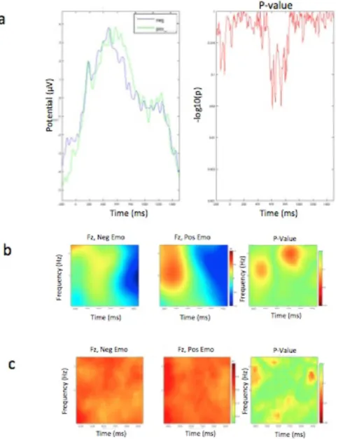
There was a main effect of framing on the LPP amplitude in response to negative stimuli, with positively-framed stimuli having a significantly (p=.03) extended peak amplitude, compared to that of negatively-framed stimuli (Figure 1a). There was also a
main effect of framing on negative stimuli in frontal theta evoked power, with positively-framed stimuli having a significantly (p=.04) higher early (400-500ms) response than
negatively-framed stimuli, and negatively-framed stimuli having a significantly higher (p=.02) later (650-850ms) response (Figure 1b) than positively-framed stimuli. There was no significant difference of framing condition on emotional stimuli for frontal theta ITC
(Figure 1c).
Framing influence on the LPP in response to neutral stimuli
(Figure 2c). There was no main effect of framing type on the LPP magnitude or frontal theta ERSP of neutral stimuli (Figure 2a, 2b).
Between group comparisons
Patients had a significantly higher LPP amplitude in response to negatively-framed negative stimuli (p=0.001), whereas controls had a significantly higher (p=.04)
amplitude to positively-framed negative stimuli (Figure 3a). Controls had significantly increased (p=.04) frontal theta ERSP from 400 to 500ms for positively-framed negative stimuli, and significantly decreased frontal theta ERSP from 600 to 800ms (p=.01) for
positively-framed negative stimuli. This effect was not seen in patients, and there was a significant interaction effect between group and frame from 400 to 500ms (p=.04) and
750 to 900ms (p=.03). Additionally, patients had significantly increased theta ITC around 500ms for positively-, rather than negatively-, framed negative stimuli (p=.01). This was not seen in controls, and there was a significant (p=.01) interaction effect between group
and frame on frontal theta ITC from 400 to 500ms (Figure 3b). Between groups, there were no significant difference of the framing type on negative stimuli for either frontal
theta ERSP (Figure 3b) or frontal theta ITC (Figure 3c).
Patients had a significantly (p=.01) increased, earlier LPP in response to positively-, rather than negatively-, framed neutral stimuli (Figure 4a). Patients had
significantly greater early (500-600ms) frontal theta ERSP (p=.02) followed by
significantly decreased (p=.02) theta ERSP later (800-1000ms) in response to negatively
Discussion
Overall, the present study has found that the framing of stimuli influences the
resultant LPP response, as well as frontal gamma-band ERSP and ITC activity. Further, these changes differ between patients with schizophrenia and healthy controls.
Consistent with the first hypothesis, there was no framing effect on LPP amplitude in neutral stimuli. However, contrary to the first hypothesis, we found that positively-framed negative stimuli actually had an extended peak LPP amplitude
compared to negatively-framed negative stimuli, across groups. While this result appears to run counter to our hypotheses regarding emotion regulation, this finding may not be
reflective of emotional salience at all, but rather an unintentional component of the paradigm, which is congruency. While previous studies (e.g. Kisley et al., 2011) using a similar paradigm included both positively- and negatively-valenced emotional images,
the present study used only negatively rated emotional images. As such, negatively-framed emotional stimuli were “congruent” with their frame, while positively-negatively-framed
emotional stimuli were “incongruent,” possibly requiring different or more effortful processing. Studies evaluating the impacts of effort on LPP have found that LPP amplitude increases as the complexity of an explanation increases (Hua, Han, & Zhou,
2015) and as task demands increase (daSilva, Crager, & Puce, 2015). Studies evaluating specifically the effects of congruency on LPP have found that mismatched affective face
Thus, it is possible that the present study’s counterintuitive findings are reflective of affective incongruency or increased processing complexity, rather than a failure of the paradigm to elicit emotion regulation.
Consistent with the possibility of the positively-framed negative stimuli necessitating increased effort of processing as a result of incongruency, frontal theta
ERSP was found to be increased in positively-framed negative stimuli from 400-500ms compared to negatively-framed negative stimuli across groups. Negatively-framed negative stimuli, however, showed increased frontal theta ERSP from 650-850ms in
compared to positively-framed stimuli, suggesting that perhaps for negatively-framed negative stimuli, some form of further processing occurs after an evaluation of
congruency, such as the degree of the congruency or a sustained attentional focus as a result of negativity bias.
Between-group analyses showed that controls had an increased LPP magnitude to
positively-framed negative stimuli compared to patients with schizophrenia, suggesting that perhaps the effect of incongruency is weaker in patients. Further, patients had a
significantly higher LPP amplitude for negatively-framed negative stimuli than controls, suggesting that the task may be more cognitively-demanding for patients, even under a congruent paradigm. This hypothesis is supported by many studies showing decreased
performance in patients with schizophrenia in domains such as working memory, which is often associated with altered neural activity as a result of cortical efficiency (Moran &
ITC, and increased, followed by decreased, frontal theta ERSP activity to positively-framed neutral stimuli, rather than negatively-positively-framed neutral stimuli. Considering the aforementioned findings of the present study, this suggests that patients with
schizophrenia may have a shifted baseline for the evaluation of neutral stimuli. In their study evaluating the effects of congruency on affective word pairs, Zhang et al. (2010)
found that LPP magnitude increased more to negatively-primed positive targets than positively-primed negative targets, and proposed that participants may feel an increased sense of incongruency to negatively-primed positive targets due to a stronger emotional
reaction to the negative primes, which were images, than the positive primes. If this is true, our results suggest that patients might perceive the neutral stimuli more negatively
than controls, leading to positively-framed neutral stimuli to be processed as “incongruent.”
Finally, while the interpretation of the present study’s results may be confounded
by the possibility of congruency affecting the evaluation of emotional stimuli, it is important to note that some findings do suggest that the paradigm is, at least in part,
evaluating emotion regulation. Notably, controls had increased frontal theta ERSP in an earlier window to positively-framed negative stimuli, and decreased frontal theta ERSP in a later window to positively-framed negative stimuli, and this was not seen in patients.
This supports the well-established finding of decreased emotion regulation in patients with schizophrenia in response to framing paradigms (Horan & Hajcak, 2013; Strauss et
al., 2013).
notably sample size. Such a small patient group (N=6) results in very low power for between-group comparisons, and makes the within-group sample highly unbalanced. However, recruitment for the study is ongoing, and successful recruitment of the target
group of 20 patients should greatly facilitate interpretation of results.
Future studies using a similar paradigm should be sure to include both positively-
and negatively-valenced target stimuli to avoid the complication of congruency. Additionally, a more nuanced paradigm will be able to better elucidate difficult to interpret findings, such as changes in ITC, which may be indicative of a process that is
being overshadowed by another process due to the complexity of the study.
Nevertheless, though the patient group is small, the findings of the present study
indicate some interesting differences between patients and controls in the evaluation of emotional stimuli and the effect of congruency, above and beyond the effects of emotion regulation. Future work should elaborate on the suggestion that patient groups may be
References
Andreasen, N.C., Pressler, M., Nopoulos, P., Miller, D., & Ho, B-C. (2010).
Antipsychotic dose equivalents and dose-years: a standardized method for comparing exposure to different drugs. Biological Psychiatry: 67(3), 255-262.
Auslander, L.A., Lindamer, L.L., Delapena, J., Harless, K., Polichar, D., & Patterson, T.L. (2001). A comparison of community dwelling older schizophrenia patients by residential status. Acta Psychiatrica Scandinavia, 103(5), 380–386
Basar-Eroglu, C., Brand, A., Hildebrandt, H., Kedizor, K.K., Mathes, B., & Schmiedt, C. (2007). Working memory related gamma oscillations in schizophrenia patients.
International Journal of Psychophysiology, 64(1), 39-45.
Blanchard, J.J., Kring, A.M., Horan, W.P., & Gur, R. (2011). Toward the next generation of negative symptom assessments: the collaboration to advance negative symptom
assessment in schizophrenia. Schizophrenia Bulletin, 37(2), 291–299.
Brantley, P.J., Waggoner, C.D., Jones, G.N., & Rappaport, N.B. (1987). A daily stress
inventory: development, reliability, and validity. Journal of Behavioral Medicine, 10(1), 61-74.
Cohen, A.S. & Minor, K.S. (2008). Emotional experience in patients with schizophrenia
re-revisited: meta-analysis of laboratory studies. Schizophrenia Bulletin, 36(1), 143–150.
Cohen, S., Kamarck, T., & Mermelstein, R. (1983). A global measure of perceived stress. Journal of Health and Social Behavior, 24, 386-396.
daSilva, E.B., Crager, K., & Puce, A. (2015). On dissociating the neural time course of
Delorme, A. & Makeig, S. (2004). EEGLab: an open source toolbox for analysis of single-trial EEG dynamics. Journal of Neuroscience Methods, 134: 9-21. Foti, D. & Hajcak, G. (2008). Deconstructing reappraisal: descriptions preceding
arousing pictures modulate the subsequent neural response. Journal of Cognitive Neuroscience, 20(6), 977–988.
Gross, J.J. (1998). The emerging field of emotion regulation: an integrative review. Review of General Psychology. 2(3), 271–299.
Gross, J.J. (2002). Emotion regulation: affective, cognitive, and social consequences.
Psychophysiology, 39(3), 281–291.
Gross J.J. & Thompson, R.A. (2007). Emotion regulation: conceptual foundations. In
Handbook of Emotion Regulation (ed. J. J. Gross), pp. 3–23. Guilford Press: New
York.
Guderian, S., Schott, B.H., Richardson-Klavehn, A., & Duzel, E. (2009). Medial
temporal theta state before an event predicts episodic encoding success in humans. PNAS, 106(13), 5365-5370.
Haenschel, C. et al. (2009). Cortical oscillatory activity is critical for working memory as revealed by deficits in early-onset schizophrenia. Journal of Neuroscience,
29(30): 9481-9489.
Hajcak, G., MacNamara, A., & Olvet, D.M. (2010). Event-related potentials, emotion, and emotion regulation: an integrative review. Developmental Neuropsychology,
Hajcak, G. & Nieuwenhuis, S. (2006). Reappraisal modulates the electrocortical response to unpleasant pictures. Cognitive, Affective, and Behavioral Neuroscience 6(4), 291–297.
Henry, J.D., Rendell, P.G., Green, M.J., McDonald, S., & O’Donnell, M. (2008). Emotion regulation in schizophrenia: affective, social, and clinical correlates of
suppression and reappraisal. Journal of Abnormal Psychology, 117(2), 473–478. Ho, B.C., Andreasen, N., & Flaum, M. (1997) Dependence on public financial support
early in the course of schizophrenia. Psychiatric Services, 48(7), 948–950
Horan, W.P., Hajcak, G., Wynn, J.K., & Green, M.F. (2013) Impaired emotion regulation in schizophrenia: evidence from event-related potentials. Psychological Medicine,
43(11), 2377-2391.
Hua, M., Zhuo, R.H., & Zhou, R. (2015). Cognitive reappraisal in preschoolers: neuropsychological evidence of emotion regulation from an ERP study.
Developmental Neuropsychology, 40(5): 279-290.
Kay, S.R., Fiszbein, A., & Opler, L.A. (1987). The positive and negative syndrome scale
(PANSS) for schizophrenia. Schizophrenia Bulletin, 13(2), 261-276.
Keil, A., Bradley, M.M., Hauk, O., Rockstroh, B., Elbert, T. & Lang, P.J. (2002) Large-scale neural correlates of affective picture processing. Psychophysiology, 39(5),
641-649.
Kimhy, D., Vakhrusheva, J., Jobson-Ahmed, L., Tarrier, N., Malaspina, D., & Gross, J.J.
Kisley, M.A., Campbell, A.M., Larson, J.M., Naftz, A.E., Regnier, J.T., & Davalos, D.B. (2011). The impact of verbal framing on brain activity evoked by emotional images. Journal of Integrative Neuroscience, 10(4), 513-524.
Kring, A.M. & Moran, E.K. (2008). Emotional response deficits in schizophrenia: insights from affective science. Schizophrenia Bulletin, 34(5), 819–834.
Kring, A.M., Gur, R.E., Blanchard, J.B., Horan, W.P., & Reise, S.P. (2013). The clinical assessment interview for negative symptoms (CAINS): final development and validation. American Journal of Psychiatry, 170(2), 165-172.
Lang, P.J., Bradley, M.M., & Cuthbert, B.N. (1999). International affective picture system (IAPS): Technical manual and affective ratings. University of Florida,
Center for Research in Psychophysiology.
Lang, P.J., Bradley, M.M., & Cuthbert, B.N. (2005). International affective picture system (IAPS): Affective ratings and instruction manual. Technical report no.
A-6, University of Florida, Gainesville, FL.
Lee, K.H., Williams, L.M., Breakspear, M., & Gordon, E. (2003). Synchronous gamma
activity: a review and contribution to an integrative neuroscience model of schizophrenia. Brain Research Reviews, 41(1), 57-78.
Leung, W.W., Bowie, C.R., & Harvey, P.D. (2008). Functional implications of
neuropsychological normality and symptom remission in older outpatients diagnosed with schizophrenia: a cross-sectional study. Journal of the
International Neuropsychological Society, 14(3), 479–488
Livingstone, K., Harper, S., & Gillanders, D. (2009). An exploration of emotion
Moran, L.V. & Hong, H.L. (2011). High vs low frequency neural oscillations in schizophrenia. Schizophrenia Bulletin, 37(4), 659-663.
Perry, Y., Henry, J.D., & Grisham, J.R. (2011). The habitual use of emotion regulation
strategies in schizophrenia. British Journal of Clinical Psychology, 50(2), 217– 222.
Rado S. (1953) Dynamics and classification of disordered behavior. American Journal of Psychiatry, 110(6), 406–416.
Roach, B.J. & Mathalon, D.H. (2008). Event-related EEG Time-Frequency Analysis: An
overview of measures and an analysis of early gamma band phase locking in schizophrenia. Schizophrenia Bulletin, 34(5): 907-926.
Sabatinelli, D., Lang, P.J., Keil, A. & Bradley, M.M. (2007) Emotional perception: correlation of functional MRI and event-related potentials. Cerebral Cortex, 17(5), 1085-1091.
Spencer, K.M., Nestor, P.G., Perlmutter, R., Niznikiewicz, M.A., Klump, M.C., Frumin, M., Shenton, M.E., & McCarley, R.W. (2004). Neural synchrony indexes
disordered perception and cognition in schizophrenia. PNAS, 101(49), 17288-17293.
Strauss, G.P., Kappenman, E.S., Culbreth, A.J., Catalano, L.T., Lee, B.G., & Gold, J.M.
(2013). Emotion regulation abnormalities in schizophrenia: cognitive change strategies fail to decrease the neural response to unpleasant stimuli. Schizophrenia
Bulletin, 39(4), 872-883.
Epidemiologic Catchment Area prospective 1-year prevalence rates of disorders and services. Archives of General Psychiatry. 50(2), 85–94.
Tiihonen, J., Lonngvsit, J., Wahlbeck, K., Klaukka, T., Niskanen, L., Tanskanen, A., &
Haukka, J. (2011). 11-year follow-up of mortality in patients with schizophrenia: a population-based cohort study (FIN11 study). Lancet, 374(9690), 620-627.
Uhlhaas, P.J. & Singer, W. (2010). Abnormal neural oscillations and synchrony in patients with schizophrenia. Nature Reviews Neuroscience, 11, 100-112.
Uhlhaas, P.J. & Singer, W. (2012). Neuronal Dynamics and Neuropsychiatric Disorders:
Toward a translational paradigm for dysfunctional large-scale networks. Neuron, 75, 963-977.
Uhlhaas, P.J., Linden, D.E., Singer, W., Haenschel, C., Lindner, M., Maurer, K., & Rodriguez, E. (2006). Dysfunctional long-range coordination of neural activity during Gestalt perception of schizophrenia. Journal of Neuroscience, 26(31),
8168-8175.
Van der Meer, L., Van’t Wout, M., & Aleman, A. (2009). Emotion regulation strategies
in patients with schizophrenia. Psychiatry Research, 170(2-3), 108–113.
Vuilleumier, P. (2005) How brains beware: Neural mechanisms of emotional attention. Trends in Cognitive Sciences, 9(12), 585-594.
Werheid, K., Alpay, G., Jentzsch, I., & Sommer, W. (2005). Priming emotional facial expressions as evidenced by event-related brain potentials. International Journal
of Psychophysiology, 55(2): 209-219.
prediction over 15 years in incidence cohorts in six European centres. Psychological Medicine, 30(5), 1155–1167.
Zhang, Q., Li, X., Gold, B.T., & Jiang, Y. (2010). Neural correlates of cross-domain
Figure 1. Framing Effects on Negative Stimuli, Across Groups. (a) LPP magnitude for
positively (green) and negatively (blue) framed negative stimuli, with collapsed groups. (b) Frontal theta ERSP for positively (right) and negatively (left) framed emotional
Figure 2. Framing Effects on Neutral Stimuli, Across Groups. (a) LPP magnitude for positively (green) and negatively (blue) framed neutral stimuli, with collapsed groups. (b)
Figure 3. Framing Effects on Negative Stimuli, Between Groups. (a) LPP magnitude for
positively (right) and negatively (left) framed negative stimuli, between groups (top row=controls, second row=patients). (b) Frontal theta ERSP for positively (right) and
Figure 4. Framing Effects on Neutral Stimuli, Between Groups. (a) LPP magnitude for
positively (right) and negatively (left) framed emotional stimuli, between groups (top row=controls, second row=patients). (b) Frontal theta ERSP for positively (right) and
negatively (left) framed emotional stimuli, between groups (top row=controls, second row=patients). (c) Frontal theta ITC for positively (right) and negatively (left) framed emotional stimuli, , between groups (top row=controls, second row=patients).