ARTIGO ORIGINAL
RESUMO
Introdução: O tromboembolismo pulmonar e trombose venosa profunda ocorrem em idade pediátrica com incidência, morbilidade e mortalidade desconhecidas. O objetivo foi rever epidemiologia, apresentação clínica, exames complementares de diagnóstico e prognóstico de doentes com tromboembolismo pulmonar e trombose venosa profunda.
Material e Métodos: Estudo retrospetivo, descritivo e analítico de doentes pediátricos internados num hospital de nível II por tromboembolismo pulmonar e trombose venosa profunda, entre 2000 e 2014. Estudaram-se características demográficas, história clínica, comorbilidades e fatores de risco.
Resultados: Foram internados 11 doentes (sete com tromboembolismo pulmonar, cinco com trombose venosa profunda e um com ambos), 64% do género feminino e idade média de 16 anos. Todos os doentes com tromboembolismo pulmonar referiam toracalgia/ dispneia, 25% síncope/palpitações e 25% febre. Todos os doentes com trombose venosa profunda referiam dor no local da obstrução, 83% edema/cianose do membro afetado e 17% febre. O estudo da trombofilia positivo foi o fator de risco mais frequente nas duas entidades. O valor médio dos D-dímeros foi 3252 ug/L e 2660 ug/L no tromboembolismo pulmonar e trombose venosa profunda, respetivamente. Todos os doentes iniciaram anticoagulação, três necessitaram de cuidados intensivos, três apresentaram sequelas e houve um óbito.
Discussão: Todos os doentes tinham pelo menos um fator de risco associado e as condições de hipercoagulabilidade herdadas foram o fator de risco mais frequentemente encontrado nos nossos adolescentes.
Conclusão: O aumento da incidência na população pediátrica descrito na literatura pode ser atribuído à crescente sensibilização para esta patologia, aos avanços médicos e aumento da sobrevida de doenças crónicas. Escasseiam recomendações baseadas na evidência que identifiquem os doentes com risco de trombose, para que as decisões possam ser tomadas de forma cuidadosa, equilibrando o risco e benefício em cada caso.
Palavras-chave: Criança; Embolia Pulmonar; Trombose Venosa
Venous Thromboembolism in Pediatric Age: A 15 Year
Retrospective Review
Tromboembolismo Venoso em Idade Pediátrica: Estudo
Retrospetivo de 15 Anos
1. Serviço de Pediatria. Centro Hospitalar Tondela-Viseu. Viseu. Portugal.
Autor correspondente: Joana Verdelho Andrade. [email protected]
Recebido: 03 de setembro de 2017 - Aceite: 02 de julho de 2018 | Copyright © Ordem dos Médicos 2018
Joana Verdelho ANDRADE1, Joana MAGALHÃES1, Catarina RESENDE1, Dora GOMES1, Gabriela LARANJO1,
Joana CAMPOS1, Elisabete SANTOS1, Cristina FARIA1
Acta Med Port 2018 Sep;31(9):489-495 ▪ https://doi.org/10.20344/amp.9639 ABSTRACT
Introduction: Pulmonary thromboembolism and deep venous thrombosis occur in pediatric age, with unknown incidence, morbidity and mortality. Our aim is to review the epidemiology, clinical presentation, complementary diagnostic tests and prognosis of patients with pulmonary thromboembolism and deep venous thrombosis.
Material and Methods: Retrospective, descriptive and analytical study of pediatric patients admitted to a Level II hospital for pulmonary thromboembolism and deep venous thrombosis, between 2000 and 2014. Demographic characteristics, clinical history, comorbidities and risk factors were studied.
Results: Eleven patients (n = 7 pulmonary thromboembolism, n = 5 deep venous thrombosis, n = 1 both), 64% females and with 16 years old average, were admitted. All patients with pulmonary thromboembolism presented symptoms of chest pain and/or dyspnea, 25% syncope/palpitations and 25% fever. All patients with deep venous thrombosis reported localized pain at the site of obstruction, 83% edema/cyanosis of the affected limb and 17% fever. The study of positive thrombophilia was the most frequent risk factor in both entities. The mean value of D-dimers was 3252 ug/dL and 2660 ug/dL in pulmonary thromboembolism and deep venous thrombosis, respectively. All patients started anticoagulation, three required intensive care, two had sequelae and one died.
Discussion: All patients had at least one risk factor, and hereditary hypercoagulability was most commonly established.
Conclusions: The increased incidence in the pediatric population described in some studies can be attributed to an increased awareness of this pathology, medical advances and increasing survival of chronic diseases. There is a lack of evidence-based recommendations identifying patients at risk of thrombosis so that decisions can be made carefully, balancing the risk and benefit in each case.
Keywords: Child; Pulmonary Embolism; Venous Thrombosis
INTRODUCTION
Venous thromboembolism (VT) includes deep venous thrombosis (DVT) and pulmonary thromboembolism (PTE) and is a potentially devastating pathology with a significant impact on the quality of life of paediatric patients as well as on associated hospital costs.1 Considering a wider age
range for paediatric attendance, the healthcare improve-ment, a longer survival from severe diseases and an
in-creasing awareness of vascular events, VT is becoming more and more recognised as a relevant paediatric issue.2
ARTIGO ORIGINAL estimated in Europe, as well as a 2% mortality rate. 4 VT physiopathology is explained by the Virchow’s triad, including venous stasis, endothelial injury/dysfunction and hypercoagulability, with a multifactorial aetiology and the presence of some underlying risk factors in most cases; only 5-10% of the patients in paediatric age groups were considered with an idiopathic condition, compared to 60% in adult patients.4-7
Different classifications are used, including (i) first or recurrent episode, (ii) symptomatic vs. asymptomatic, (iii) acute vs. chronic, (iv) veno-occlusive vs. non-occlusive, (v) idiopathic vs. with underlying risk factors. The use of exog-enous oestrogens, the use of central vexog-enous catheters, a reduced mobility and thrombophilia (including the presence of transient or persistent anti-phospholipid antibodies, ac-quired or inherited anticoagulant abnormalities, factor V Lei-den or prothrombin gene G20210A mutations) have been included as prothrombotic risk factors. The term thrombo-philia was introduced to describe the presence - usually in-herited - of an increased predisposition for the occurrence of thrombotic events.2
The contribution of acute risk factors to the development of VT, in isolation as underlying a chronic disease, has been shown. Injuries and postoperative settings are frequent risk factors for its development in children and adolescent pa-tients with no chronic disease. Other common risk factors include the presence of infectious diseases, the use of oral contraceptives, long-term parenteral nutrition and other in-flammatory conditions.7,8 The use of central venous
cath-eters, an underlying malignant disease and postoperative status (following heart surgery) were the most prevalent causes of VT described by the Canadian Childhood Throm-bophilia registry,5 while infectious diseases and the use of
central venous catheters were described by a recent US cohort study.6
Inherited and acquired thrombophilia and markers of coagulation activation have been considered as blood risk factors for paediatric VT. Inherited thrombophilia (as for in-stance factor V Leiden or prothrombin gene G20210A mu-tations) is frequently found in Caucasian populations, with an approximate prevalence of 5 and 2%, respectively. An elevated factor VIII activity with underlying significant infec-tions and inflammatory states, anticoagulant deficiencies and disseminated intravascular coagulation underlying bacterial sepsis, the production of inhibiting antibodies in viral acute infections and para-infectious disorders with the development of anti-phospholipid antibodies are common examples of acquired thrombophilia in children.2
In fact, it is estimated that 60% predisposition to throm-bosis is attributable to genetic factors.9,10 Patients with
thrombophilia usually present with an initial thrombotic event under the age of 25 and an increasing chance of re-currence with patient’s age, as well as with the association of other underlying risk factors. There is a low risk for throm-bosis under the age of 15 due to this condition, with a 2 to 4% increasing annual risk onwards.12
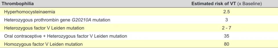
The estimated risk based on adult populations with VT is shown in Table 1, showing the relevance of VT associated with risk factors due to inherited or acquired thrombophilia.2
A high level of suspicion for an acute VT episode should be taken into consideration with the presence of clinical thrombotic risks and a family history of early VT or other vascular disorders, as well as the presence of an history of known thrombophilia traits and clinical signs and symp-toms, depending on the anatomical location and system involved and influenced by veno-occlusive characteris-tics and chronicity. The presence of unilateral limb painful oedema is the classical manifestation of DVT and signs and symptoms may include the presence of cervical and facial oedema and headache in case of involvement of superior vena cava. PTE usually presents with unexplained acute dyspnoea and underlying pleuritic chest pain, even though it may be asymptomatic or present with mild symptoms in children, mainly when limited segments of the pulmonary arteries are involved. In the case of involvement of renal veins, haematuria and thrombocytopenia are the typical clinical presentation and eventually with the presence of an abdominal mass and oligo-anuria (when bilateral). Chronic VT may be detected by accident in imaging requested due to other reasons, as in the presence of an asymptomatic thrombosis of dural venous sinuses or it may present with signs and symptoms of chronic venous obstruction or post-thrombotic syndrome secondary to limb venous thrombosis, including limb painful oedema, dilated superficial collateral veins, venous stasis dermatitis or skin ulceration.2
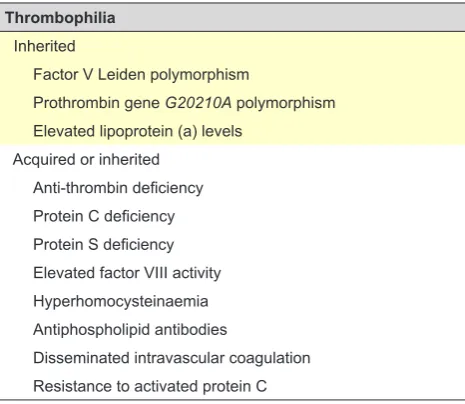
Complete blood count, coagulation, thrombophilia screening and beta-hCG blood test in post-menarche ado-lescents are included in paediatric VT workup. Thrombo-philia screening panel and risk factors for VT used in pae-diatric evaluations and recommended by the Subcommittee for Perinatal and Paediatric Thrombosis of the International Society on Thrombosis and Haemostasis for the diagnosis of acute VT in children and adolescents are shown in Table 2.2,13
Suspected distal or proximal DVT may be confirmed by Doppler ultrasonography and MRI or CT scan are frequently
Table 1 – Estimated risk of VT in selected thrombophilia traits
Thrombophilia Estimated risk of VT (x Baseline)
Hyperhomocysteinaemia 2.5
Heterozygous prothrombin gene G20210A mutation 3
Heterozygous factor V Leiden mutation 2 - 7
Oral contraceptive + Heterozygous factor V Leiden mutation 35
ARTIGO ORIGINAL
needed for the identification of the involvement of perineal and abdominal veins. The use of spiral CT scan or ventila-tion/perfusion lung scintigraphy is usually used to confirm the presence of PTE.2,13
Heparin and warfarin are the most widely used conven-tional thrombotic agents and the target levels of anti-coagulation should be based on recent paediatric recom-mendations. Hypercoagulability underlying this pathology is attenuated by these agents and therefore the risk of pro-gression of the thrombus and embolism is reduced, relying on intrinsic fibrinolytic mechanisms that allow for a progres-sive dissolution of the thrombus.14
Even though around half of the venous thrombotic events are settled with 3-6-month traditional anticoagula-tion therapy, a risk of post-thrombotic syndrome remains, probably due to complete venous obstruction and local in-flammatory disorders.15-17 While coagulation is reduced by
anticoagulant therapy, fibrinolysis is increased by thrombo-lytic therapy due to an enhanced activation of plasminogen into plasmin.2 Despite increasingly used, the indication for
thrombolytic therapy in children have not yet been clearly established. Its use has been recommended by the Ameri-can College of Chest Physicians in patients suffering from life, organ or limb-threatening venous thromboembolism even though it should not be used as routine.14 This therapy
in a paediatric age is recommended by the American Heart Association when the benefits outweigh the risks.18
In addition, surgical thrombectomy in children with life-threatening thromboembolism is recommended by the American College of Chest Physicians. Inferior vena cava filter placement is recommended in children over 10 kg and presenting with lower limb thromboembolism and a con-traindication for anticoagulation therapy. 14
Acute complications of VT may arise from antithrombotic interventions or from the own thrombotic event, while long-term complications include the presence of recurrent VT, chronic hypertension in case of the involvement of the renal veins, variceal haemorrhage related to portal vein
thrombo-sis, chronic superior vena cava syndrome and development of post-thrombotic syndrome.16
This study aimed at a revision of the epidemiology, clini-cal presentation, diagnostic workup and outcome in paedi-atric patients with pulmonary thromboembolism and deep venous thrombosis. In addition, the presence of recognised potential risk factors for this pathology has been described in our group of patients.
MATERIAL AND METHODS
This was a retrospective, descriptive and analytical study of all the patients with VT (including DVT and PTE) aged under 18 and admitted to a level-II hospital throughout a 15-year period.
Data on children and adolescent patients admitted be-tween 1 Jan 2000 and 31 Dec 2014 were collected: patient’s gender, relevant personal and family history, in addition to VT-related variables including patient’s age at diagnosis, presentation signs and symptoms, associated risk factors, comorbidities, treatment, clinical progression and complica-tions.
Patients presenting with PTE confirmed by X-ray and those with DVT confirmed by ultrasound were considered. The presence of obesity was considered for BMI (body mass index) beyond the 97th percentile of the reference
gender- and age-specific WHO (World Health Organization) curves. The study was approved by the Ethics Commission for Health of the Centro Hospitalar Tondela-Viseu E.P.E. and an informed consent was not required.
Data analysis
Data were collected and coded into a grid at an Excel®, version 2011 (Microsoft Corporation, USA) software and using the SPSS®, version 20 for Mac (SPSS, IL, USA) soft-ware for a descriptive analysis of a set of variables aimed at the characterisation of our group of patients, as well as to build up 95% confidence intervals (CI).
RESULTS
A total of 11 patients with VT were identified throughout the study period, corresponding to 13 in-hospital stays: sev-en patisev-ents with PTE, five with DVT and two patisev-ents with both pathologies, corresponding to three thrombotic events, two with PTE and one with DVT. Four events have occurred up to 2007 (31% of the patients) and nine onwards (69%) (Fig. 1), while this pathology has corresponded to 0.0004% of the hospital stays throughout this period.
Mostly female patients (64%) and a 16 year mean and median age at diagnosis (±1.4), 95% CI 15.2 – 17.0, 13-17 year range were found.
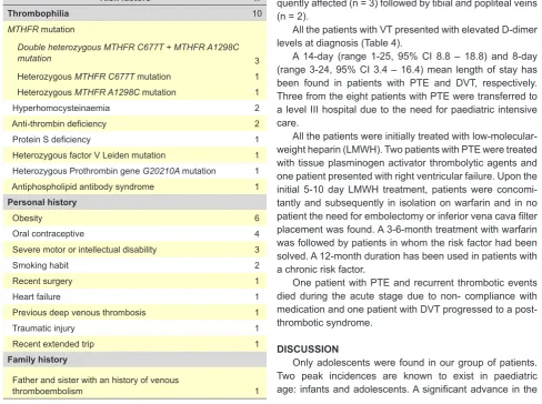
All the patients presented with at least one associated risk factor (9% presented with one, 37% with two, 18% with three, 27% with four and 9% with five) (Table 3) and the presence of thrombophilia was the most frequently found risk factor in both entities, followed by obesity and the use of oral contraceptive drugs.
All the patients with hyperhomocysteinaemia presented
Table 2 – Thrombophilia underlying an acute VT episode in the
paediatric age
Thrombophilia
Inherited
Factor V Leiden polymorphism
Prothrombin gene G20210A polymorphism Elevated lipoprotein (a) levels
Acquired or inherited Anti-thrombin deficiency Protein C deficiency Protein S deficiency Elevated factor VIII activity Hyperhomocysteinaemia Antiphospholipid antibodies
ARTIGO ORIGINAL
with a MTHFR C677T and MTHFRA1298C double-hete-rozygous mutation. In addition, two adolescents presented with concomitant thrombophilia: heterozygous MTHFR C677T mutation and protein S deficiency; double-heterozy-gous MTHFR C677T and MTHFR A1298C mutation and anti-thrombin deficiency.
Three patients presented with severe motor or intellec-tual disability related to paraplegia due to T9 spinal cord injury from a car accident, autism spectrum disorder and trisomy 21 syndrome.
As regards the patients with PTE (n = 8), one of whom with concomitant DVT, all presented with thoracalgia and dyspnoae at admission, 25% with syncope/palpitations and 25% with fever (Fig. 2). All the patients underwent a CT-scan for diagnostic confirmation and a bilateral pulmonary involvement has been found in five patients. The patient with concomitant DVT presented with iliac and femoral vein involvement.
As regards the patients with isolated DVT (n = 5), all pre-sented with pain at the obstruction site, 83% with oedema and cyanosis in the affected limb and 17% with fever (Fig. 2). All the patients underwent Doppler ultrasound for diag-nostic confirmation. Iliac and femoral veins were more fre-quently affected (n = 3) followed by tibial and popliteal veins (n = 2).
All the patients with VT presented with elevated D-dimer levels at diagnosis (Table 4).
A 14-day (range 1-25, 95% CI 8.8 – 18.8) and 8-day (range 3-24, 95% CI 3.4 – 16.4) mean length of stay has been found in patients with PTE and DVT, respectively. Three from the eight patients with PTE were transferred to a level III hospital due to the need for paediatric intensive care.
All the patients were initially treated with low-molecular-weight heparin (LMWH). Two patients with PTE were treated with tissue plasminogen activator thrombolytic agents and one patient presented with right ventricular failure. Upon the initial 5-10 day LMWH treatment, patients were concomi-tantly and subsequently in isolation on warfarin and in no patient the need for embolectomy or inferior vena cava filter placement was found. A 3-6-month treatment with warfarin was followed by patients in whom the risk factor had been solved. A 12-month duration has been used in patients with a chronic risk factor.
One patient with PTE and recurrent thrombotic events died during the acute stage due to non- compliance with medication and one patient with DVT progressed to a post-thrombotic syndrome.
DISCUSSION
Only adolescents were found in our group of patients. Two peak incidences are known to exist in paediatric age: infants and adolescents. A significant advance in the
Figure 1 – Annual distribution of patients with VT
Venous thromboembolism
-1 1
0 1
0 0 0 0 0
3
0 1
2 3
0 1
2
3 5
2000 2001 2002 2003 2004 2005 2006 2007 2008 2009 2010 2011 2012 2013 2014
Table 3 – Risk factors in patients with VT
Risk factors n
Thrombophilia 10
MTHFR mutation
Double heterozygous MTHFR C677T + MTHFR A1298C
mutation 3
Heterozygous MTHFR C677T mutation 1
Heterozygous MTHFR A1298C mutation 1
Hyperhomocysteinaemia 2
Anti-thrombin deficiency 2
Protein S deficiency 1
Heterozygous factor V Leiden mutation 1
Heterozygous Prothrombin gene G20210A mutation 1
Antiphospholipid antibody syndrome 1
Personal history
Obesity 6
Oral contraceptive 4
Severe motor or intellectual disability 3
Smoking habit 2
Recent surgery 1
Heart failure 1
Previous deep venous thrombosis 1
Traumatic injury 1
Recent extended trip 1
Family history
Father and sister with an history of venous
ARTIGO ORIGINAL
knowledge of the physio-pathological mechanisms involved in VT has occurred over the past few decades.9 Most pa-tients in the different studies involving children had some underlying clinical condition and the presence of a central venous catheter was most frequently described.10 At least
one known risk factor for VT has been found in our patients. The confirmation of hereditary hypercoagulability conditions in a large percentage of patients with DVT and PTE has been the most significant advance in the knowledge of VT physiopathology and was the most frequently found in our group of patients.
Four from seven VT patients in our group were on oral contraceptive drugs and three from these presented with thrombophilia which was unknown until the event. The risk of VT relates to the dose of oestrogens, the type of proges-tin, patient’s age, family history and the presence of throm-bophilia. The recommendations of the American College of Obstetrics and Gynecology were issued in 2010 by the Centers for Disease Control and Prevention, allowing for the consideration of the use of oral contraceptive drugs in patients with VT attributable to a reversible risk or on antico-agulation therapy. In addition, whenever a previous VT had occurred in a patient on oral contraceptives or pregnant, there was an absolute contraindication for hormone thera-py. Despite the attention that was given to thrombophilia, no universal screening was ever recommended, due to its high cost, in addition to the fact that it may provide false reas-surance, unnecessary anxiety, overtesting, additional family screening and significant and unnecessary expenditure as well as, when negative, a false reassurance regarding the development of thromboembolic events.19
A strong prevalence of obesity has been found in our
study (55%), in line with literature.20 A 32% prevalence of
overweight/obesity up to the age of 3 has been found in dif-ferent Portuguese studies21 and 35% - 37% in children and
adolescents.22,23 Considering that obesity is a recognised
risk factor for VT and has been increasing worldwide at an alarming rate, this may have contributed to an increased incidence of thrombotic events in children.
Different risk factors were found in the patient with con-comitant PTE and DVT, including a recent surgery, history of DVT, obesity and thrombophilia, in addition to non-com-pliance to medication.
There are recommendations for the assessment and classification of the risk of VT aimed at a stratified approach to its prophylaxis; this is the reason why healthcare profes-sionals should be aware of the differentiation of this risk, even though there is still little evidence supporting any spe-cific prophylactic approach in children/adolescents.24,25
Our study showed that treatment has complied with the international recommendations and there was no need for embolectomy or inferior vena cava filter placement. PTE is a potentially lethal condition although rare in the paediatric age. No multicentric studies on the progression of PTE in children have been carried out both national or inter-nationally. A 10% global mortality rate has been described by both the registries in Canada and in The Netherlands, particularly in patients with PTE, showing a 9% (2/22) and 10% (1/10) mortality rate, respectively.3,4 A 9% (1/11)
mor-tality rate has been found in our study, in line with literature. Some limitations to our study are worth mentioning. The fact that our patients have attended only one institution and the small sample do not allow for any result extrapolation. All data were retrospectively collected from the patient’s
Figure 2 – Distribution of patients with DVT and PTE according to clinical signs and symptoms
Clinical presentation
0%
100%
83%
17%
100% 100%
0% 0%
25%
0% 0% 0% 0%
25% 25%
50% 100%
Pain Oedema/Cyanosis Fever Dyspnoea Thoracalgia Syncope Palpitations
DVT PTE
Table 4 – Diagnostic workup in patients with PTE and DVT
Diagnostic workup PTE DVT
Leukocyte count > 15.0x10⁹/L 12% 20%
Platelet count < 150,000/uL 0% 0%
Activated partial thromboplastin time 13% 17%
C-reactive protein > 4.0 mg/dL 88% 80%
D-dimer level (µg/mL) Range 661 – 14,239Mean 3,252
95% CI 1,303 – 7,048
ARTIGO ORIGINAL clinical record and a multicentric analysis should be includ-ed in a further study, with a control group of patients and a standard thrombophilia screening.
CONCLUSION
VT is a rare pathology in children. An increased inci-dence described in some studies may be due to an increas-ing awareness of this pathology, to medical and technologi-cal advances and to an increasing survival from chronic diseases. However, the incidence was probably underesti-mated as PTE is frequently a silent pathology or presenting with non-specific symptoms. This study was focused on the risk factors that should increase clinical suspicion.
In addition, the diagnostic and therapeutic approaches used in children are based on studies with adult patients and therefore there are little evidence-based recommenda-tions in order to identify which paediatric patients are in risk of thrombosis and to allow for a careful decision with an adequate risk-benefit balance.
Further studies are needed to look for a reason for the increasing incidence of VT, which may be due to the longer survival of children with chronic disease, the increasing number of paediatric patients using central venous cath-eters and the increasing prevalence of obesity. The study on the progression of paediatric VT is crucial, including mor-tality, recurrent thrombotic events and pulmonary function. Finally, multicentric studies including the assessment of risk
factors and clinical presentation are needed and crucial to improved care of patients and the development of reliable and reproducible recommendations allowing for the evalua-tion of the chance of VT in paediatric patients.
OBSERVATIONS
This study was presented as oral communication to the 16º Congresso Nacional de Pediatria, held in 22-24 Oct 2015, in Albufeira.
HUMAN AND ANIMAL PROTECTION
The authors declare that the followed procedures were according to regulations established by the Ethics and Clini-cal Research Committee and according to the Helsinki Dec-laration of the World Medical Association.
DATA CONFIDENTIALITY
The authors declare that they have followed the proto-cols of their work centre on the publication of patient data.
CONFLICTS OF INTEREST
The authors declare that there were no conflicts of inter-est in writing this manuscript.
FINANCIAL SUPPORT
The authors declare that there was no financial support in writing this manuscript.
REFERENCES
1. Candrilli SD, Balkrishnan R, O’Brien SH. Effect of injury severity on the incidence and utilization related outcomes of venous thromboembolism in pediatric trauma inpatients. Pediatr Crit Care Med. 2009;10:554-7. 2. Goldenberg NA, Bernard TJ. Venous Thromboembolism in Children.
Hematol Oncol Clin N Am. 2010;24:151-66.
3. Andrew M, David M, Adams M, Ali K, Anderson R, Barnard D et al.
Venous thromboembolic complications (VTE) in children: first analyses of the Canadian Registry of VTE. Blood. 1994;83:1251–7.
4. van Ommen CH, Heijboer H, Buller HR, Hirasing RA, Heijmans HS, Peters M Venous thromboembolism in childhood: a prospective two-year registry in the Netherlands. J Pediatr. 2001;139:676–81.
5. Monagle P, Adams M, Mahoney M, Ali K, Barnard D, Bernstein M, et al. Outcome of pediatric thromboembolic disease: a report from the Canadian Childhood Thrombophilia Registry. Pediatr Res. 2000;47:763– 6.
6. Goldenberg NA, Knapp-Clevenger R, Manco-Johnson MJ, Mountain States Regional Thrombophilia Group. Elevated plasma factor VIII and D-dimer levels as predictors of poor outcomes of thrombosis in children. N Engl J Med. 2004;351:1081–8
7. Setty BA, O’Brien SH, Kerlin BA. Pediatric Venous Thromboembolism in the United States: a tertiary care complication of chronic diseases. Pediatr Blood Cancer. 2012;59:258–64.
8. Takemoto CM, Sohi S, Desai K, Bharaj R, Khanna A, McFarland S et al. Hospital-associated venous thromboembolism in children: incidence and clinical characteristics. J Pediatr. 2014;164:332-8.
9. Rosendaal FR. Venous thrombosis: a multicausal disease. Lancet. 1999;353:1167-73.
10. Fedeman DG, Kirsner RS. An update on hypercoagulable disorders. Arch Intern Med. 2001;161:1051-6.
11. Franco RS, Reitsman PH. Genetic risk factors of venous thrombosis. Hum Genet. 2001;109:369-84.
12. Laffan M, Tuddenham E. Assessing thrombotic risk. BMJ. 1998:317:520-3.
13. Manco-Johnson MJ, Grabowski EF, Hellgreen M, Kemahli AS, Massicotte MP, Muntean W, et al. Laboratory testing for thrombophilia
in pediatric patients. On behalf of the Subcommittee for Perinatal and Pediatric Thrombosis of the Scientific and Standardization Committee of the International Society on Threombosis and Haemostasis (ISTH). Thromb Hae Most. 2002;88:155–6.
14. Monagle P, Chan AKC, Goldenberg NA, Ichord RN, Journeycake JM, Nowak-Göttl U, Vesely SK. Antithrombotic therapy in neonates and children: Antithrombotic Therapy and Prevention of Thrombosis, 9th ed: American College of Chest Physicians Evidence-Based Clinical Practice Guidelines. Chest. 2012;141:e737S–e801.
15. Raffini L. Thrombolysis for intravascular thrombosis in neonates and children. Curr Opin Pediatr. 2009;21:9–14.
16. Goldenberg NA. Long-term outcomes of venous thrombosis in children. Curr Opin Hematol. 2005;12:370–6.
17. Goldenberg NA, Donadini MP, Kahn SR, Neil A, Crowther M, Kenet G, et al.Post-thrombotic syndrome in children: a systematic review of frequency of occurrence, validity of outcome measures, and prognostic factors. Haematologica. 2010;95:1952–9.
18. Jaff MR, McMurtry MS, Archer SL, Cushman M, Goldenberg N, Goldhaberet SZ, et al. Management of massive and submassive pulmonary embolism, iliofemoral deep vein thrombosis, and chronic thromboembolic pulmonary hypertension: a scientific statement from the American Heart Association. Circulation. 2011;123:1788–830. 19. Centers for Disease Control and Prevention (CDC). U S. Medical
Eligibility Criteria for Contraceptive Use, 2010. MMWR Recomm Rep. 2010;59:1–86.
20. Agha BS, Sturm JJ, Simon HK, MD, Hirsh DA. Pulmonary embolism in the pediatric emergency department. pediatrics. 2013;132:663-7. 21. Vale S, Trost SG, Rêgo C, Abreu S, Mota J. Physical activity, obesity
status, and blood pressure in preschool children. J Pediatr. 2015;167:98-102.
22. Camarinha B, Graça P, Nogueira PJ. A prevalência de pré-obesidade/ obesidade nas crianças do ensino pré-escolar e escolar na autarquia de Vila Nova de Gaia, Portugal. Acta Med Port. 2016;29:31-40.
ARTIGO ORIGINAL
24. Meier KA, Clark E, Tarango C, Chima RS, Shaughnessy E. Venous thromboembolism in hospitalized adolescents: an approach to risk assessment and prophylaxis. Hosp Pediatr. 2015;5:44-51.
25. Mahajerin A, Webber EC, Morris J, Taylor K, Saysana M. Development and implementation results of a venous thromboembolism prophylaxis guideline in a tertiary care pediatric hospital. Hosp Pediatr. 2015;5:630-6.
26. van Ommen CH, Peters M. Acute pulmonary thromboembolism in childhood. Thromb Res. 2006;118:13-25.
27. Streif W, Andrew ME. Venous thromboembolic events in pediatric patients: Diagnosis and Management. Hematol Oncol Clin North Am. 1998;12:1283-312.