© 2019 by the Serbian Biological Society 735
Benzo[
a
]pyrene-induced changes in carboxylesterase, acetylcholinesterase and heat
shock protein 70 of
Lymantria dispar
(Lepidoptera: Lymantriidae) from unpolluted and
polluted forests
Anja Grčić1,*, Larisa Ilijin1, Marija Mrdaković1, Milena Vlahović1, Aleksandra Filipović1, Siniša Đurašević2
and Vesna Perić-Mataruga1
1Department of Insect Physiology and Biochemistry, Institute for Biological Research “Siniša Stanković” - National Institute
of Republic of Serbia, University of Belgrade, Despot Stefan Blvd. 142, 11060 Belgrade, Serbia
2Faculty of Biology, University of Belgrade, Studentski trg 3, Belgrade 11000, Serbia
*Corresponding author: [email protected]
Received: June 20, 2019; Revised: July 30, 2019; Accepted: August 28, 2019; Published online: September 6, 2019
Abstract: Plant vegetation accumulates polycyclic aromatic hydrocarbons (PAHs) among which benzo[a]pyrene (B[a]P) is recognized as being very toxic, including cancerogenic. Lymantria dispar L. larvae are sensitive to changes in the envi-ronment, providing potential signs of pollutant presence. We examined the chronic effects of two concentrations of B[a] P on the activity of carboxylesterase (CaE), acetylcholinesterase (AChE) and heat shock protein 70 (Hsp70) levels in the brain tissue of two populations of L. dispar larvae, originating from unpolluted and polluted habitats. We found that the relative growth rate was significantly lower in both populations and that only larvae from polluted forests were sensitive to low B[a]P concentrations, exhibiting a significant increase in brain tissue CaE activity and Hsp70 concentration. AChE activity showed no changes in response to B[a]P exposure in either population. Examined biochemical parameters indicate that their sensitivity to chronic treatment with B[a]P was highly dependent on the pre-exposure history of L. dispar larvae, suggesting that they could be promising biomarkers of B[a]P and PAH pollution in forest ecosystems.
Keywords: Lymantria dispar L.; brain tissue; benzo[a]pyrene; esterase; Hsp70; polycyclic aromatic hydrocarbons (PAHs); environmental pollution
How to cite this article: Grčić A, Ilijin L, Mrdaković M, Vlahović M, Filipović A, Đurašević S, Perić-Mataruga V. Benzo[a]pyrene-induced changes in carboxylesterase, acetylcholinesterase and heat shock protein 70 of Lymantria dispar (Lepidoptera: Lymantriidae) from unpolluted and polluted forests. Arch Biol Sci. 2019;71(4):735-45.
INTRODUCTION
Industrial development and urbanization give momen-tum to dramatic modification of environmental condi-tions, forcing nature to adapt to the unstoppable forces of anthropogenic influence. One of the more recent negative anthropogenic contributions to environmental contamination are the polycyclic aromatic hydrocar-bons (PAHs), which are mostly generated by incom-plete combustion of fossil fuels (particularly by auto-mobiles) and wood, production of coke and charcoal, metal smelting, petroleum refining and petroleum spills [1]. PAHs are very persistent organic contaminants and ubiquitous in various environments across the world [2,3]. They are identified as pollutants of global concern as they pose a great risk to human health and wildlife, with some of them being recognized as carcinogens and mutagens, including benzo[a]pyrene (B[a]P) [4].
Also, PAHs have recently been included in official air quality standards [5], pointing to the necessity of their monitoring. About 40% of PAH emissions into the en-vironment are scavenged by vegetation via dry and wet deposition [6], and due to the large surface area and high content of lipids, foliage is considered the main access point for the accumulation of these xenobiot-ics [7]. Significant concentrations of high molecular weight PAHs, especially B[a]P, were detected in the leaves of two oak species – Quercus robur and Q. ilex
predators of L. dispar larvae, and thus via several lev-els of the food chain, they pose an indirect threat to humans. The necessity for monitoring the impact of xenobiotic concentrations on environment quality has promoted numerous studies in search for biomarkers in living systems that can serve as indicators of biological sublethal changes resulting from individual exposure to toxicants [11]. In our previous studies of the physiologi-cal responses of L. dispar to various types of stressors, we observed significant changes in biochemical param-eters, which points to their potential use as indicators of adverse environmental changes [12,13]. Many inves-tigations have shown the negative effects of PAHs on insect development and reproduction [14,15]. L. dispar
is an insect species that is a very suitable model system for this type of research, considering the uncomplicated manipulation of individuals in laboratory conditions, their short generation time, precisely defined develop-mental stages and well-known physiological processes. Furthermore, the nervous system of L. dispar has prov-en to be very sprov-ensitive to stressogprov-enic or prov-environmprov-ental stimuli and a primary activator of the stress response mechanisms [16].
Carboxylesterases (CaE) or esterases are enzymes in the carboxyl/cholinesterase gene family that hydro-lyze different types of esters [17]. They appear to be widely distributed in all insect tissues, and after ace-tylcholinesterase, they are the most abundant enzymes in the brain and nervous system [18]. In insects, CaE are implicated in many endogenous functions, such as regulation of juvenile hormone titer, general me-tabolism and mobilization of fats and energy related to fat catabolism in muscles [19]. Also, as a component of the defense system of insects, CaE are frequently implicated in insect resistance to organophosphate, carbamate and pyrethroid insecticides [20]. Since the early stage of PAH metabolism in insects activates ox-ygenation reactions and CaE activity, these enzymes are considered to be an indicator of sublethal PAH action [21]. Acetylcholinesterase (AChE) belongs to the family of cholinesterases that are specialized carboxylic ester hydrolases, with the main function of hydrolyzing the neurotransmitter acetylcholine at the cholinergic synapse, thereby blocking the nerve impulse. Organophosphate and carbamate pesticides possess a great affinity towards binding to AChE, causing enzyme inhibition and leading to the accu-mulation of the neurotransmitter acetylcholine in the
synapse, resulting in a disruption to the nervous sys-tem functioning [22]. The sensitivity of AChE activity to insecticides has been used as a biomarker of neuro-toxicity in insects [23], and recently there have been reports supporting the inhibitory effects of AChE by other xenobiotics, such as PAHs, pointing to the po-tential of the enzyme to serve as an indicator of expo-sure to these pollutants [24,25]. Heat shock proteins (Hsp) act as molecular chaperones that participate in protein folding and unfolding and are essential in the cellular response to a variety of damaging condi-tions. Among several Hsp families, based on sequence homology and typical molecular weight, members of the Hsp70 family appear to be synthesized in the gen-eral response to stress [26]. They act as integrators of diverse aspects of protein damage and are considered very useful in biomonitoring by complementing other tests that are more specific measures of toxic action. These proteins have been successfully used to assess the effects of different environmental stressors, includ-ing PAHs, on invertebrates [27,28].
We examined the effects that long-term exposure to relatively low concentrations of B[a]P in insect host plants [8] can have on several biochemical parameters: CaE, AChE and Hsp70 from the brain tissue of L. dis-par larvae originating from unpolluted and polluted environments. Also, we focused our attention on the possible differences in the responses of two insect pop-ulation, with regard to variations in their pre-exposure history and the phenotypic plasticity of biochemical parameters in response to B[a]P exposure, which can differ among different genotypes but can also serve as potential bioindicators of PAH exposure.
MATERIALS AND METHODS Insect rearing and B[a]P treatments
Lymantria dispar egg masses were collected in
free of industrial pollution. The Bor forest is contami-nated by various types of pollutants from the mining industry and smelter complex located next to it. On this locality, PAH concentration showed very high lev-els in the gas phase [29] and in the roots of blackberry [30]. L. dispar larvae originating from Majdanpek for-est are from an unpolluted forfor-est and represent the unpolluted population (UP), whereas those from Bor forest belong to a population from a polluted forest, and designated as the polluted population (PP).
Collected egg-masses were kept at 4°C until hatch-ing in April 2014 and were then grown in plastic dishes (V=200 mL) at 23°C with a 12:12 h light:dark photoperiod. The larvae were fed on a diet specially optimized for L. dispar [31]to which two concen-trations of B[a]P were added as follows:5 ng B[a]P/g dry food weight (5 ng of B[a]P was mixed with 1 g of dry diet, without any liquid), and 50 ng B[a]P/g dry food weight. Acetone was used to dissolve B[a]P to specified concentrations and was then mixed into the artificial diet. The concentration of acetone was 3% of the total diet volume. Diet mixtures were poured into wide, shallow plastic plates and left at 25°C for 4 h until the residual acetone evaporated. Two control groups of larvae were used to test the possible inter-ference of acetone on enzyme activities and Hsp70 expression. Larvae in one control group were fed on the same artificial diet, but without acetone and B[a] P, while larvae in the other control group were fed the artificial diet with only acetone. There were no sig-nificant differences between these two control groups throughout the experiment, so we treated the second group as the control. All larvae were provided with the same amount of B[a]P-spiced food, B[a]P-free food or artificial food free of B[a]P and acetone was replaced every 48 h post hatching until death on the 3rd day of
the 5th larval instar (on average after 31.67±0.33 days).
There were no significant changes in the mortality rate between the experimental groups. The two concen-trations of B[a]P were chosen based on the recorded content of B[a]P in the leaves of Quercus species [9] that are the preferred host plants of L. dispar.
Preparation of homogenates
The brain tissues were dissected out from the head capsules on ice, pooled in experimental groups (n=25 brain tissues per group) and diluted with distilled
wa-ter (1:9/w:V). The brains of larvae were homogenized on ice at 5000 rpm during three 10-s intervals, sepa-rated by 15-s pauses (MHX/E Xenox homogenizer, Germany). Homogenates were centrifuged at 10000
g for 10 min at 4°C in an Eppendorf 5417R centri-fuge (Germany). The supernatants were used for the enzyme assays, Western blotting and indirect non-competitive enzyme-linked immunosorbent assay (ELISA). The protein concentration was determined according to Bradford [32] with bovine serum albu-min (BSA) as standard.
Estimation of relative growth rate
To estimate the effects of two B[a]P concentrations on the individual performance of L. dispar larvae, the relative growth rate (RGR) was measured, from molting into the 3th instar until the 3rd day of the 5th
instar. RGR was calculated as RGR=(lnWt−lnW0)/t, where W0 and Wt are the weights of the larvae at the beginning and end of the examined period and t is the interval in days.
CaE and AChE assays
CaE activity was determined according to Main et al. [33] using the spectrophotometric assay with p -nitro-phenyl butyrate (15 mM) hydrolysis and recording the absorbance increase at 414 nm. Two replicates were used for each experimental group, 2 blanks and 2 noncatalytic probes. The reaction was performed in 50 mM Na-phosphate buffer (pH 7,5). The incuba-tion time was 4 min at 30°C and the enzyme reacincuba-tion was monitored with a UV mc2 spectrophotometer (SAFAS, Monaco City, Monaco). One unit of enzyme activity was defined as the amount of enzyme required to hydrolyze 1 µmol of p-nitro-phenyl butyrate per min per mg protein.
Monaco). All samples were measured in triplicate (ho-mogenized brain tissues pooled in each group, n=25). Blanks were run without brain tissue to correct for the absence of enzymatic activity. The rate of enzyme ac-tivity was expressed as 1 μmol of substrate hydrolyzed per min per mg protein.
Native electrophoresis
Zymography detection of CaE required electropho-retic separation of L. dispar brain homogenates (15 µg of protein per lane) on an 8% native polyacrylamide gel [35] at a constant current of 20 mA for 3 h at 4°C. For CaE visualization, the gel was soaked and shaken in a mixture of 1,1 mM a-naphthyl acetate, 1,1 mM
β-naphthyl acetate and 1,2 mM Fast Blue B salt, previ-ously dissolved in 20 mM phosphate buffer (pH 7.2) (modified from the method of Gottlieb [36]). After a few min at room temperature (25°C), pink lines cor-responding to CaE activity appeared on a transpar-ent background. Direct staining by the thiocholine method (modification of Karnovsky and Roots [37]) was used to localize AChE activity in the gel. Electro-phoretic separation of brain tissue homogenates (35 µg of protein per lane) was carried out on a 10% non-denaturing polyacrylamide gel at a constant current of 20 mM for 3 h at 4°C. After electrophoresis the gel was rinsed with distilled water and incubated overnight at room temperature (25°C) in staining solution. The regents of the solution were dissolved in the 0.1 M phosphate buffer (pH 6) in the following order: 0.1 M acetylthiocholine iodide, 0.1 M sodium citrate, 30 mM CuSO4 and 5 mM potassium ferricyanide. The next day, the gel was thoroughly rinsed with distilled water. Sites of AChE activity appeared as transpar-ent bands against a brown background. All gels were scanned with a CanoScan LiDE 120, Japan. For the purpose of qualitative image analysis, scans of all gels were converted to black and white, but only the scans of AChE zymography gels were inverted into negative. The protein band area and relative optical density in the region of the CaE and AChE activity were ana-lyzed using ImageJ 1.42q program (NIH, USA).
Hsp70 detection methods
The concentration of Hsp70s in the brain homogen-ates of L. dispar larvae was determined by Western
blotting and indirect ELISA. An indirect noncompeti-tive ELISA was used to quantify the concentrations of Hsp70s in the homogenates of larval brain tissues. Samples were diluted with carbonate-bicarbonate buffer (pH 9.6) and coated on a microplate (Multi-well immuno plate, NAXISORP, Thermo Scientific, Denmark) overnight at 4°C. Coated samples were first incubated with monoclonal anti-Hsp70 mouse IgG1 (dilution 1:5000) (clone BRM-22, Sigma Aldrich, USA) for 12 h at 4°C, and then for 2 h at 25°C with secondary anti-mouse IgG1 (gamma-chain)-HRP conjugate (dilution 1:5000) antibodies (Sigma Aldrich, USA). Chromogenic substrate 3,3’,5,5’-Tetrameth-ylbenzidine (TMB) was used as a visualizing reagent and absorption of the end product was measured on a microplate reader (LKB 5060-006, Austria) at 450 nm. Serial dilutions of standard Hsp70 (recombinant Hsp70, 50 ng/mL) were used to calculate the Hsp70 concentrations, expressed as ng/mg protein. Each data point represents the mean of three replicates (n=25, homogenized brain tissues pooled for each group). The homogenates of brain tissues were separated by sodium dodecyl sulfate (SDS) polyacrylamide gel elec-trophoresis (PAGE) on a 12% gel [35]. Protein transfer from the gel to the nitrocellulose membrane (Amer-sham Prothron, Premium 0.45 mm NC, GE Health-care Life Sciences, UK) was left overnight at 40 V and 4°C. Hsp70 expression patterns in larval brain tissue were detected using the primary monoclonal anti-Hsp70 mouse IgG1 (dilution 1:5000) (clone BRM-22, Sigma Aldrich, USA) and secondary anti-mouse IgG1 (gamma-chain)-HRP conjugate antibodies (dilution 1:5000) (Sigma Aldrich, USA). Protein bands were visualized using a chemiluminescence (ECL kit, Am-ersham). Relative band densities of the Hsp70 protein band areas were analyzed using ImageJ 1.42q program (NIH, USA).
Statistical analyses
RESULTS
L. dispar relative growth rate
In both populations of L. dispar larvae a significant decrease in the RGR was recorded but only in those reared on the diet supplemented with the lower concentration of B[a]P, 5 ng/g dry weight (DW) (Fig. 1) (one-way ANO-VA, p<0.05). Relative to the control groups, 50 ng/g DW of B[a]P did not affect the RGR of the treated larvae.
CaE and AChE activities in L. dispar brain tissue
B[a]P supplemented in diet had differ-ent effects on the two populations of
L. dispar larvae as regards brain CaE
activity (Fig. 2A). UP larvae showed
Fig. 1. RGR from molting to the 3rd instar until
the 3rd day of the 5th instar of L. dispar larvae
from unpolluted (UP) and polluted (PP) popu-lations fed with an artificial diet supplement-ed with 5 ng B[a]P/g DW and 50 ng B[a]P/g DW of benzo[a]pyrene (B[a]P). The control group of larvae was fed with an artificial diet without B[a]P (0 ng). Data are expressed as the mean±SEM, mg/day. Values marked with different letters indicate significant differences between groups (Tukey’s post hoc test, p<0.05).
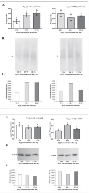
Fig. 2. A – CaE activity in brain tissue of 5th
no changes in CaE activity after expo-sure to the two B[a]P concentrations. A significant rise in brain CaE activity was observed in the group of larvae from PP fed on 5 ng/g DW of B[a]P, relative to the control group (one-way ANOVA, p<0.05). The higher concen-tration of B[a]P (50 ng/g DW) did not cause a significant change in enzyme activity. Detection of CaE activity on a native polyacrylamide gel uncovered two isoforms, I1 and I2 (Fig. 2B), with I1 being dominant in all experimental groups. It appears that the lower con-centration of B[a]P (5 ng/g DW) pro-voked a stronger expression of I1 and I2 isoforms in both L. dispar
popula-Fig. 3. A – AChE activity in brain tissue of the 5th instar L. dispar larvae from unpolluted (UP)
and polluted (PP) populations fed with an arti-ficial diet supplemented with 5 ng B[a]P/g DW and 50 ng B[a]P/g DW of benzo[a]pyrene (B[a] P). The control group of larvae was fed with an artificial diet without B[a]P (0 ng). The data are expressed as the mean±SEM, U/mg protein. Values marked with different letters indicate significant differences between groups (Tukey’s
post hoc test, p<0.05). B – Activity staining for AChE on a 10% polyacrylamide gel for UP and PP. The arrow (→) points to AChE activity of isoform I1. C – Densitometric analysis of po-lyacrylamide gels showing the relative levels of AChE isoform activities compared with the control group in UP and PP.
Fig. 4. A – Concentrations of Hsp70 and B – Western immunoblots of Hsp70 in brain tissue of 5th instar L. dispar larvae from unpolluted
tions when compared to the other B[a]P treatment and the control groups (Fig.2C).
AChE activity in L. dispar brain tissue was not significantly affected by the B[a]P dietary treatment (Fig. 3A) in either population. Native electrophoresis of brain tissue homogenates revealed the presence of the I1 isoform of AChE (Fig. 3 B). The relative inten-sity of this isoform corresponds to AChE activity in both populations of larvae, and there were no notable differences between the groups treated with different B[a]P concentrations and the control groups (Fig. 3C).
Concentration of Hsp70 in the brain of L. dispar
larvae
The concentration of Hsp70 estimated by indirect ELISA was significantly increased only in brain tissue homogenates of PP L. dispar, fed on the lower con-centration of B[a]P (5 ng/g DW) (one-way ANOVA, p<0.05). Hsp70 levels remained unchanged during the dietary treatment with B[a]P in the UP larvae, relative to the control group (Fig. 4A). Western blot analysis of Hsp70 revealed a single protein band at 70 kDa in all experimental groups of both UP and PP L. dispar
populations (Fig. 4B). Relative estimation of band den-sities followed the concentrations of Hsp70 (Fig. 4C).
DISCUSSION
Exposure to PAHs poses a great risk to herbivorous in-sects that live and feed on tree leaves in contaminated forests. Intermediates of B[a]P metabolism that can bind to nucleic acids and proteins have been shown to be very toxic as they are highly mutagenic and/or car-cinogenic for vertebrates and invertebrates, and stud-ies have demonstrated the direct impact of B[a]P on life history traits such as development and growth in insects [38]. Stress provoked by xenobiotics like B[a]P trigger the biochemical defense mechanisms (changes in enzyme activities, Hsp synthesis, etc.) that enable insect survival [39]. Large amounts of metabolic en-ergy are crucial for the successful coping with and elimination of toxicants, and the fitness-related traits of insects, development, growth and reproduction, are most commonly affected by this energy allocation [39]. As we described in our previous research [13], most energy resources are probably directed toward
the activation of the SOD-CAT system as the first line of defense against oxidative stress. Our present results showed that only the lower concentration of B[a]P (5 ng/g DW) had a harmful effect on the relative growth rate of L. dispar larvae in both populations of insects. This disruption of larval growth, which is apparently inconsistent with the applied B[a]P concentration, seems to be the characteristic response for this type of compound in L. dispar. Previous investigation of B[a]P and fluoranthene influence on L. dispar larval development and growth showed that mainly a lower concentration of these PAHs caused the greatest harm to larval fitness [15,40]. It is possible that the B[a] P mechanism of action resembles the class of com-pounds called endocrine disruptors (EDCs), which elicit reverse responses of different parameters as the concentration of disruptors increases, resulting in an U-shaped or inverted U-shaped curve [41]. Tomas [42] also found that B[a]P indeed displays effects like EDCs, specifically as an antiandrogenic compound.
PAH metabolism in insects is connected to several important enzyme systems of detoxification – micro-somal oxidases, glutathione S-transferases and carbox-ylesterases. Being a heterogeneous group of enzymes with the ability to hydrolyze different esters, CaE have a potential role in the degradation and elimination of various xenobiotics [43]. The results of the present study showed that the very low concentration of B[a] P (5 ng/g DW) significantly elevated brain CaE activ-ity in the PP L. dispar larvae, while the higher B[a]P concentration had no effect. Induction of CaE activity was not recorded in UP larvae at either B[a]P concen-tration. Native polyacrylamide gels revealed that the CaE I1 isoform is most probably responsible for B[a] P metabolism in larvae brain tissue, especially after exposure to 5 ng/g DW B[a]P in both the UP and PP
L. dispar populations. Callaghan et al. [44] proposed
that quantitative differences in CaE activity between populations are an adaptive mechanisms against en-vironmental xenobiotics. We presume that the high-er CaE sensitivity to B[a]P in the polluted L. dispar
clean forest locations. Esterase overexpression can be due to either gene amplification or upregulation, or a combination of both [43]. In our previous research, we showed a stronger induction of the midgut antioxida-tive enzymes, superoxide dismutase (SOD) and cata-lase (CAT) and the detoxification enzyme glutathione S-transferase (GST) in a polluted population than in ones from an unpolluted habitat, in response to B[a]P treatment [13]. Migula et al. [45] discovered increased CaE activity in red wood ants, Formica polyctena, from metal-polluted areas. Kapin and Ahmad [18] deter-mined that most of CaE activity in L. dispar larvae is located in the midgut, while the nerve cord and the brain contain predominantly AChE. On the basis of obtained results, it seems that in unpolluted popula-tions the applied B[a]P concentrations were too low to bypass the high concentration of CaE and other defense enzymes in the larval midgut epithelium, so that brain CaE isoforms were not activated.
The likely reason why we recorded only a signifi-cant change in CaE activity under chronic exposure to the lower B[a]P concentration (5 ng/g DW) in the polluted population may be due to the aggregation of enzyme molecules and decreased sensitivity to higher xenobiotic concentrations [46]. Also, the extended time of exposure to the higher B[a]P concentration (50 ng/g DW) probably caused direct and indirect inhibition of CaE activity due to the accumulation of reactive B[a]P intermediaries [47].
AChE is established as a major biomarker of insec-ticide and pesinsec-ticide exposure [48]. Although Jett et al. [24] and Kang and Fang [25] demonstrated that a num-ber of different PAHs, including B[a]P, inhibit AChE directly in vitro, the results of our study showed no significant changes in AChE activity under the dietary treatment with B[a]P (5 and 50 ng/g DW) in either of the two L. dispar populations. In all experimental groups, zymogram detection of AChE activity revealed discrete, barely visible lines on the gel corresponding to AChE, indicating moderate expression of this enzyme. It is very likely that this is a consequence of the high efficiency of the first line defense systems present in the midgut of L. dispar larvae [18,49] and the fact that PAHs are not natural, specific substrates for AChE. The concentrations of B[a]P present in host plant species
for L. dispar larvae used in our experiment probably
were not high enough to influence brain AChE activity
in any way. Ilijin et al. [50] found that B[a]P present in low concentrations of 2 and 10 ng/g DW in insect diet did not provoke changes in AChE activity of L. dispar
larvae from an unpolluted population, and neither did dietary intake of another PAH, fluoranthene, at low and high concentrations (6.7, 33,5 and 67 ng/g DW) [51].
Van Brummelen and Van Straalen [52] found, in their investigation of the uptake, elimination and tis-sue distribution of B[a]P in the isopod Porcellio sca-ber, that 14% of all absorbed B[a]P remains in the brain. Kapin and Ahmad [18] determined that the dominant esterase in the brain of L. dispar larvae is AChE while the content of CaE is much smaller. On the other hand, CaE are much more sensitive and re-sponsive to B[a]P presence than AChE, even in low concentrations of the contaminant. Other studies have shown that other xenobiotics have increased the affin-ity for CaE over AChE, suggesting that CaE activaffin-ity provides a more sensitive endpoint in the evaluation of environmental pollution [53,54]. The lack of inhibi-tion of AChE activity in L. dispar larvae in response to B[a]P chronic effects suggests that changes in other biochemical parameters, such as CaE, are probably a more convenient indicator of PAH exposure in this polyphagous insect species.
The adverse effects of B[a]P metabolism are par-ticularly pronounced in sensitive tissues like insect brain. Damaged proteins tend to aggregate in the cy-toplasm of the cell, which consequently provokes in-creased expression of heat shock proteins (Hsp70). By promoting the refolding of misfolded proteins and by removing excessively damaged ones, Hsp70 contrib-utes to the preservation of cell function and protects the cell from various stressors. Lee et al. [55] observed that PAHs and other types of chemical pollutants stim-ulate the expression of inducible and constitutive forms of Hsp70 in Chironomus tentans. An increase in Hsp70 expression was also recorded in aquatic invertebrates, oysters and mollusks, in response to exposure to PAHs and B[a]P [56]. Köhler et al. [27] reported a significant but transient induction of Hsp70 levels in the terres-trial isopod Oniscus asellus after exposure to B[a]P. The results of the present investigation revealed dif-ferent Hsp70 expression patterns among two L. dispar
larval brains from PP, and only under the influence of a lower concentration of pollutant (5 ng/g DW B[a]P) relative to the control group. Chronic treatment with B[a]P did not cause any meaningful changes in Hsp70 levels of UP larvae. One protein band of Hsp70 was detected by Western blot analysis in all experimen-tal groups of both populations of larvae, with discrete variations in band intensity. The obtained results are very similar to those recorded in larvae from the same populations (UP and PP) in terms of brain CaE activ-ity and the mentioned response of midgut SOD, CAT and GST enzymes [13], which indicates that L. dispar
larvae derived from a habitat burdened with pollutants demonstrate higher levels of synthesis and activity of vital, protective enzymes and proteins against the toxic influence of B[a]P. Perić-Mataruga et al. [16] also dis-covered differences in Hsp70 expression among two differently adapted L. dispar populations after exposure to temperature and heavy metal stress. Thus, larvae from the less polluted environment showed decreased amounts of Hsp70, while the polluted population ex-hibited a rise in Hsp70 concentration. It is possible that the larvae from UP also showed changes in Hsp70 levels at the beginning of the treatment with B[a]P, but since the stress protein system probably does not pos-sess the capacity to withstand long-term exposure to a toxicant, and as its prolonged expression consumes too much of the cellular energy needed for housekeeping metabolism, there was no measurable response at the endpoint of the experiment. Beside the possibility of involvement of additional detoxification systems in the elimination of the higher B[a]P concentration (50 ng/g DW), there is evidence that the nonlinear response of Hsp70 expression to pollutant dose is the consequence of the properties of B[a]P. As we indicated before, it appears that B[a]P induces a higher response from bio-chemical parameters at lower than at higher concentra-tions, which is quite similar to the patterns character-istic for endocrine disruptors. This type of behavior was observed by Dinan et al. [57] during fluoranthene treatment of Drosophila melanogaster cells. Ilijin et al. [50] reported that among six tested dietary doses of B[a]P, the largest induction of Hsp70 expression was under the chronic influence of the lowest (2 and 10 ng/g DW) B[a]P concentration. An analogous situa-tion was described in the research of Mrdaković et al. [51], where the lowest concentration of fluoranthene (6.7 ng/g DW) caused the greatest elevation of Hsp70
in the brain of L. dispar larvae. Also, Köhler et al. [27] found that high doses of B[a]P (0.5, 7.2 and 59.3 µg/g DW) induced a rise in Hsp70 in the isopod Oniscus
asellus after one day of exposure, which was followed
by a continuous decline in Hsp70. Authors have rec-ommended that for highly lipophilic compounds such as B[a]P, Hsp70 could be a good indicator of long-term exposure to sublethal toxic effects. Based on the response of Hsp70 in our present study, we suggest that it can serve as a potential biomarker of chronic exposure to B[a]P concentrations that are recorded in the environment and correspond to those used in this research. However, this possibility is only valid for L.
dispar populations that originate from habitats exposed
to some kind of pollution. Further research is advisable when we have in mind that Hsp70 is generally sensitive to cytotoxicity, responsive to a wide range of chemicals, and is involved in numerous physiological functions that take place in insects under stress [58].
CONCLUSION
Both populations of L. dispar larvae (UP and PP) were sensitive to the adverse effects of B[a]P present in the diet in two different concentrations, expressed as significantly lower RGR with respect to the control groups. Examination of the responses of biochemi-cal parameters, CaE and Hsp70 to B[a]P revealed an increase only in larvae from the polluted population. We did not detect any meaningful changes in AChE activity in response to B[a]P exposure. A very low B[a]P concentration (5 ng/g DW) provoked signifi-cant changes in brain CaE activity and Hsp70 level, reflecting their high inducibility in response to this xenobiotic. Although only the larvae population origi-nating from polluted forests were sensitive, a more comprehensive examination of the dose-dependent response of these parameters is required to support the use of these biomarkers as indicators of B[a]P and PAH pollution in forest ecosystems.
Funding: This study was funded by the Serbian Ministry of Edu-cation, Science and Technological Development, Grant No.173027.
Acknowledgments: The authors would like to thank Dragana
Matić for her moral support.
and interpreted the results. Marija Mrdaković, Milena Vlahović and Larisa Ilijin designed the study. Siniša Đurašević and Vesna Perić-Mataruga supervised the project and helped in work planning.
Conflict of interest disclosure: There is no conflict of interest.
REFERENCES
1. Douben PET. PAHs: An Ecotoxicological Perspective. West Sussex, UK: John Wiley and Sons; 2003. 392 p.
2. Fernandez P, Grimalt JO, Vilanova RM. Atmospheric gas/ particle partitioning of polycyclic aromatic hydrocarbons in high mountain regions of Europe. Environ Sci Technol. 2002;36:1163-8.
3. Wilcke W. Global patterns of polycyclic aromatic hydrocar-bons (PAHs) in soil. Geoderma. 2007;141:157-66.
4. US Environmental Protection Agency. Polycyclic organic mat-ter (POM) [Inmat-ternet]. Washington, D.C: US Environmental Protection Agency; 1992 Apr [updated 2000 Jan; cited 2013 Dec 16]. Available from: https://www.epa.gov/sites/produc-tion/files/2016-09/documents/polycyclic-organic-matter.pdf 5. EEA: Air Quality in Europe - 2012 Report, EEA Report No
4/2012 [Internet]. Copenaghen: European Environment Agency; 2012. [cited 2013 Dec 16]. Available from: https:// www.eea.europa.eu/publications/air-quality-in-europe-2012 6. Collins CD, Finnegan E. Modeling the plant uptake of organic
chemicals, including the soil-air-plant pathway. Environ Sci Technol. 2010;44:998-1003.
7. Desalme D, Binet P, Chiapusio G. Challenges in tracing the fate and effects of atmospheric polycyclic aromatic hydro-carbon deposition in vascular plants. Environ Sci Technol. 2013;47:3967-81.
8. De Nicola F, Concha Graña E, López Mahía P, Muniategui Lorenzo S, Prada Rodríguez D, Retuerto R, Carballeira A, Aboa JR, Fernández JÁ. Evergreen or deciduous trees for cap-turing PAHs from ambient air? A case study. Environ Pollut. 2017;221:276-84.
9. Howsam M, Jones KC, Ineson P. PAHs associated with the leaves of three deciduous tree species, I - concentrations and profiles. Environ Pollut. 2000;18:413-24.
10. Liebhold AM, Gottschalk KW, Muzika RM, Montgomery ME, Young R, O’Day K, Kelley B. Suitability of North American Tree Species to the Gypsy Moth: a Summary of Field and Laboratory Tests. Morgantown, WV, USA: Northeastern Forest Experiment Station, USDA Forest Service; 1995. 34 p. General Technical Report NE-211.
11. McCarty LS, Power M, Munkittrick KR. Bioindicators ver-sus biomarkers in ecological risk assessment. Hum Ecol Risk Assess. 2002;8(1):159-64.
12. Matić D, Vlahović M, Kolarević S, Perić-Mataruga V, Ilijin L, Mrdaković M, Vuković Gačić B. Genotoxic effects of cad-mium and influence on fitness components of Lymantria dispar caterpillars. Environ Pollut. 2016;218:1270-7. 13. Gavrilović A, Ilijin L, Mrdaković M, Vlahović M, Mrkonja
A, Matić D, Perić-Mataruga V. Effects of benzo[a]pyrene dietary intake to antioxidative enzymes of Lymantria dispar
(Lepidoptera:Lymantriidae) larvae from unpolluted and pol-luted forests. Chemosphere. 2017;179:10-9.
14. Van Brummelen TC, Stuijfzand SC. Effects of benzo[a]pyrene on survival, growth and energy reserves in the terrestrial iso-pods Oniscus asellus and Porcellio scaber. Sci Total Environ. 1993;134:921-30.
15. Mrdaković M, Ilijin L, Vlahović M, Todorović D, Gavrilović A, Mrkonja A, Perić-Mataruga, V. Effects offluoranthene on thefitness-related traits and antioxidative defense in Lyman-tria dispar L. Environ Sci Pollut Res. 2015;22:10367-74. 16. Perić-Mataruga V, Petković B, Ilijin L, Mrdaković M, Dronjak
Čučaković S, Todorović D, Vlahović M. Cadmium and high temperature effects on brain and behaviour of Lymantria dis-par. L. caterpillars originating from polluted and less-polluted forests. Chemosphere. 2017;185:628-36
17. Satoh T, Hosokawa M. Structure, function and regulation of carboxylesterases. Chem Biol Interact. 2006;162(3):195-211. 18. Kapin MA, Ahmad S. Esterases in larval tissues of gypsy
moth, Lymantria dispar (L.): Optimum assay conditions, quantification and characterization. Insect Biochem. 1980;10:331-7.
19. Jones BR, Bancroft HR. Distribution and probable physiologi-cal role of esterases in reproductive, digestive and fat-body tissue of adult cotton boll weevil, Anthonomus grandis. Bio-chem Genet. 1986;24:499-508.
20. Hemingway J, Hawkes N, McCarroll L; Ranson H. The molec-ular basis of insecticide resistance in mosquitoes. Insect Bio-chem Mol Biol. 2004;34(7):653-65.
21. Richardson BJ, Mak E, De Luca-Abott SB, Martin M, McLel-lan K, Lam PKS. Antioxidant responses to polyclic aromatic hydrocarbons and organochloride pesticides in green-lipped mussels (Perna viridis): Do mussels “integrate” biomarker response. Mar PollutBull. 2008;57:503-14.
22. Galloway TS, Handy R. Immunotoxicity of organophospho-rous pesticides. Ecotoxicology. 2003;12:345-63.
23. Senthil Nathan S, Choi MY, Seo HY, Paik CH, Kalaivani K, Kim JD. Effect of azadirachtin on acetylcholinesterase (AChE) activity and histology of the brown planthopper
Nilaparvata lugens (Stål). Ecotox Environ Safe. 2008;70:244-50.
24. Jett DA, Navoa RV, Lyons MAJr. Additive inhibitory actions of chlorpyrifos and polycyclic aromatic hydrocarbons on acetyl-cholinesterase activity in vitro. Toxicol Lett. 1999;105:223-9. 25. Kang JJ, Fang HW. Polycyclic aromatic hydrocarbons inhibit the activity of acetylcholinesterase purified from electric eel. Biochem Biophys Res Commun. 1997;238:367-9.
26. Lewis S, Handy RD, Cordi B, Billinghurst Z, Depledge MH. Stress proteins (HSP’s): methods of detection and their use as an environmental biomarker. Ecotoxicology. 1999;8:351-69. 27. Köhler HR, Knödler C, Zanger M. Divergent kinetics of hsp70
induction in Oniscus asellus (isopoda) in response to four environmentally relevant organic chemicals (B[a]P, PCB52, g-HCH, PCP): suitability and limits of a biomarker. Arch Environ Contam Toxicol. 1999;36:179-85.
28. Morales M, Planello R, Martínez-Paz P, Herrero O, Cortes E, Martínez Guitarte, JL, Morcillo, G. Characterization of Hsp70 gene in Chironomus riparius: expression in response to endo-crine disrupting pollutants as a marker of ecotoxicological stress. Comp Biochem Physiol C. 2011;153:150-8.
particle-bound phase in schools at different locations in Ser-bia. Chem Ind Chem Eng. Q. 2015;21:159-67.
30. Alagić S, Stankov-Jovanović V, Mitić V, Cvetković J, Petrović G, Stojanović G. Bioaccumulation of HMW PAHs in the roots of wild blackberry from the Bor region (Serbia): phy-toremediation and biomonitoring aspects. Sci Total Environ. 2016;562:561-70.
31. O’Dell TM, Butt CA, Bridgeforth AW. Lymantria dispar. In: Singh P, Moore RF, editors. Handbook of Insect Rearing. New York: Elsevier; 1985. p. 355-67.
32. Bradford MM. A rapid and sensitive method for the quanti-fication of microgram quantities of protein utilizing the prin-ciple of proteindye binding. Anal Biochem. 1976:72:248-54. 33. Main AR, Miles KE, Braid PE. The Determination of
Human-Serum-Cholinesterase Activity with o-Nitrophenyl Butyrate. Biochem J. 1961;78:769-76.
34. Ellman GL, Courtney KO, Anders V, Featherstone RM. A new and rapid colorimetric determination of acetylcholinesterase activity. Biochem Pharmacol. 1961;7:88-95.
35. Laemmli UK. Cleavage of structural proteins during the assembly of the head of bacteriophage T4. Nature. 1970;227:680-5.
36. Gottlieb LD. Genetic confirmation of the origin of Clarkia lingulata. Evolution. 1974;28:244-50.
37. Karnovsky MJ, Roots L. A “Direct-coloring” thiocho-line method for chothiocho-linesterase. J Histochem Cytochem. 1964;12:219-21.
38. Prud’homme SM, Chaumot A, Cassar E. David JP, Reynaud S. Impact of micropollutants on the life-history traits of the mosquito Aedes aegypti: On the relevance of transgenera-tional studies. Environ Pollut. 2017;220(Part A):242-54. 39. Van Straalen NM, Hoffmann, A. Review of experimental
evi-dence for physiological costs of tolerance to toxicants. In: Kam-menga J, Laskowski R, editors. Demography in Ecotoxicology. Chichester, UK: John Wiley & Sons Ltd.; 2000. p. 147-61. 40. Ilijin L, Mrdaković M, Todorović D, Vlahović M, Gavrilović
A, Mrkonja A, Perić-Mataruga V. Life history traits and the activity of antioxidative enzymes in Lymantria dispar L. (Lepi-doptera, Lymantriidae) larvae exposed to benzo[a] pyrene. Environ Toxicol Chem. 2015;34(11):2618-24.
41. Bergman A, Heindel JJ, Jobling S, Kidd KA, Zoeller RT, edi-tors. State of the science of endocrine disrupting chemicals - 2012 : an assessment of the state of the science of endocrine disruptors prepared by a group of experts for the United Nations Environment Programme (UNEP) and WHO. Geneva, Switzerland: United National Environment Pro-gramme; 2013. 296 p.
42. Thomas P. Teleost model for studying the effects of chemicals on female reproductive endocrine function. J Exp Zool Suppl. 1990;4:126-8.
43. Panini M, Manicardi GC, Moores GD, Mazzoni E. An over-view of the main pathways of metabolic resistance in insects. Invert Surviv J. 2016;13:326-35.
44. Callaghan A, Parker PJAN, Holloway GJ. The use of variance in enzyme activity as an indicator of long-term exposure test to toxicant-stressed environments in Culex pipens mosqui-toes. Funct Ecol. 1998;12:436-44.
45. Migula P, Glowacka E, Nuorteva SL, Nuorteva P, Tulisalo E. Time related effects of intoxication with cadmium and
mer-cury in the red wood ants (Formica aquilonia). Ecotoxicology. 1997;6:307-20.
46. Vlahović M, Perić-Mataruga V, Ilijin L, Mrdaković M, Mirčić D, Todorović D, Lazarević J. Changes in activity of non-spe-cific esterases in cadmium treated Lymantria dispar larvae. Ecotoxicology. 2012;21:370-8.
47. Miller KP, Ramos KS. Impact of cellular metabolism on the biological effects of benzo[a]pyrene and related hydrocar-bons. Drug Metab Rev. 2001;33:1-35.
48. Lionetto MG, Caricato R, Calisi A, Schettino T. Acetylcholin-esterase inhibition as a relevant biomarker in environmental biomonitoring: New insights and perspectives. In: Visser JE, editor. Ecotoxicology around the Globe. New York: Nova Sci-ence Publishers Inc.; 2011. p. 87-115.
49. Perić-Mataruga V. Vlahović M, Mrdaković M, Matić D, Gavrilović A, Mrkonja A, Ilijin L. Ghrelin effects on midgut tissue and antioxidative defense and gluthatione S-transferase activity in Lymantria dispar (Lepidoptera). Turkish J Biol. 2015;39:618-23.
50. Ilijin L, Mrdaković M, Vlahović M, Matić D, Gavrilović A, Mrkonja A, Perić-Mataruga V. Acetylcholinesterase and heat shock protein 70 response in larval brain tissue of Lymantria dispar L. (Lepidoptera, Limantriidae) upon chronic exposure to benzo[a]pyrene. Environ Sci Pollut Res. 2017;24(25):20818-23.
51. Mrdaković M, Ilijin L, Vlahović M, Matić D, Gavrilović A, Mrkonja A, Perić-Mataruga V. Acetylcholinesterase (AChE) and heat shock proteins (Hsp70) of gypsy moth (Lymantria dispar L.) larvae in response to long-term fluoranthene expo-sure. Chemosphere. 2016;159:565-9.
52. Van Brummelen TC, Van Straalen NM. Uptake and elimi-nation of benzo[a]pyrene in the terrestrial isopod Porcellio scaber. Arch Environ Contam Toxicol. 1996;31(2):277-85. 53. Escartin E, Porte C. The use of cholinesterase and
carboxy-lesterase activities from Mytilus galloprovincialis in pollution monitoring. Environ Toxicol Chem. 1997;16;2090-5. 54. Wogram J, Sturm A, Segner H, Liess M. Effects of parathion
on acetylcholinesterase, butyrylcholinesterase, and carboxy-lesterase in three-spined stickleback ( Gasterosteus aculea-tus) following short-term exposure. Environ Toxicol Chem. 2001;20:1528-31.
55. Lee SM, Lee SB, Park CH, Choi J. Expression of heat shock protein and hemoglobin genes in Chironomus tentans (Dip-tera, Chironomidae) larvae exposed to various environmental pollutants: a potential biomarker of freshwater monitoring. Chemosphere. 2006;65:1074-81.
56. Cruz-Rodriguez LA, Chu FLE. Heat-shock protein (HSP70) response in the eastern oyster, Crassostrea irginica, exposed to PAHs sorbed to suspended artificial clay particles and to suspended field contaminated sediments. Aquatic Toxicol. 2002;60:157-68.
57. Dinan L, Bourne P, Whiting P, Dhaidalla TS, Hutchinson TH. Screening of environmental contaminants for ecdys-teroid agonist and antagonist activity using the Drosophila melanogaster BII cell in vitro assay. Environ Toxicol Chem. 2001;20:2038-46.
![Fig. 1. RGR from molting to the 3rd instar until the 3rd day of the 5th instar of L. dispar larvae from unpolluted (UP) and polluted (PP) popu-lations fed with an artificial diet supplement-ed with 5 ng B[a]P/g DW and 50 ng B[a]P/g DW of benzo[a]pyrene (B[](https://thumb-us.123doks.com/thumbv2/123dok_us/7807016.2085291/5.595.55.351.82.719/molting-instar-unpolluted-polluted-lations-artificial-supplement-pyrene.webp)