REVIEW
Magnetic resonance imaging of sellar and juxtasellar
abnormalities in the paediatric population: an imaging review
Rachel Shields&Rajiv Mangla&Jeevak Almast& Steven Meyers
Received: 4 November 2014 / Revised: 11 February 2015 / Accepted: 17 February 2015 / Published online: 21 March 2015 #The Author(s) 2015. This article is published with open access at Springerlink.com
Abstract The sellar and juxtasellar regions in the paediatric population are complex both anatomically and pathologically, with magnetic resonance imaging (MRI) being theBgold stan-dard^ imaging modality due to the high contrast of detail. Assessment requires a detailed understanding of the anatomy, embryology, pathophysiology and normal signal characteris-tics of the pituitary gland and surrounding structures in order to appropriately characterise abnormalities. This article aims to provide an overview of the imaging characteristics of developmental/congenital and acquired disease processes which affect the sellar and juxtasellar region in the paediatric population.
Main Messages
• The sellar region is anatomically complex and covers a wide pathology spectrum.
• MRI is the key imaging modality to assess sellar and juxtasellar pathology.
• Numerous developmental anomalies may not be discovered until adulthood.
• Knowledge of pathology alerts and guides the clinician towards appropriate management.
Keywords Magnetic resonance imaging . Paediatrics . Pituitary gland . Pituitary diseases . Sella turcica
Pituitary function and anatomy
The pituitary gland is comprised of two dominant lobes, the neurohypophysis and the adenohypophysis. It is situated with-in the sella turcica, a depression with-in the sphenoid bone which is covered by the diaphragm sella with a defect for the passage of the infundibulum. The pituitary stalk extends inferiorly as a continuation of the hypothalamic infundibulum (Fig.1).
The neurohypophysis receives its hormonal supply through direct contiguity with the hypothalamus, with axons extending from the hypothalamic supraoptic and paraventricular nuclei into the neurohypophysis. The hormonal supply includes oxy-tocin and vasopressin-containing vesicles. The adenohypoph-ysis does not have axonal contiguity between the hypothala-mus and the anterior gland and relies on the hypothalamic-hypophyseal portal system. Parvocellular messengers are transported from the hypothalamic median eminence into the capillaries surrounding the anterior gland. Hormones from the adenohypophysis include: follicle stimulating hormone, lu-teinising hormone, corticotropin (ACTH), thyroid-stimulating hormone, prolactin and growth hormone.
Embryology
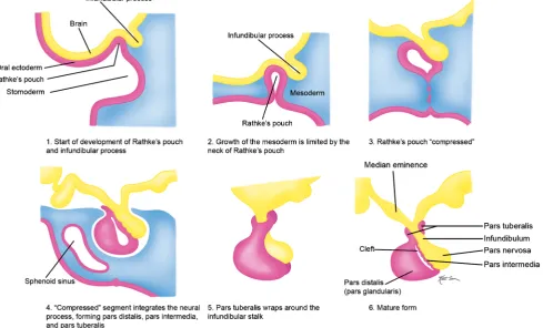
The pituitary gland develops through a complex mechanism involving the developing diencephalon and the primitive oral stomodeum [1]. The adenohypophysis derives from Rathke’s pouch, which originates rostral to the oropharyngeal mem-brane. This begins at approximately 24 days of gestation, in which Rathke’s pouch forms as an ectodermal outpouching of stomodeum and the infundibulum which forms in the floor of the diencephalon. The infundibulum grows ventrally, while simultaneously Rathke’s pouch grows dorsally. Rathke’s pouch eventually loses its connection with the stomodeum
R. Shields (*)
:
R. Mangla:
J. Almast:
S. Meyersand forms a sac which adheres to the infundibular process. This sac continues to differentiate into the adenophyophysis and the distal aspect of the infundibulum differentiates to form the neurohypophysis (Fig.2).
Imaging characteristics in the paediatric population
The pituitary gland exhibits a wide range of appearances dur-ing the first 2 years of life, with considerable variability in
Fig. 1 Normal pituitary anatomy
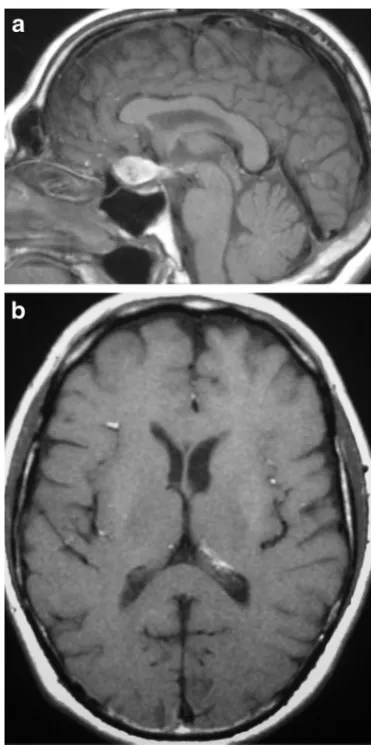
size, shape, and signal. At birth, both the anterior and posterior pituitary gland have high signal intensity on T1-weighted im-ages (Fig.3). The anterior pituitary progressively decreases in signal intensity on T1-weighted images with signal drop to near isointensity with the posterior pons by 6 weeks of age [1,2]. The posterior pituitary gland retains its high signal intensity on T1-weighted images throughout adulthood, with several papers supporting that this high signal results from neurosecretory granules or the protein neurophysin [3–5]. The normal gland enhances symmetrically after intravenous gadolinium with the anterior gland enhancement appearing more noticeable given the posterior gland is already high sig-nal at baseline.
At birth, the gland is physiologically enlarged with a cor-responding concave superior margin, flattening with age. The gland is traditionally symmetric in size; however there can be asymmetry in normally functioning glands. In preterm infants,
the gland is taller than in normal-term infants, even when correcting for gestational age. This is thought to be the result of reduced insulin-like growth factor 1 and higher levels of growth hormone in premature infants [6]. The pituitary height is fairly stable for the first 2 years of life in normal-term chil-dren. Gender-associated differences in the rate of gland growth tend to occur after 5 years of age, with linear growth of the gland up to that point in both boys and girls [7]. Typi-cally, the normal pituitary measures between 2 to 6 mm in height during childhood and up to 10 mm or slightly higher at puberty. In adults, the female glands are slightly larger than their male counter parts [1]. Standards for assessing the pitu-itary stalk size are not well established, and are based on experience rather than age-appropriate measurements. The stalk is best visualised on post-contrast T1-weighted images because of the strong enhancement.
Developmental anomalies of the pituitary gland
Agenesis of the adenohypophysis (Fig. 4) This is a rare congenital anomaly with a neonatal presentation. The neo-nates can present with metabolic acidosis, thyroid and adrenal insufficiency, hypoglycaemia, and/or seizures. Associated anomalies of the forebrain and midline craniofacial bones can be present. Newborns are normal in size at birth, which has been explained by the presence of maternal growth hor-mone. Standard treatment includes replacement of growth hormone, thyroid hormone and glucocorticoids. Magnetic res-onance imaging (MRI) findings include the absence of the anterior pituitary gland with an associated shallow sella, and variable length of the infundibulum and position of the poste-rior pituitary, ranging from the hypothalamus to the sella [8].
Fig. 3 Normal physiological T1 sagittal (a) and axial (b) hyperintensity of the anterior pituitary in a 1-month-old baby
The infundibulum may be thinned and terminate into a par-tially formed bulbous posterior pituitary gland.
Hypoplasia of the adenohypophysis (Fig.5) This rare anomaly consists of hypoplasia of the anterior pituitary gland, and also potentially the infundibulum and posterior pituitary gland. It can be associated with varying degrees of endocrine dysfunction, including panhypopituitarism and other congen-ital anomalies. Some research suggests that traumatic transec-tion of the pituitary stalk during birth or hypoxic injury to the hypothalamus could result in hypoplasia, whereas others sug-gest an error during embryogenesis [9–12]. MRI findings in-clude varying degrees of hypoplasia of the anterior pituitary gland with variable size and length of the infundibulum. As with agenesis of the adenohypophysis, the posterior pituitary position can range from the hypothalamus to the sella.
Ectopic posterior pituitary (Fig.6) This congenital anom-aly results in an aberrant position of the neurohypophysis, often located at the undersurface of the hypothalamus. It is associated with pituitary dwarfism, delayed skeletal matura-tion, Kallman syndrome, septo-optic dysplasia, dysgenesis of the corpus callosum, lobar holoprosencephaly, Chiari 1 mal-formation or a persistent/enlarged craniopharyngeal canal. It affects males more than females. MRI may show a small
anterior pituitary gland with the posteriorBbright spot^ on T1-weighted imaging (T1WI) located within the upper portion of the infundibulum or the undersurface of the hypothalamus instead of the dorsal portion of the sella [1,13–15].
Pituitary duplication (Fig.7) This congenital anomaly is related to abnormal separation of the anterior portion of the notochord and prechordal plate during embryogenesis, likely due to teratogenic factors occurring during early embryogen-esis [16, 17]. Clinical findings include precocious puberty, facial dysmorphism, hypothyroidism and/or increased prolac-tin levels. Pituitary duplication may be associated with other anomalies such as dysgenesis of the corpus callosum, absent olfactory bulbs, cerebellar hypoplasia, midline defects, and teratomas. MRI findings include duplicated pituitary stalks and glands within separate pituitary fossa in the sphenoid bone. Abnormal thickening,Bpseudohamartoma^, of the hy-pothalamus with duplication of the infundibular recess of the third ventricle is often seen [16,18]. Pituitary duplication can also be associated with duplication of the basilar artery [19].
Hypothalamic hamartoma (Figs.8and9) Congenital de-velopmental non-neoplastic grey matter heterotopia involving the tuber cinereum, inferior hypothalamus and/or mammillary bodies which are composed of small neuronal cells within a neutrophil-like stroma and scattered fibrillary astrocytes.
Fig. 5 Severe hypoplasia of the anterior pituitary gland. Pre-contrast (a) and post-contrast (b) MR images show severe hypoplasia of the anterior pituitary gland which shows only minimal contrast enhancement along the floor of the sella. The ectopic posterior pituitary is located adjacent to the optic chiasm
These lesions typically occur in children with isosexual pre-cocious puberty (0-8 years) or seizures, both gelastic or partial complex, in the second decade [20,21]. They can be associ-ated with holoprosencephaly, midline facial, cardiac and renal anomalies. MRI demonstrates sessile or pedunculated lesions at the tuber cinereum of the hypothalamus, often intermediate signal on T1WI and T2WI similar to grey matter. Lesions occasionally have slightly high signal on T2WI, with no con-trast enhancement or restricted diffusion. Rarely contain cystic and/or fatty portions may be seen in a small minority of cases. The absence of long-term change in size or signal intensity supports the diagnosis [22]. Ictal FDG-PET and SPECT show
hyperperfusion during seizures. On MR spectroscopy, there may be an elevated myoinositol peak.
Persistent craniopharyngeal canal (Fig.10) Lack of in-volution of the craniopharyngeal canal, which can contain pituitary tissue. The adenohypophysis develops from evagina-tion of ectodermal cells at the upper posterior porevagina-tion of the primitive oropharynx, Rathke’s pouch, at 4-5 weeks of gesta-tion. Rathke’s pouch passes between the developing presphenoid and basisphenoid chondrification centres at the upper clivus and sella, where it associates with the descending neuroectoderm of the infundibulum and will go on to form the neurohypophysis. The pathway of Rathke’s pouch is the craniopharyngeal canal, which progressively involutes by 6-7 weeks of gestation. The persistent craniopharyngeal canal o n M R I c a n b e a s s o c i a t e d w i t h t r a ns - s p h e no i d al meningoencephaloceles, ectopic pituitary tissue, sphenoid ter-atoma and infrasellar craniopharyngiomas [23,24]. On MRI, the persistent canal has low T1 signal intensity compared with the high T1 signal of the clivus [13].
Rathke’s cleft cyst (Fig.11) Benign cystic lesion contain-ing fluid and variable amounts of protein, mucopolysaccha-rides and/or cholesterol [25,26]. The cysts arise from epithe-lial rests of the craniopharyngeal canal. They are 2-3 times more common in women than men, and occasionally become symptomatically related to compression of adjacent structures. The frequency of these cysts on MRI in one study was 1.2 % in childhood [27]. MRI will show a well-circumscribed lesion with variable low, intermediate or high signal on T1WI and T2WI. On T2WI, a small low-signal nodule may be seen within the predominant high signal of the lesion, which does not enhance after contrast administration. The incidence of these intracystic nodules varies from 17 to 78 % [28,29]. There is no contrast enhancement centrally; however, occa-sionally there may be a thin peripheral rim of enhancement from associated inflammation. The majority are located in the intrasellar region or combined intrasellar and suprasellar re-gion with reports that only 8.3 % are exclusively suprasellar [30,31]. Most are stable in size, and some may disappear spontaneously. Pars intermedia cysts (Fig.12) can also form along the posterior margin of Rathke’s pouch, are typically less than 3 mm, with many considered Rathke’s cleft cysts on imaging as they are difficult to distinguish from one anoth-er and Rathke’s cleft cysts are more common [1].
Cephaloceles (meningoceles or meningoencephaloceles) (Fig.13) Congenital malformation involving the lack of separation of neuroectoderm from surface ectoderm with re-sultant localised failure of bone formation. There is a wide range of abnormalities which affect the skull base within the sella turcica, ranging from a persistent craniopharyngeal canal to trans-sphenoidal and sphenoethmoidal cephaloceles, many
presentations of which are extremely rare. For example, the reported incidence of trans-sphenoidal meningoencephaloceles is 1 per 700,000 live births [13,32]. Clinical findings include difficulty in feeding and nasal obstruction in the first year, and potential for cerebrospinal fluid (CSF) leaks and meningitis. MRI will show a defect in the skull, through which there is either herniation of meninges and CSF (meningocele), or me-ninges, CSF and brain tissue (meningoencephaloceles).
Epidermoid (congenital cholesteatoma) (Fig.14) Non-neoplastic congenital or acquired extra-axial off-midline le-sion filled with desquamated cells and keratinaceous debris, usually with mild mass effect on the adjacent brain. Epider-moids grow by accumulation of desquamated epithelial cells into the centre of the cyst [33–35]. MRI will show a well circumscribed spheroid or multilobulated extra-axial ectoder-mal inclusion cystic lesion with low-intermediate signal on T1WI, high signal on T2WI and no contrast enhancement. They can be distinguished from arachnoid cysts, as they are typically high signal on FLAIR and DWI sequences, with corresponding low signal on ADC [36].
Dermoid (Fig.15) Non-neoplastic congenital or acquired ectodermal inclusion cystic lesion, which like an epidermoid, is lined by squamous epithelium, and are filled with lipid material, cholesterol, desquamated cells and keratinaceous de-bris. They are distinguished from epidermoids by containing deeper dermal elements and commonly arise from the midline. They typically present in the 20- to 30-year age range, but can also be seen in adolescence, with males slightly more affected than females [36]. MRI will show a well-circumscribed spher-oid or multilobulated extra-axial lesion, usually with high sig-nal on T1WI, variable sigsig-nal on T2WI and no contrast en-hancement. Fluid/fluid or fluid/debris levels may be present. They can cause a chemical meningitis if the dermoid cyst ruptures into the subarachnoid space [37,38].
Arachnoid cyst (Fig. 16) Congenital arachnoid cysts are found in 0.16 % of all newborns with approximately 9-15 % occurring in the sellar region, with suprasellar cysts more commonly seen than intrasellar cysts. Most cases are asymp-tomatic; however, they may cause headache, optic nerve com-pression, psychomotor retardation, endocrine dysfunction or
Fig. 8 Hypothalamic
hydrocephalus [39,40]. Secondary arachnoid cysts may result from trauma, haemorrhage or infection [41]. On imaging, the cysts have low signal intensity on T1WI, FLAIR and DWI, high signal intensity on T2WI, no contrast enhancement, and may cause mild mass effect on adjacent brain.
Lipoma (Fig. 17) Benign fatty lesions resulting from a congenital malformation, often located at or near the mid-line. In the sellar region, they occur along the surface of the infundibulum, floor of the third ventricle, or adjacent to cranial nerves, and are usually discovered incidentally, un-less they cause a mass effect [42, 43]. They may contain calcifications and/or traverse blood vessels. MRI signal is
Fig. 9 Tuber cinerum hamartoma. Sagittal (a) and coronal (b) T1WI, and coronal post-contrast T1WI (c) show a non-enhancing bulky soft tissue mass in the region of the of the tuber cinerum
isointense to subcutaneous fat on all sequences, with fat suppressed images useful in distinguishing from haemorrhagic or proteinaceous lesions.
Empty sella (Fig.18) Common incidental finding involv-ing the pituitary gland and sella, which can result from a weakened or fenestrated diaphragma sella allowing CSF pul-sations to flatten the pituitary gland. They are associated with elevated intracranial pressure, a posteriorly placed optic chi-asm, and are considered a normal variant or termedBprimary
empty sella turcica^in the absence of surgery, radiation ther-apy or medically treated intrasellar tumour [44]. In children, the incidence of an empty sella is seen more often in boys and is more likely to be associated with clinical symptoms and endocrinopathies, particularly growth hormone or multiple pituitary hormone deficiencies [45]. MRI findings include a thinned/flattened pituitary gland along the floor of the sella, with the infundibulum in its normal midline position. The flattened pituitary gland has normal MRI signal characteristics and contrast enhancement.
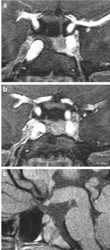
Fig. 11 Rathke’s cleft cyst. Sagittal T1WI (a) shows a lesion in the central portion of the pituitary gland which has high signal. Post-contrast sagittal fat-suppressed T1WI (b) shows lack of enhancement of the Rathke cleft cyst. The cyst has intermediate signal on T2WI (c) secondary to slightly elevated protein content
Pituitary lesions
Pituitary adenoma (Fig.19) In comparison to adults, pi-tuitary adenomas are rare in the paediatric population, ac-counting for approximately 2.7 % of all supratentorial tu-mours. Most paediatric adenomas are functioning, with only 3-6 % functioning, compared to one-third of non-functioning adenomas in adults [46–48]. Microadenomas may be associated with endocrine abnormalities related to over-secretion of hormones, with prolactinomas being the most common. Rarely, extensive haemorrhage can occur in-volving the adenoma, resulting in pituitary apoplexy (Sheehan syndrome). Macroadenomas with suprasellar extension can cause compression and displacement of the optic chiasm with associated visual disturbances (bitemporal hemianiopsia). Most pituitary adenomas arise from sporadic mutations; how-ever, 5 % can be associated with inherited disorders such as McCune Albright syndrome, Carney complex and multiple endocrine neoplasia type 1 (MEN 1).
MRI findings of a microadenoma (<10 mm) commonly have intermediate signal on T1WI and T2WI, and may show char-acteristics of cysts, haemorrhage or necrosis. They typically show less contrast enhancement than normal pituitary tissue, often best visualised with dynamic early-phase imaging. In macroadenomas (>10 mm), they commonly have intermediate signal on T1WI and T2WI similar to grey matter, and may
Fig. 13 Cephalocele in a 17-year-old girl. Coronal T2WI (a) and sagittal T1WI (b) shows a meningocele extending inferiorly into the nasopharynx through an osseous defect in the sphenoid bone posterior to the pituitary gland
also have cystic, haemorrhagic or necrotic components. They can extend into the suprasellar cistern with a waist at the dia-phragm sella or into the cavernous sinus, with occasional in-vasion into the skull base.
Juxtasellar lesions
Craniopharyngioma (Fig.20) Usually histologically be-nign locally aggressive lesions which arise from squamous epithelial rests along Rathke’s cleft, occurring in a bimodal age distribution, in children (<10 years) and adults (>40 years). Craniopharyngioma’s account for 3 % of all intracranial tu-mours. They are categorised into adamantinomatous and squamous-papillary types. The adamantinomatous type is more common and has a bimodal age distribution occurring in children and adults, whereas the squamous-papillary type usually occurs in adults. Although histologically benign, their
Fig. 15 Dermoid. Sagittal (a) and axial (b) T1WI shows an extra-axial lesion in the suprasellar cistern which has high signal. Multiple small foci with high signal are seen in the sulci and within the left lateral ventricle representing ruptured dermoid contents in the leptomeninges
Fig. 16 Arachnoid cyst. Sagittal T1WI (a) and coronal T2WI (b) show a well defined cystic mass with low T1 and high T2 signal intensity with sellar and suprasellar components
insinuating pattern of growth makes complete surgical exci-sion very difficult and not often possible.
The adamantinomatous type usually demonstrates circumscribed lobulated margins on MRI, with the suprasellar location seen at a greater frequency than intrasellar. They show variable low, intermediate and/or high signal on T1WI and T2WI, with or without a nodular or rim of contrast en-hancement. They may contain cysts, lipid components and calcifications. In children, the cystic parts tend to predomi-nate, and frequently contain highly proteinaceous fluid, which may be bright on T1WI [49].
Glioma (Fig.21) Lesions which may occur anywhere along the optic pathway and account for 4-6 % of brain tu-mours in children, with a median age of diagnosis from 5-9 years. Approximately half of patients with optic pathway gli-omas have neurofibromatosis type 1 (NF1), with symptomatic lesions only occurring in 1-5 % of patients [50]. Most tumours are slow growing WHO grade I-II astrocytomas, often the pilocytic type, however tumours may show a wide spectrum of progression. On MRI, there is fusiform and/or nodular en-largement of the optic chiasm and/or optic nerves with thick-ening of the third ventricular floor and hypothalamus. They usually have low-intermediate signal on T1WI, intermediate-high signal on T2WI and variable contrast enhancement. Larger lesions may have cystic components and grow directly into the pituitary stalk [1].
Pilomyxoid astrocytoma (Fig.22) Rare neoplasms (WHO grade II) which contain prominent mucoid matrix and angiocentric pattern of bipolar neoplastic astrocytes. Typical-ly, they occur in children in the first and second decades, with a mean age of 7 years [51]. They tend to occur at a younger age and are more aggressive with a higher rate of local recur-rence than pilocytic astromcytomas. They have a strong pre-dilection for the hypothalamus and suprasellar cistern [52]; however, they can also occur in the thalamus, cerebellum,
brainstem, temporal lobe and spinal cord. The presence of intra-tumoural haemorrhage is the most prominent imaging characteristic to suggest a pilomyxoid astrocytoma over a pilocytic astrocytoma, 25 % versus 8 %, respectively [51]. MRI demonstrates a solid/cystic focal lesion with low-intermediate signal on T1WI, high signal on T2WI and FLAIR, variable contrast enhancement and may show in-creased ADC values.
Germ cell tumours—germinoma (Fig.23) Extra-gonadal germ cell tumours include: germinoma (most common), ma-ture teratoma, malignant teratoma, yolk sac tumour, embryo-nal carcinoma and choriocarcinoma. They account for 0.6 %
Fig. 18 Empty sella. Sagittal post-contrast T1WI with fat-suppression show an enlarged sella mostly filled with CSF. The enhancing flattened pituitary tissue is located along the floor of the sella
of primary intracranial tumours, with an incidence of 0.09 per 100,000. Peak incidence occurs between 10 and 14 years, with 90 % occurring in patients less than 25 years. They occur more frequently in males. Prognosis depends on the histological subtype, with a 10 year survival rate for germinomas of over 85 %. Other germ cell tumours have a lower survival rate, particularly those containing non-germinomatous malignant cells. Tumours often have intermediate signal on T1WI, slightly high signal on T2WI, and show contrast enhance-ment. They are typically centred on the pituitary stalk with the posterior pituitary bright spot often absent. They may look similar to Langerhans’cell histiocytosis (LCH) or lymphocyt-ic hypophysitis [53]. Contrast-enhancing disseminated sub-arachnoid and/or intraventricular tumour may occur through CSF seeding.
Germ cell tumours—teratoma (Fig.24) The second most common type of germ cell tumour behind a germinoma. They occur in children, with a greater frequency in boys, accounting for 0.5-1.5 % of all childhood brain tumours, and >50 % of intracranial tumours in infants less than 2 months [54,55]. Histologically, they demonstrate benign or malignant types, composed of derivatives of ectoderm, mesoderm and/or endo-derm. On MRI, they are relatively circumscribed lesions, most common in decreasing order frequency within the pineal re-gion, suprasellar rere-gion, and third ventricle. On MRI, they
demonstrate heterogeneous signal intensity on T1WI, T2WI and post-contrast sequences, with suppression of signal on fat-saturated imaging. Marked enhancement of the solid portions of the tumour is a key feature in distinguishing a mature versus a malignant teratoma, with malignant tumours demonstrating enhancement. Additionally, they typically present without perilesional high T2 signal due to intact capsules and an un-damaged blood-brain barrier [55]. They may contain calcifi-cations with low signal on T1WI and T2WI.
Tuberculous meningitis (Fig. 25) Mycobacterium tuberculosisinfection remains a major international concern, with increasing burden of disease in developed nations and young children being the most susceptible [56,57]. MRI is increasingly being used to evaluate patients with suspected tuberculous meningitis, to aid in rapid early diagnosis [58]. MRI findings include basal leptomeningeal enhancement sec-ondary to multiple granulomas and pial tuberculomas. The granulomas can be either non-caseating with homogeneous
Fig. 21 Glioma in a 2-year-old girl with neurofibromatosis type 1. Sagittal (a) and axial (b) post contrast T1WI shows a partially enhancing lesion enlarging the optic chiasm and proximal optic nerves Fig. 20 Craniopharyngioma. Sagittal T1WI (a) shows a lobulated lesion
enhancement or caseating with rim enhancement (both T1 hypointense and T2 hyperintense) [59].
Osteosarcoma (osteogenic sarcoma)Rare malignant bone tu-mour comprised of proliferating neoplastic spindle-cells which produce osteoid and/or immature tumoural bone. They usually involve the endochondral bone-forming portions of the skull-base and are locally invasive with high metastatic potential. Over 75 % of osteosarcomas are diagnosed in pa-tients between 8 and 25 years, and account for less than 1 % of all head and neck malignant tumours in the paediatric popu-lation [60]. They occur in children as primary tumours and adults associated with Paget’s disease, irradiated bone, chronic osteomyelitis, osteoblastoma, giant cell tumour and fibrous dysplasia. MRI will show a destructive lesion involving the skull base, with low-intermediate signal on T1WI, mixed low, intermediate or high signal on T2WI. Usually contains matrix
mineralisation/ossification with low signal on T2WI and het-erogeneous contrast enhancement.
Ewing sarcomaBone tumour usually occurring between ages 5 and 30 years, with a greater prevalence in males and is the second most common primary bone malignancy affecting ado-lescents and young adults. It most commonly arises in the trunk and diaphysis of long bones, with only 4 % of cases within the head and neck region [61]. Tumours are locally invasive with a high metastatic potential. MRI will show a destructive lesion involving the skull base, with low-intermediate signal on T1WI, mixed low, intermediate or high signal on T2WI, with or without matrix mineralisation showing low signal on T2WI and heterogeneous contrast enhancement.
Esthesioneuroblastoma (olfactory neuroblastoma) (Fig.26) These malignant neoplasms of neuroectodermal origin, arise from olfactory epithelium in the upper nasal cav-ity and cribiform region. Tumours consist of immature neuroblasts with variable nuclear pleomorphism, mitoses and necrosis. Tumour cells occur in aneurofibrillary intercel-lular matrix. There is a bimodal age of occurrence in adoles-cents (11-20 years) and adults (50-60 years) [62], with a
Fig. 23 Germ cell tumours in two different children. Post-contrast T1WI (a) shows a large enhancing lesion in the suprasellar cistern extending superiorly into the third ventricle and posteriorly into the prepontine cistern. Sagittal post-contrast T1WI (b) in another child shows enhancing, disseminated germ-cell tumour in the suprasellar cistern, third and lateral ventricles, with involvement of the corpus callosum and optic chiasm
greater prevalence in males. On MRI, they present as a locally destructive lesion with low-intermediate signal on T1WI, intermediate-high signal on T2WI and prominent contrast
en-Fig. 24 Teratoma. Sagittal T1WI (a) and axial T2WI (b) show a lesion at the undersurface of the hypothalamus which has mixed low, intermediate and high signal. Axial CT image (c) shows the lesion to contain zones of calcification, fat and intermediate soft-tissue attenuation
hancement. They are commonly located within the superior nasal cavity, ethmoid air cells with occasional extension into other paranasal sinuses, orbits, anterior cranial fossa, and cav-ernous sinuses. Positron emission tomography (PET)/comput-ed tomography (CT) can be useful for staging of disease and detection of metastases [63].
Fibrous dysplasia (Fig. 27) Benign medullary fibro-osseous lesion, which can involve a single site (mono-ostotic) or multiple locations (poly-(mono-ostotic), results from devel-opmental failure in the normal process of remodelling
tive bone to mature lamellar bone with zones of immature trabeculae within dysplastic fibrous tissue. Craniofacial bones are involved in 10-20 % of patients with the monostotic disease and in 50 % of patients with the polyostotic form [64]. Patients range in age from <1 year to the eighth or ninth decades of life with 75 % occurring before the age of 30 years. MRI features depend on the proportions of bony spicules, collagen, fibro-blastic spindle cells, haemorrhagic and/or cystic changes, and if associated pathological fractures are present. Lesions are usually well-circumscribed and have low-intermediate signal on T1WI and proton-density weighted imaging (PDWI). On T2WI, lesions have variable mixtures of low, intermediate and/ or high signal often surrounding by a low signal rim of variable thickness. Internal septations and cystic changes are seen in a minority of lesions. Bone expansion with thickened and/or thinned cortex can be seen. Lesions show contrast enhance-ment varying in degree and pattern [65].
McCune-Albright syndromeRefers to an uncommon disorder in which polyostotic fibrous dysplasia is associated with pigmented cutaneous macules (café-au-lait spots) with irreg-ular indented borders, precocious puberty, and/or other endo-crine disorders such as acromegaly, hyperthyroidism, hyper-parathyroidism, and/or Cushing’s syndrome. McCune-Albright syndrome is uncommon and occurs in 2-3 % of pa-tients with polyostotic fibrous dysplasia [66].
Langerhans’cell histiocytosis (Fig.28) Disorder of the reticuloendothelial system in which bone-marrow-derived dendritic Langerhans’cells infiltrate various organs as focal lesions or in diffuse patterns. Langerhans cells have eccentri-cally located ovoid or convoluted nuclei within pale to eosin-ophilic cytoplasm. Lesions often consist of Langerhans cells, macrophages, plasma cells and eosinophils. Prevalence of 2 per 100,000 children less than 15 years [67], with only a third of the lesions occurring in adults. Localised lesions (eosino-philic granuloma) can be single or multiple in the skull, usually at the skull base. There is a striking preference for LCH to involve regions without a blood brain barrier, with the hypothalamic-pituitary axis the most frequently involved intra-cranial region [68], with patients often presenting with diabetes insipidus. MRI will typically show a fusiform or lobulated lesion with intermediate signal on T1WI and T2WI involving the pituitary stalk. The pituitary stalk is usually >3 mm in thickness [69], and is often associated with a loss of high signal on T1WI of the posterior pituitary. Lesions involving the pitu-itary usually show contrast enhancement [70].
Lymphocytic hypophysitis (Fig.29) Rare autoimmune in-flammatory process involving the pituitary gland confirmed by biopsy showing varying degrees of lymphocytic infiltra-tion and plasma cells with fibrotic changes without multinu-cleated giant cells. They are more prevalent in women (80 %)
who are in late pregnancy or postpartum but can also occur in children. Clinical findings include headaches and pituitary hormonal dysfunction, with deficiency of ACTH in adults and growth hormone in children [71]. Lesions can be associ-ated with Hashimoto thyroiditis, polymyositis, pernicious anaemia, atrophic gastritis, psoriasis, systemic lupus erythe-matosus, adrenalitis and/or ovarian failure. MRI shows a slightly lobulated lesion with intermediate signal on T1WI, heterogeneous low-intermediate and high signal on T2WI in-volving the pituitary lobe with thickened pituitary stalk, and prominent homogeneous or heterogeneous contrast enhance-ment involving the pituitary gland, pituitary stalk and dura. A
characteristic finding includes a parasellar T2 dark sign, which helps in determining lymphocytic hypophysitis from a pitui-tary adenoma [72].
Rosai-Dorfman diseaseA rare benign histiocytosis, also re-ferred to as sinus histiocytosis with massive lymphadenopa-thy. It usually occurs in children and young adults, with a male-female ratio of 3:2 [73]. Patients may present with pain-less adenopathy with one or more extranodal sites. When there is intracranial involvement, patients may present with head-aches, seizures, visual disturbances, numbness and/or paraple-gia. Involvement of the central nervous system (CNS) occurs in 4 % and usually involves the intracranial or spinal dura, rarely occurring in the sella and suprasellar region. On MRI, suprasellar/sellar masses tend to be isointense on T1WI and hypointense/isointense on T2WI with homogeneous enhance-ment, and may be mistaken as a meningioma. A central hypointensiity on T2WI, thought to be related to free radicals release by inflammatory macrophages, may help distinguish from a meningioma [74,75].
Lymphomatous leptomeningial metastases (Fig.30) Pri-mary CNS lymphoma is extremely rare in children, and usu-ally occurs as intra-axial lesions and infrequently as leptomeningeal tumour. Most cases are non-Hodgkin, high-grade B-cell lymphomas. There is a strong link in patients w i t h A I D S o r p r i m a r y i m m u n o d e f i c i e n c y [7 6] . Leptomeningeal metastases can also be seen with acute lym-phoblastic leukaemia, medulloblastomas, germ-cell tumours, ependymomas and malignant gliomas [77].
Fig. 29 Lymphocytic hypophysitis in a 16-year-old boy who presented with frequent urination, which started when he was 9. Coronal T1WI (a) shows thickening of the pituitary stalk which has intermdiate signal. Post-contrast axial (b) and sagittal (c) T1WI shows thickening of the pituitary stalk with homogeneous enhancementVascular malformations (Fig. 31) Numerous vascular malformations can be seen in the sellar/juxtasellar regions, including arterial aneurysms (saccular or giant aneurysms of the internal carotid artery [ICA]), cavernous carotid fistulas, arterial-venous malformations, haemangiomas or cavernous malformations. Cavernous malformations are the most com-mon CNS vascular lesions in children, although the incidence is lower than in adults [78]. Compared to adults, these lesions are more aggressive in children with high rates of growth and haemorrhage, and a mean age of presentation of 9-10 years
[7 9]. MRI fi ndings wil l s how s ingle o r m ul tiple multilobulated intra-axial lesions with a peripheral or irregular zone of low signal on T2WI and T2* secondary to haemosiderin, with a central zone of variable signal on T1WI and T2WI depending on ages of haemorrhagic compo-nents. Contrast enhancement is usually absent.
Conclusions
Imaging of the sellar and juxtasellar region in the paediatric population is ideally performed with MRI, and relies on a strong fundamental knowledge of the anatomy and signal characteristics of a normal gland and surrounding structures. The knowledge of key differentiating MRI characteristics in common and uncommon disease entities of the paediatric sellar and juxtasellar regions, in conjunction with the clinical findings, undoubtedly aids in alerting and guiding the clini-cian down the appropriate path of management. It is best to approach a suspected sellar or juxtasellar abnormality with an open mind and a broad differential, with MRI being theBgold standard^of imaging to accurately characterise these lesions.
Disclosures/Conflicts of Interest No funding source was used for this project. There are no relevant financial disclosures or conflicts of interest from any of the authors.
Open AccessThis article is distributed under the terms of the Creative Commons Attribution License which permits any use, distribution, and reproduction in any medium, provided the original author(s) and the source are credited.
References
1. Delman BN (2009) Imaging of pediatric pituitary abnormalities. Endocrinol Metab Clin N Am 38:673–698
2. Dietrich RB, Lis LE, Greensite FS, Pitt D (1995) Normal MR ap-pearance of the pituitary gland in the first 2 years of life. AJNR Am J Neuroradiol 16:1413–1419
3. Fujisawa I, Asato R, Nishimura K et al (1987) Anterior and posterior lobes of the pituitary gland: assessment by 1.5 T MR imaging. J Comput Assist Tomogr 11:214–220
4. Nishimura K, Fujisawa I, Togashi K et al (1986) Posterior lobe of the pituitary: identification by lack of chemical shift artifact in MR im-aging. J Comput Assist Tomogr 10:899–902
5. Fujisawa I, Asato R, Kawata M et al (1989) Hyperintense signal of the posterior pituitary on T1-weighted MR images: an experimental study. J Comput Assist Tomogr 13:371–377
6. Kiortsis D, Xydis V, Drougia AG et al (2004) The height of the pituitary in preterm infants during the first 2 years of life: An MRI study. Neuroradiology 46:224–226
7. Takano K, Utsunomiya H, Ono H et al (1999) Normal development of the pituitary gland: assessment with three-dimensional MR volumetry. AJNR Am J Neuroradiol 20:312–315
8. Cervantes LF, Altman NR, Medina LS (2006) Case 102: pituitary aplasia. Radiology 241:936–938
9. Hamilton J, Blaser S, Daneman D (1998) MR imaging in idiopathic growth hormone deficiency. AJNR Am J Neuroradiol 19:1609–1615 10. Kelly WM, Kucharczyk W, Kucharczyk J et al (1988) Posterior pi-tuitary ectopia: An MR feature of pipi-tuitary dwarfism. Am J Neuroradiol 9:453–460
11. Cohen LE, Radovick S (2002) Molecular basis of combined pituitary hormone deficiencies. Endocr Rev 23:431–442
12. Pellegrini-Bouiller I, Bélicar P, Barlier A et al (1996) A new mutation of the gene encoding the transcription factor Pit-1 is responsible for combined pituitary hormone deficiency. J Clin Endicrinol Metab 81: 2790–2796
13. Spampinato MV, Castillo M (2005) Congenital pathology of the pi-tuitary gland and parasellar region. Top Magn Reson Imaging 16: 269–276
14. Hamilton BE, Salzman KL, Osborn AG (2007) Anatomic and path-ologic spectrum of pituitary infundibulum lesions. AJR Am J Roentgenol 188:W223–W232
15. Di Iorgi N, Iorgi ND, Allegri AEM et al (2012) The use of neuroim-aging for assessing disorders of pituitary development. Clin Endocrinol (Oxf) 76:161–176
16. Slavotinek A, Parisi M, Heike C et al (2005) Craniofacial defects of blastogenesis: duplication of pituitary with cleft palate and orophgaryngeal tumors. Am J Med Genet A 135:13–20
17. Kollias SS, Ball WS, Prenger EC (1995) Review of the embryologic development of the pituitary gland and report of a case of hypophy-seal duplication detected by MRI. Neuroradiology 37:3–12 18. Manjila S, Miller EA, Vadera S et al (2012) Duplication of the
pitu-itary gland associated with multiple blastogenesis defects: duplica-tion of the pituitary gland (DPG)-plus syndrome. Case report and review of literature. Surg Neurol Int 3:23
19. Vittore CP, Murray RA, Martin LS (2005) Case 79: pituitary dupli-cation. Radiology 234:411–414
20. Boyko OB, Curnes JT, Oakes WJ, Burger PC Hamartomas of the tuber cinereum: CT, MR, and pathologic findings. AJNR Am J Neuroradiol 12:309–314
21. Saleem SN, Said A-HM, Lee DH (2007) Lesions of the hypothala-mus: MR imaging diagnostic features. Radiographics 27:1087–1108 22. Freeman JL, Coleman LT, Wellard RM et al (2004) MR im-aging and spectroscopic study of epileptogenic hypothalamic hamartomas: analysis of 72 cases. AJNR Am J Neuroradiol 25:450–462
23. Chen CJ (2001) Suprasellar and infrasellar craniopharyngioma with a persistent craniopharyngeal canal: case report and review of the lit-erature. Neuroradiology 43:760–762
24. Kaushik C, Ramakrishnaiah R, Angtuaco EJ (2010) Ectopic pituitary adenoma in persistent craniopharyngeal canal: case report and litera-ture review. J Comput Assist Tomogr 34:612–614
25. Kucharczyk W, Peck WW, Kelly WM et al (1987) Rathke cleft cysts: CT, MR imaging, and pathologic features. Radiology 165:491–495 26. Nemoto Y, Inoue Y, Fukuda T et al (1988) MR appearance of
Rathke’s cleft cysts. Neuroradiology 30:155–159
27. Takanashi JI, Tada H, Barkovich AJ et al (2005) Pituitary cysts in childhood evaluated by MR imaging. Am J Neuroradiol 26:2144– 2147
28. Byun WM, Kim OL, Kim DS (2000) MR imaging findings of Rathke’s cleft cysts: significance of intracystic nodules. AJNR Am J Neuroradiol 21:485–488
29. Binning MJ, Liu JK, Gannon J et al (2008) Hemorrhagic and nonhemorrhagic Rathke cleft cysts mimicking pituitary apoplexy. J Neurosurg 108:3–8
30. Brassier G, Morandi X, Tayiar E et al (1999) Rathke’s cleft cysts: surgical-MRI correlation in 16 symptomatic cases. J Neuroradiol 26: 162–171
31. Kim JE, Kim JH, Kim OL et al (2004) Surgical treatment of symp-tomatic Rathke cleft cysts: clinical features and results with special attention to recurrence. J Neurosurg 100:33–40
32. Jabre A, Tabaddor R, Samaraweera R, Hahn YS (2000) Transsphenoidal meningoencephalocele in adults. Surg Neurol 54: 183–188
33. Ruscalleda J (2005) Imaging of parasellar lesions. Eur Radiol 15: 549–559
34. Pisaneschi M, Kapoor G (2005) Imaging the sella and parasellar region. Neuroimaging Clin N Am 15:203–219
35. Gelabert-González M (1998) Intracranial epidermoid and dermoid cysts. Rev Neurol 27:777–782
36. Smith JK (2005) Parasellar tumors: suprasellar and cavernous si-nuses. Top Magn Reson Imaging 16:307–315
37. Venkatesh SK, Phadke RV, Trivedi P, Bannerji D (2002) Asymptomatic spontaneous rupture of suprasellar dermoid cyst: a case report. Neurol India 50:480–483
38. Indulkar S, Hsich GE (2011) Spontaneous rupture of intracranial dermoid cyst in a child. Neurology 77:2070
39. Güzel A, Trippel M, Ostertage CB (2007) Suprasellar arachnoid cyst: a 20-year follow-up after stereotactic internal drainage: case report and review of the literature. Turk Neurosurg 17:211–218
40. Rengachary SS, Watanabe I (1981) Ultrastructure and pathogenesis of intracranial arachnoid cysts. J Neuropathol Exp Neurol 40:61–83 41. Choi JU, Kim DS (1998) Pathogenesis of arachnoid cyst: Congenital
or traumatic? Pediatr Neurosurg 29:260–266
42. Di Pietro P, Debbia C, Paola Fondelli M (2004) Pediatric hypotha-lamic lipoma with hypothermia—case report. Brain Dev 26:61–62 43. Kurt G, Dogulu F, Kaymaz M et al (2002) Hypothalamic lipoma
adjacent to mamillary bodies. Childs Nerv Syst 18:732–734 44. Sage MR, Blumbergs PC (2000) Primary empty sella turcica:
a radiological-anatomical correlation. Australas Radiol 44:341– 348
45. Cacciari E, Zucchini S, Ambrosetto P et al (1994) Empty sella in children and adolescents with possible hypothalamic-pituitary disor-ders. J Clin Endocrinol Metab 78:767–771
46. Mindermann T, Wilson CB (1995) Pediatric pituitary adenomas. Neurosurgery 36:259–268, discussion 269
47. Partington MD, Davis DH, Laws ER, Scheithauer BW (1994) Pituitary adenomas in childhood and adolescence. Results of transsphenoidal surgery. J Neurosurg 80:209–216
48. Pandey P, Ojha BK, Mahapatra AK (2005) Pediatric pituitary adeno-ma: a series of 42 patients. J Clin Neurosci 12:124–127
49. Ginat DT, Meyers SP (2012) Intracranial lesions with high signal intensity on T1-weighted MR images: differential diagnosis. Radiographics 32:499–516
50. Rao AA, Naheedy JH, Chen JYY et al (2013) A clinical update and radiologic review of pediatric orbital and ocular tumors. J Oncol. doi: 10.1155/2013/975908
51. Linscott L, Osborn AG, Blaser S et al (2008) Pilomyxoid astrocyto-ma: expanding the imaging spectrum. AJNR Am J Neuroradiol 29: 1861–1866
53. Derman A, Shields M, Davis A et al (2013) Diseases of the sella and parasellar region: an overview. Semin Roentgenol 48:35–51 54. Jennings MT, Gelman R, Hochberg F (1985) Intracranial germ-cell
tumors: natural history and pathogenesis. J Neurosurg 63:155–167 55. Liu Z, Lv X, Wang W et al (2014) Imaging characteristics of primary
intracranial teratoma. Acta Radiol 55:874–881
56. Van den Bos F, Terken M, Ypma L et al (2004) Tuberculous menin-gitis and miliary tuberculosis in young children. Trop Med Int Health 9:309–313
57. Jinkins JR, Gupta R, Chang KH, Rodriguez-Carbajal J (1995) MR imaging of central nervous system tuberculosis. Radiol Clin N Am 33:771–786
58. Janse Van Rensburg P, Andronikou S, Van Toorn R, Pienaar M (2008) Magnetic resonance imaging of miliary tuberculosis of the central nervous system in children with tuberculous meningitis. Pediatr Radiol 38:1306–1313
59. Bernaerts A, Vanhoenacker FM, Parizel PM et al (2003) Tuberculosis of the central nervous system: Overview of neuroradiological find-ings. Eur Radiol 13:1876–1890
60. Gadwal SR, Gannon FH, Fanburg-Smith JC et al (2001) Primary osteosarcoma of the head and neck in pediatric patients: a clinico-pathologic study of 22 cases with a review of the literature. Cancer 91:598–605
61. Cotterill SJ, Ahrens S, Paulussen M et al (2000) Prognostic factors in Ewing’s tumor of bone: analysis of 975 patients from the European Intergroup Cooperative Ewing's Sarcoma Study Group. J Clin Oncol 18:3108–3114
62. Broich G, Pagliari A, Ottaviani F (1997) Esthesioneuroblastoma: a general review of the cases published since the discovery of the tumour in 1924. Anticancer Res 17:2683–2706
63. Broski SM, Hunt CH, Johnson GB et al (2012) The added value of 1 8 F - F D G P E T / C T f o r e v a l u a t i o n o f p a t i e n t s w i t h esthesioneuroblastoma. J Nucl Med 53:1200–1206
64. Moore AT, Buncic JR, Munro IR (1985) Fibrous dysplasia of the orbit in childhood. Clinical features and management. Ophthalmology 92:12–20
65. Shah ZK, Peh WCG, Koh WL, Shek TWH (2005) Magnetic reso-nance imaging appearances of fibrous dysplasia. Br J Radiol 78: 1104–1115
66. Bousson V, Rey-Jouvin C, Laredo JD et al (2014) Fibrous dysplasia and McCune-Albright syndrome: imaging for posi-tive and differential diagnoses, prognosis, and follow-up guidelines. Eur J Radiol 83:1828-1842
67. (1996) A multicentre retrospective survey of Langerhans’ cell histiocytosis: 348 cases observed between 1983 and 1993. The French Langerhans' Cell Histiocytosis Study Group. Arch Dis Child 75:17–24
68. Prayer D, Grois N, Prosch H et al (2004) MR imaging presentation of intracranial disease associated with Langerhans cell histiocytosis. AJNR Am J Neuroradiol 25:880–891
69. Maghnie M, Cosi G, Genovese E et al (2000) Central diabetes insipidus in children and young adults. N Engl J Med 343:998–1007 70. Louis DN, Ohgaki H, Wiestler OD et al (2007) The 2007 WHO classification of tumours of the central nervous system. Acta Neuropathol 114:97–109
71. Kalra AA, Riel-Romero RMS, Gonzalez-Toledo E (2011) Lymphocytic hypophysitis in children: a novel presentation and lit-erature review. J Child Neurol 26:87–94
72. Nakata Y, Sato N, Masumoto T et al (2010) Parasellar T2 dark sign on MR imaging in patients with lymphocytic hypophysitis. AJNR Am J Neuroradiol 31:1944–1950
73. Rosai J, Dorfman RF (1972) Sinus histiocytosis with massive lymph-adenopathy: a pseudolymphomatous benign disorder. Analysis of 34 cases. Cancer 30:1174–1188
74. Wu M, Anderson AE, Kahn LB (2001) A report of intracranial Rosai-Dorfman disease with literature review. Ann Diagn Pathol 5:96–102 75. Lou X, Chen Z, Wang F, Ma L (2012) MR findings of Rosai-Dorfman disease in sellar and suprasellar region. Eur J Radiol 81: 1231–1237
76. Batchelor T, Loeffler JS (2006) Primary CNS lymphoma. J Clin Oncol 24:1281–1288
77. Chamberlain MC (1995) A review of leptomeningeal metastases in pediatrics. J Child Neurol 10:191–199
78. Acciarri N, Galassi E, Giulioni M et al (2009) Cavernous malformations of the central nervous system in the pediatric age group. Pediatr Neurosurg 45:81–104