© 2019 by the Serbian Biological Society How to cite this article: Timganova V, Bochkova M, Khramtsov P, Kochurova S, 369 Rayev M, Zamorina S. Effects of pregnancy-specific β-1-glycoprotein on the helper T-cell response. Arch Biol Sci. 2019;71(2):369-78.
Effects of pregnancy-specific β-1-glycoprotein on the helper T-cell response
Valeria Timganova1,*, Maria Bochkova1, Pavel Khramtsov1,2, Sofia Kochurova2, Mikhail Rayev1,2,3 and Svetlana
Zamorina1,2
1Institute of Ecology and Genetics of Microorganisms Ural Branch Russian Academy of Sciences, Goleva str., 13, Perm,
614081, Russian Federation
2Perm State University, Department of Biology, 15 Bukireva str., Perm, 614990, Russian Federation
3Ural Federal University named after the First President of Russia B. N. Yeltsin, 28 Mira str., Yekaterinburg, 620078, Russian
Federation
*Corresponding author: [email protected]
Received: January 22, 2019; Revised: March 15, 2019; Accepted: March 21, 2019; Published online: March 25, 2019
Abstract: Pregnancy-specific β-1-glycoproteins (PSGs) are capable of regulating innate and adaptive immunity. As fetal antigens circulate in the blood of pregnant women, it is of particular interest to reveal the effects of PSGs on the differ-entiation of memory T cells in the context of maternal-fetal tolerance formation. We studied if, native PSG preparation affects helper T-cell proliferation, the frequencies of CD4+CD45R0+, CD4+CD45RA+, CD4+CD45RA+CD45R0+cells, naive
CD45RA+CD45R0-CD62L+ cells (NAIVE), central memory CD45RA-CD45R0+CD62L+ cells (TCM), effector memory
helper T cells (CD45RA-CD45R0+CD62L- (TEM) and CD45RA+CD45R0-CD62L- cells (TEMRA), together with IL-4 and
IFN-γ production by all СD4+ T cells. The suppressive effect of PSG on helper T-cell proliferation was established. It was
found that PSG does not influence the frequencies of CD45RA+ and CD45R0+ cells, while it decreased the percentage of
CD45RA+CD45R0+ cells. PSG increased the percentage of NAIVE cells in culture, and prevented the conversion of these
cells into TEMRA, without affecting the levels of TCM and TEM. In addition, PSG lowered the amount of IL-4 and IFN-γ in the supernatants of helper T-cell cultures. As TEMRA exhibit cytotoxic activity that is unfavorable during pregnancy, the revealed PSG effects may play a fetoprotective role in vivo.
Keywords: pregnancy-specific β-1-glycoprotein; naive CD4+T cells; helper T cells; effector memory T cells; RA+ effector
memory T cells
Abbreviations: pregnancy-specific β-1-glycoprotein (PSG); CD4+CD45RA+CD45R0-CD62L+ naive helper T cells (NAIVE);
CD4+CD45RA-CD45R0+CD62L+ central memory helper T cells (TCM); CD4+CD45RA-CD45R0+CD62L- effector memory
helper T cells (TEM); CD4+CD45RA+CD45R0-CD62L-effector memory helper T cells (TEMRA)
INTRODUCTION
Pregnancy-specific β-1-glycoproteins (PSGs) are pro-duced during pregnancy by cells of the syncytiotro-phoblast[1]. PSGs are the product of the expression of psg genes and are members of the protein family with more than 30 isoforms [2,3]. The serum levels of PSGs in pregnancy gradually reach a concentration of 200-400 µg/mL in the III trimester, which significantly exceeds the content of other known placental proteins [4-6]. PSGs have different functions: they participate in implantation and placentation; regulate vasculogenesis and angiogenesis [7,8]. PSGs are involved in the
The expression of different CD45 isoforms is direct-ly associated with T-cell status. CD45RA and CD45R0 surface molecules are commonly used to identify naive and memory T cells. CD45+СDR0- T lymphocytes
are considered to be naive and CD45R0+CDRA- are
memory T cells [15-17]. Sallusto et al. established the heterogeneity of the CD45R0+CDRA- T-cell population
according to different levels of expression of CCR7 and CD62L lymph node homing molecules. The authors described two main subsets: CCR7+CD62L+ central
memory T cells (TCM) that have confined effector functions, and CCR7−CD62L− effector memory T
(TEM) cells, which preferentially move to peripheral tissues and realize rapid effector functions [18,19]. Subsequently it was established that highly differenti-ated effector T cells can re-express CD45RA on the cell surface [20, 21]. Some reports have suggested that these cells have characteristics of end-stage differentiation, and while they were called “terminally differentiated effector memory T cells”, it was later shown that they proliferated when provided with suitable stimulatory signals [22,23] Therefore, it is probably more reason-able to call them CD4+ effector memory T cells
re-expressing CD45RA (TEMRA).
In our study, we used CD45RA, СD45R0 and CD62L molecules to determine memory cell sub-populations. Although the heterogeneous expression of CD62L on TEM was described, this is more important for CD8+ T cells [24]. Besides, it is unclear whether
CCR7 expression unambiguously distinguishes between memory and effector CD4+ T cells [25]. It is known that
myeloid-derived suppressor cells (MDSC) modulate naive T cells using ADAM 17-CD 62L interactions, and it was of particular interest to analyze the expression of L-selectin [26]. One more argument in support of our choice is that in the complex and multistep lymphocyte traffic to lymphoid organs, L-selectin turns on at the first step of the process leading to tethering and roll-ing of lymphocytes on the endothelium. Chemokine receptors (in particular CCR7) are involved in hom-ing L-selectin, integrins and Ig superfamily adhesion molecules and in mediating further cell movement along the density gradient of the corresponding ligands [27,28]. Therefore, the expression of СD62L in rela-tion to the expression of CCR7 in our opinion can be referred to as primary.
Thus, some naive CD45RA+CD45R0-CD62L+ T cells
(NAIVE) after contact with the antigen undergo conver-sion to central memory CD45RA-CD45R0+CD62L+ T
cells (TCM), which do not demonstrate effector func-tions but can respond quickly upon repeated stimulation with antigen. A portion of memory cells is transformed into CD45RA-CD45R0+CD62L- effector memory T
cells (TEM) and CD45RA+CD45R0-CD62L- effector
memory T cells (TEMRA) [20,29-33]. Both TEM and TEMRA secrete cytokines, primarily IL-4 and IFN-γ, as well as other biologically active molecules [31].
In 2017, T.E. Kieffer et al. [34] showed that phys-iological pregnancy does not affect the activation status of peripheral cytotoxic memory lymphocytes (CD45R0+CD8+), but increases the proportions of
CD4+ TEM, CD4+ TCM and activated memory T cells.
Thus, the aim of this study was to examine the role of PSG in the helper T cell response regulation. Our objectives were: to study the PSG effect on helper T cell proliferation; to evaluate the role of PSG in regu-lating helper T cell CD45RA/CD45R0 expression; to assess its effect on the conversion of naive T helpers to TCM, TEM and TEMRA, and on total IL-4 and IFN-γ production by these cells.
MATERIALS AND METHODS
The research was performed according to the World Medical Association’s Declaration of Helsinki and Council of Europe Protocol of the Convention on Human Rights and Biomedicine and approved by the Ethics Committee of the Institute of Ecology and Ge-netics of Microorganisms, Ural Branch of the Russian Academy of Sciences (IRB00010009) on 12.06.2016. Written informed consent was obtained from all the participants.
Study groups
Venous blood samples were collected from healthy do-nors (non-pregnant women, n=12, 21-39 years-of-age) by venipuncture with vacuum tubes (BD VacutainerTM,
Anti-PSG antibody production and isolation
BAP3 hybridoma (“Genovac”, Germany) was cultured in DCCM-1 medium (Biological Industries, Israel). Biospecific affinity chromatography was carried out to isolate antibodies from conditioned media. Thirty mL of antibodies containing medium (1 mg/mL) were applied to the 5-mL HiTrapTM Protein G HP column
(GE Healthcare) at a rate of 1 mL/min, and the column was washed successively with 0.15 M phosphate buffer pH 7.0, 0.02 M phosphate buffer pH 7.0 and 0.15 M sodium chloride to zero optical density. Elution of the bound protein was carried out with 0.1 M glycine-HCl buffer, pH 2.7, elution rate of 1 mL/min. The eluate was immediately neutralized. The protein concen-tration was determined by the Bradford assay [35]. Resulting antibodies were dialyzed against PBS. The homogeneity of the antibody preparation was analyzed by sodium dodecyl sulfate (SDS) polyacrylamide-gel electrophoresis (PAGE).
Native PSG isolation
Granules of Sepharose CL 4B (GE Healthcare) were conjugated with isolated anti-PSG antibodies according to the manufacturer’s instructions and further used as a biospecific sorbent. Sera of pregnant women (at a gestation period of more than 36 weeks) were pooled and centrifuged at 25000 g, mixed with the sorbent and incubated for 36-48 h at 4°C. The sorbent was washed
with PBS on an affinity column, pH 7.25, to zero value of optical density. Elution was performed with 0.1 M glycine-HCl buffer, pH 2.6. Protein-containing fractions were combined and immediately exposed to concentration and diafiltration against a normal saline solution, followed by negative chromatography in a HiTrapTM Protein G HP column (GE Healthcare). PSG
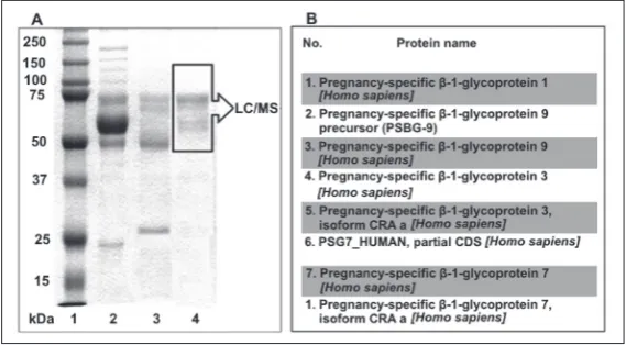
concentration in the preparation was determined using ELISA (Vector-Best, Russia). The molecular weight of the obtained preparation according to SDS-PAGE [36] was 50-75 kDa, and was in agreement with existing data [37,38]. Liquid chromatography-tandem mass spectrometry assay (LC/MS) was performed at the Israel Institute of Technology, using LTQ-Orbitrap (Thermo Fisher, USA). Several molecular forms of the PSG were detected (Fig. 1).
CD4+ cell isolation and cultivation
The experiment included several steps: isolation of CD4+ T cells, further cultivation in the presence of
PSG and T-cell activator and subsequent flow cyto-metric detection of proliferation and T-cell marker expression. CD4+ cells were obtained from PBMCs by
immunomagnetic separation with MACS® MicroBeads and MS Columns (Miltenyi Biotec, Gmbh, Germany). Isolated cells (1×10⁶ cells/mL, 200 µL) were cultured
in 96-well plates in complete medium (CM): RPMI-1640 (Sigma-Aldrich, USA) supplemented with 10% FBS (Sigma, USA), 10 mM HEPES, 2 mM L-glutamine (both from ICN Pharmaceuticals, USA), IL-2 (10 ng/mL), and 30 µg/mL of gentamy-cin (KRKA, Slovenia) in a humidified CO2 incubator at 37°C and 5% of CO2 for 48 h. T Cell Activation/Expansion Kit human, (Anti-Biotin MACSiBead™ particles loaded
with biotinylated antibodies against human CD2, CD3 and CD28, Miltenyi Biotec, Ger-many) was used to activate T cells via the T-cell receptor (TCR stimulation). Physi-ological concentrations of PSG were used (1, 10 and 100 µg/mL) that correspond to first, first-second and second-third trimesters of pregnancy, respectively [5,39]. The sample with CM instead of hormone served as a negative control. After the incubation, the viability of cells was evaluated (0.4% trypan blue, Invitrogen, USA), and was 95-98%.
Fig. 1.A – SDS-PAGE of the PSG preparation; 1 – molecular weight markers
Proliferation analysis
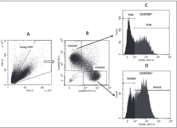
The differential gating method [40] accord-ing to the modification [41] was used to determine the proliferative status of cells. This method is a flow cytometric variant of “classic” microscopic evaluation of blast transformation by determining the pro-portion of large cells. Flow cytometry has made this method more rapid, objective and reliable. The method is based on changes in the light scatter parameters of proliferating cells. The advantages are that it does not require the use of fluorescent antibodies and allows retrospective analysis of existing flow cytometry data files. Gating strategy was as follows: three regions were determined at the FSC-H/SSC-H dot plots of CD4+-cell
cultures in accordance with their light scat-ter characscat-teristics as follows: the 1st were
non-proliferating living lymphocytes (small size and low granularity; NP); the 2nd were
proliferating living lymphocytes (larger in size and higher granularity; P); the 3rd were
dead and apoptotic (smaller size, different granularity; A). The percentages of cells in each gate from the total CD4+ cell number
were determined (Fig. 2). Data were ac-quired on the CytoFLEX S flow cytometer and analyzed in CytExpert 2.0 software (Beckman Coulter, USA).
Flow cytometry
Stained samples were analyzed by running a three-color flow cytometry with CytoFLEX S (Beckman Coulter, USA). Mathemati-cal processing of the flow cytometry data was performed by CytExpert 2.0 software
(Beckman Coulter, USA). The purity of the isolated T-helper population was checked using the two-color BD Simultest™ IMK-Lymphocyte kit (Becton, Dick-inson and Company, BD Biosciences, USA) and Cy-toFLEX S Flow Cytometer (Beckman Coulter, USA). The percentage of B-cells (CD3-CD19+) did not exceed
0.02%, of monocytes (CD45+CD14+) 0.2%, of CTLs
(CD3+CD8+) 1%, and of NK-cells (CD3-CD16/56+)
0.05%. The proportion of CD3+CD4+ cells from all
events varied within 97-99%. CD45RA+CD45R0-,
CD45RA-CD45R0+ and CD45RA+CD45R0+ subset
percentages were determined. The threshold be-tween positive and negative cells was determined using isotype controls. The following T-cell subsets were analyzed within isolated CD4+ cells: naive T cells
(NAIVE; CD45RA+CD45R0-CD62L+), TCM (CD45RA
-CD45R0+CD62L+), TEM (CD45RA-CD45R0+CD62L-),
TEMRA (CD45RA+CD45R0-CD62L-). The antibodies Fig. 2. Determination of the proliferative status of cells by the differential gat-ing method. Representative light scatter dot plots of one experiment are shown. A – control without TCR-stimulation beads; B – control with TCR-stimulation beads. Abbreviations on the dot plots: A – dead and apoptotic cell gate, NP – gate of non-proliferating cells, P – gate of proliferating cells. The numbers indicate the percentage of cells in the corresponding gate of the total number of cells.
Fig. 3. Gating strategy example. A – Gating on living lymphocytes. B – CD45RA
-CD45R0+ and CD45R0-CD45RA+ gates within the lymphocyte gate. C, D – Subsets
of interest are identified according to CD62L expression in CD45RA-CD45R0+
used were CD62L-APC (clone 145/15), CD45RA-FITC (clone HI100) and CD45R0-PE (clone UCHL1) (Bio-Legend, USA). The data are presented as percentages of naive T cells, TCM, TEM, TEMRA, CD45RA+CD45R0-,
CD45RA-CD45R0+, CD45RA+CD45R0+ cells from the
number of cells in the living lymphocyte gate, made according to FSC and SSC properties (Fig. 3). Evaluation of cytokine concentrations
IL-4 and IFN- γ concentrations in the supernatants were evaluated by enzyme-linked immunosorbent assay (ELISA) kits (Vector-Best, Russia) according to the manufacturer’s instructions using multichannel spectrophotometer Biohit BP 800 (Finland).
Statistical procedures
Data were analyzed using paired Student’s T-test (pro-liferation) and paired Wilcoxon’s test (surface mark-ers) in Statistica 8.0 (Dell, USA) and are presented as arithmetic means and standard errors of the mean (M±m) and median, first and third quartile values (Me(Q1-Q3)). Differences were considered significant when P<0.05.
RESULTS
The effect of TCR stimulation on the proliferative status of CD4+ lymphocytes
TCR stimulation led to a significant change in the proliferative status of CD4+ cells. The percentage of
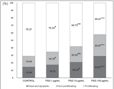
proliferating cells significantly increased, and the percentage of non-proliferating cells decreased. This indicated adequate activation of cells under these experimental conditions. With regard to dead and apoptotic cells, their percentages significantly decreased in the presence of the activating particles (Table 1). The effect of PSG on the proliferative status of activated CD4+ cells
All PSG concentrations reduced the frequency of prolif-erating CD4+ cells while simultaneously increasing the
percentage of non-proliferating lymphocytes in culture.
At concentrations of 10 and 100 µg/mL, PSG changed the percentage of cells in the dead and apoptotic gate (Fig. 4). We observed a decrease in the proliferative activity of helper T cells under the influence of PSGs. The effect of TCR-mediated activation on helper T cell percentage
TCR stimulation of CD4+ cells led to a significant
decrease in NAIVE, from 55.64% to 46.77%, Me and central memory T cells (TCM; from 29.81% to 23.62% Me, and to an increase in the percentage of TEM: 1.4-fold (from 7.42% to 11.10%, Me and TEMRA: 6.3-1.4-fold (from 0.88% to 5.56%, Me).
Fig. 4. PSG effects on the proliferative status of CD4+ cells. X-axis
– PSG concentrations; Y-axis – percentage of cells in the corre-sponding gate. Control – helper T cell culture with the activator (TCR stimulation) alone. * – P<0.05, ** – P<0.01, *** – P<0.001, paired Student’s T-test.
Table 1. The effect of TCR stimulation on the proliferative status of CD4+ lymphocytes (n=12).
Control (M±m)
TCR stimulation
(M±m)
Non-proliferating (%) 70.51±0.96 14.71±1.48P<0.0001
Proliferating (%) 9.54±0.79 74.98±1.25P<0.0001
Dead and apoptotic (%) 19.23±1.27 9.58±0.74P<0.0001
Control – T cell culture without TCR stimulation. The arithmetic means and standard errors of the mean (M±m) of cell percentages in corresponding gates from all CD4+ lymphocytes are shown. P values
The effect of PSG on the regulation of CD45 isoform expression on helper T cells
Activation of CD4+-cells with TCR stimulation beads
did not change the frequencies of CD45RA+CD45R0-
and CD45R0+CD45RA- cells in culture (Table 2). PSG
produced no significant effect on CD45 isoform ex-pression in the T-helper culture, but at the concentra-tions of 10 and 100 μg/mL reduced the frequency of CD45RA+CD45R0+ cells (Table 2).
Table 2. The effect of PSG on the regulation of CD45 isoform expression on T helpers (n=12).
CD45R
A
+CD45R0 - (%)
CD45R
A
-CD45R0 + (%)
CD45R
A
+CD45R0 + (%)
Control (48.37-60.54)56.00 (27.49-39.03)36.54 (3.55-8.48)4.81 Control
+TCR stimulation (46.27-61.19) 51.77 (25.57-40.87) 28.15 (7.22-13.63) 8.69 PSG 1 µg/mL
+TCR stimulation (50.88-59.15)53.89 (25.80-37.08)33.50 (6.22-11.88) 7.98 PSG 10 µg/mL
+TCR stimulation (51.09-60.62)54.12 (27.70-37.73)34.47 (5.27-8.60) 7.51 * PSG 100 µg/mL
+TCR stimulation (51.23-59.30)53.09 (26.92-35.31)32.30 (4.03-8.27) 6.11* Control – T cell culture without both TCR stimulation and PSG. Me, Q1 and Q3 are shown. * – P<0.05 as compared to control+TCR stimulation, paired Wilcoxon test.
Table 3. The effect of PSG on IL-4 and IFN-γ levels in T-helper culture supernatants (n = 12).
IL-4
(pg/mL) (ng/mL)IFN-γ
Control (3.36-3.62)3.42 (0.4-0.8)0.5
Control+TCR stimulation (15.63-40.45)22.16* (5.29-7.24)6.35*
PSG 1 µg/mL+TCR stimulation (17.49-32.82)23.26 (5.89-7.94)6.84
PSG 10 µg/mL+TCR stimulation (12.84-32.73)20.62** (5.56-7.43)6.83
PSG 100 µg/mL+TCR stimulation (9.62-24.30)12.71** (3.68-5.84)4.30**
Control – T cell culture without both TCR stimulation and PSG. Me, Q1 and Q3 are shown.* – P<0.05 as compared with control, paired Wilcoxon test. **– P<0.05 as compared to control+TCR stimulation, paired Wilcoxon test.
PSG effects on the frequencies of NAIVE, TCM, TEM and TEMRA in CD4+ cell culture
Introduction of 1, 10 and 100 μg/mL of PSG into the activated lymphocyte cultures did not affect the conversion of naive T helpers to TCM and TEM. However, the medium and the high concentrations of PSG (10 and 100 μg/mL) significantly reduced the percentage of TEMRA and decreased the percentage of NAIVE (Fig. 5).
The effect of PSG on the levels of IL-4 and IFN-γ in helper T cell culture supernatants
At doses of 10 and 100 µg/mL, PSG reduced the pro-duction of IL-4, and at a concentration of 100 µg/ mL reduced the production of IFN-γ in cultures of activated helper T cells (Table 3).
DISCUSSION
The effect of PSGs on lymphocyte proliferation is con-sistent with existing data [42,11]. According to these investigations, PSG1 and PSG9 suppressive effects are mediated through the TGF-β1 pathway [43,44]. This cytokine is secreted by many cells, including CD4+
Fig. 5. PSG effects on NAIVE (CD45RA+CD45R0-CD62L+), TCM
(CD45RA-CD45R0+CD62L+), TEM (CD45RA-CD45R0+CD62L-)
and TEMRA (CD45RA+CD45R0-CD62L-) frequencies in CD4+
T lymphocytes. It carries out autocrine regulation of these cells’ functions [45]. It is likely that the observed effect of T-helper proliferation suppression by PSGs was realized via activation of the latent form of TGF-β1.
Naive T lymphocytes express a high molecular weight CD45 isoform, CD45RA, which after cell acti-vation is replaced by a low molecular weight isoform, CD45R0 [46-48]. In vivo and in vitro human T cells can pass from CD45RA+ to CD45R0+ without antigen
stimulation [49]. In our study, PSG had no significant effect on the expression of the CD45 isoform in T-helper culture, but the medium and its high concen-trations (10 and 100 μg/mL) reduced the number of CD45RA+CD45R0+ cells that considered to be activated
(Table 2) [50]. In general, the mechanisms of regulation of CD45 expression have been poorly studied, and there is no evidence of ligand-induced activation of CD45. It is possible that CD45 is permanently active, and that it causes dephosphorylation of Src family kinases, maintaining them in a working, non-phosphorylated state [51].
In our study, IFN-γ and IL-4, the central cytokines determining the direction of the immune response, were evaluated. There is a so-called Th1/Th2 paradigm or “Th2 bias” hypothesis in pregnancy [52,53], based on which it would be reasonable to expect that PSG, the most abundant fetal protein in maternal bloodstream, contributes towards the secretion of antiinflammatory cytokines and inhibition of the synthesis of proinflam-matory ones. However, new data are inconsistent with this theory and an increasing number of researchers refers to this as an oversimplification [54-56]. For example, it was shown that CD4+ IFN-γ+ (Th1) and
CD4+ IL-4+ (Th2) cells increase in equal amount in
the second trimester of pregnancy, and in the third trimester a decrease is observed [57]. Because mater-nal PSG concentration peaks in the third trimester, these results overlap to some extent with our study that demonstrates the inhibitory effect of PSG at high doses on the production of both Th1 proinflammatory IFN-γ and Th2 antiinflammatory IL-4.
TEM and TEMRA make the main contribution to the production of IL-4 and IFN-γ [31,58], and the amount of IFN-γ-producing TEMRA moderately in-creases in pregnancy and strongly in preeclampsia [57]. Therefore, the decrease in the production of cytokines
mentioned above under the influence of PSG could be associated with a decrease in TEMRA percentage. It is likely that a decrease in TEMRA percentage under the influence of PSG has a fetoprotective effect in vivo, as these cells are known for their cytotoxic activity, and for this reason they are even called СD4+ cytotoxic T
lymphocytes (CTLs) [21].
The obtained data has an applied aspect. The thera-peutic potential of PSGs was demonstrated relatively recently. In particular, in vivo expression of pregnancy-specific glycoprotein inhibits the symptoms of collagen-induced arthritis and prevents dextran sodium sulfate (DSS)-induced colitis in mice [59-61]. In these studies, the involvement primarily of regulatory T (Treg) cells was shown, while memory T cells are also involved in autoimmune processes [61].
CONCLUSIONS
We previously demonstrated that PSG affects immu-nity parameters associated with immune tolerance, IDO production, Treg proportion, etс. In this study, we found that PSG regulates the functional activity of circulating CD4+ memory T cells capable of carrying out
antigen-specific responses to fetal antigens. PSG may be one of the factors preventing the immune response to fetoplacental antigens. Our findings broaden our understanding of the role of PSG in the modulation of human T-helper functions that are of particular importance in the context of immune tolerance dur-ing pregnancy.
Funding: This work was carried out within the framework of the state task, the state topic registration number: 01201353248 and supported by Russian Foundation for Basic Research [grant number 16-04-00591], and the Competitiveness Enhancement Program of the Ural Federal University (resolution no. 211 of the Russian Federation Government, contract number 02.A03.21.0006). Acknowledgments: The authors thank Professor Arie Admon (The Smoler Protein Research Center, Israel Institute of Technol-ogy «Technion», Haifa, Israel) for LC/MS analysis carrying out and LC/MS data processing.
and work guidance: Zamorina; final approval of the version to be published: all authors.
Conflict of interest disclosure: The authors certify that there is no actual or potential conflict of interest in relation to this article.
REFERENCES
1. Zhou G-Q, Baranov V, Zimmermann W, Grunert F, Erhard B, Mincheva-Nilsson L, Hammarström S, Thompson J. Highly specific monoclonal antibody demonstrates that pregnancy-specific glycoprotein (PSG) is limited to syncytiotrophoblast in human early and term placenta. Placenta. 1997;18(7):491-501.
2. Moldogazieva NT, Mokhosoev IM, Terentiev AA. Pregnancy-Specific β1-Glycoproteins: Combined Biomarker Roles, Structure/Function Relationships and Implications for Drug Design. Curr Med Chem. 2017; 24(3):245-67.
3. Moore T, Dveksler GS. Pregnancy-specific glycoproteins: complex gene families regulating maternal-fetal interactions. Int J Dev Biol. 2014;58(2-4):273-80.
4. Lin TM, Galbert SP, Kiefer D, Spellacy WN, Gall S. Character-ization of four human pregnancy-associated plasma proteins. Am J Obstet Gynecol. 1974;118:223-36.
5. Towler CM, Horne CH, Jandial V, Campbell DM, MacGilli-vray I. Plasma levels of pregnancy-specific beta1-glycoprotein in normal pregnancy. Br J Obstet Gynecol. 1976;83(10):775-79.
6. Schumacher A, Costa SD, Zenclussen AC. Endocrine Factors Modulating Immune Responses in Pregnancy. Front Immu-nol. 2014;5(196):1-12.
7. Ha CT, Wu JA, Irmak S, Lisboa FA, Dizon AM, Warren JW, Ergun S, Dveksler GS. Human pregnancy specific beta-1-gly-coprotein 1 (PSG1) has a potential role in placental vascular morphogenesis. Biol Reprod. 2010;83(1):27-35.
8. Wu JA, Johnson BL, Chen Y, Ha CT, Dveksler GS. Murine pregnancy-specific glycoprotein 23 induces the proangio-genic factors transforming growth factor beta 1 and vascular endothelial growth factor a in cell types involved in vascular remodeling in pregnancy. Biol Reprod. 2008;79(6):1054-61. 9. Snyder SK, Wessells JL, Waterhouse RM, Dveksler GS, Wess-ner DH, Wahl LM, Zimmermann W. Pregnancy-specific glycoproteins function as immunomodulators by inducing secretion of IL-10, IL-6 and TGF-beta1 by human monocytes. Am J Reprod Immunol. 2001;45:205-16.
10. Motrán CC, Díaz FL, Gruppi A, Slavin D, Chatton B, Bocco JL. Human pregnancy-specific glycoprotein 1a (PSG1a) induces alternative activation in human and mouse mono-cytes and suppresses the accessory cell-dependent T cell pro-liferation. J Leukoc Biol. 2002;72(3):512-21.
11. Motrán CC, Diaz FL, Montes CL, Bocco JL, Gruppi A. In vivo expression of recombinant pregnancy-specific gly-coprotein 1a induces alternative activation of monocytes and enhances Th2-type immune response. Eur J Immunol. 2003;33(11):3007-16.
12. Zamorina SA, Litvinova LS, Yurova KA, Dunets NA, Khaziakhmatova OG, Timganova VP, Bochkova MS, Khramtsov PV, Rayev MB. The effect of pregnancy-specific
β1-glycoprotein 1 on the transcription factor FOXP3 expres-sion by immunocompetent cells. Dokl Biochem Biophys. 2016;470(1):361-63.
13. Zamorina SA, Timganova VP, Bochkova MS, Khramtsov PV, Raev MB. Effect of pregnancy-specific β1-glycoprotein on indoleamine-2,3-dioxygenase activity in human monocytes. Dokl Biol Sci.2016;469(1):206-8.
14. Rayev MB, Litvinova LS, Yurova KA, Dunets NA, Khazi-akhmatova OG, Timganova VP, Bochkova MS, Khramtsov PV, Zamorina SA. Role of the pregnancy-specific glycoprotein in regulation of the cytokine and chemokine profiles of intact mononuclear cells. Dokl Biol Sci. 2017;475(1):180-82. 15. Michie CA, McLean A, Alcock C, Beverley PC: Lifespan
of human lymphocyte subsets defined by CD45 isoforms. Nature. 1992;360(6401):264-65.
16. Picker LJ, JR Treer, Ferguson-Darnell B, Collins PA, Buck D, Terstappen LW. Control of lymphocyte recirculation in man. I. Differential regulation of the peripheral lymph node hom-ing receptor L-selectin on T cells durhom-ing the virgin to memory cell transition. J Immunol. 1993;150:1105-21.
17. Trowbridge IS, Thomas ML. CD45: an emerging role as a pro-tein tyrosine phosphatase required for lymphocyte activation and development. Annu Rev Immunol. 1994;12:85-116. 18. Sallusto F, Lenig D, Förster R, Lipp M, Lanzavecchia
A. Two subsets of memory T lymphocytes with dis-tinct homing potentials and effector functions. Nature. 1999;401(6754):708-12.
19. Gattinoni L, Speiser DE, Lichterfeld M, Bonini C. T memory stem cells in health and disease. Nat Med. 2017;23(1):18-27. 20. Henson SM, Riddell NE, Akbar AN. Properties of end-stage human T cells defined by CD45RA re-expression. Curr Opin Immunol. 2012;24(4):476-81.
21. Tian Y, Babor M, Lane J, Schulten V, Patil VS, Seumois G, Rosales SL, Fu Z, Picarda G, Burel J, Zapardiel-Gonzalo J, Tennekoon RN, De Silva AD, Premawansa S, Premawansa G, Wijewickrama A, Greenbaum JA, Vijayanand P, Weiskopf D, Sette A, Peters B. Unique phenotypes and clonal expan-sions of human CD4 effector memory T cells re-expressing CD45RA. Nat Commun. 2017;8:1473.
22. Koch S, Larbi A, Derhovanessian E, Ozcelik D, Naumova E, Pawelec G. Multiparameter flow cytometric analysis of CD4 and CD8 T cell subsets in young and old people. Immun Age-ing. 2008;5:6.
23. Di Mitri D, Azevedo RI, Henson SM, Libri V, Riddell NE, Macaulay R, Kipling D, Soares MV, Battistini L, Akbar AN. Reversible senescence in human CD4+CD45RA+CD27− memory T cells. J Immunol. 2011;187:2093-100.
24. Unsoeld H, Pircher H. Complex Memory T-Cell Phenotypes Revealed by Coexpression of CD62L and CCR7. Journ Virol. 2005;79:4510-13.
25. Unsoeld H, Krautwald S, Voehringer D, Kunzendorf U, Pircher H. Cutting Edge: CCR7+ and CCR7- Memory T Cells
Do Not Differ in Immediate Effector Cell Function. J Immu-nol. 2002;169:638-41.
27. Camerini D, James SP, Stamenkovic I, Seed B. Leu-8/TQ1 is the human equivalent of the Mel-14 lymph node homing receptor. Nature.1989;342:78-82.
28. Gunn MD, Tangemann K, Tam C, Cyster JG, Rosen SD, Wil-liams LT. A chemokine expressed in lymphoid high endothe-lial venules promotes the adhesion and chemotaxis of naive T lymphocytes. Proc Natl Acad Sci USA. 1998;95:258-263. 29. Ahmadzadeh M, Hussain SF, Farber DL. Effector CD4 T cells
are biochemically distinct from the memory subset: evidence for long-term persistence of effectors in vivo. J Immunol. 1999;163:3053-63.
30. Ahmadzadeh M, Hussain SF, Farber DL. Heterogeneity of the memory CD4 T cell response: persisting effectors and resting memory T cells. J Immunol. 2001;166:926-35.
31. Sallusto F, Geginat J, Lanzavecchia A. Central memory and effector memory T cell subsets: function, generation, and maintenance. Annu Rev Immunol. 2004;22:745-63. 32. Paulsen E-E, Kilvaer T, Khanehkenari MR, Maurseth RJ,
Al-Saad S, Hald SM, Al-Shibli K, Andersen S, Richardsen E, Busund L-T, Bremnes R, Donnem T. CD45RO+ Memory T
Lymphocytes-a Candidate Marker for TNM-Immunoscore in Squamous Non-Small Cell Lung Cancer. Neoplasia. 2015;17:839-48.
33. Van Den Broek T, Borghans JAM, Van Wijk F. The full spectrum of human naive T cells. Nat Rev Immunol. 2018;18(6):363-73.
34. Kieffer TE, Faas MM, Scherjon SA, Prins JR. Pregnancy per-sistently affects memory T cell populations. J Reprod Immu-nol. 2017;119:1-8.
35. Bradford MM. A rapid and sensitive method for the quantita-tion of microgram quantities of protein utilizing the principle of protein-dye binding. Anal Biochem. 1976;72:248-54. 36. Laemmli UK. Cleavage of structural proteins
dur-ing the assembly of the head of bacteriophage T4. Nature.1970;227:680-5.
37. Chou JY, Sartwell AD, Wan YY, Watanabe S. Characteriza-tion of Pregnancy-Specific β1-Glycoprotein Synthesized by Human Placental Fibroblasts. Mol Endocrinol.1989;3:89-96. 38. Tschentscher P, Wagener C, Neumaier M. Distinction
of highly homologous pregnancy-specific glycoprotein (PSG) isoforms by differential absorption of antisera with recombinant PSG fusion protein domains. J Immunol Met. 1994;170:247-54.
39. Wurz H, Geiger W, Kunzig HJ, Jabs-Lehmann A, Bohn H, Luben G. Radioimmunoassay of SP1 (pregnancy-specific beta1-glycoprotein) in maternal blood and in amniotic fluid normal and pathologic pregnancies. J Perinat Med. 1981;9:67-78.
40. Vesela R, Dolezalova L, Pytlik R, Rychtrmocova H, Mareckova H, Trneny M. The evaluation of survival and proliferation of lymphocytes in autologous mixed leukocyte reaction with dendritic cells. The comparison of incorporation of
3H-thymidine and differential gating method. Cell Immunol.
2011;271:78-84.
41. Zamorina SA, Litvinova LS, Yurova KA, Khaziakhmatova OG, Timganova VP, Bochkova MS, Khramtsov PV, Rayev MB. The role of human chorionic gonadotropin in regulation
of naive and memory T cells activity in vitro. Int Immuno-pharmacol. 2018;54:33-8.
42. Harris SJ, Anthony FW, Jones DB, Masson GM. Pregnancy-specific-β1-glycoprotein: effect on lymphocyte proliferation in vitro. J Reprod Immunol. 1984;6:267-70.
43. Ballesteros A, Mentink-Kane MM, Warren J, Kaplan GG, Dveksler GS. Induction and activation of latent transform-ing growth factor-β1 are carried out by two distinct domains of pregnancy-specific glycoprotein 1 (PSG1). J Biol Chem. 2015;290:4422-31.
44. Jones K, Ballesteros A, Mentink-Kane M, Warren J, Rattila S, Malech H, Kang E, Dveksler G. PSG9 stimulates increase in FoxP3+regulatory T-cells through the TGF-β1 pathway. PLoS One. 2016;11(7):e0158050.
45. Fox FE, Ford HC, Douglas R, Cherian S, Nowell PC. Evidence that TGF-β can inhibit human Т-lymphocyte proliferation through paracrine and autocrine mechanisms. Cell Immunol. 1993;150:45-58.
46. Beverley PCL. CD45 Isoform Expression: Implications for Recirculation of Naive and Memory Cells. Immunol. 1991;Res 10:196-8.
47. Mahnke YD, Brodie TM, Sallusto F, Roederer M, Lugli E. The who’s who of T-cell differentiation: Human memory T-cell subsets. Eur J Immunol. 2013;43:2797-809.
48. Farber DL, Yudanin NA, Restifo NP. Human memory T cells: generation, compartmentalization and homeostasis. Nat Rev Immunol. 2014;14:24-35.
49. Geginat J, Sallusto F, Lanzavecchia A. Cytokine-driven proliferation and differentiation of human naive, central memory, and effector memory CD4+ T cells. J Exp Med. 2001;194:1711-9.
50. Johannisson A, Festin R. Phenotype Transition of CD4+ T Cells From CD45RA to CD45RO is Accompanied by Cell Activation and Proliferation. Cytometry. 1995;19:343-52. 51. Saunders AE, Shim YA, Johnson P. Innate immune cell CD45
regulates lymphopenia-induced T cell proliferation. J Immu-nol. 2014;193:2831-42.
52. Raghupathy R., Makhseed M, Azizieh F, Omu A, Gupta M, Farhat R. Cytokine production by maternal lymphocytes dur-ing normal human pregnancy and in unexplained recurrent spontaneous abortion. Hum Reprod. 2000;15:713-8. 53. Makhseed M, Raghupathy R, Azizieh F, Farhat R, Hassan
N, Bandar AA. Circulating cytokines and CD30 in normal human pregnancy and recurrent spontaneous abortions. Hum Reprod. 2000;15:2011- 7.
54. Lim KJ, Odukoya OA, Ajjan RA, Li TC, Weetman AP, Cooke ID. The role of T-helper cytokines in human reproduction. Fertil Steril. 2000;73:136-42.
55. Zenclussen AC, Fest S, Busse P, Joachim R, Klapp BF, Arck PC. Questioning the Th1/Th2 paradigm in reproduction: peripheral levels of IL-12 are down-regulated in miscarriage patients. Am J Reprod Immunol. 2002;48:245-51.
56. Schust DJ, Hill JA. Correlation of serum cytokine and adhe-sion molecule determinations with pregnancy outcome. J Soc Gynecol Investig. 1996;3:259-61.
normal pregnancy and preeclampsia. Journal of Obstetrics and Women’s Diseases (Russian). 2013;62:110-5.
58. Merino D, San Segundo D, Medina JM, Rodrigo E, Asensio E, Irure J, Fernández-Fresnedo G, Arias MA, López-Hoyos M. Different in vitro proliferation and cytokine-production inhibition of memory T-cell subsets after calcineurin and mammalian target of rapamycin inhibitors treatment. Immu-nology. 2016;148:206-15.
59. Falcón CR, Martínez FF, Carranza F, Cervi L, Motrán CC. In vivo expression of recombinant pregnancy-specific
glycopro-tein 1a inhibits the symptoms of collagen-induced arthritis. Am J Reprod Immunol. 2014;72:527-33.
60. Blois SM, Sulkowski G, Tirado-González I, Warren J, Fre-itag N, Klapp BF, Rifkin D, Fuss I, Strober W, Dveksler GS. Pregnancy-specific glycoprotein 1 (PSG1) activates TGF-β and prevents dextran sodium sulfate (DSS)-induced colitis in mice. Mucosal Immunol. 2014;7:348-58.