CASO CLÍNICO
A Rare Case of Spontaneous Remission and Relapse of a
Primary Central Nervous System Lymphoma
Caso Raro de Remissão Espontânea e Posterior Recidiva
de Um Linfoma Primário do Sistema Nervoso Central
Rui RAMOS1, João Soares FERNANDES2, Marta ALMEIDA3, Rui ALMEIDA1
Acta Med Port 2018 Dec;31(12):777-783 ▪ https://doi.org/10.20344/amp.10198 ABSTRACT
Primary central nervous system lymphoma remission after steroid treatment is a well-known phenomenon, but remission without any type of treatment is extremely rare. We present a rare case of spontaneous remission of a diffuse large B-cell lymphoma of the central nervous system as well as its subsequent reappearance in another location. The atypical presentation misled the neurosurgeons and neurologists, delaying diagnosis and treatment. The patient underwent brain biopsy after the relapse and started radiotherapy and chemotherapy with cytarabine + methotrexate + rituximab. As of 32 months after the diagnosis, the patient remained asymptomatic, with no focal neurological deficits and the disease in complete remission. A PubMed search of the literature up to June 2017 regarding spontaneous remission central nervous system lymphoma was also carried out.
Keywords: Central Nervous System Neoplasms/drug therapy; Central Nervous System Neoplasms/radiotherapy; Lymphoma/drug therapy; Lymphoma/radiotherapy
1. Departamento de Neurocirurgia. Hospital de Braga. Braga. Portugal. 2. Departamento de Neurorradiologia. Hospital de Braga. Braga. Portugal. 3. Departamento de Oncologia. Hospital de Braga. Braga. Portugal. Autor correspondente: Rui Ramos. [email protected]
Recebido: 06 de janeiro de 2018 - Aceite: 28 de maio de 2018 | Copyright © Ordem dos Médicos 2018 RESUMO
É bem conhecida a remissão com corticoterapia dos linfomas primários do sistema nervoso central, mas a sua remissão sem qual-quer tipo de tratamento é extremamente rara. Apresentamos um caso raro de uma remissão espontânea e posterior recidiva noutra localização de um linfoma difuso de grandes células B do sistema nervoso central. A apresentação atípica deste caso confundiu os neurocirurgiões e neurologistas, atrasando o diagnóstico e tratamento. A doente foi submetida a biópsia de uma das lesões cerebrais e, posteriormente, iniciou radioterapia e quimioterapia com citarabina + metotrexato + rituximab. Neste momento, 32 meses após o diagnóstico histológico, a doente encontra-se assintomática, sem défices neurológicos focais e com remissão completa da doença. Foi também efetuada uma pesquisa na PubMed até junho de 2017 sobre a remissão espontânea dos linfomas do sistema nervoso central. Palavras-chave: Linfoma/quimioterapia; Linfoma/radioterapia; Neoplasias do Sistema Nervoso Central/quimioterapia; Neoplasias do Sistema Nervoso Central/radioterapia
REFERENCES
1. Levison ME, Bush LM. Peritonitis and intraperitoneal abscesses. In: Schlossberg D, editor. Clinical infectious disease. 2nd ed. Cambridge:
Cambridge University Press; 2015. p. 938-9.
2. Slavoski LA, Levison ME. Peritonitis. In: Bennett J, Dolin R, Blaser M. Mandell, Douglas and Bennett’s principles and practice of infectious diseases. 8th ed. Philadelphia: Elsevier Saunders; 2014. p. 375-7.
3. Malota M, Felbinger T, Ruppert R, Nüssler N. Group A Streptococci: a rare and often misdiagnosed cause of spontaneous bacterial peritonitis in adults. Int J Surg Case Rep. 2015;6:251-5.
4. Efstratiou A, Lamagni T. Streptococcus pyogenes: basic biology to clinical manifestations [Internet]. University of Oklahoma Health Sciences Center. 2016. [Acessed 2017 Out 10]. Available at: https:// www.ncbi.nlm.nih.gov/books/NBK343616/.
5. Lappin E, Ferguson A. Gram-positive toxic shock syndromes. Lancet
Infect Dis. 2017;9:281-90.
6. Westwood D, Roberts R. Management of primary group A Streptococcal peritonitis: a systematic review. Surg Infect. 2013;14:171-6.
7. Munrós J, Alonso I, Del Pino M, Pahisa J, Almela M, Mensa J, et al. Peritonitis primaria por Streptococcus pyogenes. Rev Esp Quimioter. 2014;27:273-8.
8. Wodd T, Potter M, Jonasson O. Streptococcal toxic shock-like syndrome - the importance of surgical intervention. Ann Surg. 1993;217:109-14. 9. Montravers P, Blot S, Dimopoulos G, Eckmann C, Eggimann P, Guirao
X, et al. Therapeutic management of peritonitis: a comprehensive guide for intensivists. Intensive Care Med. 2016;42:1234-47.
10. Anthony C, Tessier J, Sanders J, Ziegler D, Duane T. Streptococcal toxic shock syndrome presenting as an acute abdomen. Surg Infect Case Rep. 2016;1:82-4.
INTRODUCTION
Malignant lymphomas can affect the central nervous system (CNS) in three ways: as primary CNS lymphoma, as a consequence of systemic lymphoma, and by intravascular
lymphomatosis.1
Primary CNS lymphoma is a rare form of extranodal
non-Hodgkin’s lymphoma.2,3 Its incidence has increased
in both immunocompetent and immunodeficient patients, and currently accounts for about 2.2% of all intracranial
tu-mors4,5 and 1% - 2% of all lymphomas.6 Most cases are
sporadic, with a minority associated with cases of
immuno-deficiencies including human immunodeficiency virus (HIV)
and iatrogenic immunosuppression.4
Primary CNS lymphoma can affect individuals of all ages, with a peak incidence in immunocompetent patients
occurring between the fifth and seventh decades of life5 and
with a mean age of 60 years.2
The clinical manifestations vary depending on the loca-tion of the lesion. The most common symptoms are psychi-atric changes, headaches, seizures, ocular symptoms and
CASO CLÍNICO
The most sensitive imaging technique is magnetic reso-nance imaging (MRI), and they frequently appear as a
high-cellular lesion, iso- or hypo-intense in T1 weighted images,8
with hypersignal in T2-weighted images and intense
con-trast uptake.5 The lesions are solitary in 65% of cases and
multiple in the remaining cases.5
The most frequent location is in the cerebral hemi-spheres, followed by the basal ganglia, corpus callosum
and cerebellum.9 However, there are no specific features
that distinguish them from other primary or secondary brain
neoplasms.2
The most used diagnostic method is a lesion biopsy and 90% – 95% are histologically classified as diffuse large
B-cell lymphoma.4
Treatment of primary CNS lymphoma, as with other brain neoplasms, involves surgery, radiotherapy (RT), and chemotherapy (CT), with the latter two being the modalities of choice. The role of surgery is mainly diagnostic. How-ever, some recent studies have reported a better prognosis in radical surgical removal of single lesions in non-eloquent brain areas. The role of RT has lost importance in recent years. Holocranial RT achieves high response rates but the duration of the effect is short and neurotoxicity is high. CT is the first therapeutic choice for newly diagnosed primary lymphomas of the CNS. It allows for high response rates and long periods without symptoms. The most commonly used drug is high-dose methotrexate alone or in combina-tion with high-dose of cytarabine, procarbazine, vincristine and rituximab. A 2009 study reported a better response with methotrexate + cytarabine compared with methotrexate
monotherapy.2
In the past, primary CNS lymphoma prognoses were typically poor, but today, long-term survival of up to 10 years
with certain treatment regimens is not uncommon,10 and
cure can be achieved in some patients.11
Brain lymphoma remission after corticosteroid therapy
is well documented9 and they are the most frequent cause
of “phantom tumor”. Other causes exist for the same phe-nomenon such as multiple sclerosis, disseminated acute
encephalomyelitis, sarcoidosis and also gliomas.12 Theories
that best explain this remission are that corticosteroids in-duce lymphocytic effects, repair of the blood–brain barrier, and the vasoconstriction that leads to a decrease in tumor
blood supply.13
When a primary CNS lymphoma is suspected, corticos-teroid therapy should be postponed until histological results are reviewed because imaging and clinical remission may delay diagnosis and initiation of treatment.
Spontaneous remission of brain lymphomas without corticosteroid therapy is extremely rare. This type of re-mission may raise other diagnostic hypotheses, namely inflammatory and demyelinating diseases and again lead to a delay in diagnosis and treatment. The most plausible explanation for this phenomenon is based on an immune
theory9,13,14 with changes in the host immunologic
compe-tence in which there is an increase in the body number of natural killer cells that may be caused by a concomitant viral infection. These natural killer cells then eliminate tumor
lym-phomatous cells.9
We present a report of a tumor that vanished without corticosteroid treatment. The diagnosis and treatment of the tumor was delayed because brain lymphoma was only sus-pected after the second brain MRI.
CASE REPORT
A 50-year-old female patient, medicated with a seroto-nin reuptake inhibitor (escitalopram), bromazepam and oral contraceptive was asymptomatic until January 2014, when
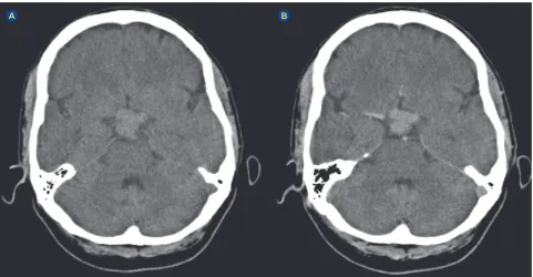
Figure 1 – Initial CT scan in January 2014, before (A) and after (B) IV contrast. A suprasellar lesion with 3 cm in diameter is seen in booth. The mass involves the optic chiasm and enhances after contrast administration.
CASO CLÍNICO she began to complain of blurred vision. She underwent
ophthalmologic evaluation and ocular correction was per-formed. A computed tomography (CT) scan revealed a 3 cm suprasellar hyperdense lesion (Figs. 1A and 1B). A hor-monal study revealed prolactin values (PRL) of 183 ng/mL and she was referred to a pituitary disease multidisciplinary consultation for suspected macroprolactinoma.
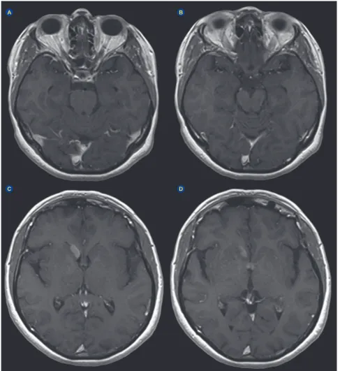
Brain MRI was performed in May 2014 (Figs 2A to 2D), without any type of medication and after clinical
improve-ment of the visual complaints. The MRI showed remission of the suprasellar lesion, but also showed two new nodular lesions, with the largest in the right caudate nucleus with subependymal expression, and the other in the anterior part of the third ventricle. A new hormonal study was performed, with no treatment having been given, and the prolactin lev-els were 76 ng/mL. The macroprolactinoma hypothesis was set aside and the patient was referred for neurologic consul-tation for suspected inflammatory disease.
Figure 2 – Initial MRI in May 2014. T1 axial weighted images with gadolinium enhancement demonstrate involution of the sellar / supra-sellar lesion (A and B). Axial T1 weighted image with gadolinium enhancement shows nodular lesion with homogeneous contrast uptake in the head of the right caudate nucleus and with subependymal expression (C). Axial T1 weighted images with gadolinium enhancement reveals a subependymal nodule in the anterior wall of the third ventricle (D).
C A
CASO CLÍNICO
In August 2014, the patient again began to experience reduced visual acuity, and a new MRI was performed with-out any treatment given (Figs. 3A to 3D). MRI revealed re-gression of the lesion in the anterior portion of the third ven-tricle, and marked enlargement of the right caudate nucleus periventricular lesion. CNS lymphoma was hypothesized, despite some atypical features, and the patient was referred for neurosurgical consultation.
In October 2014, the patient underwent a right caudate nucleus lesion biopsy guided by neuronavigation. The
his-tological result revealed a diffuse large B-cell lymphoma (Figs. 4A to 4D).
The patient completed the disease staging with cerebral spinal fluid analysis, bone marrow biopsy, thoraco-abdomi-no-pelvic CT scan, and positron emission tomography (Fig. 5). All the tests revealed that the lesion was restricted to the CNS. Viral serologies were also negative. The International Extranodal Lymphoma Study Group (IELSG) prognostic score was 2 (elevated cerebrospinal fluid proteins and el-evation of serum lactate dehydrogenase, corresponding to
Figure 3 – MRI in August 2014. Axial T1 weighted image with gadolinium enhancement shows enlargement of the right caudate nucleus periventricular lesion (A). Axial T1 weighted image with gadolinium enhancement confirms regression of the lesion in the anterior wall of the third ventricle (B). Axial diffusion (ADC map) showing restriction to diffusion in the right caudate lesion (C). Axial diffusion (b-1000 map) showing restriction to diffusion in the same lesion (D).
C A
CASO CLÍNICO
an intermediate risk).
Chemotherapy was initiated with high-dose cytarabine + high-dose methotrexate + rituximab and radiotherapy (45 gray fractionated in 4.5 gray/day, 5 times/week).
The patient was still alive at 32 months after the histological diagnosis, with the CNS lymphoma in complete remission.
DISCUSSION
Differential diagnosis of a vanishing sellar/suprasellar lesion should always include a demyelinating lesion. Other possible causes are inflammatory diseases such as au-toimmune hypophysitis, pituitary apoplexy with interval
resolution, and pituitary macroadenoma.15 Autoimmune
hypophysitis is more frequent in young female patients,
and can regress with corticosteroid treatment.15 Pituitary
apoplexy is a clinical syndrome in which more than 80% of the cases present with sudden-onset headaches. This did not occur in our case, but is also a cause of regression
in pituitary adenomas.15 There are two cases reported in
the literature of spontaneous regression of a pituitary mac-roadenoma. One of these experienced regression in 3.5 months, but this patient had only one lesion and not multiple
lesions as our case.15
In our report, there was no histological diagnosis for the vanishing sellar/suprasellar lesion but it is highly probable that it corresponds to the same diagnosis of the caudate Figure 4 – Histology of the right caudate nucleus lesion. Hematoxylin eosin with diffuse neoplasic cells with scarce cytoplasm and bulky nuclei (A). Immunocytochemistry demonstrating positivity for CD 20, ACL and BCL6 (B, C, D).
C D
B A
Figure 5 – Positron emission tomography in October 2014 showing disease located in the right caudate nucleus. C
CASO CLÍNICO
Table 1
– Pubmed search regarding “spontaneous remission central nervous system lymphoma” until June 2017.
First author Year Gender Age Symptoms Location Diagnosis procedure Histological diagnosis Treatment Survival Sonoda 15 1983 Male 28 Unknown Parieto-occipital Craniotomy Malignant lymphoma Radiotherapy Unknown Sonoda 15 1983 Female 75 Unknown Lateral ventricles Craniotomy Malignant lymphoma Unknown Unknown Rubin 16 1987 Female 56
Right side weakness
Frontal and parietal lobes
Biopsy
Dif
fuse imunoblastic
B-cell lymphoma
Radiotherapy + steroids
Unknown Sugita 17 1988 Female 63
Seizures, left upper
extremities weakness
Frontal, parietal lobes and
occipital lobes Craniotomy Non-Hodgkin, large, dif fuse type lymphoma
Whole brain radiotherapy
+ chemotherapy with
dexamethasone Unknown Galetta 18 1992 Male 72
Diplopia, gait difficulty
Third nerve, lumbar nerve
roots
Lumbar
nerve root
biopsy
Dif
fuse large B-cell
lymphoma
No specific treatment.
The
patient dies 3 days after
final diagnosis by pulmonary
embolism 15 months Kon 8 2003 Male 61 Disorientation, difficulties
with writing and walking,
gaze palsy
Corpus callosum, midbrain
tectum
Autopsy
Dif
fuse large B-cell
lymphoma Steroids 4 years Partap 14 2006 Male 45
Right side weakness,
slurred speech
Left hemisphere lesions
Brain lesion
biopsy
Large B-cell
lymphoma
Methotrexate, procarbazine,
vincristine, cytarabine +
whole brain radiotherapy
18 months Takekawa 19 2008 Female 68 General malaise, hyperventilation Occipital lobes Brain lesion biopsy B-cell lymphoma Unknown Unknown Korner 20 201 1 Male 53 Unbalance, dizziness, headache Corpus callosum Brain lesion biopsy B-cell non-Hodgkin lymphoma No treatment 12 months Rubio 9 2013 Male 65 Dizziness, vertigo Frontal, cerebellum, mesencephalon, internal capsule Autopsy Large B-cell lymphoma Steroids 9 months Sasaki 21 2015 Female 55 Facial dysesthesia Thalamus Brain lesion biopsy Dif
fuse large B-cell
lymphoma
Unknown
CASO CLÍNICO lesion.
As already mentioned, the most probable explanation for a lymphoma that vanishes without corticosteroid treat-ment is a concomitant viral infection leading to an increased number of body natural killer cells, which attack the tumor cells.
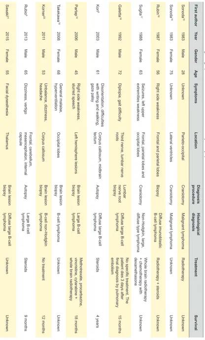
To the best of our knowledge and based on a PubMed search of the literature regarding spontaneous remission of central nervous system lymphoma up to June 2017, there are only 11 case reports of spontaneous remission/relaps-ing of central nervous system lymphomas. In some of those reports, other conditions like multiple sclerosis were sus-pected because of the spontaneous regression on diag-nostic imaging and the diagnosis and treatment were also delayed, which affected the overall survival (Table 1).
CONCLUSION
Despite being extremely rare, there are cases reported of spontaneous remission and relapse of brain CNS lym-phomas.
This case allows us to infer that differential diagnosis of primary CNS lymphoma should always be considered when a patient presents with a brain lesion that shows imaging and clinical spontaneous remission and relapse. These pa-tients should be followed at short intervals, and in suspi-cious cases, a histological diagnosis must be obtained as soon as possible to avoid delays in diagnosis and treat-ment.
ACKNOWLEDGEMENTS
The authors would like to thank Cristiano Antunes and Maria João Machado for their time and help with improving this article. Special thanks to Ren Ito for his support in the translation of the Japanese articles.
PROTECTION OF HUMANS AND ANIMALS
The authors declare that the procedures were followed according to the regulations established by the Clinical Re-search and Ethics Committee and to the Helsinki Declara-tion of the World Medical AssociaDeclara-tion.
DATA CONFIDENTIALITY
The authors declare having followed the protocols in use at their working center regarding patients’ data publica-tion.
PATIENT CONSENT Obtained.
CONFLICTS OF INTEREST
All authors report no conflict of interest.
FUNDING SOURCES
This research received no specific grant from any fund-ing agency in the public, commercial, or not-for-profit sec-tors.
REFERENCES
1. Namekawa M. [Malignant lymphoma in the central nervous system: overview]. Brain and nerve = Shinkei kenkyu no shinpo. 2014;66:907-16.
2. Roth P, Stupp R, Eisele G, Weller M. Treatment of primary CNS lymphoma. Curr Treat Options Neurol. 2014;16:277.
3. Bellefqih S, Mezouri I, Khalil J, Bazine A, Diakite A, El Kacimi H, et al. Lymphome primitif du système nerveux central : quel rôle pour la radiothérapie?. Cancer Radiother. 2014;18:685-92.
4. Phillips EH, Fox CP, Cwynarski K. Primary CNS lymphoma. Curr Hematol Malig Rep. 2014;9:243-53.
5. Schlegel U. Primary CNS lymphoma. Ther Adv Neurol Disord. 2009;2:93-104.
6. Yamamoto J, Shimajiri S, Nakano Y, Nishizawa S. Primary central nervous system lymphoma with preceding spontaneous pseudotumoral demyelination in an immunocompetent adult patient: A case report and literature review. Oncol letters. 2014;7:1835-8.
7. Stoker TB, Young AM, Patani R, Manford M. Primary cerebral lymphoma causing remitting and relapsing neurological symptoms. J Med Cases. 2013;4:420-3.
8. Kon T, Kakita A, Koide A, Mori H, Tanaka R, Takahashi H. A primary CNS lymphoma in spontaneous remission for 3.5 years after initial detection of the lesions by MRI. Brain Tumor Pathol. 2003;20:27-31. 9. Hernandez Rubio L, Giner Bernabeu JC, Perez Sempere A, Toro P.
Primary cerebral lymphoma with spontaneous remission. Neurologia. 2013;28:123-6.
10. Schorb E, Kasenda B, Atta J, Kaun S, Morgner A, Hess G, et al. Prognosis of patients with primary central nervous system lymphoma after high-dose chemotherapy followed by autologous stem cell transplantation. Haematologica. 2013;98:765-70.
11. Wang CC, Carnevale J, Rubenstein JL. Progress in central nervous system lymphomas. Brit J Haematol. 2014;166:311-25.
12. Bromberg JE, Siemers MD, Taphoorn MJ. Is a “vanishing tumor”
always a lymphoma? Neurology. 2002;59:762-4.
13. Al-Yamany M, Lozano A, Nag S, Laperriere N, Bernstein M. Spontaneous remission of primary central nervous system lymphoma: report of 3 cases and discussion of pathophysiology. J Neuro-Oncol. 1999;42:151-9.
14. Partap S, Spence AM. Spontaneously relapsing and remitting primary CNS lymphoma in an immunocompetent 45-year-old man. J Neuro-Oncol. 2006;80:305-7.
15. Sieg EP, Stepanyan H, Payne R, Ouyang T, Zacharia BE. Vanishing pituitary macroadenoma: a case report. Cureus. 2016;8:e838. 16. Sonoda H, Matsukado Y, Kaku M. [Peculiar characteristics of primary
intracranial malignant lymphoma. Report of three cases]. Neurol Med Chir. 1983;23:483-9.
17. Rubin M, Libman I, Brisson ML, Goldenberg M, Brem S. Spontaneous temporary remission in primary CNS lymphoma. Can J Neurol Sci. 1987;14:175-7.
18. Sugita Y, Shigemori M, Yuge T, Iryo O, Kuramoto S, Nakamura Y, et al. Spontaneous regression of primary malignant intracranial lymphoma. Surg Neurol. 1988;30:148-52.
19. Galetta SL, Sergott RC, Wells GB, Atlas SW, Bird SJ. Spontaneous remission of a third-nerve palsy in meningeal lymphoma. Ann Neurol. 1992;32:100-2.
20. Takekawa H, Hozumi A, Hirata K, Yamazaki K. A spontaneously vanishing primary cerebral lymphoma “ghost tumour”. J Neurol Neurosurg Psychiatry. 2008;79:1159.
21. Korner S, Raab P, Brandis A, Weissenborn K. Spontaneous regression of an intracerebral lymphoma (ghost tumor) in a liver-engrafted patient. Neurologist. 2011;17:218-21.