141
© 2018 by the Serbian Biological Society How to cite this article: Çördük N, Yücel G, Akıncı N, Tuna M, Esen O. In vitro
propagation of Silene bolanthoides Quézel, Contandr. & Pamukç. and assessment of genetic stability by flow cytometry. Arch Biol Sci. 2018;70(1):141-8.
In vitro
propagation of
Silene bolanthoides
Quézel, Contandr. & Pamukç. and assessment
of genetic stability by flow cytometry
Nurşen Çördük1,*, Gülru Yücel2, Nihan Akıncı1, Metin Tuna3 and Onur Esen4
1Department of Biology, Faculty of Arts and Sciences, Canakkale Onsekiz Mart University, 17100, Canakkale, Turkey
2Department of Biology, Faculty of Arts and Sciences, Namik Kemal University, 59030, Tekirdag, Turkey
3Department of Field Crop, Faculty of Agriculture, Namik Kemal University, 59030, Tekirdag, Turkey
4Botanic Garden & Herbarium Application-Research Center, Canakkale Onsekiz Mart University, 17100, Canakkale, Turkey
*Corresponding author: [email protected]
Received: April 10, 2017; Revised: August 9, 2017; Accepted: August 16, 2017; Published online: September 12, 2017
Abstract: Silene bolanthoides Quézel, Contandr. & Pamukç. is an endemic species from Kazdagi (Mt. Ida),
Canakkale-Balikesir, Turkey. In order to develop an efficient shoot regeneration protocol,the leaf, nodal and internodal explants of S.
bolanthoides were cultured on Murashige and Skoog (MS) medium containing benzyladenine (BA) alone or in combination
with α-naphthaleneacetic acid (NAA). The highest number of regenerated shoots (5.75±0.1) was obtained from nodal explants that were cultured on MS medium with 8.8 µM BA+0.54 µM NAA. Regenerated shoots were rooted on MS medium without plant growth regulators (PGRs). Rooted plants (2-3 cm) were separately transferred to pots containing a mixture of peat and perlite (3:1 v/v) and acclimatized successfully in a growth chamber. Genetic stability of the propagated plants was assessed by flow cytometry and cytological analysis. Flow cytometry analysis demonstrated that regenerated plants had 2.61±0.01 pg nuclear DNA (2C) and seed-derived plants had on average 2.58±0.02 pg/2C. Cytological analysis showed that the regenerated
plants had the same chromosome number as seed-derived plants of S. bolanthoides (2n=24). It was determined that
regener-ated plants were uniform in chromosome number and had a similar DNA content to the seed-derived ones, indicating that
the described efficient shoot regeneration protocol can be applied for ex situ conservation of this species.
Key words: catchfly; chromosome; endemic; genetic stability; regeneration
INTRODUCTION
The genus Silene (Caryophyllaceae) is represented by 187 taxa in Turkey, with 37% of these taxa being endemic [1-4]. This genus is divided into 31 sections. The Spergulifoliae section is represented by 11 taxa in Turkey and these species are Silene arguta, S. cappa-docica, S. montbretiana, S. oreophila, S. spergulifolia,
S. stenobotrys, S. supina with four endemic species,
S. bolanthoides Quézel Contandr. & Pamukç., S. mu-radica,S. sangaria and S. surculosa [1,2].
S. bolanthoides is an endemic species in the flora of Turkey and its natural habitats are alpine meadows and schistose rock. It is distributed on only one local-ity at an elevation of 1700 m in Kazdagi (Mt. Ida), Tur-key. The conservation status of this species is declared as CR B1ab (ii, iii) according to IUCN criteria [5]. The
fact that this plant shows a specific distribution in only one locality can lead to a decline in the number of individuals in the population, which makes it poten-tially endangered. Thus, this plant species needs to be conserved in order to prevent the disappearance of ge-netic resources and inheritance in future generations.
this unexpected and mostly undesired phenomenon in culture, propagated plants derived from organ cultures, calli and somatic embryos can be different from the donor plants with regard to phenotypic, physiological, cytological and molecular characteristics [12]. There-fore, it is necessary to analyze whether the propagated plants are genetically identical to the donor plants. Ge-netic uniformity of the plants can be detected using many techniques, such as cytogenetic analysis, flow cytometry and/or molecular markers such as amplified fragment length polymorphism (AFLP), inter simple sequence repeats (ISSR), isozymes, random ampli-fied polymorphic DNA (RAPD), restriction fragment length polymorphisms (RFLP) and simple sequence repeats (SSR) [13,14]. Among these techniques, flow cytometry has been shown to be an easy, rapid, accu-rate and economical method for screening the genetic stability of propagated plants [15-18]. Flow cytometry can be complemented by traditional cytological stud-ies, including chromosome counting.
In recent years, many studies have been devoted to in vitro cultivation, propagation or the protection of genetic resources of rare, endemic and economi-cally valuable plants [19-22]. In vitro culture of some
Silene species such as micropropagation of S. cretacea
[23], a Silene hybrid (S. polypetala x S. virginica) [24] and S. sangaria [25], suspension culture of S. vulgaris
[26], shoot regeneration of S. vulgaris [27,28] and S. thymifolia [29] have been described previously. To date, there has been no report on the in vitro culture of S. bolanthoides.
The objectives of this study were to investigate the effect of plant growth regulators (PGRs) and to evaluate the regeneration capacity of the leaf, nodal and internodal tissues in order to establish an efficient shoot regeneration system for S. bolanthoides, as well as to assess the genetic stability of regenerated plants by cytological analysis and flow cytometry.
MATERIALS AND METHODS Plant material
S.bolanthoides seeds were collected from alpine mead-ows at the Kartalcimen locality, Kazdagi, Canakkale-Balikesir, Turkey. Seeds were surface-disinfected by
stir-ring in a 1% (v/v) solution of sodium hypochlorite con-taining two drops of 0.1% Tween 20 for 10 min under sterile conditions, followed by 5 rinses in sterile water. The surface-sterilized seeds were cultured on MS basal medium (MS: M0222, Duchefa Biochemie B.V., Haar-lem, Netherlands) [30], containing 3% (w/v) sucrose and 0.8% (w/v) agar, and left in the dark for a week for germination. Germinated seedlings were subsequently transferred to a growth chamber at 25±2°C under a 16-h light and 8-h dark cycle with a light intensity of 72 µmol m-2s-1 provided by cool-white fluorescent lamps.
Induction of shoots
Shoot cultures of S. bolanthoides were established in vitro using leaf (5x5 mm), nodal (ca. 5 mm long) and internodal (ca. 5 mm long) segments excised asepti-cally from 8-week-old in vitro germinated seedlings. The explants were cultured on MS medium supple-mented with BA (0, 2.2, 4.4 or 8.8 μM) in combination with NAA (0, 0.54 or 2.69 μM) and 3% (w/v) sucrose (S0809, Duchefa). All media were gelled with 0.7% (w/v) agar (P1001, Duchefa) and the pH was adjusted to 5.75 before autoclaving. All cultures were main-tained at 25±2°C under the 16/8 h photoperiod with a light intensity of 72 µmol m-2s-1. Ten explants were cultured per Petri dish for each type of explant, and at least five replicates were used for each treatment. The mean number of regenerated shoots per explant was recorded in each culture after eight weeks.
Shoot multiplication, rooting and transfer of plants to soil
cov-ered with plastic cups to maintain a high humidity and a few holes were opened on the cups. The diameter of the holes was gradually increased over the next 2 weeks. After one month, the plants were uncovered and were then transplanted to new pots (15 mm in diameter) containing the mixture of 3 peat:1 perlite.
Flow cytometry analysis
Fresh leaf samples were collected from randomly chosen healthy and fully developed plants.Flow cyto-metric analysis was carried out using a flow cytometer (Partec CyFlowR Space) in the Laboratory of Plant Ge-netics and CytogeGe-netics at Namik Kemal University. A Partec commercial kit (CyStain PI absolute P) was used for the isolation and staining of nuclei and propidium iodide served as the fluorescent dye. Lycopersicon es-culentum Mill, which has 1.96 pg/2C DNA content [31], was used as the internal standard. Briefly, the procedure was as follows: about 20 mg of a fresh leaf sample (S. bolanthoides) and 40 mg of fresh leaf of the internal standard were placed into a Petri dish with 500 µL of extraction buffer. The tissues were chopped with a razor blade into small pieces for 30-40 s. The solution was transferred into a glass tube through a filter. Two mL of staining buffer was added to the glass tube and incubated for 1 h. The absolute DNA contents of S. bolanthoides plants were calculated according to the formula used for converting fluorescence values to the DNA content, based on ratios of the G1 peak means of sample and internal standard as follows: Sample DNA Content=[(sample G1 peak mean)/(standard G1 peak mean)] x Standard 2C DNA Content.
Three plants per sample (n=30) were analyzed to obtain the mean nuclear DNA content. The mean DNA content per plant was based on 2000 scanned nuclei.
Chromosome counts
Cytological analyses were performed on root tips of the plants used for flow cytometry. Chromosome counts were done on slides made according to the ac-etocarmine squash protocol as described by Tsuchiya and Nakamura [32] with some modifications. Root tips, approximately 1.5 cm in length, were cut and im-mediately pretreated with 8-hydroxyquinoline (Sigma, USA) for 4 h at room temperature and then fixed in
Farmer’s solution (3:1 absolute ethanol:glacial acetic acid) and stored at 4°C for at least 24 h. For mitotic analysis, root tips were stained with 2% acetocarmine and kept at least 3-4 days at 4°C. The root cap of the stained root tip was removed before squashing on a glass slide. The chromosomes were observed under a light microscope to determine the ploidy level.
Statistical analysis
One-way analysis of variance (ANOVA) was used to evaluate significant differences in the mean values of different treatments, using the Statistical Package for the Social Sciences (SPSS), version 20.0. The differ-ences between the mean values were compared by Tukey’s test (p≤0.05).
RESULTS
In vitro culture
In this study, different explant types (leaf, nodal and internodal segments) of S.bolanthoides were cultured on MS medium containing different concentrations of BA and NAA in order to achieve optimization of the shoot regeneration system. Axillary and de novo shoots directly appeared on the cut ends of the nodal and in-ternodal explants on MS medium without PGRs. How-ever, direct adventitious shoot induction from the leaf explants grown on the same medium was not achieved.
explants on MS media containing PGRs after 8 weeks in culture (Fig. 1A) and developed during culturing (Fig. 1B). Of the different concentrations of PGRs, the maximum number of shoots per explant was recorded on nodal explants cultured on MS medium containing 8.8 µM BA+0.54 µM NAA (5.75±0.11) with the high-est regeneration frequency (100%) (Table 1).
When different explant types were compared with regard to their regeneration capacity, it was found that nodal explants had a higher shoot induction capac-ity than the leaf and internodal explants (Table 1). The induction of shoots on leaf explants could not be achieved on any of the used media. Shoot forma-tion from leaf explants was only observed when the explants were cultured on MS medium supplemented with 2.2 µM BA and on MS medium with 8.8 µM BA+0.54 µM NAA. The highest number of regener-ated adventitious shoots (0.85±0.02) was observed from the leaf explants cultured on MS medium with 8.8 µM BA+0.54 µM NAA (Table 1).
Shoot induction occurred on the internodal ex-plants on MS media with PGRs except the media sup-plemented with 2.2 µM BA+0.54 µM NAA, 2.2 µM BA+2.69 µM NAA, 4.4 µM BA and 8.8 µM BA+2.69 µM NAA. However, the mean number of shoots per internodal explant was lower than the mean number of shoots per nodal explant on the media with the same growth regulators. The highest number of shoots from internodal explants occurred in the presence of 8.8 µM BA and 0.54 µM NAA (2.35±0.12), and is pre-sented in Table 1.
The shoots were propagated on MS medium con-taining the same PGRs as the shoot-induction medi-um. When the shoots were subcultured, they displayed cluster-shaped growth and their lengths were not too long. Although the mean number of shoots per explant
Table 1. Effect of α-naphthaleneacetic acid (NAA) and benzyladenine (BA) on shoot regeneration from leaf, nodal and internodal explants of S. bolanthoides. Ten explants were cultured per Petri dish for each type of explant and at least five replicates were used for each treatment.
PGRs (µM) Percentage of shoot induction (%) Number of shoots per explants (±SE) Rooting rates (%)
BA NAA Leaf Internode Node Leaf Internode Node
0 0 0 a 50±1.15d 100±0.00g 0 a 1.65±0.17bcd 2.5±0.07de 80
2.2 0 66.6±1.53e 50±±2.45d 100±0.00g 0.33±0.05 ab 1.25±0.02 abcd 5.5±0.65g 89.5
2.2 0.54 0 a 0 a 100±0.00g 0 a 0 a 3.5±0.16ef 97.83
2.2 2.69 0 a 0 a 100±0.00g 0 a 0 a 0.5±0.02ab 20
4.4 0 0 a 0 a 50±2.25d 0 a 0 a 2.5±0.19de 80.5
4.4 0.54 0 a 14.28±1.05b 100±0.00g 0 a 1.14±0.01abcd 2.5±0.08de 88.5
4.4 2.69 0 a 28.57±1.44c 100±0.00g 0 a 1.33±0.05 abcd 2.71±0.24 de 45.6
8.8 0 0 a 100±0.00g 100±0.00g 0 a 2.14±0.08 cde 4.5±0.02fg 77.55
8.8 0.54 78.65±2.15f 100±0.00g 100±0.00g 0.85±0.02 ab 2.35±0.12 cde 5.75±0.11g 60.83
8.8 2.69 0 a 0 a 0 a 0 a 0 a 0 a 0
*Means with the same letter in the columns are not significantly different at p ≤ 0.05
was not too high in the shoot-induction medium, each shoot was multiplied to a large number in the propaga-tion stage (Fig 1C). In contrast, a high multiplicapropaga-tion rate was not observed in the shoots that regenerated from nodal explants cultured on MS medium contain-ing 2.2 µM BA, even though the number of shoots per explant was statistically higher (5.5±0.65).
Single shoots were separated from the clusters and then transferred to basal MS medium for root induc-tion (Table 1). Prolonged growth on basal MS led to root formation in all shoots (Fig 1D). The highest rooting rate was observed in MS with 2.2 µM BA+0.54 µM NAA. All regenerated plants exhibited normal growth and were fully developed by the end of the culture. When the regenerated plants were compared with control plants, no anatomical or morphological differences were observed. At the end of the experi-mental series of our research, the regenerated plants were successfully acclimatized to ex vitro conditions and then transplanted to 3 peat:1 perlite (Fig 1E). The plants were successfully transferred and grown in greenhouse conditions with an approximate 86% survival rate (Fig 1F).
Genome size and chromosome counting
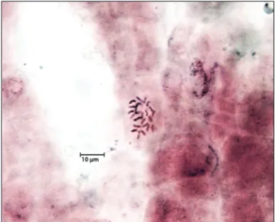
Flow cytometric analyses and chromosome counting were used to assess the genetic stability of regener-ated plants. Flow cytometric histograms indicregener-ated two peaks: the first peak representing nuclei in the G1 phase of the cell cycle belonging to the internal standard, with the second peak representing nuclei in the G1 phase of the S. bolanthoides samples (Fig. 2). It was determined that the nuclear DNA con-tent of in vitro-propagatedplants and seed-derived plants of S. bolanthoides were 2.61±0.01 pg/2C and 2.58±0.02 pg/2C, respectively. The mitotic metaphase chromosome of in vitro-propagated S. bolanthoides
was counted in root-tip cells and compared with the seed-derived plants. The number of chromosomes of propagated plants was determined to be 2n=24, like the seed-derived plants (Fig 3). Flow cytomet-ric analysis and chromosome counting revealed that the regenerated plants had a uniform chromosome number and similar DNA contents.
DISCUSSION
The leaf, nodal and internodal tissues of S. bolan-thoides were evaluated in order to establish a shoot regeneration system. Furthermore, the effects of vari-ous concentrations of BA and NAA on shoot forma-tion from these explants were investigated. The ex-plant source is an important factor for successfully
Fig 3. Mitotic chromosomes in root-tip cells of in vitro
propagated plants of S. bolanthoides(2n=24).
establishing tissue culture techniques [33]. Accord-ing to our results, the nodal explants were effective for shoot regeneration of S. bolanthoides. Our results indicated that the lowest number of regenerated ad-ventitious shoots was observed from the leaf explants. Similarly, a study by other researchers demonstrated that the lowest callus formation was obtained with leaf explants of S. sangaria and shoot regeneration could not be obtained from leaf explants cultured on MS medium containing different combinations and concentrations of PGRs [25].
Although the induction of direct shoot regenera-tion was achieved on nodal and internodal explants cultured on MS medium without growth regulators, the number of shoots per explant was limited. It was also observed that the number of these shoots did not increase during the shoot multiplication phase. The induction of shoots on MS basal medium with-out growth regulators could be presumably due to endogenous hormone levels, especially cytokinin, of the nodal and internodal explants. There are a number of reports indicating that endogenous hormone levels in explants play an important role in shoot induction, initiation and differentiation [34-36]. On the other hand, we also observed that the supplementation of PGRs to MS medium promoted callus induction and indirect shoot regeneration. These shoots propagated in a large number in the propagation stage. Similarly, it has been reported on various Silene species that PGRs supplemented to the culture medium improve the number of shoots and shoot length [23-25].
According to our results, the 8.8 µM BA in combi-nation with 0.54 µM NAA delivered the highest shoot regeneration from explants of S.bolanthoides. BA in combination with NAA has been the most widely used combination for promoting shoot induction and mul-tiplication in Silene species [27,29].
Although the explants on MS medium containing 8.8 µM BA+2.69 µM NAA produced a high frequency of calli, their compact structure and yellow color failed to regenerate shoots. This result showed that the in-crease in the amount of NAA exogenously added to the MS medium inhibited shoot formation in S. bolan-thoides. This response was similar to the previously published report on two ecotypes of S. vulgaris. In the study, it was reported that the high concentrations of
NAA acting in conjunction with BA eventually ar-rested growth and ultimately caused the callus to die over an 8-week period [27].
Our results showed that none of the regener-ated plants had genomic alterations and different polyploidy levels compared to seed-derived plants of
S.bolanthoides. There are a number of factors that induce somaclonal variation during in vitro culture. Based on the results, tissue culture conditions used in this study, such as explant tissue source, concen-tration and type of PGR, media components and the number of subcultures, could provide genomic stabil-ity of propagated plants of S.bolanthoides.
In this study, we carried out the propagation of S. bolanthoides via indirect shoot organogenesis. When unorganized cell mass growth occurs in culture, somaclonal variation may occur in these cells, and plants regenerated from the callus can be genetically different from the donor plant [37-40]. In this study, the callus stage was not prolonged, and this type of callus stage duration in culture could provide genetic stability of regenerated plants of S.bolanthoides. Ad-ditionally, a high rate of propagation of S.bolanthoides
was achieved in relatively shorter periods with more frequent subculturing.
The results of this study confirmed that flow cy-tometry is a quick and effective method for estimat-ing the nuclear DNA content of propagated plants. Flow cytometry has been used to determine genome size and ploidy levels of in vitro regenerated plants in various plant species. It was successfully used to assess genetic stability in micropropagated Puya berteroni-ana through adventitious shoots [41], in various tissue culture-derived plants of Citrus limon Burm. [13], in
Iris sibirica plants regenerated by somatic embryogen-esis and organogenembryogen-esis [42], and in regenerated plants of Eryngiumplanum L. performed through axillary bud proliferation [17]. Cytogenetic studies and chro-mosome counts can provide information about abnor-mal mitosis or changes in ploidy levels of plants [43]. In conclusion, this is the first report on efficient shoot regeneration from nodal explants of S. bolan-thoides. We showed that nodal explants can be cul-tured on MS medium with 8.8 µM BA+0.54 µM NAA with a satisfactory frequency of plant regeneration of
chromosome analysis show that the in vitro propaga-tion protocol did not cause any genetic changes in the regenerated plants. Therefore, this efficient protocol can be applied for ex situ conservation of this species.
Acknowledgments: This research did not receive any specific grant from funding agencies in the public, commercial, or not-for-profit sectors.
Author contributions: The authors make the following decla-rations regarding their contributions: conception and design of the experiments: NC; collection and identification of the plant samples of S.bolanthoides: OE; in vitro culture studies: NC; flow cytometry: GY and MT; karyotype analysis: NA. All authors con-tributed to the writing of the manuscript.
Conflict of interest disclosure: The authors declare that they have no conflict of interest.
REFERENCES
1. Davis PH. Flora of Turkey and the East Aegean Islands. Vol. 2. Edinburgh: Edinburgh University Press; 1967. p. 287. 2. Davis PH, Mill RR, Tan K. Caryophyllaceae. In: Flora of
Tur-key and the East Aegean Islands. 1st ed. Vol. 10. Edinburg: Edinburg University Press; 1988. p. 65-81.
3. Tan K, Vural M. Silene L. In: Güner A, Özhatay N, Ekim T, Başer KHC editors. Flora of Turkey and the East Aegean Islands (Suppl. 2). Vol. 11. Edinburgh: Edinburgh University Press; 2000. p. 50-3.
4. Güner A, Aslan S, Ekim T, Vural M, Babaç MT, editors. Tür-kiye Bitkileri Listesi (Damarlı Bitkiler). İstanbul: Nezahat Gökyiğit Botanik Bahçesi ve Flora Araştırmaları Derneği Yayını 1; 2012. p 356. Turkish.
5. Esen O. Kazdağı (Türkiye)’na endemik Silene bolanthoides
Quézel, Contandr. & Pamukç. (Caryophyllaceae) türünün biyolojisi. [dissertation]. [Turkey]: Institute of Natural and Applied Sciences, Canakkale Onsekiz Mart University. 2012. 59 p. Turkish.
6. Benson EE, Danaher JE, Pimbley IM, Anderson CT, Wake JE, Daley S, Adams LK. In vitro micropropagation of Primula scotica: a rare Scottish plant. Biodivers Conserv. 2000;9:711-26.
7. Rout GR, Samantaray S, Das P. In vitro manipulation and propagation of medicinal plants. Biotech Adv. 2000;18:91-120.
8. Temel A, Kartal G, Gözükırmızı N. Genetic and epigenetic variations in barley calli cultures. Biotechnol Biotechnol Equip. 2008;22(4):911-14.
9. Chinnusamy V, Zhu JK. Epigenetic regulation of stress responses in plants. Curr Opin Plant Biol. 2009;12:133-9. 10. Leljak-Levanic D, Bauer N, Mihaljevic S, Jelaska S. Changes
in DNA methylation during somatic embryogenesis in
Cucurbita pepo L. Plant Cell Rep. 2004;23:120-7.
11. Lira-Medeiros CF, Parisod C, Fernandes RA, Mata CS, Car-doso MA, Ferreira PCG. Epigenetic variation in mangrove
plants occurring in contrasting natural environment. PLOS One. 2010;5(4):e10326.
12. Rani V, Raina, SN. Genetic fidelity of organized meristem-derived micropropagated plants: A critical reappraisal. In Vitro Cell Dev Biol Plant. 2000;36:319-30.
13. Orbović V, Calović M, Viloria Z, Nielsen B, Gmitter F, Castle W, Grosser J. Analysis of genetic variability in various tissue culture-derived lemon plant populations using RAPD and flow cytometry. Euphytica. 2008;161:329-35.
14. Leva AR, Petruccelli R, Rinaldi LMR. Somaclonal variation in tissue culture: A case study with olive. In: Leva A, Laura M, Rinaldi R, editors. Recent advances in plant in vitro cul-ture. Rijeka: InTech; 2012. p. 10-7.
15. Pasqual M, Pio LAS, Oliveira ACL, Soares JDR. Flow cytom-etry applied in tissue culture. In: Leva A, Laura M, Rinaldi R, editors. Recent advances in plant in vitro culture. Rijeka: InTech; 2012. p. 109-22.
16. Singh SR, Dalal S, Singh R, Dhawan AK, Kalia RK. Evalu-ation of genetic fidelity of in vitro raised plants of Dendro-calamus asper (Shult. & Shult. F.) Backer ex K. Heyne using DNA-based markers. Acta Physiol Plant. 2013;35:419-30. 17. Thiem B, Kikowska M, Krawczyk A, Wieckowska B,
Sliwin-ska E. Phenolic acid and DNA contents of micropropagated
Eryngium planum L. Plant Cell Tiss Org. 2013;114:197-206. 18. Nybom H, Weising K, Rotter B. DNA fingerprinting in
bot-any: past, present, future. Invest Genet. 2014;5:1-35. 19. Çürük S, Çetiner S, Yalçın Mendi, Y, Carmelli-Weissberg M,
Graber E, Gaba V. Food grade sugar can promote differentia-tion in melon (Cucumis melo L.) tissue culture. In Vitro Cell Dev Biol Plant. 2012;48:600-8.
20. Izgü T, Sevindik B, Çürük P, Şimşek O, Kaçar YA, Silva JAT, Mendi YY. Development of an efficient regeneration proto-col for four Cyclamen species endemic to Turkey. Plant Cell Tiss Org. 2016;127:95-113.
21. Isah T, Mujib A. In vitro propagation and camptothecin pro-duction in Nothapodytes nimmoniana. Plant Cell Tiss Org. 2015;121:1-10.
22. Slazak B, Sliwinska E, Saługa M, Ronikier M, Bujak J, Słomka A, Göransson U, Kuta E. Micropropagation of Viola uligi-nosa (Violaceae) for endangered species conservation and for somaclonal variation-enhanced cyclotide biosynthesis. Plant Cell Tiss Org. 2015;120:179-90.
23. Kritskaya TA, Kashin AS, Spivak VA, Firstov VE. Features of clonal micropropagation of Silene cretacea (Caryophylla-ceae) in in vitro culture. Russ J Dev Biol. 2016;47(6):359-66. 24. Ault JR. In vitro propagation of a Silene hybrid (S. polypetala
x S. virginica). Hortsci. 1992;27(11):1226.
25. Erdoğan U. Tehlike altındaki Silene sangaria Coode & Cullen’nin mikroçoğaltımı [dissertation]. [Turkey]: Institute of Natural and Applied Sciences, Trakya University. 2010. 90 p. 26. Hanus-Fajerska E, Czura A, Grabski K, Tukaj Z. The effect of
conditioned medium obtained from Scenedesmus subspicatus
on suspension culture of Silene vulgaris (Caryophyllaceae). Acta Physiol Plant. 2009;31:881-7.
27. Jack EM, Anatasova S, Verkleij JAC. Callus induction and plant regeneration in the metallophyte Silene vulgaris
28. Cafuir L, Antonovics J, Hood ME. Tissue culture and quanti-fication of individual level resistance to anther-smut disease in Silene vulgaris. International Journal of Plant Sciences. 2007;168(4):415-19.
29. Panayotova LG, Ivanova TA, Bogdanova YY, Stoeva TD. In vitro cultivation of plant species from sandy dunes along the Bulgarian Black Sea Coast. Phytol Balcan. 2008;14(1):119-23. 30. Murashige T, Skoog F. A revised medium for rapid growth
and bioassay with tobacco tissue culture. Physiol Plant. 1962;15:473-97.
31. Dolezel J, Sgorbati S, Lucretti S. Comparison of three DNA fluorochromes for flow cytometric estimation of nuclear DNA content in plants. Physiol Plant. 1992;85:625-31. 32. Tsuchiya T, Nakamura C. Acetocarmine squash method for
observing sugar beet chromosomes. Euphytica. 1979;28:249-56.
33. Don Palmer C, Keller WA. Plant regeneration from petal explants of Hypericum perforatum L. Plant Cell Tiss Org. 2010;105:129-34.
34. Silué N, Koné T, Soumahoro AB, Koné M. In vitro shoot tip multiplication of bambara groundnut [Vigna subterranean
(L.) Verdc.]. Plant Cell Tiss Org. 2016;127(3):603-11. 35. Guo B, He W, Zhao Y, Wu Y,·Fu Y,Guo J. Wei Y. Changes in
endogenous hormones and H2O2 burst during shoot organo-genesis in TDZ-treated Saussurea involucrate explants. Plant Cell Tiss Organ Cult 2017;128:1-8.
36. Peng X, Zhang T, Zhang J. Effect of subculture times on genetic fidelity, endogenous hormone level and
pharma-ceutical potential of Tetrastigma hemsleyanum callus. Plant Cell Tiss Organ Cult. 2015;122(1):67-77.
37. Karp A. Origins, causes and uses of variation in plant tissue tures. In: Vasil IK, Thorpe TA, editors. Plant cell and tissue cul-ture. Dordrecht: Kluwer Academic Publishers; 1994. p. 139-52. 38. Araújo LG, Prabhu AS, Filippi MC, Chaves LJ. RAPD
analysis of blast resistant somaclones from upland rice cul-tivar IAC 47 for genetic divergence. Plant Cell Tiss Org. 2001;67:165-72.
39. Cooper C, Crowther T, Smith BM, Isaac S, Collin HA. Assess-ment of the response of carrot somaclones to Pythium violae, causal agent of cavity spot. Plant Pathol. 2006;55:427-32. 40. Sivanesan I. Shoot regeneration and somaclonal variation
from leaf callus cultures of Plumbago zeylanica Linn. Asian J Plant Sci. 2007;6:83-6.
41. Viehmannova I, Cepkova PH, Vitamvas J, Streblova P, Kisi-lova J. Micropropagation of a giant ornamental bromeliad
Puya berteroniana through adventitious shoots and assess-ment of their genetic stability through ISSR primers and flow cytometry. Plant Cell Tiss Org. 2016;125:293-302. 42. Stanišić M, Raspor M, Ninković, S, Milošević S, Ćalić D,
Bohanec B, Trifunović M, Petrić M, Subotić A, Jevremović S. Clonal fidelity of Iris sibirica plants regenerated by somatic embryogenesis and organogenesis in leaf-base culture-RAPD and flow cytometer analyses. S Afr J Bot. 2015;96:42-52. 43. Radic S, Prolic M, Pavlica M, Pevalek-Kozlina B. Cytogenetic