299
© 2018 by the Serbian Biological Society How to cite this article: Zhang J, Lin M, Chen H, Zhu Q, Linh VN, Chen X. Floral biology and pistil receptivity of the drumstick tree (Moringa oleifera Lam.). Arch Boil Sci. 2018;70(2):299-305.
Floral biology and pistil receptivity of the drumstick tree (
Moringa oleifera
Lam.)
Junjie Zhang1,2,3,4, Mengfei Lin1,2,3,4, Hanbin Chen1,2,3,4, Qin Zhu5, Vu Ngoc Linh1,2,3,4 and Xiaoyang Chen1,2,3,4,*
1State Key Laboratory for Conservation and Utilization of Subtropical Agro-Bioresources South China Agricultural University, Guangzhou, 510642, China
2Guangdong Key Laboratory for Innovative Development and Utilization of Forest Plant Germplasm, Guangzhou, 510642, China
3Guangdong Province Research Center of Woody Forage Engineering Technology, Guangzhou, 510642, China
4College of Forestry and Landscape Architecture, South China Agricultural University, Guangzhou, 510642, China
5School of Life Science, Jiaying University, Meizhou, 514015, China
*Corresponding author: [email protected]
Received: February 5, 2017; Revised: April 10, 2017; Accepted: May 8, 2017; Published online: November 20, 2017
Abstract: Drumstick (Moringa oleifera Lam.) has a wide range of uses due to its high nutritional value and the high oil content of its seeds. Many aspects of its reproductive biology remain poorly understood. We investigated the floral mor-phology of drumstick, its stigma receptivity and the structural and cytochemical features of the stigma and style at different developmental stages. The inflorescences are panicles of hermaphroditic flowers, with a pistil consisting of one open-type stigma and a hollow stylar canal. Stigma receptivity was assayed based on pollen germination, pollen tube growth and fruit set following artificial pollination. Flowers at later developmental stages exhibited greater stigma receptivity, higher percent-ages of pollen germination and a higher fruit set than those in earlier stpercent-ages. Enhanced stigma receptivity was associated with increased amounts of insoluble polysaccharides, lipids and proteins in the canal cells at later developmental stages. An ultrastructural study of the cells lining the canal indicated that they were secretory cells containing an enlarged endoplasmic reticulum, dictyosomes, mitochondria, plastids and ribosomes. Post-anthesis, these organelles exhibited degeneration at the end of the secretory phase. This study provides an important contribution to current knowledge of the anatomy and ultrastructure of the style and stigma in drumstick.
Key words: Moringa oleifera Lam.; stigma; style; stigmatic receptivity; ultrastructure
INTRODUCTION
The drumstick tree (Moringa oleifera Lam., family Moringaceae) is broadly known and widely distrib-uted because of its many uses [1-3]. This rapidly-growing, small- to medium-sized tree is indigenous to sub-Himalayan tracts of India and tropical-African countries [4,5] and has spread to many tropical and subtropical regions. Because drumstick is an economi-cally important species, it has attracted much attention in recent years. Despite extensive planting programs and research on its agronomic [6,7], nutritional [8,9] and pharmacological properties [10,11], relatively lit-tle is known about its reproductive system.
Drumstick fruit production tends to be low in com-parison with its abundant floral display; the reasons for
its low fruit set are unclear. Previous studies have shown that drumstick flowers throughout the year, with two peaks in flower production per year, and that anthesis peaks occur within one day [13-15]. The drumstick tree is a mixed mating species adapted to outcrossing, although selfing is also possible [16]. It is insect-nated, with large numbers of insects required for polli-nation [13,14]. Fruit set via open pollipolli-nation is typically 11-15%, while hand pollination yields 62-100% [15].
recep-tive during the third day post-anthesis; this receptivity lasts 2-3 days [14]. However, many anatomical and ultrastructural features of the drumstick flower stigma and style are largely unexplored. We investigated stig-ma and style structure and stigstig-ma receptivity during flower development in drumstick. The findings of this study should improve our understanding of the factors affecting the effective pollination period in drumstick, and could provide opportunities to optimize pollina-tion and increase the fruit set.
MATERIALS AND METHODS
Material and study site
This study was conducted at Guangzhou (23°8’N, 113°17’E; 43.4 m a.s.l.) in southern China at the be-ginning of July 2014. The daily mean maximum and minimum temperatures and mean temperatures per month were 28.5°C, 13.3°C and 22°C, respectively. The relative humidity averaged 77% and the annual rainfall was 1700-2000 mm. Five 3-year-old drum-stick trees were selected from a drumdrum-stick germplasm conservation farm based on flowering performance and accessibility.
Fruit set field studies
The fruit set of flowers at different developmental stages was assessed following artificial pollination. We defined anthesis as the time when the perianth is fully open. The flower stages assessed were: -2 days pre-anthesis, -1 day pre-anthesis, 0 days or anthesis, and 1, 2, 3 and 4 days post-anthesis. Flowers were emasculated approximately 2 days pre-anthesis and isolated immediately using polyester pollination bags. Flowers manipulated at -2 days and -1 day pre-anthe-sis and on the day of anthepre-anthe-sis had their own pollen removed and immediately crosspollinated artificially. At other developmental stages, the pollination time was referred to as the emasculation day, and flowers were pollinated using pollen from fresh, 0-day flowers from different trees. Pollen was visible on the stigmas once applied. Flowers that were younger or older than the stage under study were manually removed from the branches at the time of pollination. Fruit set was surveyed 3 weeks later as it became more stable.
Pollen behavior on stigma surfaces following artificial pollination
To determine the floral stage that corresponds to op-timum stigma receptivity, pollen germination on the stigma and subsequent pollen tube growth were as-sessed at various floral stages. One panicle per tree was tagged at each stigma developmental stage and artificial pollination was performed. To observe pollen tube germination on the stigma, flower samples were harvested 8 h after pollination and fixed in formalin-acetic acid-alcohol (FAA). Fixed stigma material was cleared by immersion in 1 M NaOH at room tempera-ture until most tissues became transparent. They were then rinsed thoroughly in distilled water three times and stained with 0.1% aniline blue. Using a fluores-cence microscope, germinating pollen grains on the stigma surface were observed under UV irradiation as bright yellow-green fluorescences. The outcome of each pollination event was judged according to the levels of pollen germination and pollen tube growth on the stigma. A pollen grain was considered germi-nated when its tube length was greater than the diam-eter of the pollen grain [17]. Fluorescing pollen grains without a pollen tube were considered ungerminated. We examined 30 flowers per treatment.
Microscopy
in 0.05 M of phosphate buffer. The material was post-fixed in 0.5% osmium tetroxide for 30 min, dehydrat-ed in a series of ethanol solutions and embdehydrat-edddehydrat-ed in Spurr resin. Sections were observed with a Tecnai 12 transmission electron microscope.
Data collection and analysis
The means±standard error for fruit set and pollen germination values were calculated using SPSS 19.0 software for Windows and compared using analysis of variance (ANOVA) and Duncan’s new multiple range test (significance was determined at P<0.05).
RESULTS
Morphology
The drumstick tree is hermaphroditic, with zygo-morphic gullet flowers arranged in large panicles (Fig. 1A). There was a highly significant difference in flower production per inflorescence between trees. Only 1-3% of the flowers in an inflorescence bloomed each day; thus, it took 1-2 months for the entire in-florescence to bloom, depending on its size. Flowers have one pistil and five unequal stamens. An average of 25 days was required from bud initiation (Fig. 1B)
to anthesis (Fig. 1E), presumably depending on the temperature. The flower stayed open for about 7 days.
Fruit set at different stages of stigmatic development
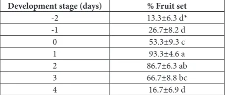
The results of the controlled pollination experiment (Table 1) indicate that fruit set was possible at all stages of stigma development, even though in some cases a low frequency was obtained. Rarely, fruit set occurred in flowers pollinated 2 days pre-anthesis or 4 days post-anthesis, suggesting nonreceptivity of the stigma dur-ing these periods. However, because deposited pollen may remain viable after the stigma becomes recep-tive, it was not possible to determine the exact timing of stigma receptivity in this study. For example, fruit set may occur even if pollen was captured 1 day pre-anthesis, when the stigma was not receptive.
Pollen germination at different stages of stigma development
The percentage of receptive stigmas, defined by pollen germination, at different stigmatic stages was varied significantly between treatments. Pollen germination was significantly higher in more developed flowers than in younger flowers. Almost no pollen germina-tion was observed on stigmas 2 days or 1 day pre-anthesis (Table 2). On the day of pre-anthesis, the germi-nated pollen grains had very short pollen tubes (Fig. 3A). Peak drumstick stigma receptivity was recorded 1 day and 2 days post-anthesis, with more than 93% of observed stigmas being receptive and pollen grains having long tubes (Fig. 3B). Stigmas observed 3 days post-anthesis exhibited high pollen germination, but
Fig. 1. Morphological characters of inflorescence and flowers of Moringa oleifera Lam. A – inflorescence; B-G – flowers at different
development stages; B – globular stage, buds were greenish and
inconspicuous; C – elongated stage, the bud enlarged and the color
became greenish-white; D – two days before anthesis, the color
was yellowish-white all over and center portion bulged out; E – anthesis, the bud split open while the anthers remained closed;
F – two days after anthesis, sepals and petals were yellowish-white and the anthers were yellow; G – flattened stage, sepals and petals were yellow and withered.
Table 1. Percentage of fruit set at different stages of stigma devel-opment after controlled pollination.
Development stage (days) % Fruit set
-2 13.3±6.3 d*
-1 26.7±8.2 d
0 53.3±9.3 c
1 93.3±4.6 a
2 86.7±6.3 ab
3 66.7±8.8 bc
4 16.7±6.9 d
had short pollen tubes and an untidy aspect, with the pollen tubes lacking clarity of appearance and growth direction.
Stigma and style structure
The drumstick stigma is perforated and hollow, with thick, elongated walled cells arranged linearly into compact masses, forming a rim-like structure at the stigma head. The cells are small near the stigma head with minute spaces that are larger below the stigma head. The linear arrangement of cells narrows towards the stigma head (Fig. 3C and D).
The hollow style is comprised of an epidermis, ground tissue and a central canal coated by canal cells with a strongly undulated cuticle (Fig. 2A and B). In most style cells of very young flowers (Fig. 1D), PAS-positive substances were abundant (Fig. 2B). On the second day of anthesis (Fig. 1F), PAS-positive sub-stances were mainly distributed in canal cells and filled the intercellular spaces between loosely arranged subepidermal canal cells with a fibrillar consistency (Fig. 2C). During the same period, the content of sub-epidermal canal cells also included lipids and proteins (Figs. 2E and F). In the flattened stage (Fig. 1G), fewer polysaccharides, lipids and proteins were detected in all style cells (Fig. 2D).
The inner layers of ground tissue and canal cells were aerenchymatic. The canal cells were approximate-ly isodiametric in transverse section, with a thicker cell wall covered by cuticle consisting of a slightly reticulate cuticle layer and the cuticle proper bordering the canal (Fig. 3G). The cytoplasm of the epithelial cells
con-Table 2. Percentage of receptive stigma (pollen-germinated stigma) at different stigma development stages after controlled pollination.
Development stage (days) % Receptive stigma
-2 3.3±3.3 d*
-1 10.0±5.6 d
0 63.3±8.9 b
1 93.3±4.6 a
2 96.7±3.3 a
3 60.0±9.1 b
4 36.7±8.9 c
*Means followed by the same letter in the same column are not sig-nificantly different from each other at P≤0.05 according to Duncan’s multiple range test.
Fig. 3. Structure of stigma and style of Moringa oleifera Lam. A – pollen grain germination on stigma of anthesis; B – pollen grain
germination on stigma at two days post anthesis; C-D – SEM of
stigma; E-J – TEM of style transverse section; E-F – two days pre anthesis; G-I – two days post anthesis; J – flattened stage. Pg, pollen grains; pt, pollen tube; er, endoplasmic reticulum; v, vesicles; p, plastid; m, mitochondria; d, dictyosome; tl, tannin-like; cl, cuticle layer.
Fig. 2. Semithin transverse section of style Moringa oleifera Lam.
A-D – PAS staining; A – elongated stage; B – two days before
tained abundant mitochondria and plastids with starch grains, free ribosomes and an endoplasmic reticulum (Fig. 3E-I). The endoplasmic reticulum and vesicles were abundant (Figs. 3H and I), with some vesicles in close contact with the plasmalemma, indicating that they are secretory cells. In very young flowers (Fig.1D), the canal cells were lined with a dense cuticle layer adhering closely to the canal cells. The epithelial cells were metabolically active, with numerous mitochon-dria and plastids of different sizes containing starch grains (Fig. 3E and F). The epidermis cells were char-acterized by the presence of a large vacuole, filled with tannin-like material, and several plastids rich in starch grains were observed. On the second day of anthesis (Fig. 1F) the canal cell cytoplasm began to atrophy and diminish (Fig. 3G). The large vacuoles became divided, forming smaller vacuoles (Figs. 3H and I). The cuticle was disrupted in places and cuticle particles could be discerned on the surface of the wall and inside the canal. Further, the plastids contained no starch; the fibrillar and granular substances formed in the canal cells passed into the canal, stretching the cuticle as de-velopment proceeded. In the flattened stage (Fig. 1G) the canal cells were loosely arranged, with a denser cytoplasm and distorted cells (Fig. 3J).
DISCUSSION
The drumstick tree has a hermaphroditic flower with little spatial and temporal separation between male and female functions. The anthers begin to dehisce be-fore the stigma becomes most receptive, and the style slightly exceeds the length of the anthers. This phe-nomenon of simultaneous ripening of the pistil and stamen is common, and protandry occurs frequently in outcrossing angiosperms such as Tectona grandis
[18], Grevillea robusta [17] and Collinsia heterophylla
[19]. In drumstick, however, the protandrous flowers still permit self-pollination. Muluvi et al. [16] studied drumstick outcrossing rates using amplified fragment length polymorphism markers and found it to have a mixed mating system with an outcrossing rate of 0.74.
The stigma is reported to be receptive at the time of anthesis in many tree species such as Malus domes-tica [20], Armeniaca vulgaris [21] and Cerasus avium
[22]. We found that stigma receptivity in drumstick flowers was delayed, with higher percentages of pollen
germination and fruit set observed in older than in younger flowers. There have been conflicting findings with respect to the optimal timing of drumstick stigma receptivity. Bhattacharya and Mandal [14] reported delayed stigma receptivity favoring cross-pollination, with which our results are consistent. However, Kan-thaswamy [15] found that the stigma was receptive 1 day pre-anthesis. The receptive period can vary among species and cultivars. In drumstick, stigma receptivity lasts only 3-5 days. Flowers of high moun-tain plants are generally long-lived; stigma receptivity can be maintained over longer periods in such plants [23]. In pear, the surfaces of immature stigmas can support pollen adhesion but cannot provide a proper hydration substrate [24]; this is also true in drumstick. Its perforated stigma facilitates pollen adhesion, but only mature stigmas can support pollen germination and pollen tube growth. We observed that drumstick pollen grains remain viable for 2-4 days, depending on the cultivar and environment (data not shown). If pollen grains are loaded 2 days pre-anthesis drumstick stigmas, even though these stigmas are not receptive at that time, as the pollen grains viability lasts 4 days they could still germinate. This may explain why 2 days pre-anthesis drumstick stigmas supported little pollen germination. Before the flowers reached 1 day post-anthesis, insufficient exudates were produced and little secretion occurred, suggesting that the ac-quisition of competence to support pollen hydration and germination defines the transition of the stigma from an immature to a mature stage.
Owens [25] distinguished between two different stigma types among 40 caesalpinioid genera. Wet, papillate stigmas were usually captitate, while wet, non-papillate stigmas were all crateriform or cham-bered. The drumstick stigma, which is perforated, is of the wet, non-papillate sort. Plant styles have been clas-sified into three types: hollow, closed and semi-closed. The drumstick style is hollow, with a canal lined with a glandular epidermis. Hollow styles are less common in angiosperms, having been documented among monocotyledonous species such as Cyphia stenopetala
[26] and Ornithogalum sigmoideum [27], as well as dicotyledonous species such as Tectona grandis [18],
Cyclamen persicum [28] and Polygala vayredae Costa
inner epidermal cells that ensure its liquid content is withdrawn once the pistil has reached maturity. Hol-low styles containing secretions have been reported
in Colophospermum mopane [30], Chrysanthemum
multicaule [31] and Ornithogalum sigmoideum [27].
This secretion can contain lipids, polysaccharides and proteins, varying in chemical composition between species. Stigma and transmitting tissue secretions ap-pear to have many functions, including facilitating pollen adhesion to stigmas [32], pollen-stigma recog-nition [33], pollen tube growth and ovule penetration [34], attraction and nourishment of floral visitors [35].
Our findings indicate significant differences in the fine structure of canal cells pre- and post-anthesis. Pre-anthesis, drumstick canal cells are rich in plastids, dictyosomes, mitochondria, ribosomes and ER; post-anthesis, the amount of cytoplasm and the number of organelles decrease. These data suggest that metabolic activity in drumstick pre-anthesis is very high, when secretory synthesis and secretory transfer are intense. Low activity post-anthesis indicates low secretory ac-tivity in the cell at that stage. These changes are similar to those that occur in Ornithogalum sigmoideum [27], but opposite to those in Lilium longiflorum [36].
Acknowledgments: This work was funded by the Forestry Technology Innovation Program, the Department of Forestry of Guangdong Province (2015KJCX009; 2017KJCX029); Guangzhou Science Technology and Innovation Commission (201707010462).
Author contributions: All authors contributed to the experi-ments, data analysis and manuscript preparations. J.J. and M.F. preformed the cytochemistry and ultrastructure observation; H.Q. and V.N. performed the phenotype observation and artificial pol-lination; X.Y. wrote the final version of the manuscript.
Conflict of interest disclosure: No potential conflict of interest is reported by the a uthors.
REFERENCES
1. Ramachandran C, Peter KV, Gopalakrishnan PK. Drumstick (Moringa oleifera): A multipurpose Indian vegetable. Econ Bot. 1980;34(3):276-83.
2. Fahey JD. Moringa oleifera: A review of the medical evidence for its nutritional, therapeutic, and prophylactic properties. Part 1. Trees Life J. 2005;1:5.
3. Anwar F, Latif S, Ashraf M, Gilani AH. Moringa oleifera:
A food plant with multiple medicinal uses. Phytother Res. 2007;21(1):17-25.
4. Popoola JO, Obembe OO. Local knowledge, use pattern and geographical distribution of Moringa oleifera Lam. ( Morin-gaceae) in Nigeria. J Ethnopharmacol. 2013;150 (2):682-91. 5. Leone A, Spada A, Battezzati A, Schiraldi A, Aristil J, Bertoli
S. Cultivation, genetic, ethnopharmacology, phytochemistry
and pharmacology of Moringa oleifera leaves: An overview.
Int J Mol Sci. 2015;16 (6):12791-835.
6. Melesse A, Steingass H, Boguhn J, Schollenberger M, Rode-hutscord M. Effects of elevation and season on nutrient com-position of leaves and green pods of Moringa stenopetala and Moringa oleifera. Agroforest Syst. 2012;86(3):505-18. 7. Mendieta-Araica B, Spörndly E, Reyes-Sánchez N,
Salm-eron-Miranda F, Halling M. Biomass production and chemi-cal composition of Moringa oleifera under different planting densities and levels of nitrogen fertilization. Agroforest Syst. 2013;87(1):81-92.
8. Makita C, Chimuka L, Steenkamp P, Cukrowska E, Madala
E. Comparative analyses of flavonoid content in Moringa
oleifera and Moringa ovalifolia with the aid of UHPLC-qTOF-MS fingerprinting. S Afr J Bot. 2016;105:116-22. 9. Qi N, Gong X, Feng C, Wang X, Xu Y, Lin L. Simultaneous
analysis of eight vitamin E isomers in Moringa oleifera Lam. leaves by ultra performance convergence chromatography. Food Chem. 2016;207:157-61.
10. Singh RSG, Negi PS, Radha C. Phenolic composition, anti-oxidant and antimicrobial activities of free and bound phe-nolic extracts of Moringa oleifera seed flour. J Funct Foods. 2013;5(4):1883-91.
11. Anand K, Gengan RM, Phulukdaree A, Chuturgoon A.
Agroforestry waste Moringa oleifera petals mediated green
synthesis of gold nanoparticles and their anti-cancer and catalytic activity. J Ind Eng Chem. 2015;21(1):1105-11. 12. Freire JEC, Vasconcelos IM, Moreno FBMB, Batista AB,
Lobo MDP, Pereira ML, Lima, JPMS, Almeida RVM, Sousa AJS, Monteiro-Moreira ACO, Oliveira JTA, Grangeiro TB.
Mo-CBP3, an Antifungal chitin-binding protein from
Mor-inga oleifera seeds, is a member of the 2S albumin family. PloS One. 2015;10(3):e0119871.
13. Jyothi PV, Atluri JB, Subba-Reddi C. Pollination ecology of Moringa Oleifera (Moringaceae). Proc Indian Acad Sci (Plant Sci.). 1990;100(1):33-42.
14. Bhattacharya A, Mandal S. Pollination, pollen germination
and stigma receptivity in Moringa oleifera Lamk. Grana.
2004;43(1):48-56.
15. Kanthaswamy V. Studies on pollination and breeding
behav-iour in moringa (Moringa oleifera Lam). Vegetable Science.
2005;32:187-88.
16. Muluvi GM, Sprent JI, Odee D, Powell W. Estimates of outcrossing rates in Moringa oleifera using Amplified frag-ment length polymorphism (AFLP). Afr J Biotechnol. 2004;3(2):145-51.
17. Kalinganire A, Harwood CE, Slee MU, Simons AJ. Floral structure, stigma receptivity and pollen viability in relation
to protandry and self-incompatibility in silky oak (
Grevil-lea robusta A. Cunn.). Ann Bot-London. 2000;86(1):133-48. 18. Tangmitcharoen S, Owens JN. Floral biology, pollination,
pistil receptivity, and pollen tube growth of teak (Tectona
19. Lankinen A, Armbruster WS, Antonsen L. Delayed stigma receptivity in Collinsia heterophylla (Plantaginaceae): genetic variation and adaptive significance in relation to pollen com-petition, delayed self-pollination, and mating-system evolu-tion. Am J Bot. 2007;94(7):1183-92.
20. Losada JM, Herrero M. Flower strategy and stigma perfor-mance in the apple inflorescence. Sci Hortic-Amsterdam. 2013;150(2):283-9.
21. Zhao S, Liao K, Da M, Xu G, Xu L, Dong S, Du R. Study on pollen viability and stigma receptivity of very later-ripening ‘Dongxing’ apricot. J Agr Sci Tech-Iran. 2015;16:954-57. 22. Fadón E, Herrero M, Rodrigo J. Flower development in
sweet cherry framed in the BBCH scale. Sci Hortic-Amster-dam. 2015;192:141-7.
23. Steinacher G, Wagner J. Flower longevity and dura-tion of pistil receptivity in high mountain plants Flora. 2010;205(6):376-87.
24. Sanzol J, Rallo P, Herrero M. Asynchronous development of stigmatic receptivity in the pear (Pyrus communis; Rosaceae) flower. Am J Bot. 2003;90(1):78-84.
25. Owens SJ. Stigma, style, pollen and the pollen-stigma inter-action in Caesalpinioideae. Advances in legume biology. In:
Proceedings of the 2nd International Legume Conference;
1986 Jun 23-27; St. Louis, Missouri. St. Louis: Missouri Botanical Garden; 1986. p. 23-7.
26. Leins P, Erbar C. Floral morphological studies in the South African Cyphia stenopetala Diels (Cyphiaceae). Int J Plant Sci. 2005;166(2):207-17.
27. Ismailoglu I, Unal M. Structure and Cytochemical Features
of Stigma and Style of Ornithogalum Sigmoideum Freyn &
Sint, Unpollinated and Pollinated Pistil. Acta Biol Cracov Bot. 2012;54(1):65-75.
28. Reinhardt S, Ewald A, Hellwig F. The Anatomy of the
stigma and style from Cyclamen persicum (Mill.) cv. “pure
white” and its relation to pollination success. Plant Biol. 2007;9(1):158-62.
29. Castro S, Silva S, Stanescu I, Silveira P, Navarro L, Santos
C. Pistil anatomy and pollen tube development in Polygala
vayredae Costa (Polygalaceae). Plant Biol. 2009;11(3):405-16. 30. Jordaan A, Wessels DCJ, Kruger H. Structure of the style and
wet nonpapillate stigma of Colophospermum mopane,
Caesal-pinioideae: Detarieae. Bot J Linn Soc. 2002;139(139):295-304. 31. Deng Y, Chen S, Teng N, Chen F, Li F, Song A, Guan Z.
Flower morphologic anatomy and embryological
charac-teristics in Chrysanthemum multicaule (Asteraceae). Sci
Hortic-Amsterdam. 2010;124(4):500-5.
32. Rejón JD, Delalande F, Schaeffer-Reiss C, Carapito C, Zien-kiewicz K, Alche JDD, Rodriguez-Garcia MI, Dorsselaer AV, Castro AJ. The plant stigma exudate: a biochemically active extracellular environment for pollen germination? Plant Sig-naling Behav. 2014;9(3):5695-705.
33. Sanchez AM, Bosch M, Bots M, Nieuwland J, Feron R, Mari-ani C. Pistil factors controlling pollination. The Plant Cell. 2004;16(suppl. 1):S98-S106.
34. Lenartowska M, Krzesłowska M, Bednarska E. Pectin
dynamic and distribution of exchangeable Ca2+ in
Haeman-thus albiflos hollow style during pollen-pistil interactions. Protoplasma. 2011;248(4):695-705.
35. Woodcock TS, Larson BMH, Kevan PG, Inouye DW, Lunau K. Flies and flowers II: Floral attractants and rewards. J Polli Ecol. 2014;12(8):63-94.