Chromatin Regulation and the Histone Code
in HIV Latency
Anne-Marie W. Turner
a,band David M. Margolis
a,b,c,d,*aUNC HIV Cure Center, University of North Carolina at Chapel Hill, Chapel Hill, NC; bDepartment of Medicine, University of
North Carolina at Chapel Hill, Chapel Hill, NC; cDepartment of Microbiology and Immunology, University of North Carolina at
Chapel Hill, Chapel Hill, NC; dDepartment of Epidemiology, University of North Carolina at Chapel Hill, Chapel Hill, NC
The formation of a latent reservoir of Human Immunodeficiency Virus (HIV†) infection hidden from immune clearance remains a significant obstacle to approaches to eradicate HIV infection. Towards an understanding of the mechanisms of HIV persistence, there is a growing body of work implicating epigenetic regulation of chromatin in establishment and maintenance of this latent reservoir. Here we discuss recent advances in the field of chromatin regulation, specifically in our understanding of the histone code, and how these discoveries relate to our current knowledge of the chromatin mechanisms linked to HIV transcriptional repression and the reversal of latency. We also examine mechanisms unexplored in the context of HIV latency and briefly discuss current therapies aimed at the induction of proviral expression within latently infected cells. We aim to emphasize that a greater understanding of the epigenetic mechanisms which govern HIV latency could lead to new therapeutic targets for latency reversal and clearance cure strategies.
*To whom all correspondence should be addressed: David Margolis, University of North Carolina at Chapel Hill, 2016 Genetic Medicine Building, CB#7042, 120 Mason Farm Road, Chapel Hill, NC, 27599-7435, Tel: (919) 966-6388,
email: [email protected].
†Abbreviations: The commonly used nomenclature for the post translational modifications of histone proteins is used. For example, the methylation of lysine 27 on histone 3 is written H3K27me. Specific modifications are indicated by me1/me2/me3, representing mono-, di-, or tri-methylation respectively.
ART, antiretroviral therapy; BET, bromo- and extra-terminal domain family; BETi, BET inhibitor; ChIP, chromatin immunoprecipitation; HDAC, histone deacetylase; HDACi, histone deacetylase inhibitor; HIV/AIDS, Human Immunodeficiency Virus/Acquired
Immunodeficiency Syndrome; LRA, latency reversal agent; LTR, long terminal repeat; ncRNA, non-coding RNA; PMBCs, Peripheral Mononuclear Blood Cells; PcG, polycomb group; PEV, position effect variegation; PKC, protein kinase C; PRC1, polycomb
repressive complex 1; PRC2, polycomb repressive complex 2; PrEP, pre-exposure prophylaxis; PTM, post translational modification; SAHA, suberanilohydroxamic acid; SEC, super elongation complex; shRNA, short hairpin RNA; siRNA, small interfering RNA; STD, sexually transmitted disease; TCR, T-cell receptor.
Keywords: HIV, chromatin, histone code, latency, epigenetic
Author contributions: A-M.W.T and D.M.M wrote the manuscript.
INTRODUCTION
Over 30 years after the identification of Human Im-munodeficiency Virus (HIV) and an intense focus on pre-vention research, there are still over 2 million new infec-tions every year across the world [1]. Improved public health and education outreach programs, increased STD
estimat-ed only 46 percent of persons living with HIV were on ART by the end of 2015 [1]. There continues to be a dis-proportionally high burden of infection in Sub-Saharan Africa, which accounts for two-thirds of new infections [1]. Even here in the United States, at the end of 2012 1 in 8 people are unaware they were living with HIV [2]. These statistics reaffirm that despite effective treatment and increasingly effective prevention methods, without a change in the status quo of tools used to respond to the HIV pandemic, the virus is and will continue to be a per-sistent human pathogen.
While there are still public health issues which result in the chronic HIV burden worldwide, diagnosis com-bined with ART compliance have resulted in the ability of HIV-infected individuals to live a normal life-span [3]. As such, the field of HIV research has taken a significant shift towards identification of a functional or sterilizing cure, defined as control of infection without need for ART or clearance of all virus from an individual respec-tively [4]. If achieved and implemented in combination with current prevention strategies, the potential exists to eradicate HIV globally. A significant obstacle to cure research is the latent reservoir of HIV. Established very early in infection in resting CD4+ T-cells, these latent
cells are indistinguishable from healthy cells and are one source of viral rebound upon halting ART. One strategy that has emerged from new research towards an HIV cure has been popularly labeled “shock and kill” [5]. A two component system, this strategy envisions the reversal of latency to reveal the latent virus reservoir, followed by clearance of these cells by a native or engineered immune response within infected individuals [5].
The latency reversal (“shock”) portion of this cure strategy has focused on inducing expression of HIV to a level detectable by immune clearance mechanisms via the use of host-targeted therapies termed latency reversal agents (LRAs) [6]. Reversal of HIV latency has focused on the two main mechanisms of transcriptional repres-sion, restriction of critical host factors and epigenetic repression of the integrated provirus. There is a signifi-cant understanding of the host factors and mechanisms that govern successful transcription of HIV [7]. However, there is still work to be done in understanding the epi-genetic mechanisms that repress the viral DNA, how the modulation of these restrictions impacts viral transcrip-tion, and if successful therapeutic induction of viral tran-scription via targeting of these epigenetic blocks might lead to recognition and clearance of latent cells. Here we will focus primarily on the role of the histone code in transcriptional activation and repression of HIV latency and highlight new discoveries in the field of epigenetics which may have functional relevance in transcriptional silencing. We will also briefly examine current LRAs, their mechanisms, and the effectiveness of these
treat-ments. Ultimately, we hope to emphasize that a greater understanding of the molecular mechanisms which gov-ern HIV latency could lead to new targets for epigene-tic-based LRAs for latency reversal and clearance cure strategies.
TRANSCRIPTIONAL ACTIVATION OF HIV The events which govern successful transcription-al activation of HIV are a well characterized cascade of events featuring both major and minor players. For the purpose of this review, we will highlight the critical fac-tors but aim to emphasize one point: successful reactiva-tion of latent HIV requires both the release of host factors restricted in resting CD4+ T-cell in combination with a
change in the repressive chromatin structure of the in-tegrated virus. In resting CD4+ T-cells, various host
pro-teins critical to driving HIV transcription such as NF-kB, NFAT, and P-TEFb are sequestered or are present at low levels [8]. T-cell activation via TCR signaling can remove these restrictions to allow recruitment to the viral promot-er. While only NF-kB and Sp1 are required for activation of the LTR, binding of these and other non-essential host transcription factors results in minimal but sufficient tran-scriptional initiation and elongation by RNA polymerase II (RNAPII) to produce the multi-spliced Tat transcript. Minimal activation of the LTR also results in unproduc-tive RNAPII transcripts approximately 60 nucleotides in length which encode the viral non-coding RNA TAR. TAR forms into a stem-loop structure that is recognized and bound by Tat. The Tat/TAR interaction helps to ini-tiate a positive feedback loop at the LTR via Tat-mediat-ed recruitment of various proteins such as P-TEFb and histone acetyltransferases which aid to drive sustained productive elongation from the viral promoter. As such, successful production of Tat is a critical driver in latency reactivation [9]. For those interested, the mechanisms of viral activation have been reviewed in far greater detail in the following [9-13].
EPIGENETIC CONTROL OF HIV TRANSCRIPTION
pairs of DNA wrapped around an octamer of four histone proteins – H2A, H2B, H3, and H4 – and acts to structur-ally organize DNA. Histone H1, which binds linker DNA between nucleosomes, is involved in spatial compaction of nucleosomes to form higher order chromatin struc-ture (reviewed in [15]). The role of H1 in HIV latency has been little characterized, although H1 appears to be present at the latent LTR and acts to repress Tat-medi-ated transcription [16,17], implicating some level of nu-cleosome compaction in latency. Each histone protein within the nucleosome has an unstructured tail which can be modified by various post-translational modifications (PTMs), the combination of which results in the histone code which governs the chromatin structure and tran-scriptional accessibility of a region of DNA [18]. PTMs found at histones tails include phosphorylation of serine and threonine and ubiquitination of lysine; however, the most studied modifications in relation to transcription are methylation and acetylation of lysine residues [19-21].
Acetylation and methylation are thought to be im-portant in the regulation of transcription due to their effect on nucleosome stability. The basic charge of the core histone proteins can be neutralized by acetylation of lysine residues on the histone tails, resulting in de-stabilization of the DNA/histone interactions, increased accessibility of the local DNA, and decreased structural stability of the overall nucleosome (reviewed in [22]). Methylation of histone tails does not change the overall charge of the proteins and certain methyl marks can act to recruit chromatin regulators which further stabilize and compact the chromatin structure. Upon integration, the HIV LTR is structured by the placement of well-defined nucleosomes. Nuc-0 is positioned at the beginning of the U3 region, followed by an unbound and nuclease sensi-tive region (DNase hypersensitivity region 1 or DHS1) around 250bp in size which includes the critical tran-scription factor binding sites for NF-kB and SP1 [23,24]. Nuc-1 is located immediately after the TSS, followed by a second nuclease sensitive region (DHS2) and nuc-2 ap-proximately 400bp downstream [23,24].
A study of global histone methylation and acetylation patterns in CD4+ T-cells identified a histone “backbone,”
a group of 17 histone marks which individually associat-ed with transcriptional activation, highly associatassociat-ed with each other, and when in combination were associated with higher gene expression [21]. These 17 marks include H2A.Z, H2BK5ac, H2BK12ac, H2BK20ac, H2BK120ac, H3K4ac, H3K4me1, H3K4me2, H3K4me3, H3K9ac, H3K9me1, H3K18ac, H3K27ac, H3K36ac, H4K5ac, H4K8ac, and H4K91ac [21]. Examination of 39 total histone lysine methylation and acetylation modifications resulted in the observation that the majority of histone ly-sine and acetylation marks associated with transcription-ally active euchromatin while only five marks associated
with repressive chromatin – H3K27me2, H3K27me3, H3K9me2, H3K9me3, and H4K20me3 [21].
HISTONES MARKS AND
TRANSCRIPTIONAL ACTIVATION OF HIV H3/H4 Acetylation
Consistent with the role of histone acetylation in the histone backbone which marks active transcription [21], one of the earliest events at the viral LTR upon activation is the recruitment of histone acetyltransferases (HATs). Proteins with known and characterized HAT activity in-cluding CBP, GCN5, and P/CAF have been observed at the LTR upon reactivation [25-28], however only CBP recruitment was shown to be Tat-independent [29]. In-deed, CBP and the closely related p300 are transcription-al coactivators of NF-kB, suggesting binding of NF-kB recruits the initial factors needed to initiate chromatin re-modeling of the integrated provirus [29,30]. Recruitment of HATs has been linked to increases in global H3 and H4 acetylation at all three nucleosomes surrounding the LTR, as well as increases of the specific marks H3K9ac, H3K-14ac, H4K5ac, H4K8ac, and H4K16ac [25-29]. HATs have also been demonstrated to modulate Tat function by acetylation of Tat itself [26,31].
minally differentiated cells. Yet, recent evidence suggests crosstalk exists between these pathways [61-63] and tran-scriptional control of HIV could be a potential example of this paradigm shift.
H3K27 Methylation and Polycomb
The Polycomb group (PcG) proteins were first iden-tified as repressors of development-related genes in Dro-sophila melanogaster (reviewed in [64]). A highly con-served mechanism also required for temporal regulation of genes during mammalian development, PcG-medi-ated repression is carried out by two complexes, PRC1 and PRC2. Polycomb Repressive Complex 2 (PRC2) is the sole complex which catalyzes H3K27 mono-, di-, and tri-methylation in mammalian cells (reviewed in [64,65]). PRC2 requires three core proteins to mediate H3K27 methylation – SUZ12, the chromatin reader EED, and the methyltransferase EZH2 – and can also associ-ate with other non-essential PcG proteins which enhance enzymatic activity or targeting of the complex [64,65]. The H3K27 mark has been linked to both active and re-pressive chromatin. Presence of H3K27me1 in concert with H3K36me3 within a gene body is associated with transcription and refractory to further H3K27 di- and tri-methylation [66]. H3K27me2/3 are strongly linked with repressive chromatin and reduced gene expression [21,65,67]. H3K27me3 at PRC2 targeted loci is main-tained via binding and reading of existing H3K27me3 by the chromodomain of EED, initiating a reinforcing loop which is proposed to help maintain the repressive mark during DNA replication [64,65]. PRC2 is also intimately linked with Polycomb Repressive Complex 1 (PRC1) to modulate chromatin structure and maintain transcription-al silencing.
PRC1 is comprised of a CBX, PCGF, RING, and PHC protein of which there are multiple homologues for each, resulting in PRC1 complexes which differ among cell types (reviewed in [68]). Regardless of composition, PRC1 functions to monoubiquitinate H2A lysine 119 (H2AK119ub1) via the ubiquitin E3 ligase activity of the RING1A/B proteins [64,65]. H2AK119ub1 has been proposed to block RNAPolII transcription, leaving it in a ‘poised’ state at PRC regulated genes [69]. Early mech-anistic characterization in D. melanogaster and in mam-malian cells identified a canonical recruitment pathway by which PRC2-mediated H3K27me3 is recognized by the chromodomain of the CBX reader proteins, leading to recruitment of PRC1 and ubiquitination at H2AK119 at chromatin domains already marked by H3K27me3 (reviewed in [67,68,70]). However in recent years, re-search has emerged suggesting the relationship between PRC1 and PRC2 recruitment appears far more complex [67,68,70]. PRC1 can also mediate chromatin compac-tion through a mechanism independent of histone tails, H3 Methylation
While acetylation of both histones and Tat are criti-cal to HIV reactivation, studies are limited with regards to other highly studied and traditionally activating marks. H3K4me at the promoter and H3K36me and K3K79me within the gene body are strongly associated with tran-scriptionally active genes. Interestingly, H3K4me3, a mark well associated with active transcription but mutu-ally exclusive to H3K4ac [19,21,47] has been linked to both repression and activation of the virus. H3K4me3 was observed in association with H3K9me3 and the bispecific H3K9/H3K4 demethylase LSD1 in microglial cells and was lost upon activation [48]. Conversely, an early study of P-TEFb observed H3K4me3 levels increased upon TNF-α activation, however this was observed well with-in the body of Gag and not examwith-ined at the LTR [49]. H3K79me3 marks active transcription, is linked to tran-scriptional elongation, and methylation is dependent on initial ubiquitination of H2BK123, a modification linked to disruption of chromatin compaction [50]. Knockdown of the only known H3K79 methyltransferase DOT1L has been linked to activation of the LTR [51], suggesting this mark may be inhibitory to viral expression. To date, no work has significantly examined a role for H3K36 meth-ylation in HIV regulation, a mark implicated exon-intron demarcation, splicing, and repression of cryptic intragen-ic transcription [52-55]. H3K36me2/3 has been observed within the coding region of HIV [49,56] and is proposed to be deposited via the recruitment of the H3K36 meth-yltransferase SETD2 by IWS1, a protein which interacts with histone chaperone SUPT6H (Spt6) [56]. It would be interesting to determine if these methylation marks con-form to established mechanisms in the context of HIV, as in the case of H3K4me3 and H3K79me3 alternate func-tions could be exploited for latency reversal strategies.
HISTONE MARKS AND TRANSCRIPTIONAL REPRESSION OF HIV
ter-ent at the LTR during latency [74]. There have been no further studies to clearly link PRC1 and H2AK119ub1 to HIV latency; however, the presence of both Polycomb complexes at the LTR suggests the symbiotic relationship between these complexes may be relevant in maintaining latency and another potential target for development of LRAs.
Another interesting and open question is the mecha-nism by which PRC1/2 are recruited to the LTR. Knock-down of PRC2 components EZH2 and SUZ12 as well as ptreatment of Jurkat cells with EZH2 inhibitors re-sulted in a decrease in establishment of latency using a reporter virus construct, implicating PRC2 in the earliest stages of chromatin repression of HIV [75]. In D. mela-nogaster, PcG recruitment is mediated by recognition of specific DNA sequences called polycomb repressive elements. Similar elements have not been found in mam-malian cells and there is still significant debate as to the mechanisms which drive initial recruitment of PRC1/2 to target loci (reviewed in [67,68,70]). Furthermore, the preferential integration of HIV into active gene bodies [76] leads to an association of epigenetic marks of ac-tive transcription [77] which are traditionally refracto-ry to PRC2, suggesting an active mechanism for PRC2 suggesting that while PTMs may aid in localization of
PRC1, they are not necessary for this activity [71]. Presence of H3K27me3 as a marker of HIV latency at the promoter and the requirement for EZH2 has been observed in both cell culture and primary cell models of latency [72-75]. In the first study to characterize the role of PRC2 in HIV latency, shRNA-mediated knockdown of EZH2 strongly reactivated HIV in Jurkat-based models of latency and synergized with known T-cell activators [72]. They also demonstrated heterogeneity in the levels of H3K27me3 at the LTR was directly related to the abil-ity of TNF-α to reactivate the virus [72], supporting the idea that different integration sites may be differentially regulated at the chromatin level which could impact the reactivation potential. Treatment of both Jurkat cells and a primary cell model of latency with the selective EZH2 methyltransferase inhibitor GSK-343 in combination with other LRAs including the HDAC inhibitor SAHA or the bromodomain inhibitor JQ1 increased levels of re-activation when compared to the individual compounds alone, suggesting H3K27 and EZH2 are active in mainte-nance of latency and that loss of H3K27me3 primed the LTR for reactivation [74]. This work also demonstrated for the first time that components of the PRC1 are
pres-Table 1. Potential mechanisms for polycomb recruitment to integrated HIV.
Mechanisms of Polycomb Recruitment
CpG Islands Both PRC1 and PRC2 are known to co-localize with CpG islands [70]. CpG islands
associate with promoter elements of active genes and remain methylation-free, in contrast to lone CpG dinucleotides which are highly subject to cytosine methylation (reviewed in
[148]). The percentage of CpG sites in the HIV LTR does not meet the canonical definition
of a CpG island, however studies of latent HIV in resting CD4+ T-cells suggests they are not subject to methylation [14,149,150] and furthermore that methylation of the LTR is highly repressive [151,152]. While the role for DNA methylation itself in latency is greatly debated [14,149-154], these elements may act as a marker for an intragenic promoter [155] and play a role in recruitment of PRC2.
ncRNAs Noncoding RNAs have been identified as major regulators of gene silencing during
development. Two of the most studied ncRNA mechanisms, X-chromosome inactivation
and HOX gene silencing by their cognate ncRNAs XIST and HOTAIR, have defined PRC2
binding and recruitment by these ncRNAs as critical for establishment of transcriptional repression (reviewed in [156]). PRC2 core proteins EZH2, SUZ12 and accessory protein JARID2 have all been demonstrated to have RNA binding domains critical to this recruitment mechanism [157,158]. Knockdown of an HIV-expressed antisense RNA has been shown to decrease EZH2 at the LTR and increase transcriptional activation, however
they were unable to establish a definitive role in a primary CD4+T-cell model [159].
H3K36 and the
HMT in latency. A later study also demonstrated siR-NA-mediated knockdown of G9a and treatment of latent-ly infected Jurkats with the G9a/GLP specific inhibitor BIX01294 resulted in loss of G9a and H3K9me3 at the LTR via ChIP, increased transcription, and synergized with the HDAC inhibitor SAHA [97]. As G9a/GLP and HP1γ localize with euchromatin regions of DNA, it may be that SETDB1 plays a greater role as the mediator of H3K9me3 at the LTR than the SUV39 HMTs. However, a role for these epigenetic marks in primary lymphoid cells has yet to be demonstrated.
Position effect variegation (PEV), whereby introduc-tion of a tradiintroduc-tionally active gene into heterochromatin results in silencing of the active gene, is another phenom-enon driven by repressive H3K9 methylation. Early Jur-kat latency models in which some integration was found near heterochromatin may have been subject to PEV [98], however the relevance of this mechanism in primary cells with integration into active genes is debatable. Regard-less, the field was further complicated by the discovery of the HUSH complex and the suggestion that this mecha-nism plays a role in HIV latency. The HUman Silencing Hub is comprised of H3K9me3 reader MPP8, TASOR, and periphilin and was identified by specifically search-ing for the proteins which were responsible for silencsearch-ing of a lentiviral-GFP reporter via PEV [99]. HUSH recruits SETDB1 to mediate H3K9me3 and heterochromatin for-mation does not appear to be dependent on any of the HP1 proteins [99]. Knockdown of HUSH components MPP8, TASOR, and periphilin was shown to activate Jurkats transduced with an HIV-Tat-LTR reporter as well as activate four different J-Lat clones, Jurkat-based laten-cy models [99]. Unfortunately, knockdown of SETDB1 was not tested or reported in the J-Lat lines, howev-er SETDB1 knockdown in a latent line established in KBM7 cells of myeloid lineage did demonstrate a strong role for SETDB1 in LTR repression [99]. Further studies of the role of HUSH in HIV regulation are needed.
New work suggesting crosstalk between the H3K9 and H3K27 methylation pathways increases the poten-tial complexity of HIV latency regulation by these path-ways. While studies of this relationship are limited, one report observed PRC2 and H3K27me increased stabili-ty and binding of HP1 proteins to H3K9me3, however a direct interaction between HP1 and PRC2 components was not observed [61]. Another identified a direct inter-action between PRC2 and G9a/GLP and demonstrated G9a deficient cells show impaired PRC2 recruitment at loci targeted by both PRC2 and G9a [62]. Interestingly, a recent study of the kinetics of HIV latency establishment observed viruses silenced within 3 days as measured by a fluorescent reporter showed increased H3K27me3 but no difference in H3K9me3 relative to the transcription-ally active population [75]. However, continued cultur-recruitment to the LTR. CpG islands, non-coding RNAs
(ncRNAs), and H3K36 methylation have all been impli-cated as mechanisms for PRC2 recruitment and all have the potential to be functionally relevant in the context of latency establishment (see Table 1).
H3K9 Methylation
Like the pattern of H3K27me, H3K9me1 is observed at the TSS of active genes while H3K9me2/3 strongly associate with constitutive heterochromatin [78]. H3K9 methylation is mediated by multiple HMTs which appear to have unique roles in the cell. SUV39H1 and SUV39H2 double knockouts show a severe impairment in H3K9me3
in vivo and loss of heterochromatin formation at pericen-tric regions, however in vitro methylation of unmodified H3K9 peptides suggests they can also mediate H3K9me1/ me2 [79-81]. PRDM3 and PRDM16 have more recently been identified as mono-H3K9 methyltransferases which co-localize with SUV39 and constitutive heterochroma-tin regions [82]. In contrast, the H3K9me1/2/3 HMT SETDB1 and H3K9me1/2 HMTs G9a and GLP show distribution patterns over euchromatin regions of the ge-nome [79,83-86]. Presence of these HMTs and addition of the H3K9me3 mark ultimately recruit heterochroma-tin protein 1 (HP1), of which there are three isoforms in mammalian cells, HP1α, HP1β, and HP1γ. HP1 is a chromatin reader which recognizes H3K9me3 and me-diates chromatin compaction via homodimerization [87-90]. HP1α and HP1β associate with pericentric heteroch-romatin while HP1γ is found with euchheteroch-romatin regions [91]. HP1 further helps to reinforce heterochromatin by recruiting DNA methyltransferases [92-94].
H4K20me1 and H4K20me3 only represent a small frac-tion of total H4K20 methylafrac-tion, a role in transcripfrac-tion- transcription-al repression and a link to pathways overlapping those already identified in HIV latency exist, suggesting these marks may be worth additional study in the context of HIV latency.
NUCLEOSOME REMODELING/HISTONE CHAPERONES
The integration of HIV into genes which are tran-scriptionally active in resting CD4+ T-cells suggests
re-pressive histone PTMs must be actively maintained in the context of elongating RNAPolII. ATP-dependent nu-cleosome remodeling complexes and histone chaperones are known to help maintain nucleosome positioning and histone PTMs during DNA replication and transcription.
ATP-dependent remodeling complexes use ATP to move, remove, or exchange nucleosomes and are thought to function to increase or decrease accessibility to DNA. Numerous complexes exist and many contain proteins which can recognize histone PTMs, a mechanism which has been implicated in how these complexes are recruit
-ed and differentially localiz-ed (review-ed in [109]). The biochemically distinct PBAF and BAF complexes are mammalian SWI/SNF-type remodelers which have been implicated in HIV latency. BAF has been linked to tran
-scriptional repression via positioning of Nuc-1 at a less energetically favorable DNA sequence [110] while PBAF is recruited by acetylated Tat and promotes efficient tran
-scription [111]. BAF has been observed to be highly re
-fractory to co-localization and co-repression with PRC2 and H3K27me [109], an interesting observation in the context of HIV latency given the strong evidence for a role for PRC2. Regardless, small molecule inhibitors of the BAF complex show promise as a new class of LRAs in both cell lines and primary cell models of latency [112].
Histone chaperones remove and deposit nucleosomes around elongating RNA polymerase to repress spurious antisense and cryptic transcription within gene bodies [113,114]. While the HIV LEDGF/p75 complex is known for its role in directing integration, additional investiga-tion has found a post-integrainvestiga-tion role in establishing and maintaining transcriptional repression in latently infected cells. LEDGF/p75 appears in complex with SUPT6H and IWS1 at the HIV LTR, whereby the H3 histone chaperone SUPT6H appears to maintain repressive nucleosomes across the HIV promoter and throughout the coding re-gion [115]. Similar to the mechanism of SUPT6H, the FAcilitates Chromatin Transcription (FACT) complex acts to remodel nucleosomes by disrupting histones H2A-H2B ahead of elongating RNA polymerase II (RNAPII) [116]. Knockdown of SUPT16H and SSRP1, the com-ponents of FACT, have been demonstrated to reactivate ing of both the inactive and active populations for 60
days resulted in eventual silencing of 60 percent of the active population and increases in H3K27 and H3K9 in both populations [75]. The inactive population marked by early H3K27me3 and later H3K9me3 was observed to be harder to reactivate than the initial active population silenced with slower kinetics, implicating a mechanism involving both of these marks which drives the virus into a deeper latency [75]. Successful reactivation of this pop-ulation may prove critical to latency reversal and clear-ance strategies and may depend on a more complete un-derstanding of the interplay between H3K9 and H3K27 repression.
H4K20 Methylation
To date, there have been no published reports impli-cating H4K20 methylation in regulation of HIV latency. The H4K20 mark has been linked to chromatin stabil-ity, DNA replication, DNA damage, and transcriptional repression (reviewed in [100]). H4K20me1 is observed across euchromatin and the inactivated X-chromosome [101] while H4K20me2 is found throughout chromatin with approximately 80 percent of H4 in the cell marked either exclusively or in some combination with this mark [101,102]. Consistent with an association with tran-scriptionally repressive chromatin [21], H4K20me3 is found co-localized with H3K9me3 at constitutive het-erochromatin including pericentric hethet-erochromatin and telomeres [101,103]. The HMT PR-SET7 (SET8) is re-sponsible for H4K20me1 and is necessary for subsequent H4K20me2/3 by the closely related SUV4-20H1/2 en-zymes [104].
vitro comparison of PKC agonists and HDACi on CD4+
and CD8+ T-cells reinforce these favorable results. PKC
agonists Bryostatin 1 and Prostratin demonstrated high levels of T-cell activation and impaired CD8+ function
while HDACi showed minimal to no impact on these pathways [133]. Thus while able to modify the restrictive chromatin structure, HDACi are likely limited in the abil-ity to alter host factor restriction and are thus less potent LRAs as compared to PKC agonists.
Bromodomain inhibitors (BETi), in clinical testing for oncology, are also being examined as LRAs and may act to both ease host factor and chromatin restrictions. Bromodomains are responsible for recognition of acetyl-ated lysine residues and are found in a wide range of pro-teins including HATs, helicases, and transcriptional medi-ators, to name a few (reviewed in [134]). In the context of HIV, bromodomain-containing protein BRD4 is known to compete with Tat for binding of P-TEFb [135]. Test-ing of JQ1, a BETi with primary specificity for BRD4, but also for BRD2 and BRD3, has been shown to induce viral reactivation, presumably by removing BRD4 and increasing p-TEFb accessibility [136-139]. However, a study has also proposed JQ1 acts in a Tat-independent mechanism by inhibiting BRD2 and that BRD2 functions as a repressor of transcription at the LTR [140]. While the specific mechanisms for JQ1 activation of HIV as well as BRD2 repression remain unclear, BETi show promise as future LRAs which may move towards the clinic.
While clinical studies of single agent LRAs have not yet achieved convincing depletion of latent infec-tion, results of such studies may be improved if LRAs are used in combination with specific viral clearance strategies [141]. However, increased LRA activity may also be needed. Mediators of NF-kB activation tested in combination with various HDACi or JQ1 ex vivo in resting CD4+ T-cells isolated from stably suppressed
in-dividuals demonstrated increased viral reactivation as compared to single agents alone, even at suboptimal dos-ing of PKC agonists [142]. However, no combinations at full dose reached equivalent levels of reactivation observed via maximal PMA/Ionomycin stimulation and
in vitro response varied between patients. In addition to these observations it has been demonstrated that maximal stimulation of resting CD4+ T-cells via small molecule agonists of T-cell activation in vitro fail to simultaneously reactivate all inducible proviruses [143]. They found no evidence to suggest these proviruses were integrated into a highly repressed chromatin region of the host genome, nor did they observe increases in repressive DNA methyl-ation at the LTR, leading to the hypothesis that induction even under maximal stimulation is stochastic [143]. In this setting, the idea of stochastic reactivation is meant to mean that a given viral promoter may respond differently to the same stimuli at two moments in time, due to mo-HIV transcription [117]. This work also observed
knock-down of SUPT6H, the remodeler CHD1, and histone chaperones ASF1a and HIRA resulted in viral reactiva-tion [117]. While these proteins are typically considered positive transcription factors in the context of normal mammalian transcription, they act to repress HIV tran-scription. This suggests a potential mechanism similar to transcriptional interference by which normal chromatin remodeling mechanisms represses access to the integrat-ed viral LTR, potentially via maintenance of establishintegrat-ed repressive histone PTMs.
CURRENT AGENTS FOR LATENCY REVERSAL AND CLEARANCE
Current LRAs act either by allowing the release of host factor restriction, modulating HIV LTR chromatin structure, or perhaps in some case both, to favor tran-scription. Within the former is the large class of protein kinase C (PKC) agonists. Induction of the PKC signaling pathway in T-cells results in the activation of NF-kB and AP-1, both which bind the viral LTR and synergize to reactivate viral transcription [118]. Prostratin [119,120], Bryostatin 1 [121], and ingenol [122] are all small mol-ecules which mimic diacylglycerol activation of PKC and are among the most potent LRAs. However, there are significant concerns regarding use of PKC agonist in patients. PKC agonists are non-specific, can activate the PKC pathway in multiple cell types, and can upregulate markers of T-cell activation (reviewed in [118,123]). A study in the 1990’s of anti-CD3, a potent T-cell activator via activation of the T-cell receptor, demonstrated signif-icant toxicity when used in patients [124]. This has led to a conservative approach in the clinic, and clinical experi-ments of this kind were not attempted until a recent study of single dose administration of Bryostatin 1 at two low concentrations showed tolerability and limited adverse effects, but failed to show PKC activation or reactivation of HIV [125].
HDAC inhibitors (HDACi) represent a class of LRAs which primarily act to modulate chromatin structure of the virus. Current HDACi are pan inhibitors which target class I and II HDACs and include three FDA approved molecules for treatment of T-cell lymphomas, Vorinos-tat (SAHA), BelinosVorinos-tat, and Romidepsin (reviewed in [126]). HDACi have been shown to induce HIV activation
infected individuals due to rarity of such cells in vivo, and the difficulties with generating a truly representative in vitro model of latency [144].
lecular, temporal fluctuations in precise biochemical and biophysical state of the promoter. However, such precise observations as to the state of the local chromatin and the repression of the DNA structure surrounding an individu-al integrated provirus cannot yet be assessed in cells from
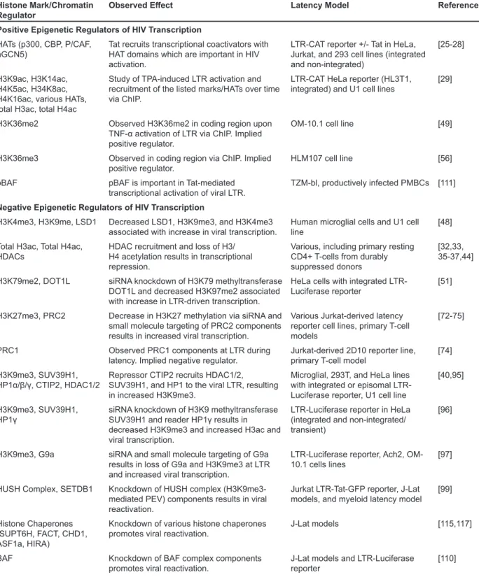
Table 2. Histone marks and the reader, writer, and eraser proteins implicated in control of HIV transcription and reactivation from latency.
Histone Mark/Chromatin
Regulator Observed Effect Latency Model Reference
Positive Epigenetic Regulators of HIV Transcription HATs (p300, CBP, P/CAF,
hGCN5) Tat recruits transcriptional coactivators with HAT domains which are important in HIV activation.
LTR-CAT reporter +/- Tat in HeLa, Jurkat, and 293 cell lines (integrated and non-integrated)
[25-28]
H3K9ac, H3K14ac, H4K5ac, H34K8ac, H4K16ac, various HATs, total H3ac, total H4ac
Study of TPA-induced LTR activation and recruitment of the listed marks/HATs over time via ChIP.
LTR-CAT HeLa reporter (HL3T1,
integrated) and U1 cell lines [29]
H3K36me2 Observed H3K36me2 in coding region upon
TNF-α activation of LTR via ChIP. Implied
positive regulator.
OM-10.1 cell line [49]
H3K36me3 Observed in coding region via ChIP. Implied
positive regulator. HLM107 cell line [56]
pBAF pBAF is important in Tat-mediated
transcriptional activation of viral LTR. TZM-bl, productively infected PMBCs [111] Negative Epigenetic Regulators of HIV Transcription
H3K4me3, H3K9me, LSD1 Decreased LSD1, H3K9me3, and H3K4me3
associated with increase in viral transcription. Human microglial cells and U1 cell line [48] Total H3ac, Total H4ac,
HDACs HDAC recruitment and loss of H3/H4 acetylation results in transcriptional repression.
Various, including primary resting CD4+ T-cells from durably suppressed donors
[32,33, 35-37,44]
H3K79me2, DOT1L siRNA knockdown of H3K79 methyltransferase DOT1L and decreased H3K97me2 associated with increase in LTR-driven transcription.
HeLa cells with integrated
LTR-Luciferase reporter [51]
H3K27me3, PRC2 Decrease in H3K27 methylation via siRNA and small molecule targeting of PRC2 components results in increased viral transcription.
Various Jurkat-derived latency reporter cell lines, primary T-cell models
[72-75]
PRC1 Observed PRC1 components at LTR during
latency. Implied negative regulator. Jurkat-derived 2D10 reporter line, primary T-cell model [74] H3K9me3, SUV39H1,
HP1α/β/γ, CTIP2, HDAC1/2 Repressor CTIP2 recruits HDAC1/2, SUV39H1, and HP1 to the viral LTR, resulting in increased H3K9me3.
Microglial, 293T, and HeLa lines with integrated or episomal LTR-Luciferase reporter, U1 cell line
[40,95]
H3K9me3, SUV39H1,
HP1γ siRNA knockdown of H3K9 methyltransferase SUV39H1 and reader HP1γ results in
decreased H3K9me3 and increased H3ac and viral transcription.
LTR-Luciferase reporter in HeLa (integrated and non-integrated/ transient)
[96]
H3K9me3, G9a siRNA and small molecule targeting of G9a results in loss of G9a and H3K9me3 at LTR and increased viral transcription.
LTR-Luciferase reporter, Ach2,
OM-10.1 cells lines [97]
HUSH Complex, SETDB1 Knockdown of HUSH complex (H3K9me3-mediated PEV) components results in viral reactivation.
Jurkat LTR-Tat-GFP reporter, J-Lat models, and myeloid latency model [99]
Histone Chaperones (SUPT6H, FACT, CHD1, ASF1a, HIRA)
Knockdown of various histone chaperones
promotes viral reactivation. J-Lat models [115,117]
BAF Knockdown of BAF complex components
Acknowledgements: This work was supported by the National Institutes of Health grants UM1 AI126619, U01AI117844, and R01HL132791 to D.M.M., and NIAID T32AI007001 to A-M.W.T. We thank all the HIV+ partic-ipants in our studies for their critical contributions to this research.
REFERENCES
1. WHO. HIV/AIDS Fact Sheet No 360 2016 [updated July 2016; cited 2016 May 24]. Available from: http://www. who.int/mediacentre/factsheets/fs360/en/.
2. CDC. Fact Sheet: HIV in the United States: At A Glance 2016 [cited 2016]. Available from: http://www.cdc.gov/hiv/ statistics/overview/ataglance.html.
3. May MT, Gompels M, Delpech V, Porter K, Orkin C, Kegg S, et al. Impact on life expectancy of HIV-1 positive individuals of CD4+ cell count and viral load response to antiretroviral therapy. AIDS. 2014;28(8):1193-202. 4. Martin AR, Siliciano RF. Progress Toward HIV Eradication:
Case Reports, Current Efforts, and the Challenges Associ -ated with Cure. Annu Rev Med. 2016;67:215-28. 5. Deeks SG. HIV: Shock and kill. Nature.
2012;487(7408):439-40.
6. Margolis DM, Garcia JV, Hazuda DJ, Haynes BF. Laten -cy reversal and viral clearance to cure HIV-1. Science. 2016;353(6297).
7. Mbonye U, Karn J. Control of HIV latency by epigen -etic and non-epigen-etic mechanisms. Curr HIV Res. 2011;9(8):554-67.
8. Oh H, Ghosh S. NF-κB: Roles and Regulation In Dif -ferent CD4(+) T cell subsets. Immunological reviews. 2013;252(1):41-51.
9. Karn J. The molecular biology of HIV latency: breaking and restoring the Tat-dependent transcriptional circuit. Curr Opin HIV AIDS. 2011;6(1):4-11.
10. Mbonye U, Karn J. Transcriptional control of HIV latency: Cellular signaling pathways, epigenetics, happenstance and the hope for a cure. Virology. 2014;454–455:328-39. 11. Siliciano RF, Greene WC. HIV Latency. Cold Spring Har
-bor Perspectives in Medicine. 2011;1(1).
12. Cary DC, Fujinaga K, Peterlin BM. Molecular mechanisms of HIV latency. The Journal of Clinical Investigation. 2016;126(2):448-54.
13. van der Sluis RM, Jeeninga RE, Berkhout B. Establish -ment and molecular mechanisms of HIV-1 latency in T cells. Curr Opin Virol. 2013;3(6):700-6.
14. Blazkova J, Murray D, Justement JS, Funk EK, Nelson AK, Moir S, et al. Paucity of HIV DNA Methylation in Latently Infected, Resting CD4+ T Cells from Infected Individuals Receiving Antiretroviral Therapy. Journal of Virology. 2012;86(9):5390-2.
15. Harshman SW, Young NL, Parthun MR, Freitas MA. H1 histones: current perspectives and challenges. Nucleic Acids Research. 2013.
16. Gadad SS, Senapati P, Syed SH, Rajan RE, Shandilya J, Swaminathan V, et al. The Multifunctional Protein Nucleo -phosmin (NPM1) Is a Human Linker Histone H1 Chaper -one. Biochemistry. 2011;50(14):2780-9.
17. O'Brien SK, Cao H, Nathans R, Ali A, Rana TM. P-TEFb
CONCLUSIONS
The proposal of the histone code came 16 years af-ter the identification of HIV as the causal agent of AIDS, yet this field of research has become integral towards understanding of the mechanisms which govern HIV latency (see Table 2). New advances in the understand-ing of chromatin regulation must force us to constantly re-examine these pathways in the context of HIV latency. The code of histone PTMs that recruit chromatin readers, writers, and erasers to modulate transcriptional activity has grown more complex as the interplay and crosstalk between these pathways becomes apparent. Furthermore, the nature of our understanding of chromatin structure and how it relates to transcription is also changing, as re-cent studies suggest the highly compact 30nm fiber pre-viously thought to characterize heterochromatin does not exist in vivo but is rather formed by irregular folding of 10nm fibers (reviewed in [145,146]). There is also evi-dence that the 3D position of integrated HIV DNA with respect to heterochromatic regions on neighboring chro-mosomes may also impact reactivation potential [147]. Rather than view chromatin in a binary open or closed fashion, this suggests a more dynamic state for chromatin which is in large part governed by the histone code.
D, Margolis DM. Expression of latent HIV induced by the potent HDAC inhibitor suberoylanilide hydroxamic acid. AIDS Res Hum Retroviruses. 2009;25(2):207-12. 34. Shirakawa K, Chavez L, Hakre S, Calvanese V, Verdin E.
Reactivation of latent HIV by histone deacetylase inhibi -tors. Trends in microbiology. 2013;21(6):277-85. 35. Coull JJ, Romerio F, Sun J-M, Volker JL, Galvin KM,
Davie JR, et al. The Human Factors YY1 and LSF Repress the Human Immunodeficiency Virus Type 1 Long Terminal Repeat via Recruitment of Histone Deacetylase 1. Journal of Virology. 2000;74(15):6790-9.
36. Margolis DM, Somasundaran M, Green MR. Human tran -scription factor YY1 represses human immunodeficiency virus type 1 transcription and virion production. J Virol. 1994;68(2):905-10.
37. Romerio F, Gabriel MN, Margolis DM. Repression of human immunodeficiency virus type 1 through the novel cooperation of human factors YY1 and LSF. Journal of Virology. 1997;71(12):9375-82.
38. He G, Margolis DM. Counterregulation of Chromatin Deacetylation and Histone Deacetylase Occupancy at the Integrated Promoter of Human Immunodeficiency Virus Type 1 (HIV-1) by the HIV-1 Repressor YY1 and HIV-1 Activator Tat. Molecular and Cellular Biology. 2002;22(9):2965-73.
39. Jiang G, Espeseth A, Hazuda DJ, Margolis DM. c-Myc and Sp1 Contribute to Proviral Latency by Recruiting Histone Deacetylase 1 to the Human Immunodeficiency Virus Type 1 Promoter. Journal of Virology. 2007;81(20):10914-23. 40. Marban C, Suzanne S, Dequiedt F, de Walque S, Redel L,
Van Lint C, et al. Recruitment of chromatin-modifying en -zymes by CTIP2 promotes HIV-1 transcriptional silencing. EMBO J. 2007;26(2):412-23.
41. Tyagi M, Karn J. CBF‐1 promotes transcriptional silencing during the establishment of HIV‐1 latency. The EMBO Journal. 2007;26(24):4985-95.
42. Williams SA, Chen LF, Kwon H, Ruiz‐Jarabo CM, Verdin E, Greene WC. NF‐κB p50 promotes HIV latency through HDAC recruitment and repression of transcriptional initia -tion. The EMBO Journal. 2006;25(1):139-49.
43. Seto E, Yoshida M. Erasers of Histone Acetylation: The Histone Deacetylase Enzymes. Cold Spring Harbor Per -spectives in Biology. 2014;6(4).
44. Keedy KS, Archin NM, Gates AT, Espeseth A, Hazuda DJ, Margolis DM. A Limited Group of Class I Histone Deacetylases Acts To Repress Human Immunodefi -ciency Virus Type 1 Expression. Journal of Virology. 2009;83(10):4749-56.
45. Smith JA, Yeung J, Kao GD, Daniel R. A role for the his -tone deacetylase HDAC4 in the life-cycle of HIV-1-based vectors. Virology Journal. 2010;7:237-.
46. Pagans S, Pedal A, North BJ, Kaehlcke K, Marshall BL, Dorr A, et al. SIRT1 Regulates HIV Transcription via Tat Deacetylation. PLoS Biology. 2005;3(2):e41.
47. Ruthenburg AJ, Allis CD, Wysocka J. Methylation of lysine 4 on histone H3: intricacy of writing and reading a single epigenetic mark. Mol Cell. 2007;25(1):15-30. 48. Le Douce V, Colin L, Redel L, Cherrier T, Herbein G,
Aunis D, et al. LSD1 cooperates with CTIP2 to promote HIV-1 transcriptional silencing. Nucleic Acids Res. Kinase Complex Phosphorylates Histone H1 to Regulate
Expression of Cellular and HIV-1 Genes. The Journal of Biological Chemistry. 2010;285(39):29713-20.
18. Strahl BD, Allis CD. The language of covalent histone modifications. Nature. 2000;403(6765):41-5.
19. Cain CE, Blekhman R, Marioni JC, Gilad Y. Gene Expres -sion Differences Among Primates Are Associated With Changes in a Histone Epigenetic Modification. Genetics. 2011;187(4):1225-34.
20. Lim PS, Hardy K, Bunting KL, Ma L, Peng K, Chen X, et al. Defining the chromatin signature of inducible genes in T cells. Genome Biol. 2009;10(10):R107.
21. Wang Z, Zang C, Rosenfeld JA, Schones DE, Barski A, Cuddapah S, et al. Combinatorial patterns of histone acetylations and methylations in the human genome. Nat Genet. 2008;40(7):897-903.
22. Mariño-Ramírez L, Kann MG, Shoemaker BA, Landsman D. Histone structure and nucleosome stability. Expert review of proteomics. 2005;2(5):719-29.
23. Verdin E. DNase I-hypersensitive sites are associated with both long terminal repeats and with the intragenic enhancer of integrated human immunodeficiency virus type 1. J Virol. 1991;65(12):6790-9.
24. Verdin E, Paras P, Jr., Van Lint C. Chromatin disrup -tion in the promoter of human immunodeficiency vi -rus type 1 during transcriptional activation. EMBO J. 1993;12(8):3249-59.
25. Benkirane M, Chun RF, Xiao H, Ogryzko VV, Howard BH, Nakatani Y, et al. Activation of integrated provirus requires histone acetyltransferase. p300 and P/CAF are coactivators for HIV-1 Tat. J Biol Chem. 1998;273(38):24898-905. 26. Col E, Caron C, Seigneurin-Berny D, Gracia J, Favier A,
Khochbin S. The histone acetyltransferase, hGCN5, inter -acts with and acetylates the HIV transactivator, Tat. J Biol Chem. 2001;276(30):28179-84.
27. Hottiger MO, Nabel GJ. Interaction of human immu -nodeficiency virus type 1 Tat with the transcriptional coactivators p300 and CREB binding protein. J Virol. 1998;72(10):8252-6.
28. Marzio G, Tyagi M, Gutierrez MI, Giacca M. HIV-1 tat transactivator recruits p300 and CREB-binding protein histone acetyltransferases to the viral promoter. Proc Natl Acad Sci U S A. 1998;95(23):13519-24.
29. Lusic M, Marcello A, Cereseto A, Giacca M. Regulation of HIV‐1 gene expression by histone acetylation and factor recruitment at the LTR promoter. The EMBO Journal. 2003;22(24):6550-61.
30. Gerritsen ME, Williams AJ, Neish AS, Moore S, Shi Y, Collins T. CREB-binding protein/p300 are transcriptional coactivators of p65. Proceedings of the National Academy of Sciences. 1997;94(7):2927-32.
31. Ott M, Schnölzer M, Garnica J, Fischle W, Emiliani S, Rackwitz H-R, et al. Acetylation of the HIV-1 Tat protein by p300 is important for its transcriptional activity. Current Biology. 1999;9(24):1489-93.
32. Archin NM, Bateson R, Tripathy MK, Crooks AM, Yang K-H, Dahl NP, et al. HIV-1 Expression Within Resting CD4+ T Cells After Multiple Doses of Vorinostat. Journal of Infectious Diseases. 2014;210(5):728-35.
65. Di Croce L, Helin K. Transcriptional regulation by Polycomb group proteins. Nat Struct Mol Biol. 2013;20(10):1147-55.
66. Ferrari KJ, Scelfo A, Jammula S, Cuomo A, Barozzi I, Stutzer A, et al. Polycomb-dependent H3K27me1 and H3K27me2 regulate active transcription and enhancer fidelity. Mol Cell. 2014;53(1):49-62.
67. van Kruijsbergen I, Hontelez S, Veenstra GJC. Recruiting polycomb to chromatin. The International Journal of Bio -chemistry & Cell Biology. 2015;67:177-87.
68. Turner SA, Bracken AP. A "complex" issue: decipher -ing the role of variant PRC1 in ESCs. Cell Stem Cell. 2013;12(2):145-6.
69. Stock JK, Giadrossi S, Casanova M, Brookes E, Vidal M, Koseki H, et al. Ring1-mediated ubiquitination of H2A restrains poised RNA polymerase II at bivalent genes in mouse ES cells. Nat Cell Biol. 2007;9(12):1428-35. 70. Blackledge NP, Rose NR, Klose RJ. Targeting Polycomb
systems to regulate gene expression: modifications to a complex story. Nat Rev Mol Cell Biol. 2015;16(11):643-9. 71. Francis NJ, Kingston RE, Woodcock CL. Chromatin Com -paction by a Polycomb Group Protein Complex. Science. 2004;306(5701):1574-7.
72. Friedman J, Cho W-K, Chu CK, Keedy KS, Archin NM, Margolis DM, et al. Epigenetic Silencing of HIV-1 by the Histone H3 Lysine 27 Methyltransferase Enhancer of Zeste 2. Journal of Virology. 2011;85(17):9078-89.
73. Pearson R, Kim YK, Hokello J, Lassen K, Friedman J, Tyagi M, et al. Epigenetic Silencing of Human Immuno -deficiency Virus (HIV) Transcription by Formation of Re -strictive Chromatin Structures at the Viral Long Terminal Repeat Drives the Progressive Entry of HIV into Latency. Journal of Virology. 2008;82(24):12291-303.
74. Tripathy MK, McManamy MEM, Burch BD, Archin NM, Margolis DM. H3K27 Demethylation at the Proviral Promoter Sensitizes Latent HIV to the Effects of Vorinostat in Ex Vivo Cultures of Resting CD4+ T Cells. Journal of Virology. 2015;89(16):8392-405.
75. Matsuda Y, Kobayashi-Ishihara M, Fujikawa D, Ishida T, Watanabe T, Yamagishi M. Epigenetic Heterogene -ity in HIV-1 Latency Establishment. Scientific Reports. 2015;5:7701.
76. Schroder AR, Shinn P, Chen H, Berry C, Ecker JR, Bush -man F. HIV-1 integration in the hu-man genome favors active genes and local hotspots. Cell. 2002;110(4):521-9. 77. Wang GP, Ciuffi A, Leipzig J, Berry CC, Bushman FD.
HIV integration site selection: analysis by massively parallel pyrosequencing reveals association with epigenetic modifications. Genome Res. 2007;17(8):1186-94.
78. Huisinga KL, Brower-Toland B, Elgin SC. The contra -dictory definitions of heterochromatin: transcription and silencing. Chromosoma. 2006;115(2):110-22.
79. Peters AH, Kubicek S, Mechtler K, O'Sullivan RJ, Derijck AA, Perez-Burgos L, et al. Partitioning and plasticity of repressive histone methylation states in mammalian chro -matin. Mol Cell. 2003;12(6):1577-89.
80. Peters AH, O'Carroll D, Scherthan H, Mechtler K, Sauer S, Schofer C, et al. Loss of the Suv39h histone methyltrans -ferases impairs mammalian heterochromatin and genome stability. Cell. 2001;107(3):323-37.
2012;40(5):1904-15.
49. Zhou M, Deng L, Lacoste V, Park HU, Pumfery A, Kashanchi F, et al. Coordination of Transcription Factor Phosphorylation and Histone Methylation by the P-TEFb Kinase during Human Immunodeficiency Virus Type 1 Transcription. Journal of Virology. 2004;78(24):13522-33. 50. Nguyen AT, Zhang Y. The diverse functions of Dot1
and H3K79 methylation. Genes & Development. 2011;25(13):1345-58.
51. He N, Chan CK, Sobhian B, Chou S, Xue Y, Liu M, et al. Human Polymerase-Associated Factor complex (PAFc) connects the Super Elongation Complex (SEC) to RNA polymerase II on chromatin. Proc Natl Acad Sci U S A. 2011;108(36):E636-45.
52. Kolasinska-Zwierz P, Down T, Latorre I, Liu T, Liu XS, Ahringer J. Differential chromatin marking of introns and expressed exons by H3K36me3. Nat Genet. 2009;41(3):376-81.
53. Simon JM, Hacker KE, Singh D, Brannon AR, Parker JS, Weiser M, et al. Variation in chromatin accessibility in human kidney cancer links H3K36 methyltransferase loss with widespread RNA processing defects. Genome Res. 2014;24(2):241-50.
54. Dhami P, Saffrey P, Bruce AW, Dillon SC, Chiang K, Bon -houre N, et al. Complex Exon-Intron Marking by Histone Modifications Is Not Determined Solely by Nucleosome Distribution. PLoS ONE. 2010;5(8):e12339.
55. Carvalho S, Raposo AC, Martins FB, Grosso AR, Sridhara SC, Rino J, et al. Histone methyltransferase SETD2 coordi -nates FACT recruitment with nucleosome dynamics during transcription. Nucleic Acids Res. 2013;41(5):2881-93. 56. Yoh SM, Lucas JS, Jones KA. The Iws1:Spt6:CTD com
-plex controls cotranscriptional mRNA biosynthesis and HYPB/Setd2-mediated histone H3K36 methylation. Genes & Development. 2008;22(24):3422-34.
57. Barth TK, Imhof A. Fast signals and slow marks: the dynamics of histone modifications. Trends in Biochemical Sciences. 2010;35(11):618-26.
58. Becker JS, Nicetto D, Zaret KS. H3K9me3-Dependent Heterochromatin: Barrier to Cell Fate Changes. Trends in genetics : TIG. 2016;32(1):29-41.
59. Beisel C, Paro R. Silencing chromatin: comparing modes and mechanisms. Nat Rev Genet. 2011;12(2):123-35. 60. Margueron R, Reinberg D. The Polycomb complex PRC2
and its mark in life. Nature. 2011;469(7330):343-9. 61. Boros J, Arnoult N, Stroobant V, Collet J-F, Decottignies
A. Polycomb Repressive Complex 2 and H3K27me3 Cooperate with H3K9 Methylation To Maintain Heteroch -romatin Protein 1α at Ch-romatin. Molecular and Cellular Biology. 2014;34(19):3662-74.
62. Mozzetta C, Pontis J, Fritsch L, Robin P, Portoso M, Proux C, et al. The histone H3 lysine 9 methyltransferases G9a and GLP regulate polycomb repressive complex 2-mediat -ed gene silencing. Mol Cell. 2014;53(2):277-89.
63. Zhang T, Cooper S, Brockdorff N. The interplay of histone modifications – writers that read. EMBO reports. 2015;16(11):1467-81.
Current Biology. 2003;13(14):1192-200.
94. Smallwood A, Estève P-O, Pradhan S, Carey M. Function -al cooperation between HP1 and DNMT1 mediates gene silencing. Genes & Development. 2007;21(10):1169-78. 95. Marban C, Redel L, Suzanne S, Van Lint C, Lecestre D, Chasserot-Golaz S, et al. COUP-TF interacting protein 2 represses the initial phase of HIV-1 gene transcrip -tion in human microglial cells. Nucleic Acids Res. 2005;33(7):2318-31.
96. du Chéné I, Basyuk E, Lin Y-L, Triboulet R, Knezev -ich A, Chable-Bessia C, et al. Suv39H1 and HP1γ are responsible for chromatin-mediated HIV-1 transcriptional silencing and post-integration latency. The EMBO Journal. 2007;26(2):424-35.
97. Imai K, Togami H, Okamoto T. Involvement of histone H3 lysine 9 (H3K9) methyltransferase G9a in the maintenance of HIV-1 latency and its reactivation by BIX01294. J Biol Chem. 2010;285(22):16538-45.
98. Jordan A, Bisgrove D, Verdin E. HIV reproducibly estab -lishes a latent infection after acute infection of T cells in vitro. The EMBO Journal. 2003;22(8):1868.
99. Tchasovnikarova IA, Timms RT, Matheson NJ, Wals K, Antrobus R, Göttgens B, et al. Epigenetic silencing by the HUSH complex mediates position-effect variegation in human cells. Science. 2015;348(6242):1481-5.
100. Jørgensen S, Schotta G, Sørensen CS. Histone H4 Lysine 20 methylation: key player in epigenetic regulation of genomic integrity. Nucleic Acids Research. 2013. 101. Schotta G, Lachner M, Sarma K, Ebert A, Sengupta R,
Reuter G, et al. A silencing pathway to induce H3-K9 and H4-K20 trimethylation at constitutive heterochromatin. Genes & Development. 2004;18(11):1251-62.
102. Pesavento JJ, Yang H, Kelleher NL, Mizzen CA. Certain and Progressive Methylation of Histone H4 at Lysine 20 during the Cell Cycle. Molecular and Cellular Biology. 2008;28(1):468-86.
103. Benetti R, Gonzalo S, Jaco I, Schotta G, Klatt P, Jenuwein T, et al. Suv4-20h deficiency results in telomere elongation and derepression of telomere recombination. The Journal of Cell Biology. 2007;178(6):925-36.
104. Nishioka K, Rice JC, Sarma K, Erdjument-Bromage H, Werner J, Wang Y, et al. PR-Set7 Is a Nucleosome-Specific Methyltransferase that Modifies Lysine 20 of Histone H4 and Is Associated with Silent Chromatin. Molecular Cell. 2002;9(6):1201-13.
105. Kapoor-Vazirani P, Kagey JD, Vertino PM. SU
-V420H2-Mediated H4K20 Trimethylation Enforces RNA Polymerase II Promoter-Proximal Pausing by Blocking hMOF-Dependent H4K16 Acetylation. Molecular and Cellular Biology. 2011;31(8):1594-609.
106. Kalakonda N, Fischle W, Boccuni P, Gurvich N, Hoya-Arias R, Zhao X, et al. Histone H4 lysine 20 monometh -ylation promotes transcriptional repression by L3MBTL1. Oncogene. 2008;27(31):4293-304.
107. Trojer P, Li G, Sims RJ, III, Vaquero A, Kalakonda N, Boccuni P, et al. L3MBTL1, a Histone-Methylation-De -pendent Chromatin Lock. Cell. 2007;129(5):915-28. 108. Beck DB, Oda H, Shen SS, Reinberg D. PR-Set7 and
H4K20me1: at the crossroads of genome integrity, cell cycle, chromosome condensation, and transcription. Genes 81. Rea S, Eisenhaber F, O'Carroll D, Strahl BD, Sun ZW,
Schmid M, et al. Regulation of chromatin structure by site-specific histone H3 methyltransferases. Nature. 2000;406(6796):593-9.
82. Pinheiro I, Margueron R, Shukeir N, Eisold M, Fritzsch C, Richter FM, et al. Prdm3 and Prdm16 are H3K9me1 methyltransferases required for mammalian heterochroma -tin integrity. Cell. 2012;150(5):948-60.
83. Schultz DC, Ayyanathan K, Negorev D, Maul GG, Rauscher FJ. SETDB1: a novel KAP-1-associated histone H3, lysine 9-specific methyltransferase that contrib -utes to HP1-mediated silencing of euchromatic genes by KRAB zinc-finger proteins. Genes & Development. 2002;16(8):919-32.
84. Wang H, An W, Cao R, Xia L, Erdjument-Bromage H, Chatton B, et al. mAM Facilitates Conversion by ESET of Dimethyl to Trimethyl Lysine 9 of Histone H3 to Cause Transcriptional Repression. Molecular Cell. 2003;12(2):475-87.
85. Tachibana M, Sugimoto K, Fukushima T, Shinkai Y. SET Domain-containing Protein, G9a, Is a Novel Ly -sine-preferring Mammalian Histone Methyltransferase with Hyperactivity and Specific Selectivity to Lysines 9 and 27 of Histone H3. Journal of Biological Chemistry. 2001;276(27):25309-17.
86. Tachibana M, Ueda J, Fukuda M, Takeda N, Ohta T, Iwan -ari H, et al. Histone methyltransferases G9a and GLP form heteromeric complexes and are both crucial for methyl -ation of euchromatin at H3-K9. Genes & Development. 2005;19(7):815-26.
87. Azzaz AM, Vitalini MW, Thomas AS, Price JP, Black -eter MJ, Cryderman DE, et al. Human H-eterochromatin Protein 1α Promotes Nucleosome Associations That Drive Chromatin Condensation. Journal of Biological Chemistry. 2014;289(10):6850-61.
88. Verschure PJ, van der Kraan I, de Leeuw W, van der Vlag J, Carpenter AE, Belmont AS, et al. In Vivo HP1 Target -ing Causes Large-Scale Chromatin Condensation and Enhanced Histone Lysine Methylation. Molecular and Cellular Biology. 2005;25(11):4552-64.
89. Cheutin T, McNairn AJ, Jenuwein T, Gilbert DM, Singh PB, Misteli T. Maintenance of stable heteroch -romatin domains by dynamic HP1 binding. Science. 2003;299(5607):721-5.
90. Lachner M, O'Carroll D, Rea S, Mechtler K, Jenuwein T. Methylation of histone H3 lysine 9 creates a binding site for HP1 proteins. Nature. 2001;410(6824):116-20. 91. Minc E, Allory Y, Worman HJ, Courvalin JC, Buendia
B. Localization and phosphorylation of HP1 proteins during the cell cycle in mammalian cells. Chromosoma. 1999;108(4):220-34.
92. Karimi Mohammad M, Goyal P, Maksakova Irina A, Bilenky M, Leung D, Tang Jie X, et al. DNA Methylation and SETDB1/H3K9me3 Regulate Predominantly Distinct Sets of Genes, Retroelements, and Chimeric Transcripts in mESCs. Cell Stem Cell. 2011;8(6):676-87.
and recombinant human IL-2 in HIV-1-infected patients on potent antiretroviral therapy. AIDS. 1999;13(17):2405-10. 125. Gutierrez C, Serrano-Villar S, Madrid-Elena N, Pe
-rez-Elias MJ, Martin ME, Barbas C, et al. Bryostatin-1 for latent virus reactivation in HIV-infected patients on antiretroviral therapy. AIDS. 2016;30(9):1385-92. 126. Mottamal M, Zheng S, Huang T, Wang G. Histone
Deacetylase Inhibitors in Clinical Studies as Templates for New Anticancer Agents. Molecules. 2015;20(3):3898. 127. Ylisastigui L, Archin NM, Lehrman G, Bosch RJ, Margo
-lis DM. Coaxing HIV-1 from resting CD4 T cells: histone deacetylase inhibition allows latent viral expression. AIDS. 2004;18(8):1101-8.
128. Van Lint C, Emiliani S, Ott M, Verdin E. Transcriptional activation and chromatin remodeling of the HIV-1 promot -er in response to histone acetylation. The EMBO Journal. 1996;15(5):1112-20.
129. Archin NM, Cheema M, Parker D, Wiegand A, Bosch RJ, Coffin JM, et al. Antiretroviral intensification and valproic acid lack sustained effect on residual HIV-1 viremia or resting CD4+ cell infection. PLoS One. 2010;5(2):e9390. 130. Sagot-Lerolle N, Lamine A, Chaix M-L, Boufassa F,
Aboulker J-P, Costagliola D, et al. Prolonged valproic acid treatment does not reduce the size of latent HIV reservoir. AIDS. 2008;22(10):1125-9.
131. Archin NM, Eron JJ, Palmer S, Hartmann-Duff A, Martin -son JA, Wiegand A, et al. Valproic acid without intensified antiviral therapy has limited impact on persistent HIV in -fection of resting CD4+ T cells. AIDS (London, England). 2008;22(10):10.1097/QAD.0b013e3282fd6df4.
132. Archin NM, Liberty AL, Kashuba AD, Choudhary SK, Kuruc JD, Crooks AM, et al. Administration of vorinostat disrupts HIV-1 latency in patients on antiretroviral therapy. Nature. 2012;487(7408):482-5.
133. Clutton G, Xu Y, Baldoni PL, Mollan KR, Kirchherr J, Newhard W, et al. The differential short- and long-term ef -fects of HIV-1 latency-reversing agents on T cell function. Sci Rep. 2016;6:30749.
134. Filippakopoulos P, Knapp S. Targeting bromodomains: epigenetic readers of lysine acetylation. Nat Rev Drug Discov. 2014;13(5):337-56.
135. Bisgrove DA, Mahmoudi T, Henklein P, Verdin E. Conserved P-TEFb-interacting domain of BRD4 in -hibits HIV transcription. Proc Natl Acad Sci U S A. 2007;104(34):13690-5.
136. Banerjee C, Archin N, Michaels D, Belkina AC, Denis GV, Bradner J, et al. BET bromodomain inhibition as a novel strategy for reactivation of HIV-1. Journal of Leuko -cyte Biology. 2012;92(6):1147-54.
137. Zhu J, Gaiha GD, John SP, Pertel T, Chin CR, Gao G, et al. Reactivation of latent HIV-1 by inhibition of BRD4. Cell Rep. 2012;2(4):807-16.
138. Li Z, Guo J, Wu Y, Zhou Q. The BET bromodomain inhibitor JQ1 activates HIV latency through antagoniz -ing Brd4 inhibition of Tat-transactivation. Nucleic Acids Research. 2013;41(1):277-87.
139. Bartholomeeusen K, Xiang Y, Fujinaga K, Peterlin BM. Bromodomain and Extra-terminal (BET) Bromodomain Inhibition Activate Transcription via Transient Release of Positive Transcription Elongation Factor b (P-TEFb) from & Development. 2012;26(4):325-37.
109. Hargreaves DC, Crabtree GR. ATP-dependent chromatin remodeling: genetics, genomics and mechanisms. Cell Research. 2011;21(3):396-420.
110. Rafati H, Parra M, Hakre S, Moshkin Y, Verdin E, Mahmoudi T. Repressive LTR nucleosome positioning by the BAF complex is required for HIV latency. PLoS Biol. 2011;9(11):e1001206.
111. Easley R, Carpio L, Dannenberg L, Choi S, Alani D, Van Duyne R, et al. Transcription through the HIV-1 nucle -osomes: effects of the PBAF complex in Tat activated transcription. Virology. 2010;405(2):322-33.
112. Stoszko M, De Crignis E, Rokx C, Khalid MM, Lungu C, Palstra RJ, et al. Small Molecule Inhibitors of BAF; A Promising Family of Compounds in HIV-1 Latency Rever -sal. EBioMedicine. 2016;3:108-21.
113. Avvakumov N, Nourani A, Côté J. Histone Chaper -ones: Modulators of Chromatin Marks. Molecular Cell. 2011;41(5):502-14.
114. Burgess RJ, Zhang Z. Histone chaperones in nucleo -some assembly and human disease. Nat Struct Mol Biol. 2013;20(1):14-22.
115. Gérard A, Ségéral E, Naughtin M, Abdouni A, Charme -teau B, Cheynier R, et al. The Integrase Cofactor LEDGF/ p75 Associates with Iws1 and Spt6 for Postintegration Silencing of HIV-1 Gene Expression in Latently Infected Cells. Cell Host & Microbe. 2015;17(1):107-17. 116. Winkler DD, Luger K. The Histone Chaperone FACT:
Structural Insights and Mechanisms for Nucleosome Reorganization. Journal of Biological Chemistry. 2011;286(21):18369-74.
117. Gallastegui E, Millán-Zambrano G, Terme J-M, Chávez S, Jordan A. Chromatin Reassembly Factors Are Involved in Transcriptional Interference Promoting HIV Latency. Journal of Virology. 2011;85(7):3187-202.
118. McKernan LN, Momjian D, Kulkosky J. Protein Kinase C: One Pathway towards the Eradication of Latent HIV-1 Reservoirs. Adv Virol. 2012;2012:805347.
119. Kulkosky J, Sullivan J, Xu Y, Souder E, Hamer DH, Pomerantz RJ. Expression of latent HAART-persistent HIV type 1 induced by novel cellular activating agents. AIDS Res Hum Retroviruses. 2004;20(5):497-505.
120. Williams SA, Chen LF, Kwon H, Fenard D, Bisgrove D, Verdin E, et al. Prostratin antagonizes HIV latency by acti -vating NF-kappaB. J Biol Chem. 2004;279(40):42008-17. 121. Mehla R, Bivalkar-Mehla S, Zhang R, Handy I, Al
-brecht H, Giri S, et al. Bryostatin modulates latent HIV-1 infection via PKC and AMPK signaling but inhibits acute infection in a receptor independent manner. PLoS One. 2010;5(6):e11160.
122. Fujiwara M, Okamoto M, Ijichi K, Tokuhisa K, Hanasaki Y, Katsuura K, et al. Upregulation of HIV-1 replication in chronically infected cells by ingenol derivatives. Arch Virol. 1998;143(10):2003-10.
123. Jiang G, Dandekar S. Targeting NF-κB Signaling with Protein Kinase C Agonists As an Emerging Strategy for Combating HIV Latency. AIDS Research and Human Retroviruses. 2015;31(1):4-12.
in long-term-infected individuals. Clinical Epigenetics. 2016;8(1):19.
155. Macleod D, Ali RR, Bird A. An alternative promoter in the mouse major histocompatibility complex class II I-Abeta gene: implications for the origin of CpG islands. Mol Cell Biol. 1998;18(8):4433-43.
156. Brockdorff N. Noncoding RNA and Polycomb recruit -ment. RNA. 2013;19(4):429-42.
157. Cifuentes-Rojas C, Hernandez Alfredo J, Sarma K, Lee Jeannie T. Regulatory Interactions between RNA and Polycomb Repressive Complex 2. Molecular Cell. 2014;55(2):171-85.
158. Kaneko S, Bonasio R, Saldaña-Meyer R, Yoshida T, Son J, Nishino K, et al. Interactions between JARID2 and Non -coding RNAs Regulate PRC2 Recruitment to Chromatin. Molecular Cell. 2014;53(2):290-300.
159. Saayman S, Ackley A, Turner A-MW, Famiglietti M, Bosque A, Clemson M, et al. An HIV-Encoded Antisense Long Noncoding RNA Epigenetically Regulates Viral Transcription. Mol Ther. 2014;22(6):1164-75.
160. Ballaré C, Lange M, Lapinaite A, Martin GM, Morey L, Pascual G, et al. Phf19 links methylated Lys36 of histone H3 to regulation of Polycomb activity. Nat Struct Mol Biol. 2012;19(12):1257-65.
161. Brien GL, Gambero G, O'Connell DJ, Jerman E, Turner SA, Egan CM, et al. Polycomb PHF19 binds H3K36me3 and recruits PRC2 and demethylase NO66 to embryonic stem cell genes during differentiation. Nat Struct Mol Biol. 2012;19(12):1273-81.
162. Musselman CA, Avvakumov N, Watanabe R, Abraham CG, Lalonde M-E, Hong Z, et al. Molecular basis for H3K36me3 recognition by the Tudor domain of PHF1. Nat Struct Mol Biol. 2012;19(12):1266-72.
7SK Small Nuclear Ribonucleoprotein. The Journal of Biological Chemistry. 2012;287(43):36609-16. 140. Boehm D, Calvanese V, Dar RD, Xing S, Schroeder S,
Martins L, et al. BET bromodomain-targeting compounds reactivate HIV from latency via a Tat-independent mecha -nism. Cell Cycle. 2013;12(3):452-62.
141. Darcis G, Kula A, Bouchat S, Fujinaga K, Corazza F, Ait-Ammar A, et al. An In-Depth Comparison of Laten -cy-Reversing Agent Combinations in Various In Vitro and Ex Vivo HIV-1 Latency Models Identified Bryosta -tin-1+JQ1 and Ingenol-B+JQ1 to Potently Reactivate Viral Gene Expression. PLoS Pathog. 2015;11(7):e1005063. 142. Laird GM, Bullen CK, Rosenbloom DI, Martin AR, Hill
AL, Durand CM, et al. Ex vivo analysis identifies effective HIV-1 latency-reversing drug combinations. J Clin Invest. 2015;125(5):1901-12.
143. Ho Y-C, Shan L, Hosmane Nina N, Wang J, Laskey Sar -ah B, Rosenbloom Daniel IS, et al. Replication-Competent Noninduced Proviruses in the Latent Reservoir Increase Barrier to HIV-1 Cure. Cell. 2013;155(3):540-51. 144. Spina CA, Anderson J, Archin NM, Bosque A, Chan J,
Famiglietti M, et al. An In-Depth Comparison of Latent HIV-1 Reactivation in Multiple Cell Model Systems and Resting CD4+ T Cells from Aviremic Patients. PLoS Pat -hog. 2013;9(12):e1003834.
145. Maeshima K, Imai R, Tamura S, Nozaki T. Chromatin as dynamic 10-nm fibers. Chromosoma. 2014;123(3):225-37. 146. Razin SV, Gavrilov AA. Chromatin without the 30-nm
fiber: Constrained disorder instead of hierarchical folding. Epigenetics. 2014;9(5):653-7.
147. Dieudonne M, Maiuri P, Biancotto C, Knezevich A, Kula A, Lusic M, et al. Transcriptional competence of the integrated HIV-1 provirus at the nuclear periphery. EMBO J. 2009;28(15):2231-43.
148. Illingworth RS, Bird AP. CpG islands--'a rough guide'. FEBS Lett. 2009;583(11):1713-20.
149. Palacios JA, Pérez-Piñar T, Toro C, Sanz-Minguela B, Moreno V, Valencia E, et al. Long-Term Nonprogres -sor and Elite Controller Patients Who Control Viremia Have a Higher Percentage of Methylation in Their HIV-1 Proviral Promoters than Aviremic Patients Receiving Highly Active Antiretroviral Therapy. Journal of Virology. 2012;86(23):13081-4.
150. Weber S, Weiser B, Kemal KS, Burger H, Ramirez CM, Korn K, et al. Epigenetic analysis of HIV-1 provi -ral genomes from infected individuals: Predominance of unmethylated CpG's. Virology. 2014;449:181-9. 151. Bednarik DP, Cook JA, Pitha PM. Inactivation of the
HIV LTR by DNA CpG methylation: evidence for a role in latency. EMBO J. 1990;9(4):1157-64.
152. Kauder SE, Bosque A, Lindqvist A, Planelles V, Verdin E. Epigenetic Regulation of HIV-1 Latency by Cytosine Methylation. PLoS Pathog. 2009;5(6):e1000495. 153. Blazkova J, Trejbalova K, Gondois-Rey F, Halfon P,
Philibert P, Guiguen A, et al. CpG methylation con -trols reactivation of HIV from latency. PLoS Pathog. 2009;5(8):e1000554.