Bat Caliciviruses and Human Noroviruses Are Antigenically
Similar and Have Overlapping Histo-Blood Group Antigen
Binding Profiles
Jacob F. Kocher,
aLisa C. Lindesmith,
aKari Debbink,
bAnne Beall,
cMichael L. Mallory,
aBoyd L. Yount,
aRachel L. Graham,
aJeremy Huynh,
a*
J. Edward Gates,
dEric F. Donaldson,
a*
Ralph S. Baric
a,caDepartment of Epidemiology, the University of North Carolina at Chapel Hill, Chapel Hill, North Carolina, USA
bDepartment of Natural Sciences, Bowie State University, Bowie, Maryland, USA
cDepartment of Microbiology and Immunology, the University of North Carolina at Chapel Hill, Chapel Hill, North Carolina, USA
dUniversity of Maryland Center for Environmental Science, Frostburg, Maryland, USA
Received20 April 2018Accepted24 April 2018 Published22 May 2018
CitationKocher JF, Lindesmith LC, Debbink K, Beall A, Mallory ML, Yount BL, Graham RL, Huynh J, Gates JE, Donaldson EF, Baric RS. 2018. Bat caliciviruses and human noroviruses are antigenically similar and have overlapping histo-blood group antigen binding profiles. mBio 9:e00869-18.https://doi.org/10.1128/ mBio.00869-18.
EditorW. Ian Lipkin, Mailman School of Public Health, Columbia University
Copyright© 2018 Kocher et al. This is an open-access article distributed under the terms of theCreative Commons Attribution 4.0 International license.
Address correspondence to Ralph S. Baric, [email protected].
*Present address: Jeremy Huynh, Washington University in St. Louis, St. Louis, Missouri, USA; Eric F. Donaldson, Food and Drug Administration, Silver Spring, Maryland, USA. ABSTRACT
Emerging
zoonotic
viral
diseases
remain
a
challenge
to
global
public
health.
Recent
surveillance
studies
have
implicated
bats
as
potential
reservoirs
for
a
number
of
viral
pathogens,
including
coronaviruses
and
Ebola
viruses.
Caliciviridae
represent
a
major
viral
family
contributing
to
emerging
diseases
in
both
human
and
animal
populations
and
have
been
recently
identified
in
bats.
In
this
study,
we
blended
metagenomics,
phylogenetics,
homology
modeling,
and
in
vitro
assays
to
characterize
two
novel
bat
calicivirus
(BtCalV)
capsid
sequences,
corresponding
to
strain
BtCalV/A10/USA/2009,
identified
in
Perimyotis
subflavus
near
Little
Orleans,
MD,
and
bat
norovirus.
We
observed
that
bat
norovirus
formed
virus-like
particles
and
had
epitopes
and
receptor-binding
patterns
similar
to
those
of
human
noroviruses.
To
determine
whether
these
observations
stretch
across
multiple
bat
caliciviruses,
we
characterized
a
novel
bat
calicivirus,
BtCalV/A10/USA/2009.
Phylogenetic
analysis
revealed
that
BtCalV/A10/USA/2009
likely
represents
a
novel
Caliciviridae
genus
and
is
most
closely
related
to
⬙
recoviruses.
⬙
Homology
modeling
revealed
that
the
capsid
sequences
of
BtCalV/A10/USA/2009
and
bat
norovirus
resembled
human
norovirus
capsid
sequences
and
retained
host
ligand
binding
within
the
receptor-binding
domains
similar
to
that
seen
with
human
noroviruses.
Both
caliciviruses
bound
histo-blood
group
antigens
in
patterns
that
overlapped
those
seen
with
human
and
ani-mal
noroviruses.
Taken
together,
our
results
indicate
the
potential
for
bat
calicivi-ruses
to
bind
histo-blood
group
antigens
and
overcome
a
significant
barrier
to
cross-species
transmission.
Additionally,
we
have
shown
that
bat
norovirus
maintains
antigenic
epitopes
similar
to
those
seen
with
human
noroviruses,
providing
further
evidence
of
evolutionary
descent.
Our
results
reiterate
the
importance
of
surveil-lance
of
wild-animal
populations,
especially
of
bats,
for
novel
viral
pathogens.
Z
oonotic diseases remain among the greatest overall threats to global public health.
Current estimates indicate that 70% of all new emerging infectious diseases are of
noroviruses
with
respect
to
binding
patterns
and
antigenic
epitopes
illustrate
the
potential
for
bat
caliciviruses
to
emerge
in
other
species
and
the
importance
of
pathogen
surveillance
in
wild-animal
populations.
KEYWORDS
calicivirus,
histo-blood
group
antigens,
noroviruses,
sialic
acid,
zoonotic
infections
zoonotic
origin
(1,
2).
Historical
examples
of
zoonotic
pathogens
include
avian
and
swine
influenza
viruses,
henipaviruses,
and
severe
acute
respiratory
syndrome
corona-virus
(SARS-CoV).
More
recently,
the
emergence
of
Ebola
virus
and
of
Middle
East
respiratory
syndrome
coronavirus
(MERS-CoV)
has
resulted
in
local
and
intercontinental
pandemics,
with
animal
reservoirs
believed
to
play
a
critical
role
in
their
emergence
(3–6).
Bats,
which
are
among
the
most
common
reservoirs
for
these
zoonotic
patho-gens,
have
been
identified
as
reservoirs
or
potential
reservoirs
for
a
number
of
highly
pathogenic
viruses,
including
SARS-CoV
(7–9),
MERS-CoV
(10),
and
Ebola
virus
(4).
A
recent
study
of
the
viral
diversity
within
Pteropus
giganteus
identified
55
viruses
spanning
nine
viral
families.
The
authors
extrapolated
these
findings
to
estimate
that
a
minimum
of
320,000
unknown
viruses
are
currently
circulating
among
wild-mammal
populations
globally
(11).
Nevertheless,
the
prevalence
of
viruses
in
wild
mammals,
particularly
among
bats,
remains
grossly
understudied.
Thus,
the
surveillance
of
local
and
global
bat
populations
can
help
identify
potentially
zoonotic
or
pandemic
patho-gens
prior
to
their
emergence.
The
Caliciviridae
family
consists
of
nonenveloped,
positive-sense
RNA
viruses
sub-classified
into
five
genera,
Vesivirus
,
Lagovirus
,
Norovirus
,
Sapovirus
,
and
Nebovirus
,
along
with
two
unclassified
genera,
⬙
Recovirus
⬙
and
⬙
Valovirus
.
⬙
Caliciviruses
infect
a
wide
range
of
hosts,
including
humans
and
wildlife
and
domestic,
companion,
and
agricul-tural
animals,
although
the
origins
and
radiation
of
these
viruses
through
mammalian
populations
remain
uncertain.
For
instance,
the
Norovirus
genus,
which
contains
the
most
common
and
well-known
caliciviruses,
consists
of
six
genogroups
and
has
an
expansive
host
range.
Primarily
identified
in
humans,
noroviruses
(NoVs)
have
also
been
identified
in
canine
(12–14),
feline
(14,
15),
swine
(16,
17),
murine
(18,
19),
ovine
(20),
and
bovine
(21)
species.
Other
caliciviruses
have
been
identified
in
sea
lions
(22–24),
minks
(25),
rabbits
(26,
27),
chickens
(28),
geese
(29,
30),
fish
(31),
and
nonhuman
primates
(32,
33),
indicating
the
broad
host
range
of
the
Caliciviridae
.
The
mechanisms
by
which
the
caliciviruses
have
expanded
their
host
range
and
emerged
to
infect
the
human
population
are
currently
unknown.
Some
human
noro-viruses
(HuNoVs),
the
most
well-known
and
prevalent
caliciviruses,
undergo
epochal
evolution,
with
a
new
pandemic
strain
emerging
every
2
to
5
years
(34);
new
strains
emerge
following
herd
immunity-induced
evolution
within
antigenic
and
receptor
binding
epitopes
(35).
The
immunocompromised
human
population
might
also
serve
as
a
reservoir
from
which
pandemic
noroviruses
might
emerge,
while
zoonotic
trans-mission
remains
likely
but
unsubstantiated
(36–39).
However,
antibodies
against
animal
caliciviruses
have
been
detected
in
the
human
population,
suggesting
the
potential
for
cross-species
transmission
events,
though
clinical
disease
has
yet
to
be
confirmed
(40–44).
Thus,
the
identification
and
study
of
animal
caliciviruses,
including
bat
calici-viruses
(BtCalVs),
and
their
potential
role
in
zoonotic
disease
and
cross-species
trans-mission
potential
represent
significant
gaps
in
global
health
preparedness.
FIG 1 BtNoV VLPs share antigenic epitopes with HuNoVs. (A) We generated VLPs for BtNoV using the VEE replicon in BHK cells. BtNoV VLPs had sizes and morphologies similar to those of GII.4.Lordsdale.1997 HuNoV VLPs. (B and C) GI (left panels) and GII.4 (right panels) HuNoV hyperimmune serums were used to determine antigenic relationships between BtNoV and HuNoV (GI.1.Norwalk.1968, GII.4.Lordsdale.1997, GII.4.Sydney.2012) VLPs. (B) Data presented are the mean absorbance values (optical density at 450 nm [OD450]) measured following subtraction of negative-control absorbance values⫾SEM for GI serum and GII.4 serum. (C) Mean EC50 ⫾SEM (1/serum dilution) for reactivity of each antibody with each VLP. Each panel is representative of results from two independent experiments conducted in duplicate.*, VLP with EC50value significantly different from EC50of BtNoV (one-way ANOVA,P⬍0.05).
to
predict
from
genome-length
sequences.
These
reports
of
novel
caliciviruses
identi-fied
in
bats
not
only
stress
the
need
for
surveillance
but
also
emphasize
the
need
for
detailed
biological
and
immunologic
characterization
of
new
caliciviruses
identified
in
wild-animal
populations,
particularly
bats,
to
provide
potential
insights
into
cross-species
transmission
potential
and
human
health.
Here,
we
evaluate
the
antigenicity
and
receptor-binding
profiles
of
two
bat
calici-viruses.
Specifically,
we
generated
virus-like
particles
(VLPs)
from
the
BtNoV
capsid
sequence
(50)
that
were
detected
with
HuNoV-derived
hyperimmune
serums,
indicat-ing
antigenic
relationships
shared
between
HuNoVs
and
BtNoV.
Further,
we
character-ize
a
novel
bat
calicivirus
capsid
sequence
isolated
from
Perimyotis
subflavus
,
the
tri-colored
bat,
in
the
Mid-Atlantic
region
of
the
United
States
(52).
Phylogenetic
analyses
revealed
that
this
bat
capsid
sequence
likely
represents
a
novel
calicivirus
that
is
most
closely
related
to
non-human
caliciviruses,
such
as
lagoviruses
and
⬙
recovi-ruses.
⬙
We
used
VLPs
from
the
newly
identified
bat
calicivirus
and
BtNoV
to
assess
their
potential
carbohydrate
patterns
in
comparison
with
those
of
human
noroviruses.
Here,
we
describe
host
carbohydrate
ligand-receptor
binding
patterns
that
overlapped
be-tween
bat
and
human
caliciviruses,
suggesting
that
bat
caliciviruses
have
the
poten-tial
to
clear
one
barrier
to
cross-species
movement.
Our
data
also
suggest
that
bat
caliciviruses
share
antigenic
epitopes
with
HuNoVs,
indicating
a
potential
linkage
via
evolutionary
descent.
RESULTS
FIG 2 BtNoV VLPs bind to HBGAs in a temperature-dependent manner similar to that seen with GII.4.Lordsdale.1997 HuNoV VLPs. We evaluated interactions between GII.4.Lordsdale.1997 HuNoV VLPs (left) and BtNoV VLPs (right) against Hs1, Hs3, A, and B HBGAs and␣-2,6 sialic acids under a gradient of physiological conditions. Data presented are mean OD450absorbance values following subtraction of negative-control absorbance⫾SEM. Each VLP-HBGA interaction was evaluated in duplicate in two independent experiments.*, VLP-HBGA at 32°C and 37°C with a mean OD450absorbance value significantly different from the mean absorbance value at 25°C (two-way ANOVA,Pⱕ0.05).
discern
the
potential
antigenic
relationships
of
BtNoV
with
HuNoVs,
we
utilized
an
enzyme
immunoassay
(EIA)
(53)
with
hyperimmune
serums
against
GI
or
GII.4
HuNoVs
and
against
VLPs
from
BtNoV
and
a
panel
of
GI
and
GII.4
HuNoVs
(Fig.
1B
and
C).
As
anticipated,
hyperimmune
GI
and
GII.4
HuNoV-derived
serums
strongly
reacted
with
their
homotypic
VLPs
(Fig.
1B).
Specifically,
GI.1.Norwalk.1968
HuNoV
VLPs
reacted
strongly
with
the
homotypic
GI
HuNoV
hyperimmune
serums
(Fig.
1C)
(50%
effective
concentration
[EC
50]
of
1/71,456);
similarly,
both
GII.4.Lordsdale.1997
and
GII.4.Sydney.
2012
HuNoV
VLPs
reacted
strongly
with
the
homotypic
GII.4
HuNoV
hyperimmune
serums
(EC50s
of
1/104,962
and
1/5,984,
respectively)
(Fig.
1C).
Interestingly,
BtNoV
VLPs
reacted
with
both
GI
and
GII.4
serums
similarly
to
the
VLPs
derived
from
heterotypic
HuNoVs
(Fig.
1B).
The
BtNoV
VLPs
required
significantly
higher
levels
of
GI
(EC
50of
1/340.4)
and
GII.4
(EC
50of
1/694.4)
serums
than
the
homotypic
VLPs
for
detection
by
EIA.
It
is
noteworthy
that
the
BtNoV
and
GII.4.Sydney.2012
VLPs
had
almost
identical
EC50s
for
binding
GI
HuNoV-derived
serums.
BtNoV
binds
histo-blood
group
antigens
similarly
to
a
historic
HuNoV.
Since
we
had
established
that
BtNoV
VLPs
are
antigenically
similar
to
HuNoV-derived
VLPs
and
since
host
receptor
ligand
differences
are
among
the
primary
barriers
to
calicivirus
cross-species
transmission,
we
next
sought
to
identify
whether
BtNoV
VLPs
could
bind
histo-blood
group
antigens
(HBGAs)
in
vitro
.
Additionally,
as
bats
are
heterothermic,
their
body
temperatures
fluctuate
from
ambient
temperature
during
torpor
to
37°C
during
their
active
state
(54,
55).
Further,
temperature
changes
have
been
shown
to
induce
conformational
changes
in
the
HuNoV
capsid,
altering
the
presentation
of
antigenic
epitopes
(56).
Thus,
we
hypothesized
that
temperature
changes
would
impact
these
potential
VLP-carbohydrate
ligand
binding
interactions
(Fig.
2).
To
address
these
issues,
we
utilized
an
in
vitro
synthetic
HBGA
carbohydrate
binding
assay
and
incubated
the
BtNoV
VLPs
in
comparison
with
GII.4.Lordsdale.1997
HuNoV
VLPs
at
a
gradient
of
temperatures
that
capture
a
range
of
physiological
temperatures
identified
in
bats:
25°C,
32°C,
and
37°C.
FIG 3 Identification of a novel North American bat viral sequence that is phylogenetically related toCaliciviridae. We identified a sequence fromPerimyotis subflavusthat was related toCaliciviridaecapsid sequences. (A) The distribution ofP. subflavusthroughout North America and Central America is marked in red. The sampling site of Little Orleans, MD, is marked by a star (inset). The map was generated using data from the International Union for Conservation of Nature (IUCN) with permission. (B and C) A maximum-likelihood cladogram (B) and radial phylograms (C) of 51 known calicivirus S domain amino acid sequences were generated based on the JTT matrix-based amino acid substitution model (SB) and the Jukes-Cantor genetic distance model (SC). BtNoV and BtCalV/A10 are indicated by yellow and blue arrows, respectively.
these
bat
viruses
may
depend
on
the
activity
level
and
body
temperature
of
bats,
and
that
bats
may
carry
or
transmit
HBGA-binding
viruses
under
a
gradient
of
physiological
conditions.
⬙
recoviruses
⬙
but likely belongs to a novel genus within
Caliciviridae
. Conversely, BtNoV
clustered with murine norovirus (MNV) and rooted a clade of viruses that included
BtCalV/A10, the animal caliciviruses, and GI and GIII noroviruses. While BtCalV/A10
clustered most closely with RHDV and next most closely with the
⬙
recoviruses,
⬙
it
maintained only 22% sequence identity and 19% to 22% sequence identity with those
viruses, respectively (see Table S1 in the supplemental material). Conversely, BtNoV
maintained 60% amino acid sequence identity with its closest relative, MNV, and only
23% similarity with BtCalV/A10. Similar phylogenetic relationships were observed in
VP1 analysis (see Fig. S1 in the supplemental material). Taken together, our
phyloge-netic results suggest that BtCalV/A10 most likely represents a genus and species
distinct from all of the previously identified caliciviruses; in contrast, based on accepted
norovirus classifications (59), BtNoV likely belongs to genogroup V (GV) with MNV.
sequences
of
GII
HuNoVs
and
feline
calicivirus,
indicating
the
identification
of
a
510-amino-acid
capsid
sequence
(GenBank
accession
number
MH259583)
of
a
bat
calicivirus,
BtCalV/A10/USA/2009
(here
referred
to
as
A10
bat
calicivirus
[BtCalV/A10]).
It
is
noteworthy
that
we
did
not
identify
similar
calicivirus
sequences
from
the
other
sampled
bat
species.
To
identify
how
BtCalV/A10
and
BtNoV
are
related
to
known
Caliciviridae
capsid
sequences,
we
analyzed
the
amino
acid
sequences
of
the
conserved
shell
(S)
domain
of
the
capsid
protein
due
to
the
extensive
variability
within
the
protruding
domain
of
the
VP1
capsid
protein
(57,
58).
We
generated
S
domain
phylogenetic
trees
and
cladograms
from
51
known
calicivirus
S
domain
sequences
(Fig.
3B
and
C).
First,
we
noted
that
BtNoV
and
BtCalV/A10
clustered
in
separate
clades.
BtCalV/A10
was
directly
rooted
by
the
nonhuman
primate
⬙
recoviruses
⬙
and
further
rooted
a
clade
containing
lagoviruses,
human
and
animal
sapoviruses,
vesiviruses,
and
neboviruses.
Based
on
this
analysis,
BtCalV/A10
is
most
closely
related
to
rabbit
hemorrhagic
disease
virus
(RHDV)
and
the
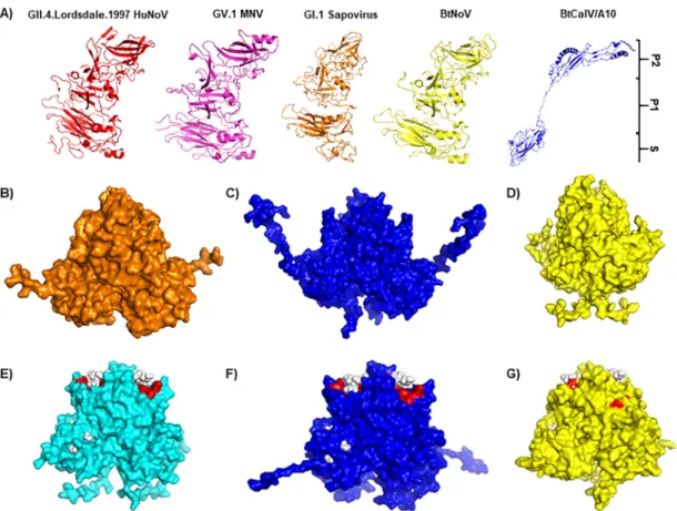
Homology
modeling
of
BtCalV/A10
and
BtNoV
using
HuNoVs
and
MNV.
Previ-ous
works
have
solved
the
capsid
and
P
domain
structures
of
several
caliciviruses
(60,
61).
To
determine
how
the
BtCalV/A10
and
BtNoV
capsids
structurally
compare
to
known
human
and
animal
norovirus
capsids,
we
utilized
predictive
structural
modeling
to
illustrate
the
full-length
BtCalV/A10
and
BtNoV
capsid
structures.
First,
we
used
PHYRE2
open
source
software
to
determine
if
BtCalV/A10
contained
known
calicivirus
capsid
structures
followed
by
structural
homology
modeling
using
Modeller
software
from
the
Max
Planck
Institute
Bioinformatics
Toolkit
(62–64).
Due
to
its
low
amino
acid
similarity
to
known
calicivirus
sequences,
we
also
modeled
the
BtCalV/A10
sequence
using
HuNoVs
(PDB
identifiers
[IDs]:
2OBT
and
4WZT
)
and
MNV
(PDB
ID:
3LQE
) as
backbones
to
capture
multiple
potential
structures
and
predict
potential
sites
of
HBGA
binding
(see
Fig.
5).
the structures in other caliciviruses, further supporting its tentative placement as a
novel genus within
Caliciviridae
.
FIG 4 Bat caliciviruses retain host ligand binding sites similar to those of HuNoVs. (A) Modeling of full-length VP1 capsid sequence from HuNoV, MNV, GI.1 human sapovirus, BtNoV, and BtCalV/A10 using the PHYRE2 Protein Recognition Server. MNV (B) and GII.4 HuNoVs (E) were used as backbones for predictive homology modeling with Modeller software from the Max Planck Institute. The P dimers of BtCalV/A10 were modeled from MNV (C) and GII.4 HuNoVs (F). The P dimers of BtNoV were modeled from MNV (D) and GII.4 HuNoVs (G). In panels E to G, ligand binding sites were predicted by the 3DLigandSite and are marked in red. (E to G) Type A trisaccharide HBGA is indicated by white spheres.
As
the
PHYRE2
modeling
software
did
not
fully
predict
the
structure
of
the
BtCalV/
A10
capsid
protein,
we
next
modeled
the
BtCalV/A10
and
BtNoV
P
dimers
from
the
most
closely
related
animal
norovirus,
MNV
(Fig.
4B
to
D).
Alignment
of
the
P
domains
revealed
that
the
BtCalV/A10
sequence
had
four
insertions
within
the
P
domain
compared
to
the
MNV
P
domain
template;
similarly,
the
BtNoV
sequence
contained
three
insertions
and
three
deletions
compared
to
MNV
(data
not
shown).
However,
as
expected,
the
homologous
models
for
both
BtCalV/A10
(Fig.
4C)
and
BtNoV
(Fig.
4D)
resembled
the
MNV
P
domain
backbone
(Fig.
4B).
We
also
modeled
the
BtCalV/A10
and
BtNoV
P
dimers
on
the
backbone
of
two
GII.4
HuNoVs
(Fig.
4E
to
G).
Compared
to
the
HuNoV
sequences,
the
BtCalV/A10
P
domain
sequence
had
six
insertions
and
four
deletions,
while
the
BtNoV
P
domain
sequence
had
three
insertions
and
five
deletions
(data
not
shown).
These
modifications
did
not
affect
modeling
of
the
P
dimers
from
HuNoVs
for
either
of
the
bat
virus
sequences.
The
BtCalV/A10
dimer
(Fig.
4F)
and
BtNoV
dimer
(Fig.
4G)
each
resembled
the
P
dimers
of
their
HuNoV
counterparts
(Fig.
4E).
Additionally,
the
open
source
software
3DLigand-Site
(65)
indicated
that
the
models
retained
ligand
binding
sites
(red)
for
type
A
HBGAs
(white
spheres)
similar
to
those
seen
with
the
GII.4
HuNoV
backbone.
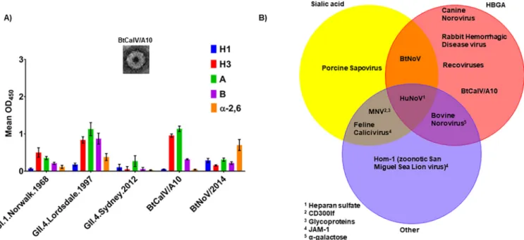
FIG 5 Bat caliciviruses retain HBGA binding patterns overlapping those of HuNoV VLPs. VLPs for BtCalV/A10 were generated using the VEE replicon in C6/36 cells. (A) The HBGA binding patterns of BtCalV/A10 and BtNoV were screened in comparison with those of HuNoVs (GI.1.Norwalk.1968, GII.4.1997.Lordsdale, and GII.4.Sydney.2012). The data presented are mean absorbance values (OD450) following subtraction of negative-control values⫾SEM. Each VLP-HBGA interaction was measured in duplicate in two independent experiments. (B) Caliciviruses bind HBGAs, sialic acids, and other host ligands. Virus-ligand interactions were included in the Venn diagram for cases in which one publication had reported binding to the host ligand.
Thus,
we
utilized
a
synthetic
carbohydrate
binding
assay
to
identify
the
potential
HBGA
receptor-binding
patterns
of
BtCalV/A10
in
comparison
with
a
panel
of
VLPs
derived
from
several
HuNoVs
(GI.1.Norwalk.1968,
GII.4.Lordsdale.1997,
and
GII.4.Sydney.2012)
and
BtNoV
(Fig.
5A).
Among
the
HuNoVs,
GI.1.Norwalk.1968
VLPs
primarily
bound
H
type
3
and
A
antigens
but
weakly
bound
the
H
type
1
and
B
antigens
and
␣
-2,6-sialic
acid.
Con-versely,
GII.4.Lordsdale.1997
VLPs
strongly
bound
H
type
3,
A,
and
B
and
␣
-2,6-sialic
acid,
with
limited
binding
to
H
type
1.
GII.4.Sydney.2012
VLPs
displayed
weak
binding
to
all
carbohydrates
but
bound
most
strongly
to
the
A
antigen.
BtCalV/A10
VLPs
bound
strongly
to
A,
B,
and
H
type
3
HBGA
carbohydrates
but
bound
weakly
to
H
type
1
and
␣
-2,6-sialic
acid.
Of
note,
BtCalV/A10
VLPs
bound
strongly
to
the
H
type
3
and
A
HBGAs
at
a
magnitude
similar
to
that
seen
with
the
GII.4.Lordsdale.1997
HuNoV
VLPs.
Con-versely,
the
BtNoV
VLPs
again
bound
to
␣
-2,6-sialic
acid
but
displayed
modest
binding
to
the
H
type
1
and
H
type
3,
A,
and
B
HBGA
carbohydrates,
as
we
showed
previously
(Fig.
2).
Each
VLP
in
our
panel
displayed
limited
or
weak
binding
to
H
type
2
antigen,
Lewis
antigens,
or
␣
-2,3-sialic
acid
(data
not
shown).
The
bat
caliciviruses
exhibited
different
binding
patterns:
the
BtCalV/A10
VLPs
strongly
bound
H
type
3,
A,
and
B
antigens,
with
limited
binding
to
H
type
1
and
␣
-2,6-sialic
acids,
while
the
BtNoV
VLPs
strongly
bound
␣
-2,6-sialic
acid,
with
moderate
binding
to
H
type
1
and
H
type
3,
A,
and
B
HBGAs,
suggesting
that
bat
caliciviruses
can
bind
to
a
range
of
host
receptors
and
offer
multiple
routes
for
cross-species
transmission.
Both
of
these
binding
patterns
overlapped
binding
patterns
that
were
previously
reported
for
other
animal
calicivi-ruses
and
HuNoVs
(Fig.
5B).
Our
results,
coupled
with
those
previous
reports,
suggest
that
multiple
routes
of
cross-species
transmission
exist
among
caliciviruses.
DISCUSSION
pop-ulations
may
have
contributed
to
the
genetic
diversity
of
noroviruses
and
caliciviruses.
Here,
we
reveal
the
potential
for
several
bat
caliciviruses
to
transmit
across
multiple
species.
Specifically,
we
found
that
a
previously
reported
but
uncharacterized
virus,
BtNoV
(50),
is
antigenically
similar
to
HuNoVs
and
can
bind
HBGAs
in
vitro
.
We
built
upon
this
finding
through
identification
of
a
bat
calicivirus
sequence
(BtCalV/A10)
from
P.
subflavus
near
an
abandoned
railroad
tunnel
outside
Little
Orleans,
MD.
We
further
characterized
both
bat
caliciviruses
through
phylogenetic
analyses,
predictive
structural
homology
modeling,
VLP
production,
and
identification
of
VLP-HBGA
binding
patterns
in
vitro
.
Our
report
suggests
that
BtCalV/A10
is
most
closely
related
to
RHDV,
a
highly
virulent
calicivirus
that
emerged
suddenly
in
China
from
preexisting
strains
prior
to
spreading
globally
(66,
67).
While
more
sequences
are
needed
for
clarification,
A10
likely
represents
a
novel
genus
within
Caliciviridae
,
while
BtNoV
likely
belongs
to
GV
with
MNV.
Structural
homology
modeling
indicated
that
both
bat
viruses
retain
ligand
binding
sites
similar
to
those
seen
with
HuNoVs,
validating
their
capability
to
bind
HBGAs.
The
capsid
sequences
of
both
bat
viruses
formed
VLPs
similar
to
those
formed
by
HuNoVs
and
showed
overlapping
HBGA
binding
profiles
with
respect
to
several
HuNoVs
and
MNV.
Importantly,
as
HBGAs
are
encoded
across
numerous
animal
species,
particularly
among
mammals
(68),
similarities
in
attachment
and
entry
cofactors
sug-gest
that
these
bat
viruses
have
the
potential
to
overcome
a
major
roadblock
to
cross-species
transmission
across
a
variety
of
mammalian
species.
Metagenomic
sequencing
has
illuminated
the
great
diversity
and
complexity
of
RNA
viruses
distributed
throughout
the
natural
world.
However,
new
strategies
are
desper-ately
needed
to
translate
this
information
into
meaningful
predictions
of
biological
processes,
disease
risk,
and
surveillance
prioritization
prior
to
epidemic
or
pandemic
emergence.
Previous
studies
on
bat
caliciviruses
have
focused
on
the
use
of
phyloge-netic
analyses
and
structural
modeling
for
classification
of
these
novel
viruses
within
the
Caliciviridae
family
(45–49).
While
these
techniques
help
contextualize
viral
relat-edness
within
the
framework
of
known
and
characterized
viruses
and
their
families,
they
do
not
necessarily
aid
in
the
understanding
of
biological
functions
or
provide
insight
into
the
emergence
potential
of
zoonotic
pathogens.
In
this
report,
we
utilized
a
novel
platform
combining
metagenomics,
phylogenetics,
immunogenic
comparisons,
and
homology
modeling
of
two
bat
viruses;
we
showed
that
these
viruses
belong
to
Caliciviridae
and
are
structurally
similar
to
known
human
and
animal
caliciviruses.
We
coupled
these
data
with
in
vitro
techniques
and
immunologic
assays
to
derive
further
relevant
biological
questions
that
can
help
to
gauge
the
potential
for
cross-species
transmission
of
two
bat
caliciviruses
to
other
species
prior
to
their
emergence
within
the
human
population.
A
similar
approach
has
identified
the
potential
for
MERS
and
SARS-like
coronaviruses
within
Asian
and
African
bat
populations
to
replicate
and
emerge
in
human
populations
and
to
evade
current
therapeutics
and
has
also
identi-fied
bat
coronaviruses
that
emerged
more
recently
and
caused
outbreaks
in
swine
(8,
9,
69,
70).
GI
and
GII
HuNoVs,
displayed
disparate
HBGA
binding
patterns,
indicating
multiple
potential
paths
for
cross-species
transmission
(Fig.
5B).
Therefore,
the
utilization
of
the
aforementioned
platform
identified
antigenic
relatedness
of
BtNoV
and
HuNoVs
as
well
as
the
potential
for
cross-species
transmission
of
two
previously
uncharacterized
or
unidentified
bat
caliciviruses.
Noroviruses
have
been
historically
classified
on
the
basis
of
capsid
sequence
ho-mology
(59);
based
on
this
strategy,
BtNoV
would
likely
be
classified
within
Norovirus
GV
alongside
MNV.
Enzyme
immunoassays
have
been
powerful
tools
for
understanding
antigenic
relationships
among
noroviruses
in
spite
of
sequence
homology,
especially
in
elucidating
how
antigenic
drift
contributes
to
the
evasion
of
herd
immunity
and
emergence
of
novel
HuNoV
strains
(35).
In
the
present
study,
EIA
performed
with
polyclonal
serums
derived
from
historic
and
current
strains
of
HuNoVs
showed
that
BtNoV
VLPs
are
antigenically
similar
(Fig.
1)
despite
the
parental
virus’s
phylogenetic
relatedness
to
MNV
(Fig.
3).
Other
bat
noroviruses
have
shown
extensive
homology
with
GIV
noroviruses
(51),
whose
antibodies
have
been
detected
in
humans
and
canine
species
(71,
72).
These
results
further
illustrate
the
importance
of
our
proposed
plat-form:
capsid
sequence
homology
alone
would
not
have
predicted
that
bat
caliciviruses
could
bind
synthetic
carbohydrates
or
be
detected
by
serums
against
human
viruses;
in
vitro
assays
revealed
novel
information
regarding
the
caliciviruses
circulating
in
bat
populations.
The
FUT2
gene,
whose
protein
product
promotes
expression
of
HBGAs
on
mucosal
surfaces,
has
a
well-established
role
in
HuNoV
infection
and
RHDV
disease
(73–75).
However,
the
receptors
for
animal
caliciviruses
are
variable
and
less
clear.
For
instance,
GIII
bovine
noroviruses
bind
␣
-galactose
(76,
77),
MNVs
bind
sialic
acids
and
CD300lf
(78–80),
porcine
sapoviruses
bind
sialic
acids
(81),
and
both
feline
calicivirus
and
the
zoonotic
San
Miguel
sea
lion
virus
Hom-1
bind
junctional
adhesion
molecule-1
(82,
83).
While
protein
receptors
seem
quite
variable,
there
is
significant
overlap
in
the
identified
carbohydrate
binding
patterns
of
all
caliciviruses
(Fig.
5),
demonstrating
multiple
potential
routes
of
cross-species
transmission
of
these
viruses.
Recent
genomics
analysis
has
further
indicated
the
potential
for
animal
caliciviruses,
including
bat
caliciviruses,
to
cross
species
barriers.
For
instance,
HBGAs
have
been
identified
or
predicted
across
numerous
animal
species
(68).
Importantly,
the
A
and
B
fucosyltransferase
enzymes
of
Myotis
lucifugus
,
a
microbat
similar
to
P.
subflavus
,
contain
amino
acid
sequences,
amino
acid
motifs,
and
levels
of
phylogenetic
related-ness
similar
to
those
of
their
human
orthologs
(68).
Similarly,
CD300lf
orthologs
have
been
predicted
in
numerous
animal
species,
including
several
Asian
bats,
such
as
Hipposideros
armiger
and
Rhinolophus
sinicus
.
In
addition
to
CD300lf,
sialic
acids
have
previously
been
implicated
as
receptors
or
cellular
cofactors
for
binding
and
entry
by
MNVs
and,
more
recently,
by
HuNoVs
(78,
84–86).
Future
studies
will
reveal
whether
the
BtCalV/A10
or
BtNoV
VLPs
also
bind
bat
or
mammalian
CD300lf
proteins.
Though
numerous
factors
mediate
the
cross-species
transmission,
replication,
and
pathogenesis
of
viruses,
our
results,
coupled
with
those
revealing
the
analogous
host
receptor
structures,
suggest
that
these
bat
caliciviruses
have
the
potential
to
overcome
host
receptor
barriers
as
a
roadblock
to
cross-species
transmission.
combined
scientific
investigations
will
pale
in
comparison
to
the
cost
of
treatment
and
eradication
of
these
pathogens
as
they
emerge
in
the
human
population.
MATERIALS
AND
METHODS
Viralgenomicsequenceisolationandidentification.Viralgenomicsequenceswereidentifiedas partofapreviousstudy(52).Briefly,batswerecollectedwithharptrapsatanabandonedrailroadtunnel nearLittleOrleans,MD,andfreshguanowascollected.Sampleswerestoredoniceuntilprocessing.The BtCalV/A10capsidsequencewasisolatedfromPerimyotis subflavus (tri-coloredbat)usingthe sequence-independentsingle-primeramplificationtechniqueand454next-generationsequencingplatform.De
novo contigswereassembledusingthreeprograms:CodonCodeAligner,Geneious,andDNAStar.Viral aminoacidsequenceswerecreatedusingVectorNTI.Assembledcontigswereanalyzedusingthebasic localalignmentsearchtool(BLAST)fromtheNationalCenterforBiotechnologyInformation(NCBI)(87). BLASTsearcheswereconductedattheaminoacidlevelusingtheprotein-proteinBLAST(blastp)function toquerynonredundantproteinsequences.
Phylogeneticanalysis.Maximum-likelihoodcladogramsof50knowncalicivirusVP1aminoacid sequencesand51knowncalicivirusSdomainaminoacidsequencesweregeneratedusingMEGA7.The cladogramsweregeneratedbasedontheJTTmatrix-basedaminoacidsubstitutionmodelandinitially derivedwiththeNeighbor-Join(NJ)andBioNJalgorithmsfollowedbypairwisedistanceestimationby theuseoftheJTTaminoacidreplacementmodel.Bootstrappingwasconductedbygenerating500 bootstrappedreplicates,andaconsensustreewasdevelopedusingMEGA7.Consensusradial phylo-gramsweregeneratedinGeneiousR11,employingthesamesequencesusedtoconstructthe clado-grams,withtheJukes-Cantorgeneticdistancemodel,theNJbuild method,nooutgroup,and 100 bootstrapreplicates.PhylogramswererenderedforpublicationinAdobeIllustratorCC2017.
Homologymodeling.Thefull-lengthBtCalV/A10,GII.4.1997.LordsdaleHuNoV,GI.1human sapovi-rus,GV.1MNV,andBtNoVcapsidsequences(50)weresubmittedtotheProteinHomology/analogy RecognitionEngineV2.0(PHYRE2)server(88)andanalyzedusingIntensiveMode.Homologymodeling oftheBtCalV/A10andBtNoVsequenceswasbasedontheknownHuNoVGII.4/VA387(PDBID:2OBT) (89) andHuNoVGII.4.2012/NSW0514boundtotypeAtrisaccharide(PDBID:4WZT) (90)andMNV(PDBID: 3LQE)structuresfromtheProteinDataBank.TheaminoacidsequenceswerealignedusingtheClustal OmegaplatformontheBioinformaticsToolkitfromtheMaxPlanckInstitute(62,91).Thepredicted tertiarystructureofthealignedsequencewasdeterminedbytheuseoftheModellerplatformfromthe BioinformaticsToolkitfromtheMaxPlanckInstituteusingGII.4.2012/NSW0514asthetemplate(63). DimericmodelswerecreatedusingThePyMOLMolecularGraphicsSystem,version1.7.4.3(Schrödinger, LLC).
Generationofvirus-likeparticles.Virus-likeparticles(VLPs)weregeneratedaspreviouslydescribed (92).Briefly,theBtCalV/A10andBtNoVVP1sequencesweresynthesizedandinserteddirectlyintothe VEE3526replicon.VP1mRNAswerederivedviaT7polymerasein vitro transcription.TheVEE-BtCalV/A10 andVEE-BtNoVmRNAswereseparatelyelectroporatedinC6/36mosquitolarva(Aedes albopictus)cells (ATCC)andBHKcells,respectively.ElectroporatedC6/36andBHKcellswereincubatedat32°Cand37°C, respectively, with5% CO2 for 26 to28 h beforeharvesting VLPs. VLPs were purified byvelocity sedimentationinsucroseandconcentratedinphosphate-bufferedsaline(PBS)using 100-molecular-weight-cutoff(MWCO)centrifugalfilterunits(Millipore).VLPformationandstructurewereconfirmedby transmissionelectronmicroscopy.Proteinconcentrations weredetermined bytheuseof the bicin-choninic acid (BCA) protein assay (Pierce). VLPs for GI.1.Norwalk.1968, GII.4.1997.Lordsdale, and GII.4.Sydney.2012HuNoVsandMNVwerepreviouslysynthesizedandstoredat⫺80°Cuntiluse.
Carbohydratebindingassays.AcarbohydratebindingassaytodetectVLPbindingwasconducted aspreviouslydescribed(93,94).Briefly,Costarenzymeimmunoassay(EIA)plates(Corning)werecoated with2g/mlofeachoftheproducedVLPsmixedwithPBSandincubatedfor 4 h at roomtemperature. Plateswereblockedovernightwithblotto(5%instantdrymilk–PBS–Tween20).Syntheticbiotinylated carbohydrates(Glycotech)dilutedinblotto(10g/ml)wereaddedtoeachwellandincubatedat37°C for1h.Forthetemperature-dependentHBGAbindingassay,theplateswereincubatedat25°C,32°C, and 37°C for 1 h.Streptavidin-horseradish peroxidase (HRP) was diluted in blotto (1:10,000) and incubatedat37°Cfor30min.TheplatesweredevelopedwithOne-StepUltra-TMB(ThermoFisher)for 15minandwerestoppedwith 2 M H2SO4priortobeingreadat450nm.Plateswerewashed3times withPBS–0.05%Tweenbetweensteps.Thefollowing11carbohydrateswereanalyzed:Htype1,Htype 2,Htype3,A,B,LewisA,LewisB,LexisX,LewisY,␣-2,3-sialicacid,and␣-2,6-sialicacid.PBSservedas anegativecontrolforVLPcoating,andblottoservedasanegativecontrolforHBGAbinding.Each HBGA-VLPinteractionwasevaluatedinduplicateinatleasttwoindependentexperiments.Dataare presentedasmeanabsorbancevalues⫾standarderrorsofthemeans(SEM)at450nmafterremovalof PBSnegative-controlabsorbancevalues.
negative-FIG S1,
TIF file, 0.2 MB.
TABLE S1,
DOCX file, 0.01 MB.
ACKNOWLEDGMENTS
We thank Victoria Madden of the Microscopy Services Laboratory, Department of
Pathology and Laboratory Medicine, University of North Carolina—Chapel Hill, for
electron microscopy assistance and expert technical support. This work was supported
by NIH grants U54AI057157 from the Southeastern Regional Center of Excellence for
Emerging Infections and Biodefense and U19AI109761 from the NIAID and a
grant-in-aid from the Burroughs Wellcome Trust.
The contents of this report are solely the responsibility of the authors and do not
necessarily represent the official views of the NIH.
REFERENCES
1. Jones KE, Patel NG, Levy MA, Storeygard A, Balk D, Gittleman JL, Daszak P. 2008. Global trends in emerging infectious diseases. Nature 451: 990 –993.https://doi.org/10.1038/nature06536.
2. Woolhouse ME, Haydon DT, Antia R. 2005. Emerging pathogens: the epidemiology and evolution of species jumps. Trends Ecol Evol 20: 238 –244.https://doi.org/10.1016/j.tree.2005.02.009.
3. Leroy EM, Epelboin A, Mondonge V, Pourrut X, Gonzalez JP, Muyembe-Tamfum JJ, Formenty P. 2009. Human Ebola outbreak resulting from direct exposure to fruit bats in Luebo, Democratic Republic of Congo, 2007. Vector Borne Zoonotic Dis 9:723–728.https://doi.org/10.1089/vbz .2008.0167.
4. Leroy EM, Kumulungui B, Pourrut X, Rouquet P, Hassanin A, Yaba P, Délicat A, Paweska JT, Gonzalez JP, Swanepoel R. 2005. Fruit bats as reservoirs of Ebola virus. Nature 438:575–576.https://doi.org/10.1038/ 438575a.
5. Haagmans BL, Al Dhahiry SH, Reusken CB, Raj VS, Galiano M, Myers R, Godeke GJ, Jonges M, Farag E, Diab A, Ghobashy H, Alhajri F, Al-Thani M, Al-Marri SA, Al Romaihi HE, Al Khal A, Bermingham A, Osterhaus AD, AlHajri MM, Koopmans MP. 2014. Middle East respiratory syndrome coronavirus in dromedary camels: an outbreak investigation. Lancet Infect Dis 14:140 –145.https://doi.org/10.1016/S1473-3099(13)70690-X. 6. Reusken CB, Haagmans BL, Müller MA, Gutierrez C, Godeke GJ, Meyer B,
Muth D, Raj VS, Smits-De Vries L, Corman VM, Drexler JF, Smits SL, El Tahir YE, De Sousa R, van Beek J, Nowotny N, van Maanen K, Hidalgo-Hermoso E, Bosch BJ, Rottier P, Osterhaus A, Gortázar-Schmidt C, Dro-sten C, Koopmans MP. 2013. Middle East respiratory syndrome corona-virus neutralising serum antibodies in dromedary camels: a comparative serological study. Lancet Infect Dis 13:859 – 866.https://doi.org/10.1016/ S1473-3099(13)70164-6.
7. Li W, Shi Z, Yu M, Ren W, Smith C, Epstein JH, Wang H, Crameri G, Hu Z, Zhang H, Zhang J, McEachern J, Field H, Daszak P, Eaton BT, Zhang S, Wang LF. 2005. Bats are natural reservoirs of SARS-like coronaviruses. Science 310:676 – 679.https://doi.org/10.1126/science.1118391. 8. Menachery VD, Yount BL, Jr., Debbink K, Agnihothram S, Gralinski LE,
Plante JA, Graham RL, Scobey T, Ge XY, Donaldson EF, Randell SH, Lanzavecchia A, Marasco WA, Shi ZL, Baric RS. 2015. A SARS-like cluster of circulating bat coronaviruses shows potential for human emergence. Nat Med 21:1508 –1513.https://doi.org/10.1038/nm.3985.
9. Menachery VD, Yount BL, Jr., Sims AC, Debbink K, Agnihothram SS, Gralinski LE, Graham RL, Scobey T, Plante JA, Royal SR, Swanstrom J, Sheahan TP, Pickles RJ, Corti D, Randell SH, Lanzavecchia A, Marasco WA, Baric RS. 2016. SARS-like WIV1-CoV poised for human emergence. Proc Natl Acad Sci U S A 113:3048 –3053.https://doi.org/10.1073/pnas .1517719113.
10. Memish ZA, Mishra N, Olival KJ, Fagbo SF, Kapoor V, Epstein JH, Alhakeem R, Durosinloun A, Al Asmari M, Islam A, Kapoor A, Briese T, Daszak P, Al
Rabeeah AA, Lipkin WI. 2013. Middle East respiratory syndrome coronavirus in bats, Saudi Arabia. Emerg Infect Dis 19:1819 –1823.https://doi.org/10 .3201/eid1911.131172.
11. Anthony SJ, Epstein JH, Murray KA, Navarrete-Macias I, Zambrana-Torrelio CM, Solovyov A, Ojeda-Flores R, Arrigo NC, Islam A, Ali Khan S, Hosseini P, Bogich TL, Olival KJ, Sanchez-Leon MD, Karesh WB, Goldstein T, Luby SP, Morse SS, Mazet JA, Daszak P, Lipkin WI. 2013. A strategy to estimate unknown viral diversity in mammals. MBio 4:e00598-13.https:// doi.org/10.1128/mBio.00598-13.
12. Mesquita JR, Barclay L, Nascimento MS, Vinjé J. 2010. Novel norovirus in dogs with diarrhea. Emerg Infect Dis 16:980 –982. https://doi.org/10 .3201/eid1606.091861.
13. Bodnar L, Di Martino B, Di Profio F, Melegari I, Lanave G, Lorusso E, Cavalli A, Elia G, Bányai K, Marsilio F, Buonavoglia C, Martella V. 2016. Detection and molecular characterization of sapoviruses in dogs. Infect Genet Evol 38:8 –12.https://doi.org/10.1016/j.meegid.2015.11.034. 14. Soma T, Nakagomi O, Nakagomi T, Mochizuki M. 2015. Detection of
norovirus and Sapovirus from diarrheic dogs and cats in Japan. Microbiol Immunol 59:123–128.https://doi.org/10.1111/1348-0421.12223. 15. Di Martino B, Di Profio F, Melegari I, Sarchese V, Cafiero MA, Robetto S,
Aste G, Lanave G, Marsilio F, Martella V. 2016. A novel feline norovirus in diarrheic cats. Infect Genet Evol 38:132–137.https://doi.org/10.1016/j .meegid.2015.12.019.
16. Wang QH, Han MG, Cheetham S, Souza M, Funk JA, Saif LJ. 2005. Porcine noroviruses related to human noroviruses. Emerg Infect Dis 11: 1874 –1881.https://doi.org/10.3201/eid1112.050485.
17. Shen Q, Zhang W, Yang S, Cui L, Hua X. 2012. Complete genome sequence of a new-genotype porcine norovirus isolated from piglets with diarrhea. J Virol 86:7015–7016.https://doi.org/10.1128/JVI.00757-12.
18. Wobus CE, Karst SM, Thackray LB, Chang KO, Sosnovtsev SV, Belliot G, Krug A, Mackenzie JM, Green KY, Virgin HW. 2004. Replication of noro-virus in cell culture reveals a tropism for dendritic cells and macro-phages. PLoS Biol 2:e432.https://doi.org/10.1371/journal.pbio.0020432. 19. Karst SM, Wobus CE, Lay M, Davidson J, Virgin H. 2003. STAT1-dependent innate immunity to a Norwalk-like virus. Science 299:1575–1578.https:// doi.org/10.1126/science.1077905.
20. Wolf S, Williamson W, Hewitt J, Lin S, Rivera-Aban M, Ball A, Scholes P, Savill M, Greening GE. 2009. Molecular detection of norovirus in sheep and pigs in New Zealand farms. Vet Microbiol 133:184 –189.https://doi .org/10.1016/j.vetmic.2008.06.019.
21. Liu BL, Lambden PR, Günther H, Otto P, Elschner M, Clarke IN. 1999. Molecular characterization of a bovine enteric calicivirus: relationship to the Norwalk-like viruses. J Virol 73:819 – 825.
22. Li L, Shan T, Wang C, Côté C, Kolman J, Onions D, Gulland FM, Delwart E. 2011. The fecal viral flora of California sea lions. J Virol 85:9909 –9917. https://doi.org/10.1128/JVI.05026-11.
controlabsorbance. Todetermine EC50, EIA datawere log transformedand fitted usingsigmoidal dose-responseanalysisofnonlineardatainGraphPadPrism7.02(GraphPadSoftware,Inc.,LaJolla,CA). EC50swerecomparedusingone-wayanalysisofvariance(ANOVA)withDunnett’sposttest.