TRANSLATIONAL INVESTIGATIONS OF REWARDING SUBSTANCES USING THE CURVE-SHIFT METHOD OF INTRACRANIAL SELF-STIMULATION IN MICE
J. Elliott Robinson
A dissertation submitted to the faculty of the University of North Carolina at Chapel Hill in partial fulfillment of the requirements for the degree of Doctor of Philosophy in
the Graduate School (Biological and Biomedical Sciences Program, Neurobiology Curriculum).
Chapel Hill 2014
Approved by: C. J. Malanga Tom Kash
iii ABSTRACT
J. Elliott Robinson: Translational Investigations of Rewarding Substances Using the Curve-Shift Method of Intracranial Self-Stimulation in Mice
(Under the direction of C. J. Malanga)
Drug and alcohol abuse represents a major public health burden in the United States and is responsible for a significant loss of productivity among U.S. citizens. As such, effective strategies will need to be developed that address the social and biomedical consequences of drug abuse in the United States, including the
discovery of pharmacotherapies and cognitive behavioral interventions that
decrease drug or alcohol intake, help maintain sobriety, and prevent or address the neurobiological sequelae associated with repeated drug exposure. These pre-clinical studies employed the curve-shift method of intracranial self-stimulation in mice to probe the behavioral effects of rewarding substances on mesocorticolimbic circuitry involved in positive reinforcement in order to 1) describe the
iv
variants associated with substance abuse or treatment efficacy (e.g. the mu opioid receptor gene (OPRM1) A118G polymorphism). Taken together, these
v
Everett, someday – probably several years from now – you may find this doctoral dissertation dusty and forgotten, restlessly waiting for you amid the clutter of a quiet room. I hope you read these words aloud and hear me calling out to you from the past: Find your calling. Have goals you want to achieve so badly that they keep you up at night. Aim high, be charitable and kind, and always try to work harder than your peers. Ask for help when you need it and never let anyone tell you that an ambition is unachievable. If you will it, it is no dream.
vi
ACKNOWLEDGEMENTS
vii
TABLE OF CONTENTS
LIST OF TABLES...………... ix
LIST OF FIGURES……… x
LIST OF ABBREVIATIONS AND SYMBOLS ………. xii
CHAPTER 1: INTRODUCTION……….. 1
References……….. 15
CHAPTER 2: MEPHEDRONE (4-METHYLMETHCATHINONE) AND INTRACRANIAL SELF-STIMULATION: COMPARISON TO COCAINE…... 22
Introduction………. 22
Methods………... 24
Results………... 27
Discussion………... 29
Conclusion….………..… 33
Figures………. 35
References……….. 40
CHAPTER 3: POTENTIATION OF BRAIN STIMULATION REWARD BY MORPHINE: EFFECTS OF NEUROKININ-1 RECEPTOR ANTAGONISM… 44 Introduction……….……. 44
Methods………... 48
Results………. 53
viii
Figures………... 61
References………... 67
CHAPTER 4: LEVETIRACETAM HAS OPPOSITE EFFECTS ON ALCOHOL AND COCAINE RELATED BEHAVIORS….……….. 75
Introduction……….…. 75
Methods.…….……...……….…. 78
Results………. 84
Discussion……….……….. 89
Figures and Tables……….. 100
References……… 109
CHAPTER 5: ALTERED RESPONES TO ALCOHOL AND MORPHINE IN A HUMANIZED MOUSE MODEL OF THE OPRM1 A118G POLYMORPHISM………...… 116
Introduction………... 116
Methods………. 120
Results……….…….. 128
Discussion………. 136
Figures and Tables……….. 152
References……… 166
CHAPTER 6: DISCUSSION……… 180
ix
LIST OF TABLES
x
LIST OF FIGURES
Figure 1.1 - Mesocorticolimbic circuits involved in the rewarding and
motivational effects of positive reinforcers………... 4 Figure 2.1- Placement of intracranial self-stimulation monopolar electrodes
and representative rate-frequency curves in C57BL/6J mice....……… 35 Figure 2.2 - Dose-response relationship for the effects of mephedrone and
cocaine onthe frequency that supported half-maximal responding in
C57BL/6J mice……….……….. 37 Figure 2.3 - Dose-response relationship for the effects of mephedrone and
cocaine onthe brain stimulation reward threshold in C57BL/6J mice……... 38 Figure 2.4 - Dose-response relationship for the effects of mephedrone and
cocaine onthe maximum response rate in C57BL/6J mice……….….. 39 Figure 3.1 - Placement of intracranial self-stimulation monopolar electrodes
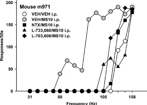
in C57BL/6J mice………... 61 Figure 3.2 - Responding for different frequencies of brain stimulation reward
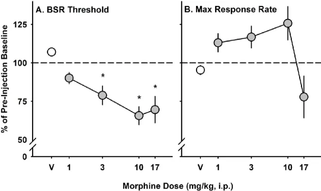
for an individual C57BL/6J mouse……….. 62 Figure 3.3 - Dose-response relationship for morphine on BSR threshold and
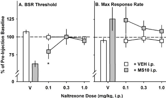
maximum operant response rate in C57BL/6J mice……… 63 Figure 3.4 - Effect of naltrexone pre-treatment on morphine-induced changes
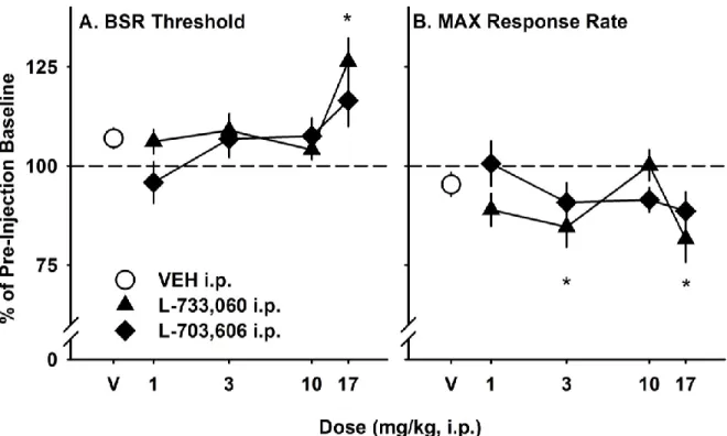
in BSR threshold and maximum response rate in C57BL/6J mice……… 64 Figure 3.5 - Dose-response relationship for the neurokinin-1 receptor
antagonists L-733,060 and L-703,606 on BSR threshold and maximum
response rate in C57BL/6J mice………. 65 Figure 3.6 - Effect of L-733,060, L-703,606, or saline vehicle pre-treatment
on morphine-induced changes in BSR threshold and maximum response
rate in C57BL/6J mice………... 66 Figure 4.1 - Intracranial self-stimulation (ICSS) electrode tip locations and
representative rate frequency curves………... 101 Figure 4.2 - Effects of levetiracetam (LEV) pretreatment on alcohol-affected
xi
in C57BL/6J mice………...……. 105 Figure 4.4 - Effect of bath-applied levetiracetam (LEV) on excitatory
neurotransmission in nucleus accumbens medium spiny neurons and
ventral tegmental area dopaminergic neurons (VTA) in vitro………... 107 Figure 5.1 - Comparison of ICSS behavior in h/mOprm1 118AA and h/mOprm1
118GG mice………. 152 Figure 5.2 - Effects of alcohol and naltrexone on ICSS in h/mOprm1 118AA
and h/mOprm1 118GG mice……….……… 153 Figure 5.3 - Effects of opioid agonists and cocaine on ICSS in h/mOprm1
118AA and h/mOprm1 118GG mice……….... 156 Figure 5.4 - Effects of MOR agonists DAMGO and morphine on mIPSCs in
ventral tegmental area neurons from h/mOprm1 118AA and h/mOprm1
118GG mice………. 159 Figure 5.5 – Baseline mIPSC frequency and amplitude during recordings
from dopaminergic neurons in the ventral tegmental area in midbrain
slices from h/mOprm1 118AA and 118GG mice…………...………... 161 Figure 5.6 - Pharmacological characterization of h/mOprm1 118A and 118G
receptors………... 163 Figure 5.7 - Replicate determination of surface fraction (P2) binding sites in
xii
LIST OF ABBREVIATIONS AND SYMBOLS
5-HT Serotonin
A112G Adenine-to-guanine substitution at Oprm1 position 112 A118G Adenine-to-guanine substitution at OPRM1 position 118 aCSF Artificial cerebrospinal fluid
ALC Alcohol
ANOVA Analysis of variance AUD Alcohol use disorder BSR Brain stimulation reward BUP Buprenorphine
cAMP Cyclic adenosine monophosphate CNS Central nervous system
COC Cocaine
CPP Conditioned place preference
CRISPR Clustered regularly interspaced short palindromic repeats DA Dopamine
DAMGO [D-Ala2, N-MePhe4, Gly-ol]-enkephalin DAT Dopamine transporter
EF50 Frequency that maintains half-maximal responding EPSC Excitatory postsynaptic current
ERK Extracellular signal-regulated kinase FENT Fentanyl
xiii
GIRK G protein-coupled inwardly-rectifying potassium channels GRK2 G protein coupled receptor kinase 2
ICSS Intracranial self-stimulation JNK c-Jun N-terminal kinase 2 LEV Levetiracetam
MAX Maximum operant response rate MDMA Methylenedioxymethamphetamine MFB Medial forebrain bundle
mIPSC Miniature inhibitory postsynaptic current MOR Mu opioid receptor
MS Morphine sulphate MSN Medium spiny neuron NAc Nucleus accumbens
NET Norepinephrine transporter NK1R Neurokinin-1 receptor NMDA N-methyl-d-aspartate OPRM1 Mu opioid receptor gene OXY Oxycodone
P1 Pellet 1 (nuclear/cytosolic fraction) P2 Pellet 2 (surface fraction)
xiv POMC Proopiomelanocortin
SAL Saline
SERT Serotonin transporter
SNc Substantia nigra pars compacta SNP Single nucleotide polymorphism STR Dorsal striatum
SUD Substance use disorder SV2A Synaptic vesicle 2A TH Tyrosine hydroxylase VEH Vehicle
VTA Ventral tegmental area β-END β-Endorphin
1
CHAPTER 1: INTRODUCTION
Drug and alcohol abuse represents a major public health burden in the United States and is responsible for a significant loss of productivity among U.S. citizens. Recent analysis by the National Center on Addiction and Substance Abuse at Columbia University suggests that 40 million Americans are addicted to nicotine, alcohol, or other drugs, while another 80 million people engage in substance use in ways that threaten their health and safety or the health and safety of others (CASA, 2012). The economic impact of tobacco, alcohol, and other drug abuse in this country is substantial and has been estimated to cost society over $500 billion in the form of health care spending, productivity loss, crime, incarceration, and drug
2
Substance use disorder (SUD) is characterized by a pathological pattern of behavior related to use of the substance, including impaired control and risky use; social impairment; prolonged drug intake; a persistent desire to cut down or regulate substance use, which may be accompanied by multiple unsuccessful efforts to decrease or discontinue use; and a significant dedication to obtaining the substance, using the substance, or recovering from its effects (American Psychiatric
Association, 2013). Very few pharmacotherapies have received approval by the Food and Drug Administration for the treatment of SUD; of these, only the opioid antagonist naltrexone for alcohol dependence, the atypical antidepressant bupropion or the nicotinic acetylcholine receptor partial agonist varenicline for nicotine
dependence, and methadone or buprenorphine maintenance therapy for opioid dependence have gained widespread acceptance among clinicians (Corelli & Hudmon, 2002; Rastegar, Kunins, Tetrault, Walley, & Gordon, 2013). Because of the high failure rate of clinical trials and costs associated with central nervous system drug development, many pharmaceutical companies have begun to rely on investigators in academic settings to identify and validate promising treatment targets for neuropsychiatric conditions, including addiction, through preclinical translational research (Bartlett & Heilig, 2013; Hurko & Ryan, 2005).
In 2003, the U.S. National Institutes of Health Annual Report, released by then director Elias Zerhouni, prioritized ‘translational research’ that would translate basic science discoveries into new therapeutic treatments and improved patient care by emphasizing multidisciplinary efforts between basic scientists and clinical
3
2003). Recent technological advances that make ‘-omic’ information (e.g. genomic, proteomic, transcriptomic, epigenomic, etc.) readily available to basic scientists has greatly expanded the number of druggable targets beyond the approximately five hundred that comprise the current pharmacopeia (Hurko & Ryan, 2005; Schadt, Monks, & Friend, 2003). As such, interest in developing pharmacotherapies for SUD has increased, and ongoing investigations using rodent, primate, and other
mammalian and non-mammalian models of disease should yield tangible benefits to patient populations in the upcoming decades. Pre-clinical drug development is aided by the fact that several aspects of addictive behavior can be modeled in laboratory animals, including escalation of drug intake, the emergence of neurocognitive deficits with prolonged use, drug seeking that is resistant to
extinction, increased motivation to consume drugs, drug preference over non-drug rewards, and continued drug consumption despite negative consequences or punishment (Vanderschuren & Ahmed, 2013). When these behavioral models are used in conjunction with molecular biological, neurochemical, and/or cellular physiological techniques, one can elucidate pathophysiological mechanisms
underlying substance abuse, validate drug targets in the context of perturbed neural signaling, and predict the utility of experimental therapeutics.
Drugs of abuse are potent positive reinforcers (i.e. they increase the
4
of well-being (Balster & Bigelow, 2003; de Wit & Phillips, 2012) and appear to be due to actions on neural circuits that evolved to promote behaviors necessary to survival (e.g. obtaining food or water) (Nestler, Hyman, & Malenka, 2001). Given that human subjects who report feelings of euphoria or liking are more likely to self-administer drugs or alcohol in a laboratory setting (Chutuape & de Wit, 1994; de Wit, 1998), there has been great interest in developing pharmacotherapies that decrease intake by attenuating drug or alcohol reward (Heilig et al., 2010), such as opioid receptor antagonists for the treatment of alcohol dependence (Anton et al., 2006).
Like natural reinforcers, drugs of abuse promote positive reinforcement through coordinated activity of limbic motor and brain reward circuits (Figure 1). These regions include the mesencephalic ventral tegmental area (VTA) and substantia pars compacta
(SNc), which send
dopaminergic projections to the forebrain nucleus accumbens (NAc) and dorsal striatum (STR); the limbic neocortex, which
includes the medial prefrontal (PFC), orbitofrontal, and anterior cingulate cortex; the hippocampus (HIPP); and the amygdala (AMYG) (Wise, 2005). Midbrain
dopaminergic projections are particularly important in appetitive behavior, reward expectancy, and motivated or goal-oriented behaviors (Di Chiara & Imperato, 1988; Salamone, Kurth, McCullough, Sokolowski, & Cousins, 1993; Schultz, Dayan, &
5
Montague, 1997; Wise, 2005). These projections are regulated by diverse inputs, including local GABAergic interneurons; inhibitory afferents from the NAc and rostromedial tegmental nucleus (RMTg); and excitatory afferents from the PFC, other tegmental nuclei, lateral hypothalamus, etc. (Geisler & Wise, 2008; Wise, 2005) While brain reward circuits are normally activated by sensory cues associated with natural reinforcers, drugs of abuse act on these pathways directly to produce supraphysiological neurotransmission, leading to enhanced motivational states and conditioned reinforcement of drug taking (Di Chiara & Imperato, 1988; Salamone, et al., 1993; Wise, 2005). Although drug-induced pleasurable states promote initial drug or alcohol consumption, these euphorigenic effects may become blunted during prolonged use and promote escalated intake (Koob & Le Moal, 2008; Robinson & Berridge, 1993). As a result, the existence of evolutionarily conserved systems that promote desirable behaviors via reinforcement by natural rewards has made
humans vulnerable to addiction (Nestler, et al., 2001).
While the measurability of hedonic processes in rodents is debatable, several behavioral methods have been developed that attempt to model these subjective rewarding effects in laboratory animals, including conditioned place preference (CPP) and intracranial self-stimulation (ICSS) procedures. CPP methods measure an animal’s preference for a previously drug-paired environment and infer that these effects are due to positive subjective drug experiences that occur during the
6
Spanagel, 2006). While there is utility in measuring the ability of a possible
pharmacotherapy to alter the emotional valence (e.g. reward or aversion) of a drug of abuse by blocking the development or expression of place preference, this methodology rarely produces graded or dose-dependent effects that are needed to characterize drug potency in vivo. Furthermore, there is heterogeneity in the ability of a drug to produce CPP that depends on species, strain, route of drug
administration, and dose that does not always reflect a drug’s ability to produce positive reinforcement under similar conditions (Sanchis-Segura & Spanagel, 2006). As a result, one must use caution when interpreting CPP data in the context of other drug and alcohol related behaviors.
7
anhedonic effects in human subjects, such kappa opioid receptor agonists (Pfeiffer, Brantl, Herz, & Emrich, 1986), devalue BSR in rodents (Carlezon et al., 2006). Several pharmacological agents diminish or enhance the effects of abused drugs on ICSS (e.g. (Bain & Kornetsky, 1987; Tzchentke & Schmidt, 2000)), and
drug-pretreatment studies allow for the preclinical evaluation of potential pharmacotherapies to alter the subjective effects of drugs or alcohol.
Several brain regions will sustain operant responding for BSR, including the VTA, SNC, PFC, NAc, etc. The medial forebrain bundle (MFB) at the level of the lateral hypothalamus is one of the most commonly used electrode implantation sites because it produces reliable self-stimulation at low current intensities without motor side effects (Carlezon & Chartoff, 2007; Liebman, 1983). The MFB is a complex fiber system that courses through the lateral hypothalamus and carries ascending and descending fibers from several sites, including the olfactory tubercle, central amygdala, lateral septal nucleus, bed nucleus of the stria terminalis, nucleus
accumbens, frontal cortex, ventral tegmental area, lateral preoptic area, and several hypothalamic nuclei (Nieuwenhuys, Geeraedts, & Veening, 1982; Veening,
8
produces robust, time-locked dopamine release in the nucleus accumbens that is independent from transients associated with the presentation of reward cues (Cheer et al., 2007). As such, ICSS is attenuated by dopamine D1- and D2-like receptor antagonists (Duvauchelle, Fleming, & Kornetsky, 1998; R. U. Esposito, Faulkner, & Kornetsky, 1979; Nakajima & Patterson, 1997) and potentiated by both D1 and D2 receptor agonists (Gilliss, Malanga, Pieper, & Carlezon, 2002; Hunt, Atrens, & Jackson, 1994; Malanga, Riday, Carlezon, & Kosofsky, 2008; Ranaldi & Beninger, 1994). Dopamine receptor antagonists block the effects of cocaine (Kita, Shiratani, Takenouchi, Fukuzako, & Takigawa, 1999; Vorel et al., 2002), opioids (Kornetsky & Duvauchelle, 1994; Kornetsky & Porrino, 1992), alcohol (Malanga, unpublished observations), nicotine (Huston-Lyons, Sarkar, & Kornetsky, 1993), and
amphetamine (Gallistel & Karras, 1984) on ICSS, indicating the involvement of mesencephalic dopaminergic projections in the effects of drugs of abuse on ICSS.
Although effects of the pharmacological agents on ICSS of the lateral
hypothalamus are largely due to their ability to interact with midbrain dopaminergic projections to forebrain targets, several strategies can be used to investigate the role of other loci in brain reward function. The most straightforward of these strategies involves the use of alternative electrode placement sites that may or may not
promote evoked striatal dopamine release, yet produce positive reinforcement. This caveat is important because ICSS could hypothetically be maintained by negatively reinforcing electrical stimulation e.g. stimulation that provides relief from an
9
are also helpful in determining the involvement of specific circuits in brain reward function, as these treatments often change the pattern and frequency of lateral hypothalamic self-stimulation. For example, NMDA receptor antagonists lower reward thresholds when injected into the nucleus accumbens shell region (Carlezon & Wise, 1996), yet injection of these drugs into the VTA attenuates reward cue-mediated phasic NAc dopamine release and increases the latency to respond for BSR (Sombers, Beyene, Carelli, & Wightman, 2009). This example demonstrates that a single class of pharmacological agents can have bi-directional effects on ICSS depending on the injection site, and similar approaches may help identify circuits that are involved in different aspects of the complex behavioral response to a systemically administered drug.
10
of acute drug effects (Bauco, Wang, & Wise, 1993; Bauco & Wise, 1997; R. Esposito & Kornetsky, 1977; Frank, Martz, & Pommering, 1988; Frank & Zubrycki, 1989). This phenomenon is particularly striking when examined in the context of the locomotor sensitization that occurs with repeated psychostimulant exposure. For example, mice will display progressive increases in the locomotor response to 15.0 mg/kg cocaine when dosed repeatedly, yet the potency of that drug dose to
potentiate BSR is consistent across several challenges in the same experimental animals (Riday, Kosofsky, & Malanga, 2012). Because desensitization or
sensitization is not typically observed with ICSS, a within subjects design can be used to assess drug effects on BSR, which reduces data variability and the number of subjects needed per experiment. This represents a major advantage of this
method over CPP, which requires a between subjects design to assess dose-effects. When discussing the utility of ICSS, one must consider differences in
methodology that are often employed to measure the subjective effects of abused drugs. In ICSS, experimental animals – usually rats or mice – are implanted with monopolar or bipolar electrodes in a brain region of interest and trained to respond for reinforcing electrical stimulation using a bar (often employed with rats), wheel (mice), nose poke, or other manipulandum. Early investigations employed response rate (e.g. number of lever presses) as the primary dependent variable using
continuous reinforcement or low fixed-ratio schedules. In these experiments,
11
psychomotor stimulation or ability to perform the operant task (Kornetsky & Bain, 1992). In order to address this concern, new variations of ICSS were developed that are less sensitive to changes in maximum operant response rate. In the curve-shift method of ICSS, rodents are trained to respond during discrete response periods in which the current intensity is fixed, but the frequency of stimulation declines in 0.05-log unit steps from period to period. This testing method produces characteristic rate-frequency curves that are similar to dose-response curves in pharmacology. As such, positive reinforcers with rewarding effects tend to produce leftward shifts in the rate-frequency curve, which are indicated by decreases in EF50 (the frequency that produces half-maximal responding) or θ0 (or BSR threshold; the lowest frequency that sustains responding, defined as the x-intercept of the least squares regression line through the frequencies that sustain 20%, 30%, 40%, 50%, and 60% of the maximum response rate (Rompre & Wise, 1989)), while drugs with anhedonic effects produce leftward shifts. Because a decrease in maximum operant response rate can alter EF50, changes in θ0 are typically used to describe drug effects
(Carlezon & Chartoff, 2007; Miliaressis, Rompre, Laviolette, Philippe, & Coulombe, 1986).
12
noncontingent prime. If the animal fails to respond within 7.5s, the trial is
terminated. By varying current intensity (uA) in a stepwise fashion in ascending and descending order, response-intensity curves can be generated to calculate BSR threshold (Kornetsky & Bain, 1992). By increasing the force necessary to turn a wheel manipulandum, Markou and Koob showed that performance manipulations did not alter BSR thresholds, although they did observe changes in response
latency, number of extra responses, and responses during intertrial timeout periods. Interestingly, reward manipulations – changes in the train duration of electrical stimulation – altered BSR threshold without altering other measures of ICSS (Markou & Koob, 1992). While the psychophysical method has been widely
employed to study brain reward mechanisms in rats, it has not been widely adopted for use in mice, possibly due to difficulty learning the task. Kornetsky and
colleagues reported that restraining experimental mice improved training efficiency, and they hypothesized that difficulties were due to a relatively short attention span in mice compared to rats (Gill, Knapp, & Kornetsky, 2004). As such, most
investigations that measure ICSS in mice employ curve-shift methodology. In these investigations, I used several behavioral, pharmacological, and electrophysiological techniques in conjunction with the curve-shift method of ICSS in mice to probe the effects of drugs of abuse on circuits involved in positive
reinforcement. For the most part, these pre-clinical studies fell into one or more of three general categories:
13
2) The assessment of neuroactive substances that modulate drug or alcohol related behaviors, subjective states, or cellular effects.
3) Assessments of the role of known human genetic variants to alter drug responses or treatment efficacy using transgenic or ‘humanized’ animals.
As such, the specific aims of this dissertation were:
Aim 1: To characterize reward-potentiating effects of the designer
psychostimulant mephedrone (4-methylmethcathinone; Chapter 2), which – along with other cathinone-derivatives – has emerged during the last five years as an easily obtained, recreational drug of relatively unknown abuse potential (Gunderson, Kirkpatrick, Willing, & Holstege, 2013).
Aim 2: To investigate novel uses of two FDA-approved drugs to attenuate the acute rewarding effects of several drugs of abuse. These studies examined the ability of neurokinin-1 receptor (NK1R) antagonists to alter the reward-potentiating effects of morphine (Chapter 3). I also examined the effects of the synaptic vesicle 2A inhibitor levetiracetam on alcohol- or cocaine-related behaviors using several behavioral assays, including ICSS (Chapter 4).
14
Taken together, these investigations reflect the neuropharmacological utility of ICSS as a translational tool to help address unmet needs in the treatment of
15
REFERENCES
American Psychiatric Association. (2013). Diagnostic and statistical manual of mental disorders : DSM-5 (5th ed.). Washington, D.C.: American Psychiatric Publishing.
Anton, R. F., O'Malley, S. S., Ciraulo, D. A., Cisler, R. A., Couper, D., Donovan, D. M., et al. (2006). Combined pharmacotherapies and behavioral interventions for alcohol dependence: the COMBINE study: a randomized controlled trial. JAMA, 295(17), 2003-2017.
Bain, G. T., & Kornetsky, C. (1987). Naloxone attenuation of the effect of cocaine on rewarding brain stimulation. Life Sci, 40(11), 1119-1125.
Balster, R. L., & Bigelow, G. E. (2003). Guidelines and methodological reviews concerning drug abuse liability assessment. Drug Alcohol Depend, 70(3 Suppl), S13-40.
Bartlett, S., & Heilig, M. (2013). Translational approaches to medication development. Curr Top Behav Neurosci, 13, 543-582.
Bauco, P., Wang, Y., & Wise, R. A. (1993). Lack of sensitization or tolerance to the facilitating effect of ventral tegmental area morphine on lateral hypothalamic brain stimulation reward. Brain Res, 617(2), 303-308.
Bauco, P., & Wise, R. A. (1997). Synergistic effects of cocaine with lateral
hypothalamic brain stimulation reward: lack of tolerance or sensitization. J Pharmacol Exp Ther, 283(3), 1160-1167.
Carlezon, W. A., Jr., Beguin, C., DiNieri, J. A., Baumann, M. H., Richards, M. R., Todtenkopf, M. S., et al. (2006). Depressive-like effects of the kappa-opioid receptor agonist salvinorin A on behavior and neurochemistry in rats. J Pharmacol Exp Ther, 316(1), 440-447.
Carlezon, W. A., Jr., & Chartoff, E. H. (2007). Intracranial self-stimulation (ICSS) in rodents to study the neurobiology of motivation. Nat Protoc, 2(11), 2987-2995.
Carlezon, W. A., Jr., & Wise, R. A. (1996). Microinjections of phencyclidine (PCP) and related drugs into nucleus accumbens shell potentiate medial forebrain bundle brain stimulation reward. Psychopharmacology (Berl), 128(4), 413-420.
16
and Substance Abuse at Columbia University.
Check, E. (2003). NIH 'roadmap' charts course to tackle big research issues. Nature, 425(6957), 438.
Cheer, J. F., Aragona, B. J., Heien, M. L., Seipel, A. T., Carelli, R. M., & Wightman, R. M. (2007). Coordinated accumbal dopamine release and neural activity drive goal-directed behavior. Neuron, 54(2), 237-244.
Chutuape, M. A., & de Wit, H. (1994). Relationship between subjective effects and drug preferences: ethanol and diazepam. Drug Alcohol Depend, 34(3), 243-251.
Corelli, R. L., & Hudmon, K. S. (2002). Medications for smoking cessation. West J Med, 176(2), 131-135.
de Wit, H. (1998). Individual differences in acute effects of drugs in humans: their relevance to risk for abuse. NIDA Res Monogr, 169, 176-187.
de Wit, H., & Phillips, T. J. (2012). Do initial responses to drugs predict future use or abuse? Neurosci Biobehav Rev, 36(6), 1565-1576.
Di Chiara, G., & Imperato, A. (1988). Drugs abused by humans preferentially
increase synaptic dopamine concentrations in the mesolimbic system of freely moving rats. Proc Natl Acad Sci U S A, 85(14), 5274-5278.
Duvauchelle, C. L., Fleming, S. M., & Kornetsky, C. (1998). Prefrontal cortex infusions of SCH 23390 cause immediate and delayed effects on ventral tegmental area stimulation reward. Brain Res, 811(1-2), 57-62.
Esposito, R., & Kornetsky, C. (1977). Morphine lowering of self-stimulation thresholds: lack of tolerance with long-term administration. Science, 195(4274), 189-191.
Esposito, R. U., Faulkner, W., & Kornetsky, C. (1979). Specific modulation of brain stimulation reward by haloperidol. Pharmacol Biochem Behav, 10(6), 937-940.
Frank, R. A., Martz, S., & Pommering, T. (1988). The effect of chronic cocaine on self-stimulation train-duration thresholds. Pharmacol Biochem Behav, 29(4), 755-758.
Frank, R. A., & Zubrycki, E. (1989). Chronic imipramine does not block cocaine-induced increases in brain stimulation reward. Pharmacol Biochem Behav, 33(3), 725-727.
17
effects on the reward summation function. Pharmacol Biochem Behav, 20(1), 73-77.
Geisler, S., & Wise, R. A. (2008). Functional implications of glutamatergic
projections to the ventral tegmental area. Rev Neurosci, 19(4-5), 227-244. Gill, B. M., Knapp, C. M., & Kornetsky, C. (2004). The effects of cocaine on the rate
independent brain stimulation reward threshold in the mouse. Pharmacol Biochem Behav, 79(1), 165-170.
Gilliss, B., Malanga, C. J., Pieper, J. O., & Carlezon, W. A., Jr. (2002). Cocaine and SKF-82958 potentiate brain stimulation reward in Swiss-Webster mice. Psychopharmacology (Berl), 163(2), 238-248.
Gratton, A., & Wise, R. A. (1985). Hypothalamic reward mechanism: two first-stage fiber populations with a cholinergic component. Science, 227(4686), 545-548. Gunderson, E. W., Kirkpatrick, M. G., Willing, L. M., & Holstege, C. P. (2013).
Substituted cathinone products: a new trend in "bath salts" and other designer stimulant drug use. J Addict Med, 7(3), 153-162.
Heilig, M., Thorsell, A., Sommer, W. H., Hansson, A. C., Ramchandani, V. A., George, D. T., et al. (2010). Translating the neuroscience of alcoholism into clinical treatments: from blocking the buzz to curing the blues. Neurosci Biobehav Rev, 35(2), 334-344.
Hunt, G. E., Atrens, D. M., & Jackson, D. M. (1994). Reward summation and the effects of dopamine D1 and D2 agonists and antagonists on fixed-interval responding for brain stimulation. Pharmacol Biochem Behav, 48(4), 853-862. Hurko, O., & Ryan, J. L. (2005). Translational research in central nervous system
drug discovery. NeuroRx, 2(4), 671-682.
Huston-Lyons, D., Sarkar, M., & Kornetsky, C. (1993). Nicotine and brain-stimulation reward: interactions with morphine, amphetamine and pimozide. Pharmacol Biochem Behav, 46(2), 453-457.
Kita, K., Shiratani, T., Takenouchi, K., Fukuzako, H., & Takigawa, M. (1999). Effects of D1 and D2 dopamine receptor antagonists on cocaine-induced
self-stimulation and locomotor activity in rats. Eur Neuropsychopharmacol, 9(1-2), 1-7.
Koob, G. F., & Le Moal, M. (2008). Addiction and the brain antireward system. Annu Rev Psychol, 59, 29-53.
18
drug-induced euphoria. NIDA Res Monogr, 62, 30-50.
Kornetsky, C., & Bain, G. (1992). Brain-stimulation reward: a model for the study of the rewarding effects of abused drugs. NIDA Res Monogr, 124, 73-93. Kornetsky, C., & Duvauchelle, C. (1994). Dopamine, a common substrate for the
rewarding effects of brain stimulation reward, cocaine, and morphine. NIDA Res Monogr, 145, 19-39.
Kornetsky, C., & Porrino, L. J. (1992). Brain mechanisms of drug-induced reinforcement. Res Publ Assoc Res Nerv Ment Dis, 70, 59-77.
Kuhr, W. G., Wightman, R. M., & Rebec, G. V. (1987). Dopaminergic neurons: simultaneous measurements of dopamine release and single-unit activity during stimulation of the medial forebrain bundle. Brain Res, 418(1), 122-128. Lester, B. M., & Lagasse, L. L. (2010). Children of addicted women. J Addict Dis,
29(2), 259-276.
Liebman, J. M. (1983). Discriminating between reward and performance: a critical review of intracranial self-stimulation methodology. Neurosci Biobehav Rev, 7(1), 45-72.
Malanga, C. J., Riday, T. T., Carlezon, W. A., Jr., & Kosofsky, B. E. (2008). Prenatal exposure to cocaine increases the rewarding potency of cocaine and
selective dopaminergic agonists in adult mice. Biol Psychiatry, 63(2), 214-221.
Markou, A., & Koob, G. F. (1992). Construct validity of a self-stimulation threshold paradigm: effects of reward and performance manipulations. Physiol Behav, 51(1), 111-119.
McCarter, B. D., & Kokkinidis, L. (1988). The effects of long-term administration of antidepressant drugs on intracranial self-stimulation responding in rats. Pharmacol Biochem Behav, 31(2), 243-247.
McGeehan, A. J., & Olive, M. F. (2003). The anti-relapse compound acamprosate inhibits the development of a conditioned place preference to ethanol and cocaine but not morphine. Br J Pharmacol, 138(1), 9-12.
Miliaressis, E., Rompre, P. P., Laviolette, P., Philippe, L., & Coulombe, D. (1986). The curve-shift paradigm in self-stimulation. Physiol Behav, 37(1), 85-91. Mucha, R. F., van der Kooy, D., O'Shaughnessy, M., & Bucenieks, P. (1982). Drug
19
Nakajima, S., & Patterson, R. L. (1997). The involvement of dopamine D2 receptors, but not D3 or D4 receptors, in the rewarding effect of brain stimulation in the rat. Brain Res, 760(1-2), 74-79.
Nestler, E. J., Hyman, S. E., & Malenka, R. C. (2001). Molecular neuropharmacology : a foundation for clinical neuroscience. New York: McGraw-Hill, Medical Pub. Div.
NIDA (Producer). (2008) Addiction Science: From Molecules to Managed Care. retrieved from http://www.drugabuse.gov/publications/addiction-science Nieuwenhuys, R., Geeraedts, L. M., & Veening, J. G. (1982). The medial forebrain
bundle of the rat. I. General introduction. J Comp Neurol, 206(1), 49-81. Olds, J. (1958). Satiation effects in self-stimulation of the brain. J Comp Physiol
Psychol, 51(6), 675-678.
Olds, J., & Milner, P. (1954). Positive reinforcement produced by electrical stimulation of septal area and other regions of rat brain. J Comp Physiol Psychol, 47(6), 419-427.
Pfeiffer, A., Brantl, V., Herz, A., & Emrich, H. M. (1986). Psychotomimesis mediated by kappa opiate receptors. Science, 233(4765), 774-776.
Ranaldi, R., & Beninger, R. J. (1994). The effects of systemic and intracerebral injections of D1 and D2 agonists on brain stimulation reward. Brain Res, 651(1-2), 283-292.
Rastegar, D. A., Kunins, H. V., Tetrault, J. M., Walley, A. Y., & Gordon, A. J. (2013). 2012 Update in addiction medicine for the generalist. Addict Sci Clin Pract, 8, 6.
Riday, T. T., Kosofsky, B. E., & Malanga, C. J. (2012). The rewarding and
locomotor-sensitizing effects of repeated cocaine administration are distinct and separable in mice. Neuropharmacology, 62(4), 1858-1866.
Robinson, T. E., & Berridge, K. C. (1993). The neural basis of drug craving: an incentive-sensitization theory of addiction. Brain Res Brain Res Rev, 18(3), 247-291.
Rompre, P. P., & Wise, R. A. (1989). Opioid-neuroleptic interaction in brainstem self-stimulation. Brain Res, 477(1-2), 144-151.
Salamone, J. D., Kurth, P. A., McCullough, L. D., Sokolowski, J. D., & Cousins, M. S. (1993). The role of brain dopamine in response initiation: effects of
20
Sanchis-Segura, C., & Spanagel, R. (2006). Behavioural assessment of drug reinforcement and addictive features in rodents: an overview. Addict Biol, 11(1), 2-38.
Sartor, R. (1991). The social impact of drug abuse on community life. Med Law, 10(2), 205-208.
Schadt, E. E., Monks, S. A., & Friend, S. H. (2003). A new paradigm for drug discovery: integrating clinical, genetic, genomic and molecular phenotype data to identify drug targets. Biochem Soc Trans, 31(2), 437-443.
Schultz, W., Dayan, P., & Montague, P. R. (1997). A neural substrate of prediction and reward. Science, 275(5306), 1593-1599.
Sombers, L. A., Beyene, M., Carelli, R. M., & Wightman, R. M. (2009). Synaptic overflow of dopamine in the nucleus accumbens arises from neuronal activity in the ventral tegmental area. J Neurosci, 29(6), 1735-1742.
Stein, L., & Ray, O. S. (1960). Brain stimulation reward "thresholds" self-determined in rat. Psychopharmacologia, 1, 251-256.
Tzchentke, T. M., & Schmidt, W. J. (2000). Effects of the non-competitive NMDA-receptor antagonist memantine on morphine- and cocaine-induced
potentiation of lateral hypothalamic brain stimulation reward. Psychopharmacology (Berl), 149(3), 225-234.
Vanderschuren, L. J., & Ahmed, S. H. (2013). Animal studies of addictive behavior. Cold Spring Harb Perspect Med, 3(4), a011932.
Veening, J. G., Swanson, L. W., Cowan, W. M., Nieuwenhuys, R., & Geeraedts, L. M. (1982). The medial forebrain bundle of the rat. II. An autoradiographic study of the topography of the major descending and ascending components. J Comp Neurol, 206(1), 82-108.
Vorel, S. R., Ashby, C. R., Jr., Paul, M., Liu, X., Hayes, R., Hagan, J. J., et al. (2002). Dopamine D3 receptor antagonism inhibits cocaine-seeking and cocaine-enhanced brain reward in rats. J Neurosci, 22(21), 9595-9603. Wise, R. A. (2005). Forebrain substrates of reward and motivation. J Comp Neurol,
493(1), 115-121.
21
22
CHAPTER 2: MEPHEDRONE (4-METHYLMETHCATHINONE) AND INTRACRANIAL SELF-STIMULATION IN C57BL/6J MICE: COMPARISON TO
COCAINE1
INTRODUCTION
Recreational use of cathinone-derived synthetic stimulants, more commonly known as “bath salts”, has increased in prevalence during the last five years. Of these, mephedrone (4-methylmethcathinone or “meow-meow”) is popular among recreational users, most likely due to its availability and ability to elevate mood and produce euphoria (Freeman et al., 2012). Mephedrone use is associated with several stimulant-like drug effects, including increased concentration, talkativeness, psychomotor stimulation, reduced appetite, and insomnia (Freeman, et al., 2012). Recent studies have described compulsive drug taking (Winstock et al., 2011), and several deaths have been attributed to mephedrone use (Maskell, De Paoli,
Seneviratne, & Pounder). Not surprisingly, several countries, including the United States, have recently banned the production, possession, and sale of mephedrone and other cathinone derivatives (Fass, Fass, & Garcia, 2012).
Activation of mesocorticolimbic dopamine circuits is a common effect of drugs of abuse, and these circuits play a critical role in motivated behaviors, drug
1This chapter previously appeared as an article in Behavioural Brain Research; doi:
23
reinforcement, and drug seeking (Wise, 2005). The effects of drugs of abuse on these circuits can be modeled in laboratory animals using several behavioral conditioning techniques, including intracranial self-stimulation (ICSS) (Kornetsky & Bain, 1992; J. Olds & Milner, 1954). ICSS measures the effects of drugs on operant responding for electrical stimulation of several brain regions, particularly the medial forebrain bundle (MFB). The MFB carries ascending dopaminergic projections from the ventral tegmental area (VTA) to targets in the nucleus accumbens (NAc) and prefrontal cortex (PFC), as well as descending glutamatergic and GABAergic fibers to the midbrain (Wise, 2005). Stimulation of the MFB is potently reinforcing
(Valenstein & Campbell, 1966) and enhances dopamine release in terminal fields (Kuhr, Wightman, & Rebec, 1987). Drugs of abuse, especially psychomotor stimulants, reduce the amount of stimulation required to sustain responding, as measured by the stimulation frequency that supports half-maximal responding (EF50) or the brain stimulation reward (BSR) threshold, θ0 (Carlezon & Chartoff, 2007; Gallistel & Freyd, 1987; Kornetsky & Bain, 1992).
24
METHODS
Mice
Male C57BL/6J mice (n = 6; Jackson Laboratories, Bar Harbor, ME) weighing at least 25 g were housed individually in polycarbonate cages (28 × 17 × 14 cm) with food and water freely available through wire lids. Cob-bedding was changed weekly, and the vivarium was 21°C with a 12 hour light cycle (lights on at 8:00 PM).
Procedures, approved by the University of North Carolina Institutional Animal Care and Use Committee (IACUC), were conducted according to the Guide for the Care and Use of Laboratory Animals (NIH publication No. 85-23, revised 2011) between 8:30 AM and 12:30 PM.
Surgery
Under ketamine (120 mg/kg) and xylazine (18 mg/kg) (Sigma, St Louis, MO) anesthesia, mice were stereotaxically implanted with insulated monopolar stainless steel electrodes (0.28 mm diameter, Plastics One, Roanoke, VA) aimed at the right medial forebrain bundle at the level of the lateral hypothalamus (coordinates relative to bregma: AP -1.2 mm, ML -1.0 mm, DV -5.0 mm) (Franklin & Paxinos, 2008), grounded to a stainless steel skull screw and secured to the skull with dental cement.
Intracranial Self-Stimulation
25
MedAssociates, St Albans, VT) containing operant conditioning boxes with a grid floor (ENV-005A; MedAssociates), wheel manipulandum (ENV-113AM;
MedAssociates) and house light (ENV-315W; MedAssociates). MED-PC software for Windows (v4.1; MedAssociates) controlled electrical stimulation (500 ms train of unipolar cathodal square-wave current 100 μs pulses and a trial-dependent
frequency) through a stimulator (PHM-150B/2; MedAssociates) connected to a swivel commutator and insulated wire (Plastics One, Roanoke, VA) attached to the stimulating electrode. Each response (1 response = ¼ turn of the wheel
manipulandum) activated the house light and produced a stimulation. During the 500 ms stimulation period, wheel responses were recorded but did not earn additional stimulation.
Mice were initially conditioned to respond for brain stimulation reward (BSR) at a single stimulus intensity and frequency, after which stimulus intensity remained constant for each mouse. Mice were subsequently trained to respond for 15
decreasing stimulation frequencies (0.05 log10 steps) presented in three series. Each frequency was available for 50 seconds and separated by a 10-second timeout in which 5 non-contingent priming stimulations were delivered. For each response series, the maximum response rate was measured, and the frequency that maintains half-maximal responding (EF50) and sustains responding (BSR threshold or θ0) were estimated through least squares regression. Daily baseline values were calculated from responses during the second and third series. When ICSS
26
baseline responding, the mice were removed from the conditioning chambers, injected with drug, and returned immediately for four 15-minute response series (i.e. 60 minutes of testing). Post-injection ICSS measures were expressed as a
percentage of the pre-injection baseline on that day.
Histology
At the end of the experiment, 50 μm coronal brain sections were collected from each mouse following anesthesia with sodium pentobarbital (120 mg/kg i.p.) and intracardiac perfusion with 0.9% saline followed by 4% paraformaldehyde in 0.1 M phosphate buffered saline. Sections were stained with cresyl violet for Nissl, and electrode locations were confirmed by direct microscopic visualization. One mouse died before the end of the study, and electrode placements were unavailable for this subject.
Drugs
27
United States Drug Enforcement Agency on October 21, 2011.
Data Analysis
One-way repeated measures analysis of variance (ANOVA) determined the effects of mephedrone and cocaine on measures of ICSS. Bonferroni-corrected post hocs were performed when p < 0.05.
RESULTS
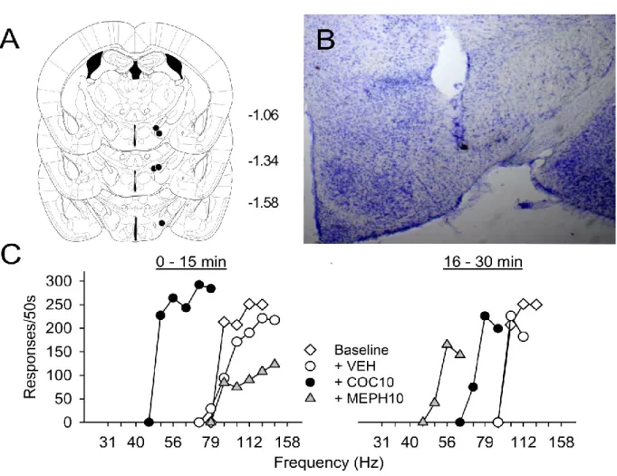
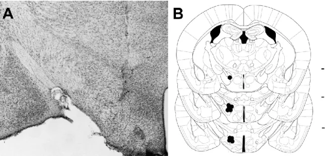
The electrode placements are shown in Figure 1A-B. All 6 mice that were implanted responded for electrical stimulation of the medial forebrain bundle within 2 sessions. Although electrodes were implanted at the AP (skull, relative to bregma) coordinate of -1.3 mm, tip locations varied from -1.06 to -1.58 mm. The average baseline EF50 and response threshold (θ0) in these mice prior to all drug
experiments expressed as charge delivery was -0.31 ±0.028 μC and -0.36 ±0.036 μC, respectively. The average baseline maximum response rate prior to all drug experiments was 162 ±20.6 responses/50s. The mice responded in a frequency-dependent manner, and mephedrone and cocaine produced parallel leftward shifts the rate-frequency curves, despite having different effects on maximum response rate (Figure 1C). No significant differences were detected between drug replicates for each drug, dose, and response series.
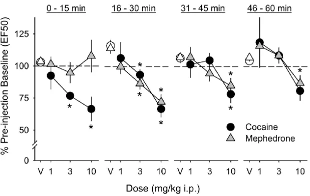
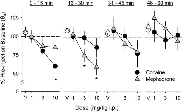
Mephedrone dose-dependently lowered EF50 (Figure 2) and BSR threshold (θ0;Figure 3) during the second (F3,15 = 19.0, p < 0.001; F3,15 = 7.4, p = 0.003,
28
fourth (F3,15 = 12.6, p < 0.001; F3,15 = 3.7, p = 0.03, respectively) 15-minute
post-injection response series when compared to saline vehicle. There was no significant effect on responding during the first 15-minutes of testing. Post hoc analysis
revealed that the 3.0 mg/kg (i.p.) mephedrone dose significantly lowered EF50 during the second 15-minute post-injection response series, while the 10.0 mg/kg dose (i.p.) significantly lowered EF50 during the second, third, and fourth response series. The 3.0 and 10.0 mg/kg (i.p.) mephedrone dose significantly lowered θ0 during the second 15-minute post-injection response series.
Cocaine dose-dependently lowered EF50 (Figure 2) and BSR threshold (θ0; Figure 3) during the first 15-minute post-injection response series when compared to saline (F3,12 = 7.1, p = 0.005; F3,12 = 6.3, p = 0.008, respectively). Cocaine lowered
the EF50, but not BSR threshold, during the second (F3,12 = 9.0, p = 0.002) and third
(F3,12 = 7.4, p = 0.006) 15-minute post-injection response series when compared to
saline. These doses did not significantly affect responding during the final 15
minutes of testing. Post hoc analysis revealed that the 3.0 mg/kg (i.p.) cocaine dose significantly lowered EF50 during the first and second 15-minute post injection series when compared to saline vehicle. The 10.0 mg/kg (i.p.) cocaine dose significantly lowered EF50 during the first, second, and third response series and lowered the BSR threshold in the first series only.
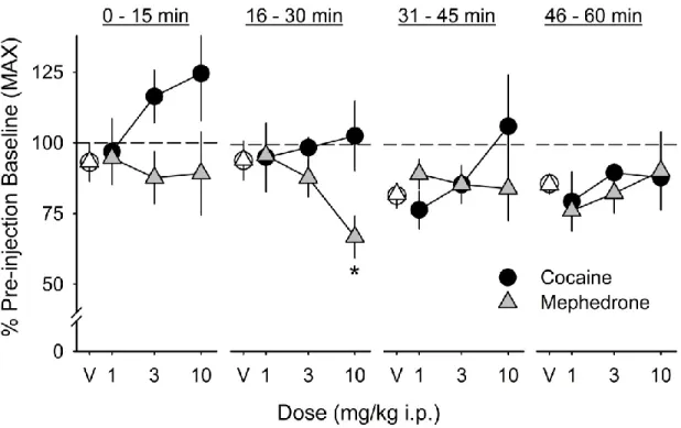
Mephedrone dose-dependently lowered maximum response rate (Figure 4) during the second 15-minute post-injection response series (F3,15 = 6.8, p = 0.004);
only the 10.0 mg/kg dose was significantly different from the saline vehicle.
29
period, but this effect was not significant (Figure 4).
DISCUSSION
The intracranial self-stimulation (ICSS) method has been used since the 1950’s to characterize brain reward system function in rodents (Wise, 2005). Drugs with abuse potential, regardless of their pharmacological class, potentiate brain
stimulation reward (BSR) (Kornetsky & Bain, 1992). Using ICSS, we examined the behavioral effects of the synthetic stimulant mephedrone in C57BL/6J mice and compared them to cocaine. We found that across the dose range tested (1.0, 3.0, and 10.0 mg/kg i.p.), cocaine and mephedrone both lowered EF50 and θ0 with similar potency. Mephedrone also decreased maximum response rate during the second 15-minute response series, while cocaine produced a non-significant trend toward increased maximum response rate during the first 15 minutes of testing. Although the behavioral effects of mephedrone were detected 15 minutes after the effects of cocaine, the duration of action of each drug on measures of ICSS was similar.
In these studies, we used the “curve-shift” method of ICSS to test the behavioral effects of mephedrone and cocaine on brain reward circuitry. During each testing session, mice responded for descending frequencies of brain
stimulation, generating characteristic rate-frequency curves. Drugs that potentiate
30
rate-frequency curve and increase the EF50 and θ0. Since drugs are given non-contingently in ICSS, one can use this method to quantify the behavioral effects of a substance on mesocorticolimbic reward circuitry independent of drug consumption. In these experiments, both cocaine and mephedrone produced robust parallel leftward shifts in the rate frequency curve (e.g. Figure 1C), and the magnitude of these effects on EF50 and θ0was similar for both drugs. The maximum effect of the 10.0 mg/kg cocaine dose on EF50 and θ0was (66.4 ± 9.4%; 60.1 ± 12.7% of pre-injection baseline, respectively), while the maximum effect of 10.0 mg/kg
mephedrone dose on EF50 and θ0was (72.3 ± 5.8%; 59.6 ± 10.9% of pre-injection baseline, respectively). These results suggest that mephedrone potentiates
responding for BSR in C57BL/6J mice similarly to cocaine, which may be indicative of its abuse potential.
31
for the dopamine transporter (DAT), serotonin transporter (SERT), and norepinephrine transporter (NET) and is similar to
3,4-methylenedioxymethamphetamine (MDMA or “ecstasy”) in selectivity and potency (Baumann, et al., 2012). In ICSS, drugs that enhance dopaminergic
neurotransmission lower response thresholds (Nakajima & O'Regan, 1991), while serotoninergic agents have diverse effects on ICSS responding depending on the receptor subclass they target (Hayes & Greenshaw, 2011). For example, 5-HT1A receptor agonists lower thresholds (A. A. Harrison & Markou, 2001), while 5-HT1B agonists (Hayes, Graham, & Greenshaw, 2009), 5-HT2C agonists (Hayes, Clements, & Greenshaw, 2009), and the selective serotonin re-uptake inhibitor fluoxetine (A. A. Harrison & Markou, 2001) increase thresholds. MDMA, which produces similar elevations of extracellular DA and 5-HT as mephedrone (Baumann, et al., 2012; Kehr, et al., 2011), robustly lowers ICSS thresholds in rats (Hubner, Bird, Rassnick, & Kornetsky, 1988). Although mechanisms underlying the observed effects of mephedrone in these studies are unknown, the enhancement of dopaminergic, as well as serotonergic, neurotransmission in loci associated with positive
reinforcement (e.g. NAc) may contribute to the potentiation of BSR by the drug. Future studies using electrophysiological or voltammetric techniques would be required to support this hypothesis.
32
Pavlovian conditioning in which laboratory animals display preference for previously drug-paired environments. For example, the psychostimulant cocaine, as well as methamphetamine (Carney, Landrum, Cheng, & Seale, 1991) and amphetamine (Elmer, Pieper, Hamilton, & Wise, 2010), is readily self-administered by C57BL/6J mice (Carney, et al., 1991) and produces robust conditioned place preference (Miner, 1997). Although the behavioral effects of mephedrone have not been investigated in mice using these methods, the drug is intravenously
self-administered by Sprague-Dawley rats using a fixed-ratio reinforcement schedule (Hadlock et al., 2011). In planaria, mephedrone induces place preference and causes abstinence-induced withdrawal-like behavior (Ramoz et al., 2012). The current findings, when examined in the context of other behavioral effects of the drug, suggest that mephedrone is a potent reinforcer that requires further behavioral characterization in animal models of drug abuse.
33
mephedrone may have induced behaviors that interfered with operant responding, such as stereotypies, which are observed following MDMA exposure (Baumann, Clark, & Rothman, 2008). Most likely, divergent effects of cocaine and mephedrone on maximum operant response rate reflect differences in their mechanism of action. Unlike the monoamine re-uptake inhibitor cocaine, mephedrone produces a robust transporter-mediated increase in striatal 5-HT levels (Baumann, et al., 2012; Kehr, et al., 2011), which is associated with reduced ICSS response rate (e.g. following fenfluramine administration (M. E. Olds & Yuwiler, 1992)). Given that selective DAT blockade by GBR-12909 enhances responding for BSR (Phillips, Blaha, & Fibiger, 1989), the effects of cocaine on maximum response rate are probably due to its effects on extracellular dopamine levels. Future studies that further characterize the psychomotor effects of mephedrone in the context of dopaminergic and serotonergic neurotransmission may help explain the observed effect of mephedrone on
maximum response rate.
CONCLUSION
34
Controlled Substances Act. Schedule I status denotes substances that have a high potential for abuse without having a legitimate therapeutic use (Fass, et al., 2012). This study examined the ability of acutely administered cocaine and mephedrone to alter responding for electrical stimulation of the medial forebrain bundle using the curve-shift method of ICSS in C57BL/6J mice. We demonstrate that mephedrone, like cocaine and amphetamines, potentiates brain stimulation reward, which may indicate that it has a high potential for abuse. Future studies will be necessary to fully characterize the cellular and behavioral effects of acute and chronic
35
FIGURES
Figure 1. Placement of intracranial self-stimulation monopolar electrodes (Panels A-B) and representative rate-frequency curves (Panel C) in C57BL/6J mice.
36
37
Figure 2. Dose-response relationship for the effects of mephedrone and cocaine on the frequency that supported half-maximal responding (EF50) in C57BL/6J mice. EF50 was measured before and after intraperitoneal injection with saline vehicle (white symbols), mephedrone (1.0, 3.0, and 10.0 mg/kg, i.p.; gray triangles; n = 6), or cocaine (1.0, 3.0, and 10.0 mg/kg, i.p.; black circles; n = 5). Results are
presented as mean percentage of pre-injection baseline during four 15-minute post-injection response series (0-15, 16-33, 31-45, and 46-60 minutes post-post-injection) ± S.E.M. Asterisks indicate significance (p < 0.05) vs. saline vehicle.
38
39
Figure 4. Dose-response relationship for the effects of mephedrone and cocaine on the maximum response rate (MAX) in C57BL/6J mice. Maximum response rate was measured before and after intraperitoneal injection with saline vehicle (white
40
REFERENCES
Angoa-Perez, M., Kane, M. J., Francescutti, D. M., Sykes, K. E., Shah, M. M., Mohammed, A. M., et al. (2012). Mephedrone, an abused psychoactive component of 'bath salts' and methamphetamine congener, does not cause neurotoxicity to dopamine nerve endings of the striatum. J Neurochem, 120(6), 1097-1107.
Baumann, M. H., Ayestas, M. A., Jr., Partilla, J. S., Sink, J. R., Shulgin, A. T., Daley, P. F., et al. (2012). The Designer Methcathinone Analogs, Mephedrone and Methylone, are Substrates for Monoamine Transporters in Brain Tissue. Neuropsychopharmacology, 37(5), 1192-1203.
Baumann, M. H., Clark, R. D., & Rothman, R. B. (2008). Locomotor stimulation produced by 3,4-methylenedioxymethamphetamine (MDMA) is correlated with dialysate levels of serotonin and dopamine in rat brain. Pharmacol Biochem Behav, 90(2), 208-217.
Carlezon, W. A., Jr., & Chartoff, E. H. (2007). Intracranial self-stimulation (ICSS) in rodents to study the neurobiology of motivation. Nat Protoc, 2(11), 2987-2995.
Carney, J. M., Landrum, R. W., Cheng, M. S., & Seale, T. W. (1991). Establishment of chronic intravenous drug self-administration in the C57BL/6J mouse. Neuroreport, 2(8), 477-480.
Elmer, G. I., Pieper, J. O., Hamilton, L. R., & Wise, R. A. (2010). Qualitative
differences between C57BL/6J and DBA/2J mice in morphine potentiation of brain stimulation reward and intravenous self-administration.
Psychopharmacology (Berl), 208(2), 309-321.
Fass, J. A., Fass, A. D., & Garcia, A. S. (2012). Synthetic cathinones (bath salts): legal status and patterns of abuse. Ann Pharmacother, 46(3), 436-441. Fish, E. W., Riday, T. T., McGuigan, M. M., Faccidomo, S., Hodge, C. W., &
Malanga, C. J. (2010). Alcohol, cocaine, and brain stimulation-reward in C57Bl6/J and DBA2/J mice. Alcohol Clin Exp Res, 34(1), 81-89.
Franklin, K. B. J., & Paxinos, G. (2008). The mouse brain in stereotaxic coordinates (3rd ed.). Amsterdam ; New York: Elsevier/Academic Press.
41
Gallistel, C. R., & Freyd, G. (1987). Quantitative determination of the effects of
catecholaminergic agonists and antagonists on the rewarding efficacy of brain stimulation. Pharmacol Biochem Behav, 26(4), 731-741.
Hadlock, G. C., Webb, K. M., McFadden, L. M., Chu, P. W., Ellis, J. D., Allen, S. C., et al. (2011). 4-Methylmethcathinone (mephedrone): neuropharmacological effects of a designer stimulant of abuse. J Pharmacol Exp Ther, 339(2), 530-536.
Harrison, A. A., & Markou, A. (2001). Serotonergic manipulations both potentiate and reduce brain stimulation reward in rats: involvement of serotonin-1A receptors. J Pharmacol Exp Ther, 297(1), 316-325.
Hayes, D. J., Clements, R., & Greenshaw, A. J. (2009). Effects of systemic and intra-nucleus accumbens 5-HT2C receptor compounds on ventral tegmental area self-stimulation thresholds in rats. Psychopharmacology (Berl), 203(3), 579-588.
Hayes, D. J., Graham, D. A., & Greenshaw, A. J. (2009). Effects of systemic 5-HT(1B) receptor compounds on ventral tegmental area intracranial self-stimulation thresholds in rats. Eur J Pharmacol, 604(1-3), 74-78.
Hayes, D. J., & Greenshaw, A. J. (2011). 5-HT receptors and reward-related behaviour: a review. Neurosci Biobehav Rev, 35(6), 1419-1449. Hubner, C. B., Bird, M., Rassnick, S., & Kornetsky, C. (1988). The threshold
lowering effects of MDMA (ecstasy) on brain-stimulation reward. Psychopharmacology (Berl), 95(1), 49-51.
Kehr, J., Ichinose, F., Yoshitake, S., Goiny, M., Sievertsson, T., Nyberg, F., et al. (2011). Mephedrone, compared with MDMA (ecstasy) and amphetamine, rapidly increases both dopamine and 5-HT levels in nucleus accumbens of awake rats. Br J Pharmacol, 164(8), 1949-1958.
Kornetsky, C., & Bain, G. (1992). Brain-stimulation reward: a model for the study of the rewarding effects of abused drugs. NIDA Res Monogr, 124, 73-93. Kuhr, W. G., Wightman, R. M., & Rebec, G. V. (1987). Dopaminergic neurons:
simultaneous measurements of dopamine release and single-unit activity during stimulation of the medial forebrain bundle. Brain Res, 418(1), 122-128. Maskell, P. D., De Paoli, G., Seneviratne, C., & Pounder, D. J. Mephedrone
42
The curve-shift paradigm in self-stimulation. Physiol Behav, 37(1), 85-91. Miner, L. L. (1997). Cocaine reward and locomotor activity in C57BL/6J and 129/SvJ
inbred mice and their F1 cross. Pharmacol Biochem Behav, 58(1), 25-30. Motbey, C. P., Hunt, G. E., Bowen, M. T., Artiss, S., & McGregor, I. S. (2012).
Mephedrone (4-methylmethcathinone, 'meow'): acute behavioural effects and distribution of Fos expression in adolescent rats. Addict Biol, 17(2), 409-422. Nakajima, S., & O'Regan, N. B. (1991). The effects of dopaminergic agonists and
antagonists on the frequency-response function for hypothalamic self-stimulation in the rat. Pharmacol Biochem Behav, 39(2), 465-468. Olds, J., & Milner, P. (1954). Positive reinforcement produced by electrical
stimulation of septal area and other regions of rat brain. J Comp Physiol Psychol, 47(6), 419-427.
Olds, M. E., & Yuwiler, A. (1992). Effects of acute and chronic fenfluramine on self-stimulation and its facilitation by amphetamine. Eur J Pharmacol, 216(3), 363-372.
Phillips, A. G., Blaha, C. D., & Fibiger, H. C. (1989). Neurochemical correlates of brain-stimulation reward measured by ex vivo and in vivo analyses. Neurosci Biobehav Rev, 13(2-3), 99-104.
Ramoz, L., Lodi, S., Bhatt, P., Reitz, A. B., Tallarida, C., Tallarida, R. J., et al. (2012). Mephedrone ("bath salt") pharmacology: insights from invertebrates. Neuroscience, 208, 79-84.
Ritz, M. C., Lamb, R. J., Goldberg, S. R., & Kuhar, M. J. (1987). Cocaine receptors on dopamine transporters are related to self-administration of cocaine. Science, 237(4819), 1219-1223.
Robinson, J. E., Fish, E. W., Krouse, M. C., Thorsell, A., Heilig, M., & Malanga, C. J. (2012). Potentiation of brain stimulation reward by morphine: effects of
neurokinin-1 receptor antagonism. Psychopharmacology (Berl), 220(1), 215-224.
Schifano, F., Albanese, A., Fergus, S., Stair, J. L., Deluca, P., Corazza, O., et al. (2011). Mephedrone (4-methylmethcathinone; 'meow meow'): chemical, pharmacological and clinical issues. Psychopharmacology (Berl), 214(3), 593-602.
Valenstein, E. S., & Campbell, J. F. (1966). Medial forebrain bundle-lateral
43
Winstock, A., Mitcheson, L., Ramsey, J., Davies, S., Puchnarewicz, M., & Marsden, J. (2011). Mephedrone: use, subjective effects and health risks. Addiction, 106(11), 1991-1996.
Wise, R. A. (2005). Forebrain substrates of reward and motivation. J Comp Neurol, 493(1), 115-121.
44
CHAPTER 3: POTENTIATION OF BRAIN STIMULATION REWARD BY
MORPHINE: EFFECTS OF NEUROKININ-1 RECEPTOR ANTAGONISM2
INTRODUCTION
Morphine, an alkaloid isolated from the Papaver somniferum plant, has been widely used since the early 19th century as an analgesic. In addition to its
antinociceptive effects, morphine and other opioid narcotics, such as heroin, fentanyl, and oxycodone, result in euphoria and alleviate psychological distress (Goodman, Brunton, Chabner, & Knollmann, 2011). It is therefore not surprising that abuse of these drugs is a significant public health concern. According to the
National Center for Health Statistics, the number of deaths from the misuse of prescription opioid analgesics has tripled from 1999 to 2006 (Warner, Chen, & Makuc, 2009), and approximately 1.7 million Americans meet the DSM-IV criteria for abuse or dependence of these drugs (Substance Abuse and Mental Health Services Administration 2010). As part of a solution to this problem, new pharmacological strategies are needed to diminish the reinforcing effects of opioids and prevent their abuse.
Morphine is a potent agonist of the mu opioid receptor (MOR), which is
2This chapter previously appeared as an article in Psychopharmacology; doi:
45
expressed in many areas of the mammalian central nervous system, including the ventral tegmental area (VTA), nucleus accumbens (NAc), periaquaductal grey, inferior colliculus, interpeduncular nucleus, and the molecular layer of the cerebellum (Maurer, Cortes, Probst, & Palacios, 1983). Opioid signaling in the VTA and NAc alters activity of the mesolimbic dopamine system, which is hypothesized to mediate the rewarding effects of drugs of abuse (Bozarth & Wise, 1986; Kornetsky, 1995). In the VTA, MOR activation causes hyperpolarization of inhibitory interneurons
(Johnson & North, 1992) and increases dopamine release in the NAc (Di Chiara & Imperato, 1988; Spanagel, Herz, & Shippenberg, 1990). This mechanism plays a critical role in reward perception and behavioral reinforcement by opioids (Phillips & LePiane, 1980, 1982; Spyraki, Fibiger, & Phillips, 1983).
The rewarding and reinforcing effects of morphine can be modeled in
laboratory animals using several behavioral conditioning techniques, including drug self-administration (Ross, Laska, & Fennessy, 1978; Wilson, Hitomi, & Schuster, 1971), conditioned place preference (CPP) (Mucha, et al., 1982), and intracranial self-stimulation (ICSS) (Kornetsky & Bain, 1992; J. Olds & Milner, 1954).
46
excitatory cholinergic projections to the VTA (Wise, 2005). Direct electrical stimulation of this pathway is potently reinforcing (Valenstein & Campbell, 1966), and results in release of dopamine in forebrain targets (Cheer et al. 2007). Drugs of abuse enhance the activity of these fibers and reduce BSR threshold, which is the minimum amount of electrical stimulation necessary to sustain responding (Carlezon & Chartoff, 2007; Kornetsky & Bain, 1992).
The effects of opioid drugs on ICSS have been measured in rats (R. Esposito & Kornetsky, 1977; R. U. Esposito, McLean, & Kornetsky, 1979; Holtzman, 1976; Koob, Spector, & Meyerhoff, 1975; M. E. Olds, 1979; Schaefer & Holtzman, 1977) and mice (Bendani & Cazala, 1988; Criswell, 1982; Elmer, Pieper, Goldberg, & George, 1995; Elmer, et al., 2010). Early studies using a discrete trial-based procedure in rats demonstrated that morphine lowers the threshold for ICSS responding at low doses but becomes aversive at higher doses (Marcus & Kornetsky, 1974). Interestingly, the acute reward-potentiating effect of morphine does not sensitize with repeated administration (R. Esposito & Kornetsky, 1977), although more recent studies have demonstrated that cycles of withdrawal from heroin progressively raises baseline ICSS threshold (Kenny, Chen, Kitamura, Markou, & Koob, 2006). Activation of the delta opioid receptor and kappa opioid receptor also alters ICSS responding. DOR agonists lower BSR threshold (Duvauchelle, Fleming, & Kornetsky, 1996), whereas KOR agonists raise the threshold (Todtenkopf, Marcus, Portoghese, & Carlezon, 2004).
47
Elmer at al. (2010) found that C57BL/6J mice are sensitive to the
reward-potentiating effects of morphine, whereas DBA/2J mice display elevations of BSR threshold suggestive of an aversion to the drug. DBA/2J mice also fail to self-administer morphine intravenously, which C57BL/6J mice do readily (Elmer, et al., 2010). These results are consistent with previous results that show that C57BL/6J mice are more sensitive to the drug’s locomotor stimulating effects (Cunningham, Niehus, Malott, & Prather, 1992; Oliverio & Castellano, 1974), although both strains readily acquire a place preference to morphine paired environments (Cunningham, et al., 1992).
Recent evidence has suggested that substance P, a peptide neurotransmitter of the tachykinin family, and its target, the neurokinin-1 receptor (NK1R), are
involved in modulating the rewarding effects of opioids (reviewed by (Commons, 2010)). NK1R-null mice on a C57BL/6J background (De Felipe et al., 1998) display morphine reward deficits in the conditioned place preference model (CPP) (Gadd, Murtra, De Felipe, & Hunt, 2003; Murtra, Sheasby, Hunt, & De Felipe, 2000) and self-administer less morphine than wild type littermates (Ripley, Gadd, De Felipe, Hunt, & Stephens, 2002). However, animals lacking NK1R display no difference in CPP or the self-administration of cocaine (Gadd, et al., 2003; Murtra, et al., 2000) or food reinforcers (Murtra, et al., 2000), suggesting that the observed reward deficits are specific to opioids. In addition, these mice are less sensitive to the locomotor-stimulating effects of morphine and fail to display behavioral sensitization to
48
study, we investigated the ability of neurokinin-1 receptor antagonists to alter the rewarding effects of morphine using ICSS. BSR thresholds were determined in C57BL/6J mice before and after treatment with the opioid antagonist naltrexone, or after one of two NK1R antagonists, L-733,060 and L-703,606. We also tested the ability of naltrexone, L-733,060, and L-703,606 to alter the effects of morphine on BSR threshold. C57BL/6J mice were chosen for this study because they exhibit robust responses to morphine with ICSS (Elmer, et al., 2010) and were used as the genetic background for NK1R deletion mutants (De Felipe, et al., 1998).
METHODS
Mice
Male C57BL/6J mice (n = 15) were purchased from Jackson Laboratories (Bar Harbor, ME), individually housed in polycarbonate cages (28 × 17 × 14 cm) with wire lids, and allowed to acclimate to the vivarium for one week. Mice had free access to food and water throughout all stages of the experiment and were housed in a