MURINE TRANSGENE INSERTIONAL MUTATION INVOLVING RUNX1T1 AND
GM11823 GENES AND THEIR CONTRIBUTION TO CLEFT PALATE AND RIB
ANOMALIES
Melanie Fawaz Alazzam
A dissertation submitted to the faculty at the University of North Carolina at Chapel Hill in partial fulfillment of the requirements for the degree of Doctor of Philosophy in the School of
Dentistry (Oral Biology).
Chapel Hill 2014
Approved by: Eric T. Everett
Sylvia Frazier-Bowers P. Emile Rossouw John van Aalst
© 2014
Melanie Fawaz Alazzam ALL RIGHTS RESERVED
ABSTRACT
Melanie Fawaz Alazzam: Murine Transgene Insertional Mutation Involving Runx1t1 and Gm11823 Genes and their Contribution to Cleft Palate and Rib Anomalies
(Under the direction of Eric Everett)
Objective: Cleft Palate (CP) is a common birth defect in humans occurring in 6.35/10,000 live births and it has been repeatedly shown that animal models are useful in dissecting
molecular etiologies of CP. The OVE1328 mouse line develops CP as a consequence of transgene (Tg) insertion mutagenesis. Preliminary data shows Tg complex integration at
chromosome 4 band A2. The goal of this work was to characterize the mutation in the OVE1328 transgenic mouse line. Methods: Genotyping microarrays and RNA-seq were used to identify the Tg insertion site. The insertion site was further validated by conventional PCR. Histology and skeletal staining were used to phenotype CP (OVE1328 (Tg/Tg)) and Wt embryos. Results: In OVE1328 embryos the transgene disrupts Runx1t1 gene at intron 12 (13,876,840 bp). The integration is associated with a deletion mutation of part of intron 12 and whole exons 13 and 14 of Runx1t1. Runx1t1 (also known as Cbfa2t1h, Eto, Mtg8) is mainly studied for its role in acute myelogenous leukemia as a fusion gene in humans and gut development in mice. The Tg
insertion disruption extends to the Gm11823 gene which encodes a long non coding RNA. Little is known regarding the normal function and expression of Gm11823. The Tg integrates at intron 2 (13,949,713 bp) of Gm11823 and induces the ectopic expression of an altered message.
Homozygous disruption of both genes is associated with CP (100% of OVE1328 (Tg/Tg) embryos) and rib anomalies (supernumerary ribs, 86% of OVE1328 (Tg/Tg) embryos.
To my lovely parents (Fawaz and Diana), who dedicated themselves to make me happy To my sweethearts (Mohammad and Mariam), whose sacrifices made this dream come true
ACKNOWLWDGEMENTS
I would like to thank my mentor Dr. Eric Everett for his continuous support and his mentorship. I am so grateful to have such brilliant advisor with great capabilities. I would like as well to thank my committee members: Dr. Sylvia Frazier-Bowers, Dr. Emile Rossouw, Dr. John van Aalst, and Dr. Tim Wright for their valuable comments and contribution to putting the dissertation in its final outcome. Each of you was an inspiration to me. I would like to
acknowledge Dr. Paul Overbeek for his precious gift of the OVE1328 transgenic line. I would like to thank Dr. Ceib Phillips for her help with some of the statistical analysis done. My
acknowledgement also goes to National Institute of Dental and Craniofacial Research (NIDCR) and Jordan University of Science and Technology for supporting this project.
My deepest gratitude goes to my soul mates baba (Fawaz), mama (Diana), my husband (Mohammad) and (Mariam), sister (Nissreen) and brother (Adam)! Thank you so much for the tremendous unconditional love you surrounded me with. Thank you for believing in my abilities. Thank you for all the prayers you made, for the sacrifices you had to do to help me achieve my dream! Thanks for all the happiness you filled my heart with! You all are an inspiration to me and I would not have been able to achieve this without the help given by each of you!
My thanks go to my friends and colleagues at UNC, thank you for making me feel at home. I really had great moments with you!
TABLE OF CONTENTS
LIST OF TABLES………..xi
LIST OF FIGURES………...xii
LIST OF ABBREVIATIONS………....xv
CHAPTER 1: REVIEW OF THE LITRITURE ……….1
1.1Development of the Oral Cavity; an Overview……….1
1.1.1 Development of the Upper Lip………...2
1.1.2 Development of the Secondary Palate (Palatogenesis)………4
1.1.3 Human vs Mouse; A Comparison of Lip and Palate Structures…………..6
1.1.4 Lip and Palate Development: Interspecies Variation………...9
1.2Oral Clefts in Humans………...11
1.3Common Problems Associated with Oral Clefts………...13
1.4Etiology of CP………...16
1.4.1. Genetic Factors………16
1.4.2. Environmental Factors………...21
1.4.3. Gene-Environment Interaction………...22
1.5Types of Cleft Palate Mouse Models………...23
1.5.1 Spontaneous CP Mouse Models………24
1.5.2 1.5.2 Induced CP Mouse Models………...24
1.6.1 Patterning the Axial Skeleton………...29
1.7Rib Anomalies (Supernumerary Ribs)………...31
1.8Etiology of Supernumerary Ribs………...32
1.9SNR Mouse Models………...33
1.10Specific Aims………...34
CHAPTER 2: PHENOTYPIC CHARACHTERISTICS OF OVE1328 TRANSGENIC LINE...35
2.1 Introduction………...35
2.1.1 Insertional Mutagenesis in Mice………...35
2.1.2 General Background of OVE1328 Transgenic Line………... 38
2.1.3 Generation of OVE1328 Transgenic Line………... 39
2.1.4General Characteristics of OVE1328 Transgenic Line……….40
2.2 Materials and Methods……….42
2.3 Results………..46
2.4 Discussion………56
CHAPTER 3: MOLECULAR CHARACHTERIZATION OF THE INSERTION MUTATION IN OVE1328 TRANSGENIC LINE……….58
3.1 Introduction………..58
3.1.1 Transgene Integration Process………..59
3.1.2 Transgene Integration: Effects on Transgene and Genomic Loci at Insertion Site………...59
3.1.3 Transgene Integration in OVE1328 Transgenic Line………...60
3.1.4 Runx1t1 a Member of ETO (Eight Twenty One) Gene Family………62
3.1.5 ETO Gene Family and Acute Myelogenous Leukemia (AML)…………...64
3.1.6 Murine Runx1t1 Gene, mRNA and Protein Structure………..64
3.1.7 Orthologs of Runx1t1………....66
3.1.8 Runx1t1 Expression………..66
3.1.9 Subcellular Location of Runx1t1………... 67
3.1.10 Biological Functions of Runx1t1………68
3.1.11 OVE1328 Transgenic Line and Runx1t1 Knockout Model………... 69
3.1.12 Runx1t1 Neighborhood: Gm11823 Gene………....71
3.1.13 Gm11823: Potential Roles as a Long Intergenic Non-Coding RNA Gene………..71
3.1.14 Runx1t1, Gm11823 and Their Alignment to the Human Genome………..73
3.2 Materials and Methods……… 75
3.3 Results………..79
3.4 Discussion………... 93
APPENDIX 2.1: TYROSINASE PRIMER PAIRS (SEQUENCE)………....100
APPENDIX 2.2: TYROSINASE PRIMER PAIRS (GENOMIC COORDINATES)………….101
APPENDIX 2.3: PROTOCOL MODIFICATIONS FOR SKELETAL CLEARING AND STAINING………...102
APPENDIX 2.4: RIB CAGE DATA………...103
APPENDIX 2.5: MEAN LENGTH OF RIGHT FORELIMB AND HINDLIMB BONES DATA………104
APPENDIX 2.6: MEAN LENGTH OF LEFT FORELIMB AND HINDLIMB BONES DATA………105
APPENDIX 2.7: MEAN TMD DATA (RT AND LT HUMERUS) / UNIVERSAL THRESHOLD………..106
APPENDIX 2.8: MEAN HEAD MEASUREMENTS DATA………....107
APPENDIX 3.1: RUNX1T1 CDNA PRIMER PAIRS (SEQUENCE)………...108
APPENDIX 3.2: RUNX1T1 cDNA PRIMER PAIRS (GENOMIC COORDINATES)……….109
APPENDIX 3.3: RUNX1T1 EXON-INTRON JUNCTION PRIMER PAIRS PRIMER
PAIRS (SEQUENCE)………..110
APPENDIX 3.4: RUNX1T1 EXON-INTRON JUNCTION PRIMER PAIRS (GENOMIC COORDINATES)………111
APPENDIX 3.5: GM11823 PRIMER PAIRS (SEQUENCE SECTION)………..112
APPENDIX 3.6: GM11823 PRIMER PAIRS (GENOMIC COORDINATES)……….113
APPENDIX 3.7: CONTIG PRIMER PAIRS (SEQUENCE)………..114
APPENDIX 3.8: CONTIG PRIMER PAIRS (GENOMIC COORDINATES)………...115
APPENDIX 3.9: THERMAL CYCLER PROGRAMMING CONDITIONS AND GENERAL PRIMER INFORMATION………116
APPENDIX 3.10: RUNX1T1 ANTIBODIES: CLONALITY I, MMUNOGEN SEQUENCE AND EPITOPE BINDING SITE………...117
APPENDIX 3.11: PART OF 5’ END (A) AND 3’END (B) MESSENGER RNA SEQUENCE ALIGNMENTS OF RUNX1T1………..118
APPENDIX 3.12: PROTEIN SEQUENCE ALIGNEMNTS AND DOMAIN STRUCTURE OF RUNX1T1 ISOFORMS………119
APPENDIX 3.13: DEFINITIONS………...120
REFERENCES………121
LIST OF TABLES
Table 1.1- Interspecies variation in the embryonic origin of secondary palate………...9
Table 1.2-Interspecies variation in palatogenesis………...10
Table 1.3- Examples of syndromic CP in humans……….17
Table 1.4- Examples of genes associated with non-syndromic CP in humans………...18
Table 2.1- Murine insertional mutations are helpful to understand developmental processes………36
Table 2.2. Single, double and paralogous null mutations in Hox genes that contribute to L1 supernumerary ribs………...………...54
Table 3.1- List of candidate genes present on chromosome 4 band A2 ± 2,000,000bp…………57
Table 3.2- Murine orthologs of ETO family members and their aliases………...60
Table 3.3- ETO family members are closely related proteins………...60
Table 3.4- Orthologs of Runx1t1………...63
Table 3.5- Comparison of OVE1328 and Runx1t1 knockout models………...67
Table 3.6- Human syndromes with clinical manifestations of CP and supernumerary ribs...97
LIST OF FIGURES
Figure 1.2- Types of oral clefts in humans………12
Figure 1.3- Functional classification of genetic factors contributing to oral clefts (including CP) in humans and mice……….18
Figure 1.4- Types of CP murine models………23
Figure 2.1- General physical differences between transgenic OVE1328 line and Wt mice………...39
Figure 2.2- CP phenotype in OVE1328 line………...43
Figure 2.3- E18 Embryo genotype frequencies of OVE1328 (Tg/+) intercrosses………44
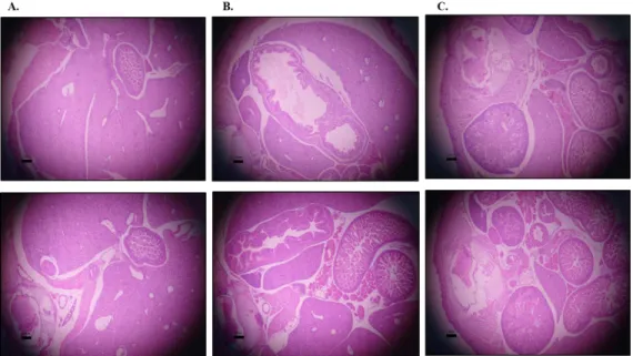
Figure 2.4- Histological analysis (H&E stained sections) of the heart, lungs and thymus gland ……….45
Figure 2.5- Histological analysis (H&E stained sections) of some major organs………...45
Figure 2.6- Histological analysis (H&E stained sections) of the intestines in Wt and OVE1328 (Tg/Tg) E18 embryos……….46
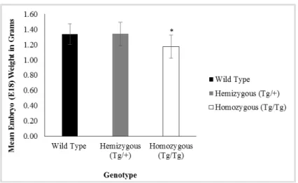
Figure 2.7- Average E18 embryo weights……….46
Figure 2.8- Cleared skeletons of E18 embryos………...47
Figure 2.9- Mean lengths of forelimb and hindlimb bones of E18 Wt (n=10), OVE1328 (Tg/+) (n=12) and OVE1328 (Tg/Tg) (n=12) embryos………48
Figure 2.10-Average TMD of right and left humerii from E18 Wt, OVE1328 (Tg/+) and OVE1328 (Tg/Tg) embryos (n=6/genotype/side)………...49
Figure 2.11-Comparison of E18 embryo head measurements between Wt, OVE1328 (Tg/+) and OVE1328 (Tg/Tg) embryos………...50
Figure 2.12. Supernumerary ribs in OVE1328 (Tg/Tg) E18 embryos………..51
Figure 2.13-Micro-CT X-ray projection image of OVE1328 (Tg/Tg) E18 embryo rib cage………..51
Figure 2.14- Supernumerary ribs in OVE1328 (Tg/Tg) embryos………..………...52
Figure 3.1- Diversity genotyping microarray data……….59
Figure 3.2- Runx1t1 gene, mRNA structure………..62
Figure 3.3- Domain structure of RUNX1T1 protein……….63
Figure 3.4- Gene regulation by lncRNAs………..70
Figure 3.5- Graphical alignment of Gm11823 (lincRNA) to RP11-122C21.1 human (lincRNA) on chromosome 8……….71
Figure 3.6- Syntenic regions between mouse chromosome 4 and human chromosome 8……....72
Figure 3.7- Primer map for Runx1t1 exon-intron junction primers………...77
Figure 3.8- Conventional PCR data of exon-intron junction amplifications……….78
Figure 3.9- Primer map for Runx1t1 message (variant1)………...79
Figure 3.10-Conventional PCR data of Runx1t1 message………...79
Figure 3.11-RNA-Seq data………80
Figure 3.12-Contig 15360 primer map………..81
Figure 3.13-Conventional PCR data of 15360 contig primer pair……….81
Figure 3.15-Interrogation of exon 12- intron 12 junction of Runx1t1………...83
Figure 3.16-Northern blot data………..84
Figure 3.17-Western blot data………85
Figure 3.18-Epitope map for RUNX1T1 antibodies used for Western blotting………85
Figure 3.19-Contig 4550 primer map………87
Figure 3.20-Gm11823 primer map………87
Figure 3.21-Conventional PCR data of Gm11823 genomic locus……….88
Figure 3.22-Conventional PCR data of Gm11823 message………..89
Figure 3.23-Conventional PCR data using 4550 contig primer pair……….90
LIST OF ABBRIVIATIONS AND SYMBOLS
µA Microamperes
µm Micrometers
AML Acute myelogenous leukemia
bp Base pair
cDNA Complementary DNA
CL/P Cleft lip with/without cleft palate CLO Cleft lip only
CLP Cleft lip and palate
CO2 Carbon dioxide CP Cleft palate
DIG Digoxigenin E Embryonic day
EDTA Ethylenediaminetetraacetic acid
FISH Fluorescence in situ hybridization gDNA Genomic DNA
H&E Hematoxylin and eosin IPD Interpupillary distance
Kb Kilobase
Kd Kilodaltons kV Kilovolts
LncRNA Long non coding RNA
Lt Left
MgCl2 Magnesium chloride
Min Minutes mM Millimolar ms Milliseconds
NaCl Sodium chloride NaOH Sodium hydroxide
NAT Natural antisense transcripts nmole Nanomoles
Nt nucleotide
PCR Polymerase chain reaction ROI Region of interest
Rt Right
SDS Sodium Dodecyl Sulfate
SNPs Single nucleotide polymorphisms
SOD Snout occiput distance SSC Saline sodium citrate
SW Snout width
Tg Transgene
TMD Tissue mineral density
TUNEL Terminal deoxynucleotidyl transferase dUTP nick end labeling Tyr Tyrosinase
V Volts
Wt Wild type
CHAPTER 1: REVIEW OF THE LITRETURE
1.1 Development of the Oral Cavity; an Overview
The formation of the orofacial region in the developing embryo is demarcated by the appearance of the oropharyngeal membrane and subsequently the primary mouth (also known as the stomodeum). The oropharyngeal membrane seen at stage 11 Carnegie, 24 days of
gestation in human, and E9 mouse embryo, is formed from a region that is cranial to the notochord where the ectoderm and endoderm fuse together in the trilaminar embryo. The ectoderm of the membrane will form the mucosal lining of the future oral cavity and the
endoderm will form the future lining of the pharyngeal mucosa. The oropharyngeal membrane, a temporary weak membrane, separates the primary mouth from the foregut. The membrane becomes perforated at this stage where the oral cavity and the pharyngeal space become connected.1-3
The primary mouth is limited by 5 developing processes (or prominences). The processes are composed of a mesenchyme covered by an ectoderm. The mesenchyme is populated by cranial neural crest cells (CNC) (originating from the crest of the folding neural tube) which start migrating at stage 10 towards their future destination. The CNC cells contribute to the formation of the facial skeleton. In addition to CNC cells, cells of mesodermal origin populate the
mesenchyme which will contribute to the formation of facial musculature. These processes are; 1 median frontonasal process (rostrally), 2 maxillary processes (laterally) and 2 mandibular
processes (caudally) (the maxillary and mandibular processes develop from the first branchial arch).2-4
1.1.1 Development of the Upper Lip
At stage 13-15 Carnegie,fourth to fifth week of gestation in human, the formation of the paired telencephalic vesicles (future cerebral hemispheres) from the forebrain results in the widening of the frontonasal process with the formation of the median groove. Meanwhile, the 2 mandibular processes grow and merge together in a caudal to rostral aspect. The fusion of those processes results in the formation of lower jaw and the low lip.2,4
At stage 14 Carnegie, around 32 days of gestation in human, and E10 mouse embryo, two ectodermal thickenings form at the inferior lateral corners of the frontonasal processes. Those thickenings will form the nasal placodes. The placodes will develop into nasal pits due to the growth and bulging of the frontonasal processes and the formation of a horseshoe like medial and lateral nasal processes. The medial and lateral nasal processes are separated and the nasal pit is in continuity with the stomodeum. Furthermore, the median groove is seen in between the two medial nasal processes. At this stage the maxillary processes become discernable.2,4
At stage 15 Carnegie, 35 days of gestation in human, and E10.5 mouse embryo, further growth of the maxillary process pushes the medial nasal processes medially and results in the wedging of the lateral nasal process between the maxillary and medial nasal processes. By this stage, nasal pits are seen as distally pointed slits.2,4
At stage 16 Carnegie, 38 days of gestation in human, and E11 mouse embryo, rapid growth of the maxillary processes and the medial nasal processes results in two fusion events: 1.
Fusion of the medial nasal process and the lateral nasal process 2. Fusion of the medial nasal process and the maxillary process. These fusion events continue through stage 16-18 Carnegie, beginning in the seventh week of human gestation, and to E11.5 to E12 in a mouse embryo. The aforementioned fusion events require the involvement of the epithelial cells in several cellular processes represented by apoptosis, formation of filopodia and epithelial mesenchymal transformation (EMT). However, for the median groove, growth and confluence of the medial nasal and maxillary processes will fill and smoothen the median groove in between the medial nasal processes.2,4,5
At Stage 19, around 48 days of gestation in human, and in E12.5 mouse embryo, the upper lip development is complete. The distal part of the medial nasal processes, the
intermaxillary segment, forms the central part of the lip. Further growth of the intermaxillary segment into the oral cavity results in the formation of the primary palate which later fuses with the secondary palate. 2
1.1.2 Development of the Secondary Palate (Palatogenesis)
At 6 weeks of gestation in human and E11 mouse embryo, palatogenesis starts as an oral outgrowth of the developing maxillary processes known as palatal shelf primordia. Palatal shelf primordia, as other facial primordia, is composed of a mesenchyme (mainly of cranial neural crest (CNC) origin) with an overlying epithelium of an ectodermal origin. 6-8
In the same week (6 weeks of gestation) in human and at E12.5-14 mouse embryo, the palatal shelves will grow downward (however, it should be noted that there is growth in the antero-posterior and medio-lateral aspects as well) around the developing tongue.6-8
At 7-8 weeks of gestation in human and E14.5-15 in mouse embryo, palatal shelves will undergo rapid morphological changes that will eventually result in reorienting the growth pattern of the shelves from vertical to horizontal plane. It was proposed that the elevation of palatal shelves is due to remodeling in which a protrusion of the medial wall and a regression in the ventral end of the shelves results in their elevation.6 Recent evidence has indeed revealed that cells expressing medial edge epithelium (MEE) markers were detected along the medial side of the developing palatal shelves.6,9 Furthermore, a recent study supported the tissue remodeling hypothesis for certain regions of the elevating shelves (mid-posterior region) but suggested that there might be other mechanisms involved in the elevation of the anterior and most posterior end of the shelves.9 It was noticed that the elevation starts at the mid-posterior region of the palatal shelves and then moves to the anterior region, and once elevated, the elevation of the most posterior end region of the shelves is achieved.10 The elevation process is associated with other morphological changes in the craniofacial region where the head is extended and there is an increase in the vertical dimension of the head with the downward positioning of the tongue.11
By this time the palatal shelves are in the horizontal plane. The two layered epithelium on the medial side of the pre-fusion palatal shelves is known as the medial edge epithelium (MEE) and is composed of a basal layer of cuboidal cells lying on a basal lamina and an outer layer of flat cells facing the amniotic fluid, known as the periderm.7,12It is agreed that the fusion of the
basal layer of both shelves contributes to the formation of the midline epithelial seam (MES).12 On the other hand, the fate of the periderm layer is still not ascertained. Several mechanisms were suggested: the periderm layer undergoes desquamation (peeling off), can be trapped in the MES, and undergo apoptosis or migrates to the oral and nasal epithelial triangles.7,12,13 Once the MES forms it will undergo degeneration to allow for the confluence of the mesenchyme in order for the fusion process to be completed.6,7However, there is still inconclusive evidence of how the MES is removed. There has been evidence supporting different fates of the MES. The MES can undergo apoptosis as some studies demonstrated that many MES cells were terminal
deoxynucleotidyl transferase dUTP nick end labeling (TUNEL) positive and Caspase 3
positive.6,14 Other studies showed the MES cells in the middle palatal region had strong TUNEL
positive staining while in the anterior region (where the primary and the secondary palate fuse in the anterior palate region) had few TUNEL positive cells.15 In addition to apoptosis, it was
proposed that MES cells can also undergo migration.7,16 or epithelial mesenchymal
transformation. However, the available evidence regarding the latter fate is still controversial.6 By 8 weeks of gestation in human and E15.5 in mouse embryo, the MES has disappeared and the palatal mesenchyme is confluent with the initiation of palatal bone
formation by intramembranous ossification. The adult hard palate (of secondary palate origin) is formed of the palatal process of the maxilla and the horizontal plate of the palatine.6,17 In the developing embryo bone formation of the palatal process of the maxilla starts at a new
ossification center which is initially separate from the maxillary ossification center. The
horizontal plate of palatine bone formation occurs as an extension of the osteogenic front of the palatine6,18. By 10 weeks of gestation in human and E16.5 in mice, palate formation has been completed and successful separation of the developing oral and nasal cavities takes place.8,19,20
Though the formation of the oral cavity seems straightforward, development and morphogenesis of oral structures depend on successful spatiotemporal regulation of different cellular processes. A disruption or alteration at any of the stages explained above will prevent the normal fusion of the lip and/or the palate resulting in a cleft.
1.1.3 Human vs Mouse; A Comparison of Lip and Palate Structures
In spite of the prominent external differences between humans and mice, lip and palate structures demonstrate many similarities. The next section will focus on comparing humans and mice in 3 aspects: lip and palate development, lip and palate anatomy and lip and palate cleft types.
Lip and Palate Development
During early craniofacial development, 32 day human and E10 mouse embryos, not only are human and mouse embryos similar in their shape and size but the formation of cleft and palate structures is basically the same.20 However, lip and palate formation in humans (lip: ~4 weeks and palate: ~4-6 weeks) requires more time to complete when compared to that of mice (lip: ~2.5 days and palate: ~4-4.5 days).20 Other developmental differences are seen during
palatal shelf elevation. There is evidence that the site of palatal shelf elevation might differ depending on the species studied.10 For example, in rats and humans, palatal shelves start to
analysis of palatal shelf elevation in mice has shown that the shelves start to elevate at the mid-posterior region and then move to the anterior region. Once this takes place the most mid-posterior end of the palatal shelves (presumptive soft palate) elevates.10
In addition to palatal shelf elevation, the fusion process might also differ between humans and mice. Evidence demonstrates that palatal shelves fuse first anteriorly (at the incisive foramen region) and then proceed posteriorly.21 Following fusion, the epithelial nests, or remnants formed
in the midline (due to the generation of the MES), remain till birth after which they completely degenerate.21,22 On the other hand, there is conflicting evidence for the site of palatal shelf fusion in mice. Some evidence supports that fusion takes place anteriorly (at the incisive foramen region) and then proceeds posteriorly in a zipper like mechanism as in humans.23-25 Other evidence supports the notion that the fusion process posteriorly can occur independently of the fusion process anteriorly as seen in Shox2 knock out mice.26 Furthermore, the epithelial seam disappears completely and no epithelial remnants persist until birth.22
Lip and Palate Anatomy (Selected Differences)
Despite the fact that lip embryogenesis in mice and humans is similar at early stages, the gross anatomy is clearly different. In contrast to our lips, close examination of the lips in mice reveal that their upper lips are normally fissured, exposing the upper incisors. Furthermore the lips are devoid of the vermillion border seen in humans. This border separates the skin from the oral mucosa and is rich in vascular supply. 27
As far as the anatomy of the hard palate, both species are anatomically similar. The anterior two thirds of the hard palate are composed of the palatal process of the maxilla and the posterior one third is formed by the horizontal plate of the palatine bone. The two bones are
joined by the transverse palatine suture. In the sagittal plane the bones of the hard palate are joined by the mid palatal and the interpalatine sutures, respectively. However, the midpalatal suture in mice is straight while in humans is interdigitated.27,28
Another difference is seen in the soft palate- in mice no uvula is seen at the midline whereas in humans, the uvula forms at the midline of the soft palate.27
Lip and Palate Cleft Types
Oral clefts in humans are diverse and present with varying degrees of severity: unilateral or bilateral cleft lip only (CLO), unilateral or bilateral cleft lip and palate (CL/P) or cleft palate only (CPO). In CL/P, the cleft can involve the alveolar ridge and the primary palate or can be more extensive, involving the alveolar ridge, primary palate and secondary palate as well. In the case CPO, the cleft can involve the hard palate or the soft palate (bifid uvula) or involve both the hard and soft palate.20,29-32 Closer examination of the type of clefts seen in mice reveals that mice do have similar phenotypes to humans. For example, CPO and CL/P are seen in mice. However, in contrast to humans, CPO is more commonly seen in mice than CL/P.20 Furthermore, CLO is
extremely rare in mice. Only one mouse model was found to have CLO where the cleft did not involve the primary palate and there was no evidence that the alveolus was involved. Though the cleft did not involve the primary palate, a cleft of the secondary palate was seen33. Other
examples of CL in mice are more commonly seen involving the lip and both the primary and secondary palates20. Clefts of both the lip and the primary palate are seen less commonly.34,35 It
is crucial to distinguish the fact that mouse models with midline clefts of the lip and the
midfacial region36,37 are considered a different entity than the cleft lip mouse models discussed
here38. The pathogenesis of a midline cleft lip differs from what was previously discussed regarding cleft lip. Midline cleft lip is seen when both medial nasal processes fail to fuse.38,39
1.1.4 Lip and Palate Development: Interspecies Variation
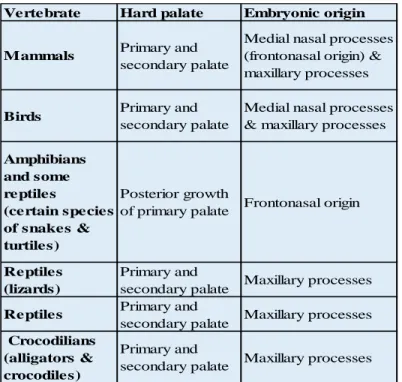
The basic steps involved in palatal shelf formation vary across different species. In some species CP is considered as being the norm such as in birds and some reptiles. Tables (1.1 and 1.2) address some of the known variations. The content of the tables 1.1 and 1.2 were adapted from Ferguson et al.40
Table 1.1 Interspecies variation in the embryonic origin of the secondary palate. Content adapted from Ferguson et al.40
Vertebrate Hard palate Embryonic origin
Mammals Primary and secondary palate
Medial nasal processes (frontonasal origin) & maxillary processes
Birds Primary and
secondary palate
Medial nasal processes & maxillary processes
Amphibians and some reptiles
(certain species of snakes & turtiles)
Posterior growth
of primary palate Frontonasal origin
Reptiles (lizards)
Primary and
secondary palate Maxillary processes
Reptiles Primary and
secondary palate Maxillary processes Crocodilians
(alligators & crocodiles)
Primary and
secondary palate Maxillary processes
1.2 Oral Clefts in Humans
Oral clefts are among the most common congenital anomalies in humans with a
prevalence of 1/700 live births.41-44 Oral clefts include a group of anomalies in which a cleavage of the lip and/or the palate is seen due to the abnormal embryonic development of these
structures. Oral clefts can be divided based on the embryological origin of the affected structures into: 1. Cleft lip with/without cleft palate (CL/P) 2. Cleft palate (CP). There is a great variation in the prevalence of CL/P and CP among different geographical regions, races, ethnic groups and socioeconomic status. For example, higher prevalence of CL/P was seen in Latin America and Asia compared with lower prevalence in Israel, South Africa and Southern Europe44. On the
other hand, higher prevalence rates of CP were seen in Canada and parts of Northern Europe and lower rates were seen in pats of Latin America and South Africa. Furthermore, the prevalence of CL/P was the highest among Asians and Native Americans (1/500), intermediate among
Europeans (1:1000) and the lowest among African American populations (1:2500).30,32,44,45 Depending on the severity/ extent of the defect, CL/P can be divided into cleft lip only (CLO) and Cleft lip and Palate (CLP) (Fig 1.2). In CLO, the cleft can be complete (extending to the nostril and loss of Simonarts band) or incomplete (in which the nostril is not affected and Simonarts band is seen).46-48 As CLO, cleft lip and palate (CLP) has variable manifestations based on the severity: cleft lip combined with clefting of the alveolar ridge, cleft lip combined with clefting of both alveolar ridge and primary palate, cleft lip combined with clefting of the alveolar ridge, primary palate and the secondary palate (Fig 1.1). CP shows such variations in severity in which the cleft affects both the hard and soft palate or in which the hard palate only or the soft palate only are affected (Fig 1.1). CL/P is further categorized, based on the side being affected, into unilateral and bilateral clefts. Unilateral clefts are seen in 90% of CLO with the left
side being mostly affected (2/3 of the cases). However, a greater proportion of CLP patients (30.2%) show a bilateral involvement of the lips compared to the bilateral involvement seen in CLO patients (10.3%).20,41,44,49 CL/P is seen more in males compared to females at a 2:1 ratio. In contrast, CP tends to be more frequently seen in females compared to males.44
Figure 1.1. Types of oral clefts in humans. A. a schematic representation of oral clefts in humans. B. Clinical cases representing variable severity of oral clefts in humans.
Figure reprinted by permission from Macmillan Publishers Ltd: Nature Reviews Genetics (Dixon, M. J., Marazita, M. L., Beaty, T. H. & Murray, J. C. Cleft lip and palate: understanding genetic and environmental influences. Nat Rev Genet 12, 167-178, doi:10.1038/nrg2933 (2011), copyright 2011.
CL/P and CP can be associated with other anomalies and can be classified accordingly. The greatest prevalence of associated anomalies are seen with CP, followed by CLP, and the least prevalence is seen in CL. Congenital cardiac defects, limb and vertebral anomalies were the anomalies often seen in CL/P and CP.44 If no other anomalies are associated with CL/P and CP,
then the cleft is known as isolated-non syndromic CL/P or CP.30,45,50 The frequency of isolated CL/P was around (76.8%). The rest of CL/P cases were either associated with other anomalies (15.9%) or were recognized as part of a syndrome (7.3%) in such case known as syndromic CL/P. On the other hand, the frequency of isolated-non syndromic CP was (54.8%), while the frequency of CP with other anomalies was (27.2%) and CP with anomalies as part of a
recognizable syndrome (syndromic CP) were (18%).44
1.3 Common Problems Associated with Oral Clefts
Patients born with oral clefts suffer from different problems and complications. This
section comes across the common problems associated with oral clefts in humans. These are: Feeding problems: Proper feeding in a newborn requires normal sucking and swallowing
mechanisms. A negative intraoral pressure is required to produce efficient sucking. The negative pressure is formed by a lip seal, elevation of the soft palate to close the nasopharynx, and expanding the intraoral cavity through contraction of the tongue or the movement of the mandible. A baby with CL/P will have an inefficient sucking mechanism because this negative intra oral pressure is lost. CL/P baby, depending on the type and affected structures, will not have a lip seal and/or will have abnormal anatomy of the soft palate muscles, the most important ones are levator veli palatani and tensor veli palatani. Because of the abnormal anatomy of the lip and palate the baby can have inefficient negative pressure and sucking, excessive air intake, regurgitation of milk
into the nasal cavity, choking, inadequate milk intake, failure to gain weight, prolonged feeding time and fatigue.51,52
Upper respiratory tract infections, recurrent ear infections and hearing loss: the lack of separation between the oral and nasal cavities in CLP and CP patients results in the regurgitation of food and milk into to the nasal cavity and aspiration, inducing upper and lower respiratory tract infections.53,54Furthermore, CLP and CP patients suffer continuous or recurrent otitis media with
effusion (OME) that can lead to conductive hearing loss. In OME, the fluids build up in the middle ear for 3 months or more results in recurrent ear infections and damage to the ear drum. Patients with CLP and CP are more prone to OME due to A. regurgitation of food and milk into the nasal cavity that induces edema and inflammation of the orifice of the tube causing its blockage. B. the structure of the Eustachian tube due to multiple reasons one of which is the abnormal anatomy of the levator veli palatani and tensor veli palatani.55,56
Dental anomalies: CL/P patients are known to have a higher prevalence of dental anomalies compared to non-cleft population.57-60 Some of the dental anomalies encountered in
CL/P patients are missing teeth (with the lateral incisor to be the most common tooth to be absent), supernumerary teeth, microdontia (such as peg shaped lateral incisors, malformed teeth (enamel hypoplasia), taurodontism, dilacerations and others.57-60 It is evident that the prevalence of those anomalies varies between different studies in different populations. For example, several studies demonstrate that that agenesis of lateral incisors at the cleft side and supernumerary teeth are the most common and the second most common anomalies encountered in CL/P patients, respectively.57,58 On the other hand, the developmentally missing lateral incisor at the cleft side
and taurodontism were the most common and the second most common dental anomalies encountered in a group of CL/P patients.61 Furthermore, there is evidence that the type of
anomalies seen can vary among different types of oral clefts.57 For example, Ackam et al found
that dilaceration, taurodontism and dens evaginatus was only seen in unilateral left cleft lip and palate patients (UCLP).57
Dental malocclusion: CL/P patients suffer abnormal growth of the craniofacial structures. However, there are few differences seen in the type of malocclusion among operated and non-operated CP patients. In general, non-operated CL/P patients have concave faces, midfacial deficiency and Class III skeletal relation with varying degrees in severity (mild to severe). The antero-posterior, transverse and vertical dimensions of the maxilla are deficient. Dentally, anterior and posterior cross bites are seen as well50,62. It is believed that the maxillary deficiency in operated
cleft patients is due to: the tissue deficiency associated with cleft itself, the scarring associated with the corrective surgery, or the inherited genetic makeup of the patient.62 On the other hand,
the maxilla in non-operated cleft patients (specifically UCLP) is normal or prognathic. The prognathism of the maxilla is seen on the non-cleft side therefore resulting in a hemifacial maxillary prognathism with the cleft side being retruded. Posterior molar relation tend to be normal with less frequency of posterior cross bites.62 In addition, there is evidence that the severity of malocclusion increases with the severity of the cleft.63 For example, in one study the frequency of
Class III malocclusion with CLP (5.5 times) CP (3.5 times) higher than that seen in CL patients.64 Speech problems: the anatomic abnormalities affecting the velopharyngeal function, the chronic/recurrent ear infections and hearing loss, dental abnormalities and malocclusion in CL/P patients contribute to a wide variety of speech problems. Some of the speech problems
encountered in CL/P patients are resonance disorders (hyper and/or hyponasality), dentalizing alveolars (s,t).65,66
1.4 Etiology of CP
It is agreed that the etiology of oral clefts (CL/P and CP) is multifactorial involving genetic and environmental factors. The etiology is even more complex as both genetic and environmental factors interact to modify the risk for developing oral clefts.20,30,45,67,68 Furthermore, CL/P and CP
are not only developmentally distinct anomalies but also etiologically distinct.30,68 This belief originates from the observation that CL/P and CP do not segregate in the same family indicating that each of them has distinct etiologic factors. However, exceptions are seen where CL/P and CP are segregating in the same family with the same mutation contributing to CL/P or CP (known as mixed clefting).30,43,68 Mixed clefting is seen in cases involving mutations of IRF6, Msx1, and
FGFR1.30,68 Since CP and the genetic factors regulating palatogenesis are within the scope of interest of this thesis, I will be focusing on the contribution of genetic factors to CP. I will touch on some of the environmental factors associated with increased risk of CP in humans.
1.4.1. Genetic Factors
It was observed that the risk of CP is 56 times higher in the first degree relatives of a patient with CP compared to individuals with no family history of CP.41 There is a higher concordance rate in the CP phenotype in monozygotic twins (33%) compared with dizygotic twins (7%).69 These observations indicate that the genetic make-up of humans contributes greatly, but not solely, to the CP phenotype in humans.41,69
CP in humans is seen with other developmental or cognitive defects where it is known as syndromic CP.30,67 More than 275 syndromes have oral clefting as a primary feature and are due to single gene mutations, chromosomal abnormalities or teratogens. About 75% of these
yndromes have an identified genetic component.67,70 Some examples of human syndromes with
CP are presented in (Table 1.3) which was adapted and modified from Gritli- Linde20
CP can be seen as an isolated anomaly i.e. with no other structural, cognitive abnormalities where it is known as non-syndromic CP. (Table 1.4) shows genes that are implicated in non-syndromic CP in humans.
As shown in (Table 1.3) and (Table 1.4), the products of the genes implicated in syndromic and non-syndromic CP can be subdivided into 4 categories (Fig 1.2): 1. Signaling proteins and receptors. 2. Transcription factors and nuclear proteins. 3. Cytoplasmic and
membrane bound proteins.4. Extracellular matrix proteins.20,71 An example of each subcategory is given in the following section. The genes that are chosen represent those associated with CP in humans and mice.
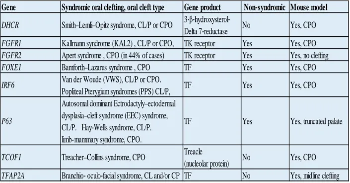
Table 1.3. Examples of syndromic CP in humans. Note that the mouse model in some examples has either no cleft phenotype or different phenotype than human species. The table demonstrates that some syndromic CP genes can contribute to non-syndromic forms of CP. Content adapted from Gritli-Linde.20
Gene Syndromic oral clefting, oral cleft type Gene product Non‐syndromic Mouse model DHCR Smith–Lemli–Opitz syndrome, CL/P or CPO 3‐β‐hydroxysterol‐
Delta 7‐reductase No Yes, CPO FGFR1 Kallmann syndrome (KAL2) , CL/P or CPO, TK receptor Yes Yes, CPO FGFR2 Apert syndrome , CPO (in 44% of cases) TK receptor Yes Yes, no clefting
FOXE1 Bamforth–Lazarus syndrome , CPO TF Yes Yes, CPO
IRF6 Van der Woude (VWS), CL/P or CPO.
Popliteal Pterygium syndromes (PPS) CL/P, TF Yes Yes, CPO
P63
Autosomal dominant Ectrodactyly–ectodermal dysplasia–cleft syndrome (EEC) syndrome,
CL/P. Hay‐Wells syndrome, CL/P. limb-mammary syndrome, CPO.
TF Yes Yes, truncated palate
TCOF1 Treacher–Collins syndrome, CPO Treacle
(nucleolar protein) No Yes, CPO TFAP2A Branchio‐ oculo‐facial syndrome, CL and/or CP TF No Yes, midline clefting
Table 1.4 Genes associated with non-syndromic CP in humans. Note that some of those genes are associated with syndromic forms of CP in humans.* PTCH associated mouse model is a conditional knock out. *SUMO1 associated mouse model is a haploinsufficient. Content adapted and updated from Gritli-Linde.20
Figure 1.2 Functional classification of genetic factors contributing to oral clefts (including CP) in humans and mice. An example for each sub-category and its contribution to CP in humans and mice is provided in the following section.
Gene Human (Oral cleft type) Cytogenetic location Gene function Available mouse model, (Oral cleft type)
FGFR1 CL and CP or CP 8p11.23-p11.22 Receptor Yes, CP
FGFR2 CL and CP or CP 10q26 Receptor Yes, CP
IRF6 CL or CP 1q32.3-q41 Transcription factor Yes, CP
MSX1 CL or CP 4p16.2 Transcription factor Yes, CP
PTCH CL/P or CP 9q22.3 Receptor Yes, CL*
SATB2 CL/P or CP 2q33 Transcription factor Yes, CP
TBX22 CP Xq21.1 Transcription factor Yes, SMCP and ~7% CP
TGFβ3 CP 14q24 Signaling protein (growth factor) Yes, CP
TGFA CP 2p13 Signaling protein (growth factor) Yes, no clefting defect
SUMO1 CLP or CP 2q33 cytoplasmic and membrane bound protein Yes, CLP*
ESR1 CL or CP 6q25.1 Transcription factor (estrogen receptor) Yes, no clefting defect
PVR CL/P or CP 19q13.2 Poliovirus receptor Yes, no clefting defect
Cell Surface Receptors: FGFR1 and FGFR2 Mutations
In humans, loss of function mutations in FGFR1 are associated with Kallman syndrome in which CP or CLP is one of its features. Gain of function mutation in FGFR1 is associated with craniosynostosis features in humans.72 Mutations in FGFR2 specifically (S252W, S252F, and
P253R) are seen in most cases of Apert syndrome.72 It is proposed that syndromes associated genetic mutations could contribute to non-syndromic CP by having hypomophic variants of these genes.72 Mutations in FGFR1 and FGFR2 were shown to contribute to non-syndromic CP and CLP in humans. 73,74 The role of these genes in human CP can possibly be better understood from Fgfr2b (-/-) and Fgf10 (-/-) mouse models which demonstrate CP phenotype beside other craniofacial and general abnormalities.75,76 In these murine models, it appears that the FGF signaling pathway plays an important role in palatal shelf growth. A significant reduction in the proliferation of epithelial (where Fgfr2 is highly expressed) and mesenchymal cells (where Fgf10 expression is limited) of palatal shelves contributes to the CP phenotype. Another
observation was the reduced expression of Shh in the epithelium of both Fgfr2b /-) and Fgf10 (-/-) mice. This observation revealed a possible role for FGF-SHH signaling network in palatal
shelf growth. The positive feedback between FGF and SHH signaling is also demonstrated in mesenchymal knockout of SMO. In this case it was shown that Fgf10 expression was
significantly reduced in the mesenchyme.6
Transcription Factors and Nuclear Proteins: IRF6 Mutations
Mutations in IRF6 gene are associated with van der Woude syndrome (VWS) and popliteal pterygium (PPS) syndromes in humans in which CL/P or CP is a major characteristic feature in humans. Mutations in IRF6 were also found to be associated with non-syndromic forms of CP in humans.77 The possible role of Irf6 in palatogenesis was discovered by studying
Irf6 (-/-) and Irf6 (R84C) knock in mouse models.6,78 Irf6 mutant mice showed an abnormally
hyperproliferative epithelium which did not differentiate. The associated epithelial defect
resulted in abnormal oral adhesions, CP and other associated abnormalities (abnormal limbs and skin).78 In addition, studies demonstrated that Irf6 (epithelial expression) is regulated by p63
transcription factor (epithelial expression). In humans, mutations in p63 are associated with syndromic CP such as ectrodactyly ectodermal dysplasia-cleft lip/palate syndrome (EEC), ankyloblepharon ectodermal dysplasia clefting (AEC), and nonsyndromic split-hand/foot malformation (SHFM).79 In Mice, a null mutation for p63 results in abnormally thin, undifferentiated epidermis and CP. In p63 (-/-) mice, the expression of Irf6 in the palatal epithelium is reduced and p63 is found to bind to an enhancer sequence upstream of Irf6 and to promote a luciferase reporter expression driven by an Irf6 enhancer.6,77
Nuclear Membrane Bound Proteins: SUMO1
SUMO1 (small ubiquitin-related modifier) is part of the SUMO family (SUMO1-4). The
SUMO family are responsible for the reversible posttranslational modification of proteins by SUMOlyation. As in ubiquitination, SUMOlyations requires will require other enzymes that results in the activation of SUMO and its addition to its target protein. In most of the cases a single SUMO is added or sometimes a poly-SUMO chain is added.20,80 In humans mutations in SUMO1 were found to be associated with both non-syndromic CL/P and CP.81,82 Interestingly, haploinsufficiency of SUMO1 in a mouse model (Sumo1Gt/+ heterozygous) resulted in an
incompletely penetrant CP or oblique facial cleft (8.7%).83 Furthermore, SUMO1 is expressed in both the mesenchyme and the epithelium of mouse palatal sheleves.20,83 There is evidence that
several proteins important for palatogenesis are being SUMOylated.20
Extracellular Matrix Proteins: Collagens (COL2A1 and COL11A2)
In humans, mutations in collagens (COL2A1 or COL11A2) are associated with Robin sequence. Robin sequence has a combination of 3 out of 4 features: micrognathia, glossoptosis, obstructive apnea, cleft palate. Robin sequence is commonly seen as part of a syndrome (80%) such as Stickler syndrome, velocardiofacial syndrome. Non-syndromic forms of Robin sequence are less frequent (20%).84The role of collagens and their relevance to palatogenesis is
demonstrated by examining the (cho/cho) homozygous mice which carry an autosomal recessive mutation in collagen (Col11a1). These mice demonstrate shortened heads and mandibles, U shaped cleft palates, short limbs. The cleft phenotype in these mice is believed to be due to the abnormal skeletal growth leading to a smaller mandible and a higher tongue position which prevents the palatal shelves from adhering. It was shown that cultured palatal shelves of (cho/cho) mice were able to adhere and fuse normally (no remnants of MES).71,85
1.4.2. Environmental Factors
The finding that the concordance rate of CP and CL/P phenotypes did not reach 100% in monozygotic twins demonstrates that non-genetic factors (such as environmental factors) contribute to such birth defects.45,68,69 This does not exclude the possibility that such
dis-concordance can be due to genetic and cytogenetic and epigenetic differences in one of the twins.69 However, the finding that the concordance in dizygotic twins is higher than the
concordance rate among singleton siblings further supports the role of environmental factors in such birth defects.69
Multiple environmental factors are associated with higher risk of CP or had a potential association with CP in the population studied such as smoking,49,86,87 alcohol (well established
with oral clefts in fetal alcohol syndrome),49,86 vitamin A,88 maternal hyperthermia and viral
infections during the first trimester49, anticonvulsant drugs such as diazepam, phenobarbital and phenytoin,49,86 corticosteroid therapy during pregnancy,49,86 nutritional deficiencies such vitamin B6 and zinc.49 Other environmental factors are associated with a lower risk of developing CP or
possibly having a protective effect against CP such as folic acid supplements, multivitamin supplements.49
Identifying environmental factors contributing to CP helps to identify the metabolic pathways that could be possibly altered and could be contributing to the formation of such defects.45
1.4.3. Gene-Environment Interaction
The cross talk between genetic and environmental factors and their effect on oral clefting was demonstrated in humans and mice. For example, evidence suggested that mothers who smoke have an increased risk (2 fold) of having babies with CP.86,89 However, if the infant had TGFα TaqI C2 allele variant and the mother was a smoker during pregnancy (≤10 cig/day) there was an increased risk of (6.16 fold) to have CP.86,90 Furthermore in the same study, if this
specific polymorphism in TGFα was present but the mother was not a smoker the risk for having CP (0.9) fold higher therefore showing how the presence of an environmental factor can modify the risk for developing CP with specific genotype.90
Moreover, experiments using mouse models demonstrated such cross talk. For example, upon examining the effect of cortisone on CP frequency in two mouse strains (different genetic backgrounds) A/J strain and C57BL/6, 100% of A/J mice offspring had a CP phenotype while
only 17% of C57BL/6 had CP. The two mouse strains obviously had different responses to the same teratogen under similar experimental conditions.20,91
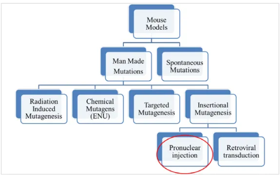
1.5 Types of Cleft Palate Mouse Models
Mouse models are very useful for dissecting the genetic basis of palatogenesis.20,92 Our
knowledge of how genetic factors contribute to oral clefts in humans was greatly advanced by studying CP murine models.20 Mouse models used to study CP can be divided into two main
categories: Spontaneous CP mouse models and Induced CP mouse models (Fig 1.3).93
Figure 1.3. Types of CP murine models. CP mutations studied in mice are either spontaneous (no intervention) or Induced (i.e. manmade mutations).
1.5.1 Spontaneous CP Mouse Models
Mouse models in which CP occurs spontaneously (without human intervention) at a stable frequency are known as spontaneous CP mouse model.94 Examples of CP mouse models with the frequency of CP are: SW/Fr (6%), CF1 (3%), J/Glw (25%). The genetic cause of CP in theses strains has not been studied yet.50,95
1.5.2 Induced CP Mouse Models
Induced CP mouse models are produced through human intervention. Induced mutations are further subdivided into 4 main subcategories based on the strategy used: radiation induced mutation, chemical mutagenesis, targeted mutagenesis and insertional mutagenesis.
Ionizing Radiation Induced Mutations
Ionizing radiation is known to induce DNA damage (base pair damage, single and double strand breaks). Mutations induced by ionizing radiation can vary from point mutations to small deletion mutations, and can be more severe and result in major genomic rearrangement.96-98 An example of ionizing radiation induced CP mouse model is (p4THO-I/p4THO-I) line which has a radiation induced deletion mutation at pink eyed dilution locus (p). These mice had pink eyes and CP with no other craniofacial abnormality (95%). The mutation was mapped Gabrb3 locus (β3 subunit of GABAA receptor). 20,99
Chemical Mutagen Induced Mutations
The N-ethyl-N-nitrosourea (ENU) is the most potent chemical mutagen in mice. ENU is an alkylating agent which can add an alkyl group to different atoms in a DNA molecule such as N-1, N-3, and N-7 groups of adenine; the O2 and N-3 groups of cytosine; the N-3, O6, and N-7
groups of guanine; the O2, N-3, and O4 groups of thymine; and the phosphate groups of the DNA
backbone.100,101 ENU is known to induce point mutations, rarely small deletion mutations, randomly all around the genome.100,101 An optimal ENU dose can result in one mutation every 1-1.5 Mb.100 The point mutations are mostly seen at A-T base pairs resulting in a transversion
mutations (AT or TA) or transition mutations (A-TG-C).100,101 An example of a CP mouse model due to ENU mutagenesis is the cleft secondary palate 1 (csp1) mutant mice due to a point mutation in Prdm16 gene. The CP is due to the failure of palatal shelves elevation as a consequence to tongue obstruction and micrognathia.102
Targeted Gene Mutations
Gene targeting involves modifying a specific genetic locus by the use of homologous recombination between a targeting vector and an endogenous gene in mouse embryonic stem (ES) cells.103 The genetically modified ES cells are then microinjected into embryos at the blastocyst stage (8 cell stage) and are then transferred to a pseudopregnant female mouse. The resulting chimeric progeny (with the targeted mutation) are then crossed with mice of the same genetic background from which ES cells were derived.104 If the gene of interest is no more functional, i.e. not expressed or altered then the mouse model is known as a knockout model. On the other hand, if the line has a duplication of a target gene or a tailored mutation (such as point mutations) then the mouse model is known as a knockin model.104,105 The TGFβ3106 and Msx1107 mouse models and Irf6R84C 108mouse model are examples of knockout and knockin CP mouse
models, respectively.
Ubiquitous gene targeting is sometimes associated with early embryonic lethality, therefore preventing researchers from examining the effects of gene targeting on the developing palatal shelves.93,104 A solution to this problem was the advent of conditional gene targeting in
which the target gene is modified only in a specific tissue and at a certain developmental stage. An example of a conditional gene targeting system is the Cre-loxP system. In this system, mice expressing the Cre recombinase enzyme in a specific tissue or cell line (i.e. driven by tissue specific promoter) are bred to another loxP mouse harboring loxP sites flanking the gene of interest. Breeding a Cre-mouse and a loxP mouse results in a progeny expressing Cre and having a target gene flanked with loxP sites. If the loxP sites are oriented in the same direction, the Cre recombinase would result in deleting the DNA segment. If the loxP sites are directed in opposite directions, the Cre recombinase would result in inverting the DNA segment flanked by the loxP sites.104 Tgfβr2 (K14- Cre)109 and Shox2 (Wnt1-Cre).110
Insertional Mutagenesis
Insertional mutations take place when the integration of an exogenous DNA results in the disruption or alteration of a functional gene; the mutation seen is due to the physical damage at the integration site rather than the expression of the transgene itself.111,112 Random insertions can be produced by microinjection of the transgene or viral infection (retroviral/lentiviral infection) into a zygote (single cell embryo).111 One example of an insertional mutant CP mouse model, generated by microinjection technique, is OVE427113. A detailed explanation of insertional
mutations produced by this microinjection technique is present in chapter 2.
Random insertions can be produced by the gene trap approach in embryonic stem cells which uses a trapping vector that has promoter-less marker/reporter with an upstream splice acceptor site and downstream poly-A signal.114,115 The trapping vector can integrate into an exon/ intron of a gene.116 Transcriptional activation of the trapped gene results in a fusion transcript
containing part of the original transcript fused to the reporter. The resulting fusion transcript represents an alteration of the original gene and at the same time a readout for the pattern of this
altered expression (due to the expression of the reporter).114,115 An example of a CP mouse model
generated by gene trap approach is Sumo1(Gt/+) mutants.83
Another approach for the generation of random insertions (random to a certain extent) is the use of DNA transposons, as the Sleeping Beauty (SB) transposon system.93,114 The system is
composed of the transposase enzyme and the transposon substrate. The transposase identifies inverted repeats that flank the transposable element and then cuts the transposable element. This is followed by pasting the element (inserting) at another site with a TA dinucleotide. Therefore the transposition is done in a “cut-paste”.93,114 An example of a CP mouse model generated by such approach is the Smoc1 mouse model.117
1.6 Development of the Vertebral Column and the Ribs
The term axial skeleton refers to head (skull and facial bones), vertebral column, and rib cage (ribs and sternum).118 This section focuses on the development of the vertebral column and the ribs due to their relevancy to the thesis. During gastrulation, paraxial mesoderm forms on each side of the axial structures (notochord and neural tube) due to the ingression of the
epiblastic cells in the developing embryo.119,120 Paraxial mesoderm undergoes segmentation (is
chopped off) to form blocks of epithelial cells known as somites.120,121 The pace of the
segmentation process is unique across different species. For example in humans a new pair of somites is formed every 4-5 hours while in mice every 120 min.120,122,123 Somitogenesis (the
formation of somites) is seen between 25-35 days post conception in humans and E8-E13 in mice.121,124 After the formation of the somites, 42 pairs in humans and around 65 pairs in mice125,
they undergo differentiation. The cells on the ventromedial aspect of the somite will differentiate into the sclerotome while cells on the dorso-lateral part of the somite will differentiate in to the dermomyotome. The dermomyotome further differentiates into dermatome (produces the dermis of the back) and the myotome (gives rise to the body and limb musculature). The sclerotomal cells delaminate then surround the neural tube and the notochord. These cells will condense and differentiate into chondrocytes that form the cartilaginous skeleton which is then replaced by bony tissue i.e. the vertebral skeleton and the ribs which are formed by endochondral
ossification.120,126
The development of vertebra does not follow one somite one vertebra relation, but rather each vertebra is formed by a caudal half of one somite and the rostral half of the following somite.120 This takes place due to another segmentation process known as re-segmentation which happens in the sclerotomal compartment. The sclerotomal cells will rearrange into two cellular
compartments, rostral and caudal cells. The caudal cells from one somite will fuse with the rostral cells from the following somite and both contribute to the formation of the developing vertebra.121
The resulting vertebrae differ in their shape and size and can be generally categorized based on their anatomical position to the cervical, thoracic, lumbar, sacral and coccygeal (caudal) vertebrae. The formula in humans is 7 cervical, 12 thoracic, 5 lumbar, 5 sacral and 4 coccygeal. The ribs are attached to the thoracic vertebrae and are classified as true ribs (1-7), false ribs (8-10) and floating ribs (11-12).127,128 On the other hand, the mouse vertebral formula is 7 cervical, 13 thoracic, 6 lumbar, 4 sacral and 30 caudal vertebrae. As in humans, ribs attach to the thoracic vertebrae whereas only the first 7 ribs are attached to the sternum.120,129
1.6.1 Patterning the Axial Skeleton
During somitogenesis, the somites that are formed look similar. However, the somites eventually contribute to the formation of the vertebral column with characteristic morphological features.130 Genetic factors are known to regulate the segmental identity i.e. the type of vertebra
each somite pair will form and its position along the anterior-posterior axis. Genetic factors regulating segmental identity and therefore the patterning of the axial skeleton can be subdivided mainly to transcription factors and signaling molecules.120
Transcription Factors
The classical example of a transcription factor regulating the identity of the developing vertebra is the Hox genes.120 These homeo-box containing genes are considered of the major players regulating the patterning of the developing vertebral column.129 In humans and mice,
respectively. There are 13 paralogous groups with 2-4 members in each group. 131-133 Hox genes
generally demonstrate a spatial-temporal colinearity in terms of their expression; the 3’ genes in each cluster are expressed earlier and contribute to determining the identity of anterior structures and the 5’ genes are expressed later and contribute to identity formation of more posterior structures.120,133 Little is known about how exactly Hox genes regulate the identity of the developing vertebral column.120,133
Targeting Hox genes in mice revealed the significance of Hox3-Hox11 to the normal development of the axial skeleton. Some single Hox genes mutants in mice had phenotypes consistent with their colinear expression such as Hoxd3 which had defects in C1 and C2 vertebra and Hoxd11 had defects in patterning the sacral vertebra. On the other hand, other single and multiple mutants demonstrated a phenotype not consistent with the colinear expression pattern of the Hox genes. for example, single and multiple mutants of Hox5, Hox6, Hox7, Hox8, and Hox9 group genes had defects in the rib on T1.133
Loss of function mutations of single or multiple mutants are sometimes associated with anterior homeotic transformation. An example of such anterior homeotic transformation is seen in the case of Hoxb9 mutants (14th rib on first lumbar vertebra) and in Hoxa7/b7 double mutants
(T1 to C7 transformation). Posterior homeotic transformations are also seen due to the loss of function of single mutants such as the extra rib seen on C7 in Hoxa5 and Hoxa6 mutants.133
Evidence revealed that there is functional redundancy among paralogous Hox genes. It was also demonstrated that a synergistic phenotype is seen when more than one gene in a paralogous group is mutated.133 For example, single mutants of Hox4a, Hox4b and Hox4d had
incompletely penetrant defects in C1 and C2 vertebrae. However, a combined triple mutant of Hox4a, Hox4b and Hox4d result in a fully penetrant anterior homeotic transformation in C2-C5.
By now, complete paralogous mutants have been generated for Hox5, Hox6, Hox7, Hox8, Hox9, Hox10 and Hox11 paralogous gene groups. In these mutants anterior homeotic transformation is
seen.133
Another example of transcription factors regulating the identity of the developing vertebral column is Caudal type homeobox gene family composed of Cdx1,2and 4.120 Cdx1 null mutants show anterior homeotic transformations, not as severe as those with Hox mutants, in the cervical and upper thoracic vertebrae. Cdx2 null mutation is embryonically lethal but
heterozygote null mutants demonstrate anterior homeotic transformation in the cervical to thoracic vertebra transition. Cdx4 null mutants did not affect the axial skeleton. However, a combined null mutants of Cdx1 and 4 or Cdx2(+/-) and 4 resulted in a more severe anterior transformations.134
Signaling Molecules and Receptors
An example of cell surface receptors regulating the anterior posterior patterning is Fgfr1. Fgfr1 null mutation results in early embryonic lethality. However, hypomorphic Fgfr1 mutations were associated posterior truncations and anterior homeotic transformations (L1 has an extra pair of ribs). On the other hand, gain of function mutations in Fgfr1 were associated with posterior homeotic transformations (such as C7 vertebrae with ribs attached, T13 vertebrae lacking their ribs and becoming like L1 vertebrae).120,135
1.7 Rib Anomalies (Supernumerary Ribs)
Rib anomalies in humans include supernumerary ribs (SNR), forked ribs, fused ribs and pseudoarthrosis of the first rib.118,136 These anomalies are seen either in isolation or in association
its relevance to this project. SNR (known as accessory ribs, extra ribs) can be categorized based on their anatomic position into cervical SNR, intrathoracic SNR, lumbar SNR, pelvic ribs. Intrathoracic and pelvic ribs are rare anomalies.118,136 Cervical ribs can be unilateral or bilateral and have a prevalence ranging from (0.04-4.5%).118 Lumbar SNR manifest as a unilateral or
bilateral extra ribs with a prevalence ranging in (0.04%-5.8%) with one study reporting a prevalence as high as 15.8%.118,137 Cervical and lumbar ribs are commonly asymptomatic.
However, their presence can cause some problems.118 For, example cervical ribs can cause a condition known as thoracic outlet disease which has vascular (cerebral embolism) and neurologic symptoms (extreme pain, migraine, and Parkinson’s like symptoms). The vascular symptoms seen are due to compression and associated reduction in the blood flow through subclavian artery and vein, and carotid arteries. The neurologic symptoms are associated with altered positioning of stellate ganglia, sympathetic ganglia and C7-T1 nerve roots.118,136,138 Moreover, lumbar ribs can be associated with pain in the lumbar region and disc degeneration in L4-5.118,138
1.8 Etiology of Supernumerary Ribs
Little is known about the etiology of SNR in humans.118,137 However, evidence from
animal models and humans suggests that both genetic and environmental factors contribute to the formation of SNR.118 For example, supernumerary cervical ribs were seen in a family across two generations. Furthermore, different mouse models targeting Hox genes (single and multiple) and other genes (Cdx and Fgfr1) demonstrate SNR (cervical and lumbar). The role of environmental factors in the etiology SNR is inferred mainly from animal models.118 Exposure to certain
environmental factors was shown to cause SNR in mice, such as retinoic acid, valproic acid, salicylate and maternal stress.118,138
1.9 SNR Mouse Models
As in CP mouse models, spontaneous SNR mouse models are available. For example, CD1 mice in which the frequency of SNR was between 14.3%- 25%. 118 SNR induced mutations in mice are also seen such as the radiation induced SNR model Pgap1otomice have
supernumerary cervical rib at C7, 139 ENU induced SNR model such as Rpl38Rbt mice which have
an lumbar rib140, targeted SNR models such as Hoxa9 and Hoxb9 single and double mutants141
and gene trap induced SNR models such as BC055757-/- line which had an extra pair of ribs on
L1.142
1.10 Specific Aims
OVE1328 line, is an insertional mutant mouse model generated by microinjecting the tyrosinase transgene complex into an FVB/NJ single cell embryo. The integration site of the transgene is mapped to chromosome 4 (near the centromere). The recessive homozygous mutation in OVE1328 mice is initially found to be associated with perinatal lethality and CP. This projects aims to:
• Aim 1: identify the phenotypic differences between OVE1328 (homozygous and hemizygous) line and the wild type FVB/NJ strain.
• Aim 2: determine the gene/genes disrupted by the transgene integration and characterize the resulting mutation.