CONNECTING NUCLEAR ORGANIZATION AND GENE EXPRESSION: THE ROLE
OF THE HISTONE LOCUS BODY IN HISTONE MRNA BIOSYNTHESIS
Deirdre Catherine Tatomer
A dissertation submitted to the faculty of the University of North Carolina at Chapel Hill in partial fulfillment of the requirements for the degree of Doctor of Philosophy in the Department of
Biology
Chapel Hill 2014
ii
©
2014iii
ABSTRACTDEIRDRE CATHERINE TATOMER: Connecting Nuclear Organization and Gene Expression: the Role of the Histone Locus Body in Histone mRNA Biosynthesis
(Under the direction of William F. Marzluff and Robert J. Duronio)
Execution of gene expression involves multiple reactions, many of which are mediated through cis elements in DNA or RNA. Organization within the nucleus, both in the arrangement of DNA and the concentration of trans factors in discrete nuclear environments called nuclear bodies, facilitates aspects of gene expression such as transcription, RNP metabolism and pre-mRNA processing. These substructures, which are visible under the light microscope,
potentially promote the efficiency and fidelity of a reaction. To investigate this proposed function of nuclear bodies (NBs), I studied the relationship between the Drosophila replicative histone genes and their associated nuclear body, the histone locus body (HLB). The genes encoding the five histone proteins are tandemly arrayed in 100 copies. The replicative histone genes are highly expressed during S phase of the cell cycle, and encode the only known mRNAs that do not end in a poly (A) tail. We defined a 297 bp sequence that encompassed the H3-H4
pre-iv
mRNA processing factor, Symplekin (Sym), involved in histone pre-mRNA processing, cleavage and polyadenylation and cytoplasmic polyadenylation. I established an in vivo system for testing candidate separation of function Sym mutations. Sym was first discovered at tight junctions, and I provide evidence that this localization indicates likely participation in cytoplasmic
v
ACKNOWLEDGMENTS
I am deeply grateful to all of the people who have supported and encouraged me along my path towards this degree. I would first like to thank my mentors, Bill Marzluff and Bob Duronio. By allowing me to join both labs, they gave me the intellectual and physical space to learn how to define scientific questions and best approach them. I appreciate their patience with my experimental wandering and that they allowed me the time to learn, with their guidance, how to navigate a project to completion. Their infectious love of science and enthusiasm for teaching fosters a wonderful environment that I hope will exist in my future workplaces. Thank you Bob and Bill for an invaluable experience.
I would also like to acknowledge the amazing scientific community that I have had the privilege of participating in during my time at UNC. This includes, but is not limited to, members of the Marzluff and Duronio labs, my committee (Greg Matera, Alan Fanning and Kevin Slep), Jeff Sekelsky and his lab, the Lieb lab, and Tony Perdue, who taught me how to use a
microscope to capture the images present in this dissertation. I also thank three wonderful undergraduates, Michelle Lin, Alison Witkowski, and Anna Orlando for their hard work and optimism. They all contributed data to this thesis and enriched my time in the Marzluff and Duronio labs. In general, the time everyone took to brainstorm solutions for hurdles in my projects, dispense advice and discuss science with me is greatly appreciated.
vi
opportunity to work with Christina and Aleksandra in his lab at UCSF. I am so lucky to have them as lifelong mentors and friends.
I also thank all of my friends who have entertained, distracted and supported me through graduate school. This group encompasses members of both labs, fellow graduate students and lovely locals that I have gotten to know during my time here in North Carolina. They have brilliantly complemented my academic experience.
vii
TABLE OF CONTENTS
LIST OF TABLES ...xii
LIST OF FIGURES ...xiii
LIST OF ABBREVIATIONS...xv
CHAPTER 1. Introduction ...1
Overview ...1
Nuclear Body Assembly ...3
Nuclear Body Function ...5
The Impact of 3’ end Formation on Gene Expression ...7
Mechanisms of 3’ end Formation ...9
Cleavage and Polyadenylation ...9
Cleavage followed by STAR-PAP Polyadenylation...11
Histone pre-mRNA Processing ...12
Components of the HLB ...17
Dissertation Goals...20
2. A Sequence In The DrosophilaH3-H4 Promoter Triggers Histone Locus Body Assembly And Biosynthesis of Replication-Coupled Histone mRNAs ...22
Preface ...22
Introduction...22
Materials And Methods...26
Drosophila Strains ...26
viii
Immunofluorescence ...27
Analysis of Histone Deletion Embryos...27
Results ...31
An HLB can Assemble at a Single, Active Histone Gene Repeat ...31
Histone Gene Expression Correlates with HLB Assembly ...34
The H3-H4 Promoter is Necessary and Sufficient for HLB Formation ...38
mRNA Processing Signals are Dispensable for HLB Assembly ...41
Transcription Stimulates HLB Maturation ...43
Stable Assembly of the HLB During Development Requires the Histone Gene Cluster ...47
Discussion ...51
How do HLBs Form During Development? ...51
Model for HLB Assembly ...53
HLBs Compared with Other Nuclear Bodies ...54
What are the Functions of the HLB? ...55
Acknowledgements ...59
3.Concentrating pre-mRNA Processing Factors at the Histone Locus Body Facilitates Efficient Histone Gene Expression ...60
Preface ...60
Introduction...60
Materials And Methods...63
Drosophila Strains ...63
Immunofluorescence and Fluorescent in situ Hybridization ...64
ix
Immunoblotting ...66
in vitro Processing Assay ...66
RNA pol II Visualization ...66
Results...67
The essential function of Drosophila FLASH is Histone mRNA 3’ End Formation ...67
The FLASH NH2-terminus is Necessary for Histone mRNA 3’ End Formation ...72
Concentrating FLASH in the HLB Facilitates ...76
Histone mRNA 3’ End Formation High Concentrations of FLASH are Necessary for U7 snRNP Accumulation in the HLB ...79
FLASH is not Sufficient for U7 snRNP Accumulation in the HLB ....85
The HLB Promotes Rapid co-transcriptional Histone pre-mRNA Processing...87
Discussion ...90
The HLB Contributes to Multiple Steps in Histone mRNA Biosynthesis ...90
Concentrating Factors within the HLB Ensures Efficient histone mRNA Synthesis ...90
Multiple Domains of FLASH are Required for Rapid Histone pre-mRNA Processing...92
Cellular Microenvironments that Enhance Biological Processes ...93
Acknowledgements ...95
4.Characterization of Drosophila Symplekin to Establish an in vivo System for Testing Histone Cleavage Complex Assembly ...96
Preface ...96
Introduction...96
x
DNA PCR Analysis ...100
Drosophila Genetics ...101
Western Blot ...102
Quantitative RT-PCR ...102
S1 protection Assay...102
Immunofluorescence ...103
GST Pull Down ...103
Symplekin D 64 Construct ...104
RNAi Knockdown ...104
Results ...104
In Symplekin Mutants the Developmental Phenotype Correlates with Level of Symplekin ...104
Reducing Symplekin Affects both Poly(A) and Histone pre-mRNA Processing...108
Symplekin Localization Is Dynamic ...111
Localization of Symplekin to the Junction Depends on YPS...113
Candidate Symplekin Separation of Function Mutation ...116
Discussion ...118
Assembling Symplekin into the Histone pre-mRNA Processing Complex......118
The Role of Symplekin at the Septate Junction...120
Acknowledgments ...121
5.Overall Conclusions And Significance ...122
The Histone Locus Participates in HLB Assembly ...123
xi
Understanding cis Control of Histone mRNA Biosynthesis ...130
Assembly of the Histone Processing Complex in vivo ...132
Concluding Remarks ...134
xii
LIST OF TABLES
Table 2.1. Boundaries for Each Histone Locus Construct ...28
Table 2.2. Primers used in RT-PCR ...29
Table 2.3. Tissue Preparation for Antibody Staining ...30
Table 2.4. Antibody Concentrations...30
Table 2.5. Identification of Histone Deletion Genotypes ...51
Table 3.1. Drosophila Strains ...63
Table 3.2. Construct Features ...63
Table 3.3. PCR Primers ...64
Table 3.4. Tissue Preparation for Antibody Staining for Immunofluorescence ...64
Table 3.5. Antibody Concentrations...64
Table 3.6. Fluorescent in situ Hybridization Probe Sequences ...65
Table 3.7. Quantification of Transgenic Rescue of the FLASHPBac/Df Mutant ...72
Table 4.1. Primer Sequences ...100
xiii
LIST OF FIGURES
Figure 1.1 Trans factors involved in histone pre-mRNA processing
and cleavage and polyadenylation ...16
Figure 1.2 Overview of the structure of the Drosophila replicative histone genes ...17
Figure 2.1 An HLB forms at an ectopic locus containing one histone gene repeat unit ...33
Figure 2.2 The H3-H4 genes assemble an ectopic HLB ...37
Figure 2.3 The H3-H4 Promoter Assembles an Ectopic HLB ...40
Figure 2.4 The H3 and H4 coding region and 3’ processing signals are not required for HLB assembly ...42
Figure 2.5 Transcription is required for HLB assembly...45
Figure 2.6 CORE and COREΔT Assemble an Ectopic HLB ...46
Figure 2.7 HLB assembly in the absence of the Histone Locus ...49
Figure 2.8 HLB assembly in the absence of the Histone Locus ...50
Figure 2.9. Model for HLB assembly and maintenance ...56
Figure 2.10 Conservation of the H3-H4 Intergenic Region ...58
Figure 3.1 Histone pre-mRNA processing is the essential in vivo FLASH function...70
Figure 3.2 Location of the FLASHLL01602 PBac Insertion ...71
Figure 3.3 Characterization of FLASH N-terminus function in histone pre-mRNA processing ...75
Figure 3.4 Characterization of FLASH N-terminus contribution to histone pre-mRNA processing ...76
Figure 3.5 Concentrating FLASH in the HLB promotes histone 3’ end formation ...79
Figure 3.6 FLASH contribution to HLB assembly ...82
Figure 3.7 Localization of transgenic FLASH proteins...84
Figure 3.8 HLB concentration of FLASH and/or U7 snRNP promotes efficient histone pre-mRNA processing...86
xiv
Figure 3.10 Model of HLB participation in histone pre-mRNA processing ...94
Figure 4.1 Characterization of Symplekin Allelic Series ...107
Figure 4.2 Characterization of Symplekin mutant RNA phenotypes ...110
Figure 4.3 Characterization of Symplekin Localization During Development ...112
Figure 4.4 Characterization of Symplekin Localization to the Septate Junction ...115
xv
LIST OF ABBREVIATIONS
aa amino acid
bcd bicoid
cas castor
CB cajal body
cDNA complementary DNA CFIm Cleavage factor I
CF IIm Cleavage factor II
CPSF Cleavage and polyadenylation specificity factor CstF Cleavage stimulation factor
CTD C-terminal domain
FLASH FLICE-associated huge protein
grk gurken
HCC histone cleavage complex HDE histone downstream element HLB histone locus body
HO-1 haem oxygenase LacI Lac Repressor LacO LacOperator
MPEF mouse primary embryonic fibroblast
mxc multi sex combs
ncRNA non coding RNA NB nuclear body
NELF negative elongation factor
nos nanos
xvi
PAP poly (A) polymerasePAS poly (A) signal
PIPKIα type I phosphatidylinositol 4-phosphate 5-kinase
PML promyelocytic leukemia poly (A) polyadenosine
PtdIns4,5P2 Phosphatidylinositol-4,5-bisphosphate
scaRNA small CB-specific RNA
SL stem loop
SL1 promoter selectivity factor SLBP stem loop binding protein
snRNP small nuclear ribonucleoprotein particle SRE Smaug response element
Star-PAP Speckle Targeted PIPKIα Regulated-Poly(A) Polymerase sym symplekin
tBHQ tert-butyl hydroquinone TBP TATA Binding Protein
1
CHAPTER 1: INTRODUCTION
Overview
Every cell in a complex organism contains an identical blueprint encoded in its DNA, and variation in how this information is accessed and employed generates diversity in cellular function. The genome contains both regulatory sequences as well as those that code for genes. Many factors involved in gene expression function by recognizing and interacting with
sequences in DNA. This theme of directing activity through motifs or structures in a nucleic acid can be extended to RNA, as many of the events required to form a mature RNA are mediated through signals in the transcript. Therefore, a fundamental question in the field of gene
2
Overlaid on the chromatin scaffold are structures visible under the light microscope. First described by Santiago Ramón y Cajal in 1903, these structures, termed “accessory bodies” and subsequently nuclear bodies (NB), contain high concentrations of protein and often nucleic acid, but are not bound by a membrane (Matera et al., 2009). A number of NBs have been identified and characterized, many of which contain factors involved in gene expression (Morimoto and Boerkoel, 2013). The presence of NBs implies that the genome is not in contact with a homogeneous pool of trans factors but instead is surrounded by discrete environments. This varied accessibility to trans factors provides another mode of regulating interplay between a factor and target gene. Understanding how nuclear bodies interact with the genome and nascent RNA transcripts is an open area of investigation.
Here, I use the replicative histone genes as model to explore the relationship between a nuclear body and gene expression. The metazoan replicative histone genes are the only known messenger RNAs that do not end a polyadenosine (poly (A)) tail but instead end in a conserved stem loop structure (Marzluff et al., 2008). Factors involved in the biosynthesis of this special class of mRNA are concentrated in a nuclear body, the histone locus body (HLB) (Liu et al., 2006). This thesis project addresses three questions: Is the formation of the HLB a cause or consequence of histone mRNA expression? Does localizing a trans factor to the HLB affect the rate of histone 3’end formation? How does a protein involved in generating both poly (A) and histone mRNA 3’ends, Symplekin, specifically incorporate into the histone processing
machinery and localize to the HLB?
In this chapter, I will introduce our current understanding of nuclear body assembly and function. I will also describe the known mechanisms of 3’end formation and how this aspect of mRNA biosynthesis influences gene expression. Finally, I will focus on the Drosophila
3
Nuclear Body AssemblyNuclear bodies are defined by their components, both because they mark the structure and because the reactions that the concentrated factors participate in suggest possible biological function(s) for each nuclear body. Discovery of NB components is an ongoing and valuable endeavor as it provides new tools for studying NBs and expands the mechanistic details of nuclear body function (Fong et al., 2013). Understanding the assembly and maintenance of nuclear bodies lays the foundation for assessing NB function.
Nuclear bodies are built through interactions between components (Matera et al., 2009). Different experimental approaches have been used to determine the nature of assembly. One strategy is to first deplete or genetically remove a factor from a cell or animal and then
determine if the remaining known components of the NB co-localize. This approach was used to characterize coilin, a fundamental component of the Cajal body (CB). Coilin mutant flies are viable (Liu et al., 2009). Coil -/- mice are also fertile, however they were not detected in expected mendelian ratios and displayed reduced fertility and fecundity (Tucker et al., 2001; Walker et al., 2009). Although there were differences in developmental phenotypes,
immunostaining in both systems revealed that other CB markers such as SMN and components of snRNPs did not form foci in the absence of Coilin. These studies indicated that coilin was a core component of the Drosophila CB.
Another nuclear body, the promyelocytic leukemia (PML) body, was also shown to have a core protein component, PML (Bernardi and Pandolfi, 2007). In mouse primary embryonic fibroblasts (MPEFs), PML colocalizes with Sp100, Daxx, and ISG20 in discrete foci. Removal of PML through gene targeting resulted in diffuse localization of these factors in PML-/- MPEFS, suggesting a role for this protein in assembling or maintaining the NB (Ishov et al., 1999; Zhong et al., 2000). These examples indicate a role for specific proteins in NB assembly.
4
remaining components to that location (Kaiser et al., 2008; Shevtsov and Dundr, 2011). In the initial study, CB components such as Coilin and SMN were fused to the Lac Repressor (LacI) and GFP. LacI binds to the Lac Operator (LacO), and expression of the fusion protein in a cell with 256 LacO repeats resulted in a detectable ectopic focus (Kaiser et al., 2008). Antibody staining for endogenous CB components was then used to screen for co-localization with the LacO array to indicate NB formation. These experiments showed the ability of these tethered components to nucleate the CB, but only in the presence of Coilin and SMN in the cell.
A subsequent study expanded these observations to other NBs and emphasized the tethering of RNA (Shevtsov and Dundr, 2011). RNA was localized to the repeat through inclusion of an MS2 loop in the transcript and expression of a GFP-LacI-MS2 fusion protein. These experiments again indicated the ability of individual NB components to nucleate structures such as the HLB, speckle, paraspeckle and nuclear stress body. For example, Shevtsov and Dundr (2011) showed that histone H2b mRNA could recruit HLB components NPAT and FLASH. Overall, these studies showed that protein or RNA components could participate in assembling a NB. While these experiments show what can happen in a cell, they do not necessarily reflect the endogenous biogenesis of a NB.
5
pre-rRNA. Incomplete assembly of the structure in the absence of the coding region of the genes, as well as dispersal of nucleolar compartments after inhibition of transcription and RNA processing suggest a role for transcription or nascent RNA in nucleolus formation and
maintenance (Hernandez-Verdun, 2006).
Although the CB is not consistently associated with a single genomic location, it has been detected at the U1 and U2 snRNA genes, U3 snoRNA genes and the replicative-histone genes (Callan and Gall, 1991; Frey and Matera, 1995; Gall et al., 1981; Gao et al., 1997; Smith et al., 1995). Insertion of an ectopic array of repeated U2 snRNA genes resulted in association of the exogenous DNA and a CB (Frey et al., 1999). The association required an intact promoter as well as the coding region of the snRNA (Frey and Matera, 2001). This suggests that a
nascent RNA plays a role in CB dynamics.
Transcription was also required for assembly of another nuclear body, the paraspeckle. Induction of a non coding RNA (ncRNA), Men e/b, resulted in the colocalization of paraspeckle components PSP1, P54nrb, PSF and PSP2 with the ectopic RNA (Mao et al., 2011). All of these approaches have contributed to our understanding of NB formation. This collection of
experiments also highlights the role of both protein and nucleic acid in assembling a NB. Identifying new components of NBs and understanding how they contribute to forming and/or maintaining a microenvironment is an ongoing pursuit of the NB field. Defining the domain(s) that localizes a factor to a NB is one of the next steps in understanding NB function as it opens up experimental approaches for isolating the contribution of a NB to its related biological process.
Nuclear Body Function
6
hyperedited mRNAs (Mao et al., 2011). How the environment created by a NB contributes to a biological reaction is still an open question. Concentrating the elements of a common process can increase the probability of interactions between components, and this is a proposed function for nuclear bodies. Increasing the local concentration of factors is also postulated to influence the rates of biological reactions (Machyna et al., 2013; Mao et al., 2011; Matera et al., 2009).
Studies of the CB provide evidence for the hypothesis that NBs increase the rate of a biological reaction. One function of the CB is to mediate small nuclear ribonucleoprotein particle (snRNP) assembly. An efficient splicing reaction requires the preassembled U4/U6.U5 tri-snRNP. Stepwise production of this complex first requires SART3 to mediate formation of the U4/U6 di-snRNP intermediate (Stanek and Neugebauer, 2004). Prp31 is then necessary for the subsequent U5 addition to complete tri-snRNP assembly (Schaffert et al., 2004). Cell biological measurements combined with mathematical modeling show that the rate of di and tri-snRNP assembly increased by an order of magnitude in the CB (Klingauf et al., 2006; Novotny et al., 2011). Experiments in zebrafish indicate that CB function is essential during embryogenesis. Depletion of Coilin by morpholino injection resulted in splicing defects and embryonic lethality. snRNP levels did not change; however, their assembly was affected and this lethal manipulation of CB function could be rescued by injection of pre-assembled human snRNPs (Strzelecka et al., 2010).
7
suggesting that concentrating factors in the CB was dispensable for this reaction (Deryusheva and Gall, 2009). Whether snRNA modification happened as efficiently in the absence of the CB is yet to be determined.
The Impact of 3’ End Formation on Gene Expression
Production of a mature eukaryotic mRNA requires addition of an RNA 7-methylguanosine cap, removal of introns (if present) by splicing and, with the exception of the histone mRNAs, cleavage and polyadenylation. The 3’ UTR of an mRNA often contains information that
regulates the stability, localization and translation efficiency of the transcript. Therefore, where the processing machinery defines the end of an mRNA post transcriptionally affects expression of the gene.
Studies in Drosophila provide numerous examples of these mechanisms.The first seven nuclear divisions of Drosophila development occur in the absence of transcription (Pritchard and Schubiger, 1996); therefore during this time all gene expression is mediated through regulation of transcripts deposited into the egg by the mother. Four genes expressed at this stage of development have been extensively characterized and are critical for establishing the embryonic axis: oskar (osk), nanos (nos), bicoid (bcd), and gurken (grk). The transcripts from each of these genes are positioned during oogenesis, and their translation ultimately establishes protein gradients that provide distinct cellular environments in the early syncytial embryo (Kugler and Lasko, 2009), and the 3’UTRs of these mRNAs are essential for this process.
8
the oocyte and grk mRNA localizes to the anterodorsal oocyte corner (Kugler and Lasko, 2009). This positioning localizes subsequent protein accumulation to distinct regions of the developing animal.
Detection of a message does not necessarily indicate presence of a protein, as numerous mechanisms of translational control have also been described. Multiple aspects of translational regulation control osk expression (Wilhelm and Smibert, 2005). The protein CUP, which binds the initiation factor eIF4E-BP, represses osk translation by preventing assembly of the translation initiation complex on the message. The length of the poly (A) tail at the end of an mRNA can also affect translation (Gebauer and Richter, 1995). A critical component of this machinery is CPEB, which binds to the 3’ UTR of an mRNA and can recruit the cleavage and polyadenylation machinery, as well as a poly (A) polymerase to extend the poly (A) tail. The Drosophila homologue of CPEB is Orb, and this protein is required for osk translation. These mechanisms contribute to temporal and localized production of Osk.
Stability of an mRNA can also affect expression. The 3’UTR of the nos mRNA contains two Smaug response elements (SREs) that mediate Smaug binding to the message. This protein recruits the CCR4-Not deadenylation complex, which removes the poly (A) tail, initiating mRNA decay (Zaessinger et al., 2006). It was also shown that the piRNA pathway also
mediates degradation of nos mRNA in the Drosophila embryo (Rouget et al., 2010). This aspect of mRNA also ensures precise Nos expression.
Overall, post transcriptional regulation of mRNA levels and function contributes extensively to gene expression. While the examples provided were from Drosophila, these mechanisms are widespread among tissues and species. As RNA sequencing experiments indicate an increasing number of mRNAs containing alternative polyadenylation sites,
9
Mechanisms of 3’ End FormationCurrently, there are three characterized mechanisms for mRNA 3’ end formation: canonical cleavage and polyadenylation, cleavage followed by STAR-PAP polyadenylation in stress conditions, and histone pre-mRNA processing. All three of these processes are directed by cis elements in the 3’UTR of an mRNA and ultimately recruit and activate CPSF-73, the
endonuclease that cleaves the pre-mRNA to form the end of the mRNA. The trans factors that recognize the encoded sequences provide specificity for the reaction, assembling distinct molecular machines that define the 3’end. Here, I will summarize the requirements for and participants in each of these reactions (Figure 1.1).
Cleavage and Polyadenylation
The untemplated addition of polyadenosine to the 3’ end of every mature mRNA, with the exception of histone mRNA, requires two reactions: cleavage and polyadenylation (Fig. 1.1A). Hybridization of oligo-dT to the poly (A) tail allowed for purification of mRNA as well as a means for priming a message to synthesize complementary DNA (cDNA) (Aviv and Leder, 1972; Proudfoot, 1976). Evaluating the globin genes indicated that eukaryotic mRNA contained a non coding sequence (3’UTR) and a sequence that was soon after shown to be critical for 3’ end formation, AAUAAA (Proudfoot and Brownlee, 1976; Proudfoot and Longley, 1976). This poly (A) signal (PAS) and the G/U rich downstream element, flank the cleavage site, which primarily terminates the encoded message in a CA (Chen et al., 1995; Gil and Proudfoot, 1984; Gil and Proudfoot, 1987; McLauchlan et al., 1985). The G/U rich element was identified through deletion of sequences after the cleavage site, however a motif for this element is not always well
10
al., 2005). How sequences other than the two critical cis elements in the 3’UTR modulate the site of cleavage and polyadenylation is still an open area of investigation. As RNA sequencing data amasses, it is clear that many genes contain multiple cleavage and polyadenylation sites, and usage often changes with cell type (Hu et al., 2005; Lianoglou et al., 2013). How sequences in the pre-mRNA mediate these differences is yet to be determined.
A number of trans factors assemble on the 3’ end of a pre-mRNA to direct cleavage and polyadenylation. Identification of the poly(A) polymerase (PAP) preceded discovery of the PAS (Winters and Edmonds, 1973; Winters and Edmonds, 1973), however a functional assay for cleavage and polyadenylation proved instrumental in biochemically isolating the complexes involved in 3’end formation (Moore and Sharp, 1985). Incubating in vitro transcribed mRNA with HeLa cell nuclear extracts resulted in cleavage and polyadenylation, and this assay provided a means for identifying active components of the machinery from purified cell fractions (Christofori and Keller, 1988; Gilmartin and Nevins, 1989; Takagaki et al., 1988; Takagaki et al., 1989). Along with PAP, four complexes and an additional protein were isolated: Cleavage and polyadenylation specificity factor (CPSF, 5 proteins), Cleavage stimulation factor (CstF, 3 proteins), Cleavage factor I (CF Im, 3 proteins), Cleavage factor II (CF IIm2 proteins), and the
protein Symplekin (Mandel et al., 2008).
11
machinery is mediated through interaction with CPSF160 (Murthy and Manley, 1995) and hFIP (Kaufmann et al., 2004). Contacts between members of CPSF and CstF as well as with CF Im,
CF IIm and Symplekin are required for efficient 3’ end formation (Mandel et al., 2008). A recent
study identified the proteins associated with the SV40 late and adenovirus L3 pre-mRNAs by mass spectrometry and the number of proteins associated with the 3’ end increased from 14 to 85 (Shi et al., 2009). While a subset of these proteins, such as WRD33 (which is a previously undiscovered subunit of CPSF), were potential homologues of known yeast poly(A) factors, the vast majority of assembled proteins may be involved in coordinating cleavage and
polyadenylation with other transcriptional events.
Interactions between the cleavage and polyadenylation machinery and factors involved in upstream transcriptional mechanisms such as initiation and elongation suggest coordination of transcription and 3’ end formation. CPSF-160, the protein that binds the PAS, interacts with a core promoter element, TFIID (Dantonel et al., 1997). CPSF-160, as well as CstF components CstF-50 and CstF-77, also associate with the C-terminal domain (CTD) of RNA polymerase (McCracken et al., 1997). The CTD of RNA Pol II is subject to various posttranslational modifications and provides a dynamic platform for complex assembly as RNA polymerase moves along a gene (Heidemann et al., 2013). The association of cleavage and polyadenylation components with a gene before the transcription of cis regulatory sites potentially provides a regulatory mechanism for defining the 3’ end of the message, particularly in the cases where multiple signals are present in the 3’ UTR. Overall, the processes of cleavage and
polyadenylation require a variety of inputs ensuring expression of the correct transcript.
Cleavage Followed by STAR-PAP Polyadenylation
12
significant portion of the messages encoded genes involved in detoxification and / or stress response. Validation and further analysis of one of the targets, haem oxygenase-1 (HO-1) showed that STAR-PAP directly bound to the pre-mRNA upstream of the PAS and depletion of this enzyme resulted in read-through HO-1 transcripts. The same group subsequently showed that STAR-PAP is also required for cleavage of the HO-1 transcript. Immunoprecipitation of STAR-PAP indicated that a subset of poly (A) proteins, CPSF-160, CPSF-100, CPSF-73,
CPSF-30, CstF-64 and Symplekin assembled into this complex (Laishram and Anderson, 2010). These data showed that while the core processing machinery (CPSF and CstF) is required for polyadenylation, the processing machinery for polyadenylated mRNA is not uniform for every transcript.
STAR-PAP was identified by yeast two hybrid through its interaction with the nuclear speckle-targeting region of a type I phosphatidylinositol 4-phosphate 5-kinase (PIPKIa). Notably, both of these proteins co-localize in a nuclear structure, the nuclear speckle. PIPKIa generates Phosphatidylinositol-4,5-bisphosphate (PtdIns4,5P2) and this signaling molecule stimulates
STAR-PAP polymerase activity (Mellman et al., 2008). Treatment of cells with tert-butyl hydroquinone (tBHQ) induces an antioxidant response in cells, including induction of STAR-PAP mediated HO-1 expression (Mellman et al., 2008). Therefore, a component of the stress response involves activating a unique 3’ end processing mechanism. While the STAR-PAP reaction is distinct, all factors but the polymerase are involved in canonical cleavage and polyadenylation. Understanding why this complex is required for RNAs that have a consensus PAS and determining if concentrating this enzyme in the nuclear speckle affects the reaction remain intriguing questions.
Histone pre-mRNA Processing
13
conserved sequence is present at the 3’ end of histone transcripts (Marzluff et al., 2008). After identification of the SL and purine rich element as critical for histone mRNA 3’ end formation (Birchmeier et al., 1982), it was subsequently shown by injection of exogenous histone pre-mRNA into frog oocytes that the histone genes were generated by endonucleolytic cleavage, and not transcription termination (Krieg and Melton, 1984). In fact, this was the first experiment to identify endonucleolytic cleavage as the means of defining the end of an mRNA and
separated 3’ end formation from transcription termination. Also, like polyadenylated mRNAs, these experiments show that the histone mRNA cleavage site is also flanked by two cis elements that assemble the complex that ultimately results in cleavage of the mRNA.
14
The other cis element required for histone pre-mRNA processing is the stem loop. The identity of the protein that binds this structure, Stem Loop Binding Protein (SLBP) was
discovered by the yeast-three hybrid system (Martin et al., 1997; Wang et al., 1996). Recently, the molecular details of this interaction have been revealed by crystallography (Tan et al., 2013). SLBP is required for histone pre-mRNA processing (Dominski et al., 1999; Sullivan et al., 2001). In addition to a role in histone pre-mRNA processing, SLBP is also required for export (Sullivan et al., 2009), translation (Sanchez and Marzluff, 2002), and degradation (Mullen and Marzluff, 2008) of histone mRNA. Therefore, SLBP mediates the histone mRNA lifecycle.
Cleavage of a histone pre-mRNA substrate by mammalian nuclear extracts provided a system for biochemical identification of additional components involved in histone pre-mRNA processing (Gick et al., 1986). Heat inactivation of the extract abolished processing activity, however this inactivated extract could complement a nuclear extract depleted of Sm proteins (Gick et al., 1987). This suggested the presence of additional factor(s), termed the heat labile factor (HLF) in the histone pre-mRNA cleavage complex.
A critical shared component of all pre-mRNA 3’ end formation mechanisms is the
endonuclease CPSF-73. Crosslinking experiments with a histone pre-mRNA substrate identified CPSF-73 as the histone cleavage factor (Dominski et al., 2005). At this time, CPSF was the suspected endonuclease for cleavage of polyadenylated mRNA (Ryan et al., 2004) and this was confirmed by crystallography (Mandel et al., 2006).
Just after the discovery of CPSF-73 as the histone pre-mRNA cleavage factor,
15
This study showed that, in addition to CPSF-73, the histone pre-mRNA processing complex and cleavage and polyadenylation machinery share components.
Encoded in the Drosophila intergenic region after every HDE are cryptic polyadenylation signals (Lanzotti et al., 2002; Sullivan et al., 2001) (Fig. 1.2). Disruption of histone pre-mRNA processing, either by mutation or depletion of factors such as SLBP or U7snRNP, results in readthrough transcripts that are polyadenylated (Godfrey et al., 2006; Godfrey et al., 2009; Lanzotti et al., 2002; Sullivan et al., 2001) (Figure 1.2). Mis-processed histone mRNA is exported and translated, minimally supporting development of mutant animals to wandering third instar larvae, thus permitting genetic studies of the histone pre-mRNA processing reaction(Burch et al., 2011; Godfrey et al., 2006; Godfrey et al., 2009; Lanzotti et al., 2002; Sullivan et al., 2001). This feature of the fly genome enabled a genome-wide screen to identify histone pre-mRNA processing factors, which included Symplekin, CPSF-73 and CPSF-100 (Wagner et al., 2007).
How general factors are recruited to the histone pre-mRNA 3’ end is still an open
question. Recent studies indicate that assembly of the cleavage complex requires an interaction between Lsm11 and another histone processing factor called FLASH (Sabath et al., 2013; Yang et al., 2013). FLASH was identified through a yeast-two hybrid screen for binding partners of the N-terminus of Lsm11 (Yang et al., 2009). Complementary to the HLF experiment (Kolev and Steitz, 2005), recombinant fragments of pre-bound FLASH and Lsm11 immunoprecipitated components of CPSF, CstF and Symplekin from mammalian and Drosophila nuclear extracts. Two proteins Symplekin and CstF-64 associated with the FLASH-Lsm11 complex under stringent conditions. Mammalian Symplekin and CstF-64 bind directly (Takagaki and Manley, 2000) and this interaction is likely required for histone pre-mRNA processing but dispensable for cleavage and polyadenylation (Ruepp et al., 2011). These results suggest residues in
16
Figure 1.1
G - C U - A C - G C - G U - A U U U C
SLBP
Sym
73
100
10FLASH
HCC
11 U7 snRNP73
100
Sym
160
Fip
30
CPSF
Clp1
Pcf11
CF II
CstF
m77
64
50
PAP
CF I
m68
25
AAUAAA
G / U
CA
U-Rich G-RichPAS
DSE
cleavage site pre-mRNAA
B
Cleavage and Polyadenylation
(adapted from Mandel, 2008)
17
Figure 1.2
Components of the HLB
The HLB was identified and named in 2006 by Joe Gall, based on studies in Drosophila (Liu et al., 2006). Factors involved in the biosynthesis of histone mRNA accumulate in the HLB. This was first observed by in situ hybridization for U7snRNA in Xenopus oocyte germinal vesicles. U7 snRNA was enriched in the C snurposome, which was later recognized as a body that also contained coilin (Gall, 2000; Wu and Gall, 1993). Coilin positive foci were detected in
mammalian cells and co-localized with the U1, U2 and histone gene loci (Frey and Matera, 1995). It was also shown that a subset of CBs in normal human fibroblasts that contain Coilin also contained the protein NPAT, identified as a protein of unknown function that was
phosphorylated by cyclin E/cdk2 (Ma et al., 2000; Zhao et al., 1998; Zhao et al., 2000).
The$
Drosophila
$Replica+ve$Histone$Genes$
Stem%Loop% HDE% poly(A)%
cleavage%
*% AAAAAAAAAAAAAA$
100$
($
)$
18
Characterization of the CB in Drosophila indicated that the CB, marked by dU2, dU85, dSMN and fibrillarin, and the HLB, visualized with components of the U7 snRNP, were distinct nuclear bodies (Liu et al., 2006). Since the initial visualization of the HLB with U7 snRNA, the list of components detected at the histone genes has expanded.
In Drosophila, the replicative histone genes are clustered and repeated 100 times on the second chromosome (Lifton et al., 1978) (Fig.1.2). Four HLB components accumulate primarily at the histone genes and also remain associated with the locus in the absence of histone mRNA transcription (outside of S phase). One of these components is the protein encoded by multi sex combs (mxc). Mxc is the fly homologue of NPAT (White et al., 2011). In both species, this protein is a Cyclin E-Cdk2 substrate, and Mxc is the major MPM-2 reactive component of the Drosophila HLB (White et al., 2007; White et al., 2011; Zhao et al., 2000). MPM-2 is an antibody that recognizes phosphorylated epitopes, and was shown to react with an HLB protein during S phase in a Cyclin E-Cdk2 dependent manner (White et al., 2007). Another factor, Mute, is enriched at the histone genes and is thought to negatively regulate histone mRNA expression (Bulchand et al., 2010). An addition component of the histone pre-mRNA processing machinery, FLASH, is also enriched at the histone locus (Burch et al., 2011; Godfrey et al., 2009; Liu et al., 2006; Yang et al., 2009).
19
consequence of high expression of the histone genes. It has not been determined if HLB localization of the other general factors is also confined to S phase.
In addition to the poly (A) factors described above, the genome-wide screen for factors required for processing in vivo also identified a new HLB component, MCRS1 (Wagner et al., 2007). Validation of a subset of these newly identified participants in histone 3’ end formation involved visualizing localization of tagged, ectopically expressed protein. These results showed enrichment of myc-tagged MCRS1 and Symplekin in the HLB, but not accumulation of CPSF-73 and CPSF-100. Another protein identified in the screen, negative elongation factor (NELF), was independently visualized by ectopic expression of a NELF-A-EYFP fusion protein in HeLa cells and accumulated at the histone locus in mammalian cells (Narita et al., 2007). It is yet to be determined if these multifunctional proteins localize temporally to the histone locus like the general transcription factors, or are constitutive components of the HLB.
Depletion experiments performed in S2 cultured cells identified FLASH and Mxc as critical components of the HLB (White et al., 2011). Treatment of cells with dsRNA targeted to the mxc coding region prevented accumulation of FLASH, Mute and Lsm11 in foci and the cells no longer contained MPM-2 reactive foci. FLASH depletion resulted in a similar loss of foci with Mute, Lsm11 and MPM-2 as well as reduced enrichment of Mxc. Depletion of Mute did not affect FLASH, Lsm11 or Mxc accumulation in the HLB, and similar results were observed after reduction of Lsm11. These experiments suggest that the HLB forms in stages with Mute and U7 snRNP requiring FLASH and Mxc for enrichment at the histone locus.
20
of histone mRNA processing. We hypothesize that the HLB plays a critical, measurable role in histone mRNA biosynthesis.
Dissertation Goals
Using the Drosophila replicative-histone genes as a model, my studies investigate how nuclear organization contributes to mRNA 3’ end formation.
In Chapter 2, together with Harmony Salzler, I investigate HLB assembly and identify a sequence within the histone locus that is essential for Drosophila HLB assembly. We show that FLASH and Mxc nucleate the HLB and that transcription from the H3-H4 promoter is required for maturation of this nuclear body. In the course of these experiments, we also found that histone pre-mRNA processing factors are recruited to the H3-H4 promoter in the absence of the cis elements required for histone pre-mRNA processing. This work implies that the HLB
assembles to promote histone gene expression, possibly by coordinating transcription and 3’ end formation, rather than as a consequence of histone gene expression.
I next asked if localizing a factor to the HLB affected the rate of histone pre-mRNA processing. In Chapter 3, I experimentally evaluate the consequences of preventing FLASH accumulation in the HLB. In the course of these experiments, I characterized the in vivo role of FLASH in histone pre-mRNA processing. This structure function analysis uncovered a role for the HLB in histone 3’ end formation.
21
22
CHAPTER 2:A SEQUENCE IN THE DROSOPHILAH3-H4 PROMOTER TRIGGERS HISTONE LOCUS BODY ASSEMBLY AND BIOSYNTHESIS OF REPLICATION-COUPLED
HISTONE MRNAS
Preface
This work was previously published as a research article. The study was done in collaboration with a former graduate student Harmony Salzlar, who initiated the project. Dr. Salzlar designed and built all transgenic constructs. With the exception of the salivary glands that I stained in Figure 2.5B and 2.6, she performed all of the cell biology experiments in this tissue. Dr. Salzler quantified co-localization of our panel of HLB markers for each construct. Pamela Malek performed the quantitative RT-PCR analysis and Prem Fort performed the western blot presented in Figure 2.7G. With the help of undergraduate student, Anna Orlando, I performed all of the other RNA analysis and also characterized the early embryonic phenotype in the histone deletion embryos. Stephen McDaniel contributed the embryo image in Figure 2.7F. Dr. Salzler wrote the initial draft of the paper in her dissertation, which was then edited and
expanded by Dr. William Marzluff, Dr. Robert Duronio and myself.
Salzler, H.R*., Tatomer, D.C*., Malek, P.Y., McDaniel, S.L., Orlando, A.N., Marzluff, W.F. and Duronio, R.J (2013) A sequence in the Drosophila H3-H4 promoter triggers Histone Locus Body assembly and biosynthesis of replication-coupled histone mRNAs. Dev. Cell 24(6):623-34.
(*) These authors contributed equally
INTRODUCTION
23
bodies (HLBs) where factors involved in processes such as transcription, RNA processing and maturation, and DNA replication and repair are concentrated (Carmo-Fonseca and Rino, 2011; Handwerger and Gall, 2006; Matera et al., 2009; Misteli, 2007) . Despite a role for NBs in a wide range of biological processes, a complete understanding of the relationship between NB
formation and the associated biochemical reactions (e.g. transcription and pre-mRNA splicing/processing) is lacking.
NBs are thought to enhance the efficiency of reactions by concentrating reaction components (Matera et al., 2009; Misteli, 2007) . While there is some evidence for this idea (Chen et al., 2010; Strzelecka et al., 2010; Wagner et al., 2007) , the importance of the
contribution that NBs make to their associated processes is not always clear. The Cajal body, a NB involved in snRNP biogenesis, provides a good example. Mutation of the gene encoding Coilin, a critical assembly component of Cajal bodies, is lethal in zebrafish due to failure to form sufficient snRNPs (Strzelecka et al., 2010), but not in flies (Liu et al., 2009). Moreover, while Coilin mutant mice are not fully viable or fertile (Tucker et al., 2001; Walker et al., 2009), coilin mutant flies, which lack detectable Cajal bodies, are fertile and correctly perform several snRNA modifications that are specified by scaRNAs normally localized to Cajal bodies (Deryusheva and Gall, 2009).
Determining how NBs form is critical for understanding how NBs affect their associated biochemical processes. Current evidence suggests that NBs form by a process of
24
al., 2008; Shevtsov and Dundr, 2011). In contrast, genetic evidence supports a hierarchical model, which posits that NBs assemble in a particular order with assembly of some components predicated on prior assembly of other components (Rajendra et al., 2011; White et al., 2011). Recently, a hybrid model has emerged from studies of the HLB and paraspeckles suggesting that the stochastic and hierarchical models of nuclear body formation are not mutually exclusive (Dundr, 2011; Mao et al., 2011; White et al., 2011).
The HLB is an excellent model for investigating both the mechanism and function of NBs. HLBs assemble at replication-coupled histone genes in animal cells and contain factors associated with the transcription and processing of histone mRNA (Bongiorno-Borbone et al., 2008; Bongiorno-Borbone et al., 2010; Frey and Matera, 1995; Ghule et al., 2008; Liu et al., 2006; Nizami et al., 2010; White et al., 2007; White et al., 2011) . HLBs contain factors such as U7 snRNP and FLASH that are necessary for an endonucleolytic cleavage of histone pre-mRNA resulting in a unique 3’ stem-loop structure that mediates all aspects of histone pre-mRNA regulation, rather than a poly(A) tail (Marzluff et al., 2008). In fact, HLBs were originally defined through studies of the localization of the U7 snRNP specific proteins, Lsm10 and Lsm11, as well as U7 snRNA (Liu et al., 2006). HLBs also contain the protein NPAT, a substrate of Cyclin E/Cdk2 that is concentrated at the two clusters of human histone genes (Ma et al., 2000; Zhao et al., 2000) . NPAT is essential for entry into S-phase and for expression of histone mRNA, although the precise molecular basis of NPAT action is not understood (Ma et al., 2000; Miele et al., 2005; Wei et al., 2003; Ye et al., 2003; Zhao et al., 2000) . There is no evidence that NPAT directly binds DNA, and more likely it acts as a cofactor for histone gene transcription and possibly coordinates the multiple steps in histone mRNA biosynthesis.
25
Hartwell, 1986) , suggesting that the S phase role of the HLB in histone biosynthesis is likely to impact a wide range of genomic functions. The HLB is also present during G1 and G2 phase, when histone mRNAs are not actively synthesized (White et al., 2011; White et al., 2007), indicating that HLB assembly and/or maintenance is not strictly dependent on active transcription and/or mRNA processing. Histone gene expression is activated in cycle 11 of Drosophila embryogenesis, the same time as the HLB forms. A subset of HLB components, FLASH and Mxc, the Drosophila ortholog of NPAT, accumulates at the histone locus before the onset of histone gene expression in the early Drosophila embryo, and we term this complex a “proto-HLB” (White et al., 2011).
Because HLBs assemble only at histone genes, we hypothesized that a sequence element(s) within or associated with the histone locus would drive HLB assembly. Here we present the surprising result that despite the fact that all five replication-dependent histone genes are coordinately transcribed and processed, only a single element in the Drosophila histone gene locus is capable of nucleating the HLB. We demonstrate that formation of the Drosophila HLB depends on a sequence in the 300 nt histone H3-H4 bidirectional promoter, and that this sequence is essential for expression of other histone genes in the cluster. A proto-HLB assembles on the minimal sequence in the absence of transcription, but transcription driven by this sequence is necessary for formation of a complete HLB. In addition, we show that proto-HLBs form transiently even in the absence of histone genes, indicating that some HLB
26
MATERIALS AND METHODSDrosophila Strains
DNA sequences from the histone locus were engineered for insertion into pattB (Gift from the Basler lab) with restriction enzymes Kpn1 and XbaI. Table 2.1 summarizes the
boundaries for each insert and also any internal features represented in the different constructs. Histone locus sequences were integrated into either 86Fb (BDSC 23648) or 102D (BDSC 24488) by φC31 mediated recombination (BestGene, Inc).
Histone Expression Analysis
Total RNA was extracted from tissues with Trizol (Invitrogen). For the RT-PCR, Total RNA was extracted with Trizol (Invitrogen) from wandering 3rd instar salivary glands. Following DNase treatment (Fermentas), either 250 ng of total RNA was reverse transcribed with random hexamers for qPCR analysis or 1 µg of total RNA was reverse transcribed with a combination of oligodT (250ng) and random hexamers (50ng) to synthesize cDNA with RevertAid reverse transcriptase (Fermentas). cDNA was used for SYBR green (Fermentas) mediated qPCR quantification with an Applied Biosystems 7900HT PCR machine. qPCR results are presented as an average of at least 3 biological replicates; error bars represent SEM.. Histone-Vector hybrid transcripts and genomic DNA control template were amplified and resolved on a 2% agarose gel. Primers are provided in Table 2.2. To analyze histone RNA expression by S1 nuclease protection assay, Total RNA was extracted with Trizol (Invitrogen) from wandering 3rd instar salivary glands, whole 3rd instar larvae, ovaries and 3-6h embryo collections. We used
RNA from glands from 20 larvae, or the indicated amount for each assay. The probes were created by digesting plasmids containing the FLAG-tagged histone H2a gene with XhoI or the FLAG-tagged H4 gene with NcoI. After removing the 5’ phosphate with Calf intestinal
phosphatase, the fragments were labeled at the 5’ end with γ-32P-ATP and T4 polynucleotide
27
gel purified. The probe was hybridized to either total salivary gland RNA or control yeast tRNA followed by digestion with S1 nuclease as described and were resolved on a 6%
polyacrylamide-7M urea gel and detected by autoradiography (Lanzotti et al., 2002). The ratio of expression of the transgene to endogenous histone mRNA was quantified with ImageQuant.
Immunofluorescence
Polytene squashes were prepared as described (Paro, 2008) . Staining conditions and antibodies used are summarized in Tables 2.3 and 2.4. All samples were blocked with Enhancer Reagent (Invitrogen) at room temperature for 30 min and all samples were stained with 4',6-diamidino-2-phenylindole (DAPI) (1:1000) for 1 min to visualize DNA. Images were obtained on a Zeiss 510 confocal microscope. Ectopic HLBs were quantified by determining the percentage of ectopic foci in nuclei of 15-20 1µM sections of a 150 µM2 area in the posterior of 7-10 salivary
glands. Graphs represent the mean and SEM for each indicated transgene.
Analysis of Histone Deletion Embryos
28
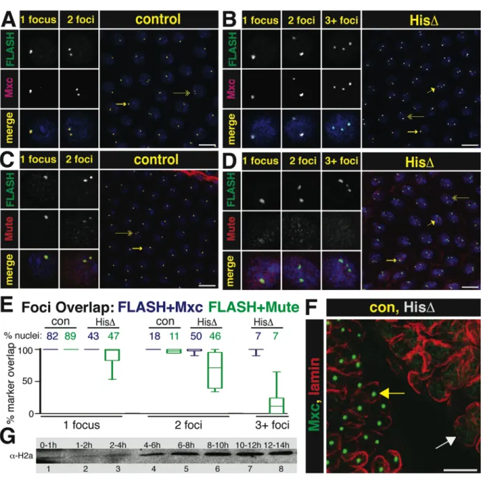
positive if all FLASH foci co localized with the other marker. Nuclei contained 1, 2 or 3+ foci and the graph presents the percent of total overlap out of the total number of nuclei counted for each of these classes within a genotype for either FLASH/Mxc or FLASH/Mute. The results are presented as a box (25th -75th quartiles) and whiskers (10-90th percentile) plot. (GraphPad Software, La Jolla California, USA)
Table 2.1. Boundaries for Each Histone Locus Construct
Construct Boundaries
HL-FL F: ggtacctaatgcatatgtggcgaggccatgtgttaactgaagaatgtgt R: ccatgtgttaactgaagaatgtgttctagatgtcgaagtttgcttgaagtg
HLT-FL See HL-FL
H3-H4 F: ggtaccaccaataaaattaatact R: tctagaaaagttataaatagtcggcaac H2a-H2b F: ggtacctcatattcgatgattggt
R: tctagattacaacaaattgccaagcta H1 F: ggtaccgccgactatttataacttta
R: tctagagttttattgttgctgcgaac H3-H4PS See H3-H4
H2a-H2bPS See H2a-H2b
H3-H4P H3 5’UTR: attgtgttttcaaacgtgaagtagtgaacgtgaactttagtgaaacccaaatcgg H4 5’UTR: ttcactgttctatactattatacacgcacagcacgaaagtcactaaagaactaatt CORE F: ggtaccgtttcatgtcatgaattac
R: tctagaaaagttataaatagtcggcaac CORETATA See CORE
29
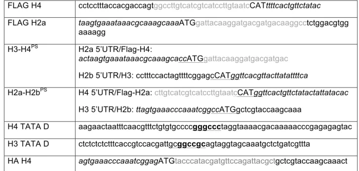
FLAG H4 cctcctttaccacgaccagtggccttgtcatcgtcatccttgtaatcCATttttcactgttctatac
FLAG H2a taagtgaaataaacgcaaagcaaaATGgattacaaggatgacgatgacaaggcctctggacgtgg aaaagg
H3-H4PS H2a 5’UTR/Flag-H4:
actaagtgaaataaacgcaaagcaccATGgattacaaggatgacgatgac
H2b 5’UTR/H3: cctttccactagttttcggagcCATggttcacgttacttatattttca
H2a-H2bPS H4 5’UTR/Flag-H2a: cttgtcatcgtcatccttgtaatcCATggttcactgttctatactattatacac H3 5’UTR/H2b: ttagtgaaacccaaatcggccATGgctcgtaccaagcaaa
H4 TATA D aagaactaatttcaacgtttctgtgtgccccgggccctaggtaaaacgacaaaaacccgagagagtac H3 TATA D ctctctctctttcaccgtccacgattgcggccgcagtaggtagcaaatgctctgatcgttta
HA H4 agtgaaacccaaatcggagATGtacccatacgatgttccagattacgctgctcgtaccaagcaaact
The following features are annotated in the sequences: engineered restriction sites are underlined; tags are grey; UTR sequence is italicized; TATA mutations are bold.
Table 2.2. Primers used in RT-PCR
Primer Name Sequence
H2a R 5’- gcagctaggtaaactggag-3’ H4 R 5’- gtaggtcacggcatcacg-3’ FLAG F 5’-gattacaaggatgacgatgacaag-3’ Actin F 5’- ggtcacgataccgtgctc-3’
Actin R 5’- aacggctctggcatgtg-3’
30
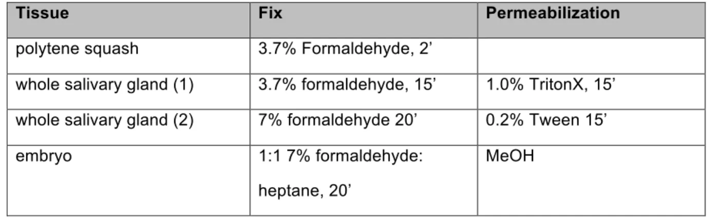
Table 2.3. Tissue Preparation for Antibody StainingTissue Fix Permeabilization
polytene squash 3.7% Formaldehyde, 2’
whole salivary gland (1) 3.7% formaldehyde, 15’ 1.0% TritonX, 15’ whole salivary gland (2) 7% formaldehyde 20’ 0.2% Tween 15’
embryo 1:1 7% formaldehyde:
heptane, 20’
MeOH
Table 2.4. Antibody Concentrations
Primary Antibody
Raised In Source Concentration Incubation
α-FLASH rabbit (Yang et al., 2009) 1:2000 4 C, overnight α-Mxc guinea pig (White et al., 2011) 1:2000 4 C, overnight α-Mute guinea pig (Bulchand et al.,
2010)
1:2000 4 C, overnight
α-MPM-2 mouse Millipore 1:2000 4 C, overnight
C1A9 α-HPI mouse Developmental
Studies Hybridoma Bank
1:1000 4 C, overnight
α-GFP chicken Upstate 1:1000 4 C, overnight
α-Lsm11 rabbit (Liu et al., 2006) 1:1000 room
temperature, 2h Secondary
Antibody
Recognizes Source Concentration Incubation
Alexa-488 rabbit-IgG Invitrogen 1:2000 room
temperature, 2h
Cy5 guinea pig IgG Jackson 1:1000 room
temperature, 2h
Cy3 mouse IgG Jackson 1:1000 room
31
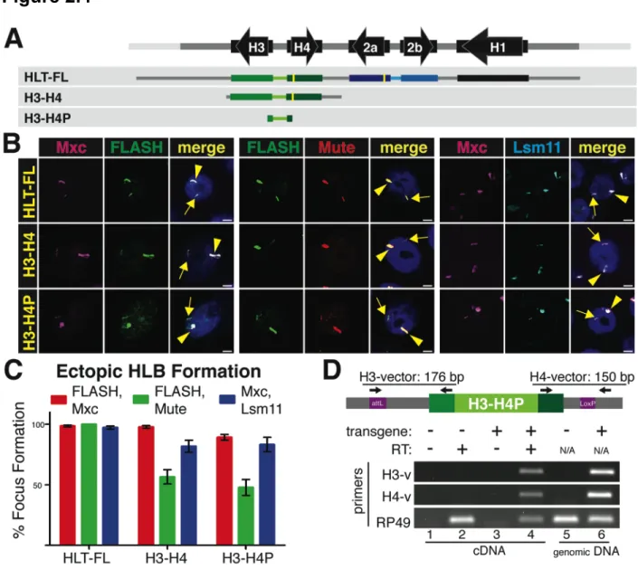
RESULTSAn HLB Can Assemble at a Single, Active Histone Gene Repeat
The Drosophila replication-coupled histone genes are present in a single locus on chromosome 2L as a tandem 5 kB repeat present in about 100 copies. Each repeat unit
contains one copy of each of the five histone genes (Fig. 2.1A). The H2a-H2b and H3-H4 gene pairs are divergently transcribed, while the H1 gene is located about 1.5 kB 3’ of the H3 gene, and ends about 300 nts before the 3’ end of the H2b gene (Fig. 2.1A). To determine whether sequences within a single repeat unit were sufficient to direct the formation of an HLB, we used a construct containing 1.2 copies of the repeat unit such that all contiguous sequences 500 nts long were represented in the construct (Histone Locus-Full Length, or HL-FL; Fig. 2.1A) and generated transgenes at specific loci in the Drosophila genome by ΦC31-mediated integration
(Bateman and Wu, 2007; Bischof et al., 2007) . To test whether the chromatin environment around the ectopic histone genes can influence expression, HL-FL was inserted into two specific sites: a euchromatic site on chromosome 3 (86Fb) and a heterochromatic site on chromosome 4 (102D).
32
We analyzed chromosome spreads from 3rd instar larval salivary gland cells. In these polyploid cells the genome reaches more than 1000C and individual chromatids line up in register, resulting in polytene chromosomes that provide high resolution for cytological experiments (Fig. 2.1B). Using antibodies to Lsm11, Mute, and FLASH, we observed HLB assembly at the ectopic HL-FL locus at 86Fb on chromosome 3, as well as at the endogenous histone locus at 39D-E on chromosome 2 (Fig. 2.1B). In contrast, when the repeat was located at 102D on chromosome 4, HLB assembly was not observed (Fig. 2.1C), although its genomic presence was confirmed by PCR (not shown). We conclude that one copy of the histone repeat is sufficient to assemble an HLB at a euchromatic but not a heterochromatic site.
34
Figure. 2.1. An HLB forms at an ectopic locus containing one histone gene repeat unit. A. Diagram of the histone repeat (chromosome 2). The 5.1 kB histone repeat unit is indicated by parentheses. A fragment containing 1.2 repeat units (HL-FL) was cloned and inserted into the Drosophila genome at either site 86Fb on chromosome 3, or at site 102D on chromosome 4. The yellow bars in the HLT-FL construct represent N-terminal FLAG tags in H2a and H4. B. Chromosome squashes from salivary glands of third instar larvae containing the HL-FL at 86Fb (left; n=15) or no transgene (right; n=7) stained with Mute (red), FLASH (green) and HP1 (pink, top panel) or Lsm11 (green) and HP1 (pink, bottom panel). The insets show a higher magnification of the 86Fb chromosome region except the panel with * which shows
chromosome 4 (102D). The arrowhead indicates the endogenous HLB and the arrow indicates chromosomal position 86Fb. Bars = 10 mm.
C. Chromosome 4 from salivary glands of 3rd instar larvae containing the HL-FL transgene at
position 102D (arrow; n=8) stained with HP1 (pink) and either FLASH (green, top) or Lsm11 (green, bottom). The endogenous histone locus near the chromocenter is indicated by the arrowhead.
D. RT-PCR analysis of H2a and H4 expression from HLT-FL located at 86Fb (chr3) and 102D (chr4) compared to no transgene (NT). Histone gene expression was normalized relative to the expression of actin mRNA. Error bars represent SEM.
Histone Gene Expression Correlates with HLB Assembly
The formation of HLBs at ectopic sites that expressed histone genes provided us with an opportunity to define sequences within the histone gene repeat that direct HLB assembly and histone gene expression, and to determine how these two processes are functionally related. We made transgenic flies with constructs inserted at 86Fb that contain only the H3-H4 gene pair, the H2a-H2b gene pair, or the histone H1 gene plus the long intergenic region between it and the 3’ end of the H3 gene (Fig. 2.2A). We assessed HLB formation by quantifying the presence of ectopic HLBs in intact salivary gland nuclei (Wagner et al., 2007) using multiple pairs of HLB markers (Fig. 2.2B). Ectopic HLBs were defined by co-localization of two or three HLB components in a focus (arrows, Fig. 2.2B), in addition to the endogenous HLB (arrowhead, Fig. 2.2B). For each experiment, ectopic HLBs were quantified using 1mm sections of a
35
HLT-FL supported HLB formation in nearly 100% of nuclei with all marker pairs (Fig. 2.2B,C). The H3-H4 construct formed ectopic HLBs in 40% to 95% of the cells depending on the marker pair examined (Fig. 2B,C). In contrast, less than 20% of cells containing either the H2a-H2b gene pair or the H1 gene formed an ectopic HLB, similar to non-transgenic controls (Fig. 2.2C). All four markers were present in the HLBs that formed on the H3-H4 gene pair, which was similar in size to the H2a-H2b or the H1 transgenes. These data indicate that a specific sequence(s) within the H3-H4 gene pair directs HLB assembly.
To test whether HLB assembly was important for histone gene expression, we developed an S1 nuclease protection assay using a 5’ end-labeled probe (P) containing the FLAG-tagged H2a or H4 genes (Fig. 2.2D). These probes detect both the endogenous (E) H2a and H4 histone mRNAs and the longer mRNAs produced by the transgenes (T) (Fig. 2.2D). This assay is quantitative and allowed us to determine the relative level of ectopic versus
endogenous histone mRNA accumulation by comparing signal intensities between the S1 nuclease protected fragments in each sample. To validate the assay, we analyzed varying amounts of total RNA isolated from dissected salivary glands, 3-6 hr old embryos (diploid cells), and ovaries and whole 3rd instar larvae (mixed diploid and polyploid cells). The ectopic H2a and
H4 genes in HLT-FL were expressed at ~7% the level of the endogenous genes in salivary glands, compared to 2.5% in ovaries and 1% in embryos and whole larvae (Fig. 2.2D). While the basis for these differences is not known, they may be due to under-replication of the endogenous histone genes relative to the rest of the salivary gland genome (Hammond and Laird, 1985). The relatively high expression of the ectopic histone mRNA as measured by S1 nuclease protection assay made salivary gland RNA the best source to carry out subsequent experiments.
36
38
Fig 2.2. The H3-H4 genes assemble an ectopic HLB.A. Diagram of the four constructs inserted into chromosomal location 86Fb. The yellow bars represent N-terminal FLAG tags in H2a and H4.
B. HLB assembly for each construct (indicated at left) was assessed by confocal microscopy of intact salivary gland nuclei stained with Mxc and FLASH (left), Mute and FLASH (center) or Mxc and Lsm 11 (right). The endogenous HLB contained Mxc (pink), FLASH (green), Mute (red) and Lsm11 (blue) in all samples (arrowhead). Note the assembly of an ectopic HLB with each marker for nuclei containing the HLT-FL or the H3-H4 transgenes (arrow). Scale bar indicates 10 mm.
C. Quantification of ectopic HLB formation. Error bars depict SEM.
D. Expression of histone mRNA from the HLT-FL transgene was assessed throughout development by 5’ S1 nuclease protection assay using a 32Pend-labeled (red star) probe (P) complementary to either the H2a or H4 endogenous and ectopic transcripts. Numbers above the gel indicate the amount of RNA (mg, except glands which were total number of glands) in each reaction. The S1 nuclease assay is diagrammed below the gel. Numbers indicate the length in nt of the probe (P), ectopic (T) and endogenous (E) protected H2a or H4 transcripts. The black triangle indicates nuclease cleavage of the probe at the point where the RNA (vertical dashed line below probe) is not complementary.
E. Expression of histone mRNA was assessed in salivary glands by 5’ S1 nuclease protection assay. Roman numerals indicate the transgene inserted in each sample (depicted in A). Note that ectopic histone expression (T) was detected from constructs carrying HLT-FL and H3-H4.
F. Relative histone mRNA expression was measured for H2a (light blue columns) and H4 (light green columns) by qRT-PCR and quantification of the S1 protection assay (dark columns). Both assays are presented as fold expression compared to HLT-FL, which was set at 1.0. Error bars depict SEM.
The H3-H4 Promoter is Necessary and Sufficient for HLB Formation
39
In addition to transferring the capability for HLB assembly, the promoter swap also resulted in expression of the H2a transgene as determined by S1 nuclease protection and qRT-PCR (Fig. 2.3D,E). The H2a gene in H2a-H2bPS was expressed at levels similar to the H4 gene in the H3-H4 transgene (about 15% of the intact repeat; Fig. 2.3D, E), while the H4 gene in H3-H4PS was no longer expressed. Note that the promoter swap results in a smaller protected fragment with the H2a probe (Fig. 2.3D, lane 4) because the chimeric H2a gene now contains the H4 5’UTR, causing the S1 nuclease to cleave at the end of the FLAG tag rather than at the end of the H2a 5’ UTR (Fig. 2.3D, diagram). Also note that with the overloading of the
41
Figure 2.3. The H3-H4 Promoter Assembles an Ectopic HLB.
A. Diagram of the five constructs inserted into chromosomal location 86Fb. The yellow bars represent N-terminal FLAG tags in H2a and H4. The promoter swap (PS) includes the bidirectional promoter and 5’UTR from each gene pair.
B and C. HLB assembly for the indicated constructs was assessed with the indicated markers and quantified as in Figure 2. V5 antibody was used to detect Lsm11 in a strain where V5-Lsm11 replaces the endogenous protein. Note that the H3-H4 promoter (H3-H4, H2a-H2bPS) assembles an HLB, regardless of the associated transcript.
D and E. Histone gene expression was assessed and quantified as in Figure 2. Numbers in the diagram indicate the length in nt of the probe and possible protected fragments. Roman
numerals refer to the depicted transgene, and NT is the no transgene control. Note robust histone mRNA expression from HLT-FL, H3-H4 (T) and H2a-H2bPS (T*)
mRNA Processing Signals are Dispensable for HLB Assembly