DROSOPHILA NEUROLIGIN 2 COORIDNATES PRE- AND POST-SYNAPTIC DEVELOPMENT, DIFFERENTIATION AND NEUROTRANSMISSION
Yu-Chi Chen
A dissertation submitted to the faculty of the Curriculum in Genetics and Molecular Biology at the University of North Carolina at Chapel Hill in partial fulfillment of the
requirement for the degree of Doctor of Philosophy
Chapel Hill 2012
Approved by:
Manzoor A. Bhat, Ph.D.
Stephen T. Crews, Ph.D.
Jay E. Brenman, Ph.D.
ii © 2012 Yu-Chi Chen
iii
ABSTRACT
YU-CHI CHEN: Drosophila Neuroligin 2 Coordinates Pre- and Post-synaptic Development, Differentiation and Neurotransmission
(Under the direction of Manzoor A. Bhat)
Many cognitive functions including emotion, attention, language, social behavior, learning and memory depend on proper synaptic connectivity in the brain. Synapses are specialized asymmetric cellular junctions responsible for communication between neurons. Synaptic adhesion molecules, neuroligins and their binding partners, neurexins, have been suggested to play an important role in bridging the pre- and post-synaptic machineries across the synaptic cleft. However, detailed molecular mechanisms of how neuroligins function at the synapse in vivo still remain unclear. Recently, neuroligins and neurexins have drawn increasing attention due to the link between mutations in human NEUROLIGINS and familial autism spectrum disorders (ASDs) (Jamain et al., 2003). Therefore, understanding the role of neuroligins at the synapse may not only improve our knowledge of how synapses are
organized but provide insights into the molecular basis of the pathology and etiology of ASDs.
iv
v
vi
ACKNOWLEDGEMENTS
vii
PREFACE
The second chapter of this dissertation has been recently published. I appreciate all the co-authors for their contribution to the manuscript.
viii
TABLE OF CONTENTS
LIST OF TABLES ...x
LIST OF FIGURES ... xi
CHAPTER 1 Introduction...1
1.1 Synapse structure ...1
1.2 Synaptic development and maturation ...3
1.3 Synaptic adhesion molecules ...5
1.4 Neuroligin ...6
1.5 Neurexin ...8
1.6 Function of neuroligins and neurexins ...9
1.7 Implications in neurological disorders ...11
1.8 Using Drosophila neuromuscular junction as a model system ...13
1.9 Overall Goal and Hypothesis ...14
CHAPTER 2 Drosophila Neuroligin 2 is Required Presynaptically and Postsynaptically for Proper Synaptic Differentiation and Synaptic Transmission ... 19
2.1 Introduction ... 19
2.2 Experimental Procedures ...21
ix
2.4 Discussion ...40
CHAPTER 3 Conclusion and Future Directions ...60
3.1 Conclusion ...60
3.2 Future directions ...62
x
LIST OF TABLES
xi
LIST OF FIGURES
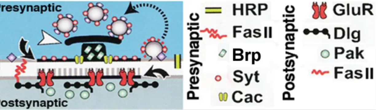
1.1 Diagram of trans-synaptic adhesion between neurexins and neuroligins ...15
1.2 Schematic displaying known components at the synapse of Drosophila NMJ ...16
1.3 Phylogenetic analysis of human, Drosophila, Apis and Caenorhabditis Neuroligin proteins. ...17
2.1. Generation of dnlg2 mutants ...46
2.2. Synaptic bouton growth at NMJs is reduced in dnlg2 mutants ...48
2.3. dnlg2 and dnrx mutants display similar NMJ developmental defects ...50
2.4. dnlg2 mutants display synapse differentiation defects with severely disorganized postsynaptic areas ...51
2.5. Dnlg2 forms a biochemical complex with Dnrx ...53
2.6. Dnlg2 is required pre- and post-synaptically for proper synaptic growth at NMJs ....54
2.7. Dnlg2 expression is required in pre- and post-synaptic areas for rescue of synaptic transmission defects in dnlg2 mutants ...56
S2.1 Genetic crossing scheme for the generation of dnlg2 null mutants ...58
CHAPTER 1
Introduction
1.1 Synapse structure
Our abilities to think, to process feelings and to memorize experiences all depend on the neural networks in our brain. These neural networks are composed of numerous neurons that interconnect with each other through highly specialized junctions called synapses (Li and Sheng, 2003). To insure that the signals are transduced from one neuron to the next in a proper direction, the structure of a synaptic junction is asymmetric. The presynaptic terminals contain synaptic vesicles that are filled with neurotransmitters. When an action potential arrives and induces the opening of Ca2+ channels, synaptic vesicles fuse with the plasma membrane and release neurotransmitters into the synaptic cleft. The receptors on the
postsynaptic membranes can bind to neurotransmitters, then transduce the signal and lead to series of events at the postsynaptic terminal (Sanes and Lichtman, 2001).
In order to perform this complex process, well-organized presynaptic and
postsynaptic machineries are required. Here, I will describe the synaptic apparatuses and the developmental process using the glutamatergic synapse at the vertebrate central nervous system (CNS) as an example (Fig. 1.1).
2
glutamatergic synapses, and proton pumps that are required for maintaining the
electrochemical gradient that drives neurotransmitter uptake. In addition, synaptic vesicles also contain proteins that are responsible for vesicle trafficking and fusion: Synaptotagmins serve as Ca2+ sensors, which translate the signal of Ca2+ influx into transmitter release. Rab3 aids the cycling and docking of the vesicles. Synapsin is involved in regulating the synaptic vesicle pool and vesicle recycling (Evergren et al., 2007). Synaptobrevin is part of the SNARE complex, a protein complex that directs the fusion of synaptic vesicle membranes to presynaptic plasma membranes. Vesicle fusion occurs when synaptobrevin binds to syntaxin and SNAP-25, two SNARE complex components on the plasma membrane.
The synaptic vesicle release site, named active zone, is a specialized region with protein complexes clustered at the presynaptic membranes. Ultrastructurally, the active zone is characterized by the electron-dense material on the presynaptic membrane directly
adjacent to the synaptic cleft. The synaptic active zone in vertebrate CNS is usually a disk of diameter 0.2–0.5 um (Südhof, 2012). The major components of active zones include RIM, Munc13, RIM-BP, α-liprin, ELKS, piccolo and bassoon. These proteins together form a large platform that clusters Ca2+ channels and enables docking, priming and recycling of synaptic vesicles and, therefore, play an important role in mediating synaptic plasticity (Owald and Sigrist, 2009; Südhof, 2012). α-liprin can interact with scaffolding protein complex, CASK/Mint1/Veli, which further connect the active zone matrix to synaptic adhesion molecules and actin cytoskeleton (Butz et al., 1998).
The postsynaptic terminal is equipped with machineries to receive the
3
postsynaptic terminal and form an electron-dense thickening at the postsynaptic membrane called the postsynaptic density (PSD), which is located directly opposed to the active zone (Sheng and Hoogenraad, 2007; Chen et al., 2008). The PSD of the glutamatergic synapses are composed of a large amount of proteins essential for propagating the signals: AMPA ( -amino-3-hydroxy-5-methyl-4-isoxazolepropionic acid) receptors are responsible for the fast synaptic transmission. NMDA (N-methyl-D-aspartate) receptors mediate synaptic plasticity. Metabolic glutamate receptors modulate synaptic activity and plasticity via signaling
cascades that involve G-proteins (Niciu et al., 2012). In addition, the PSD also contain signaling molecules, such as CaMKII, small GTPase and their regulators, which help transduce the signals from the receptors to the downstream effectors to regulate synaptic activity and morphology. The major scaffolding proteins at the PSD is PSD-95 family proteins which interact with synaptic adhesion molecules, signaling proteins and receptors as well as other scaffolding proteins which further link this whole complex to actin cytoskeleton (Sheng and Kim, 2011).
1.2Synaptic development and maturation
4
synapse (McAllister, 2007). Studies showed that presynaptic proteins are pre-assembled as multi-molecule complexes in the cytoplasm and transported to the nascent synapse in a saltatory fashion (Zhai et al., 2001; Shapira et al., 2003). Two types of these complexes were observed at the axonal growth cones. The complex that arrives first at the nascent synapse contains active zone proteins such as Piccolo, Bassoon, RIM, Synatxin, SNAP-25,
N-cadherin, Munc13, Munc18. Then, the next complex arriving at the synapse contains synaptic vesicle proteins, VAMP, voltage-gated Ca2+ channel, synapsin (Ahmari et al., 2000). Both of these precursor complexes reach at the nascent synapse prior to postsynaptic assembly. Synaptic scaffolding proteins, PSD-95, GKAP and Shank, appear to be the first postsynaptic protein complex assembled at the nascent synapse, followed by NMDA receptors, and then AMPA receptors (Washbourne et al., 2002, 2004; Li and Sheng, 2003; Gerrow et al., 2006; McAllister, 2007).
After the assembly of the key pre- and postsynaptic molecules, the newly-formed synapse gradually grows in size and changes its morphology. At the same time, the synapse strength is also increased mostly through the recruitment of more AMPA receptors (Li and Sheng, 2003). During maturation, these synapses then undergo activity-dependent
modification that refines the neural networks in response to external stimuli and activity levels. Therefore, active synapses are stabilized or strengthened, while others become silenced or eliminated (Scheiffele, 2003). Recent studies suggest that the activity-dependent synaptic plasticity is mediated by multiple mechanisms (Malenka and Bear, 2004). One of the most extensively studied mechanisms is the NMDA receptor-dependent synaptic
5
depolarized, which removes the Mg2+ block of NMDA receptors. Therefore, NMDA receptors can function as indicators for the timing of sequential synaptic activities. Upon the opening of NMDA receptors, Ca2+ influx activates CaMKII as well as many other
downstream signaling cascades, which then result in a series of events at the postsynaptic terminal, including phosphorylation and recruitment of AMPA receptors as well as remodeling of actin cytoskeletons and adhesion molecules, thus increasing the synaptic strength and altering the size and morphology of the synapse (Lamprecht and LeDoux, 2004; Malenka and Bear, 2004; Sheng and Hoogenraad, 2007). In addition, studies suggested that the glutamate release machinery at the presynaptic terminal can also be potentiated through the retrograde signaling mediated by trans-synaptic adhesion molecules (Choi et al., 2000; Zakharenko et al., 2002; Malenka and Bear, 2004). Synaptic plasticity allows adjustments of synaptic strength in response to different activity patterns, and thus, the neural networks in the brain can store information acquired from previous experiences. Therefore, synaptic plasticity has been a popular candidate mechanism mediating experience-dependent
development, learning and memory (Sheng and Hoogenraad, 2007; Citri and Malenka, 2008).
1.3Synaptic adhesion molecules
6
proteins. Thus, synaptic adhesion molecules not only can serve as molecular “glue” of the presynaptic and postsynaptic terminals (e.g. cadherins and syndecan, nectins, integrins), they have also been implicated in regulating the initial steps of synapse formation (e.g. ephrinB, EphB2, SynCAM, SYGs, sidekicks, Dscam and neurofascin), specifying synapse
connectivity, maintaining and aligning mature synapses (e.g. SynCAM, neuroligins, neurexins, LRRTMs and pentraxins) and modulation synaptic plasticity (e.g. N-cadherin, neuroligins, neurexins and NCAMs) (Yamagata et al., 2003; Waites et al., 2005; Craig et al., 2006; Sudhof, 2008; Tallafuss et al., 2010; Missler et al., 2012). Numerous studies tried to identify the roles of these molecules in specific steps of synapse development; however, it appears that each synaptic adhesion molecule involves in multiple processes throughout synapse development and maturation. Multiple redundant pathways could cooperate to organize synapse formation (McAllister, 2007). Among these pathways, neuroligins and their binding partners, neurexins, have received the most attention in this decade. Especially since mutations in human NEUROLIGINS were found in autism patients (Jamain et al., 2003), many attempts have been made to study their roles in synapse development and the underlying signaling events (Craig and Kang, 2007; Sudhof, 2008).
1.4Neuroligin
Neuroligins are a family of single-pass transmembrane proteins localized in the postsynaptic membranes in the mammalian central nervous system (CNS). Neuroligin 1 was first discovered as a ligand of neurexin (Ichtchenko, 1995; Song et al., 1999). Most
7
NEUROLIGIN 4Y or NEUROLIGIN 5 (Bolliger et al., 2001). Neuroligin-1 and -2 are
exclusively localized to the excitatory and inhibitory synapses respectively, while neuroligin-3 expresses in both excitatory and inhibitory synapses (Prange et al., 2004; Varoqueaux et al., 2004; Chih et al., 2005; Levinson et al., 2005; Budreck and Scheiffele, 2007). All
mammalian neuroligins consist of a single extracellular domain which is homologous to acetylcholinesterase (AChE), but are catalytically inactive due to changes in several crucial amino acids (Ichtchenko, 1995). Through this AChE-like domain, neuroligins form dimers and bind to neurexins (Fig.1.1). All neuroligins contain one alternative splicing site in the AChE-like domain (neuroligin 1 contains an additional splicing site), which regulate their binding affinities to neurexins (Ichtchenko et al., 1996; Boucard et al., 2005). The AChE-like domain is linked to the transmembrane domain by a glycosylated linker sequence.
Intracellularly, neuroligins contain the sites for interacting with other synaptic proteins. The tyrosine-based motif binds to gephyrin, a scaffolding protein at the GABAergic and
8
1.5Neurexin
Neurexins are a family of single-pass transmembrane proteins that were first
discovered as the receptors for α-latrotoxin, one of the components in the venom of the black widow spiders that leads to substantial neurotransmitter release from the presynaptic terminal (Ushkaryov et al., 1992). Unlike neuroligins, neurexins are mostly found in presynaptic terminals (Ushkaryov et al., 1992; Berninghausen et al., 2007). There are three neurexin genes reported in mammalian genomes, each containing two promoters driving the
expression of α- and β-neurexins (Ushkaryov and Südhof, 1993; Tabuchi and Südhof, 2002). Extracellularly, α-neurexins contain six LNS (laminin, neurexin, sex-hormone-binding globulin) domains that are interspersed by three EGF-like domains, while -neurexins only contain one LNS domain that are equivalent to the sixth LNS domain of α-neurexins. Although the binding properties are different, both - and -neurexins are capable of interacting with neuroligins through the last LNS domain of -neurexins and the only LNS domain of -neurexins in a Ca2+-dependent manner. Through the LNS domain, two
9
(Boucard et al., 2005; Chih et al., 2006; Comoletti et al., 2006). In addition to neuroligins, the extracellular region of neurexins can also bind to other postsynaptic adhesion molecules. LRRTMs (leucine rich repeat transmembrane proteins) have recently been identified as a binding partner of neurexins at excitatory synapses (de Wit et al., 2009; Ko et al., 2009). Similar to neuroligins, they can bind to both - and -neurexins. LRRTMs also interact with PSD-95 and they regulate the localization of AMPA receptors at the PSD and excitatory synaptic transmissions. In addition, neurexins can also interact with other postsynaptic binding partners, such as cerebellins, dystroglycan and neurexophilin (Missler et al., 1998; Sugita et al., 2001; Uemura et al., 2010). The cytoplasmic region of neurexins interacts with the synaptic vesicle protein synaptotagmin (Hata et al., 1993) and the CASK/MINT1/VELIs complex, which is coupled to synaptic vesicle exocytosis machinery and actin cytoskeletons (Hata et al., 1996; Butz et al., 1998; Biederer and Südhof, 2001).
1.6Function of neuroligins and neurexins
Since the discovery of the trans-synaptic neuroligins and neurexins complex, many efforts have been contributed to unravel their role during synaptic development and
10
overexpression of neuroligins or neurexins in cultured neurons results in increased synapse numbers (Prange et al., 2004; Chih et al., 2005; Chubykin et al., 2007). Specifically, overexpression of neuroligin 1 induces excitatory synapse formation, and neuroligin 2 induces inhibitory synapse formation (Prange et al., 2004; Chih et al., 2005; Chubykin et al., 2007). The above studies suggest the trans-synaptic neurexin-neuroligin complex may play an important role in the initiation of synapse formation (Dean and Dresbach, 2006; Chubykin et al., 2007; Missler et al., 2012). However, knockout mice studies show that neuroligins and neurexins may function at the mature synapse for proper synaptic transmission but are dispensable for the initial synapse formation (Missler et al., 2003; Varoqueaux et al., 2006; Chubykin et al., 2007). Neuroligin-1, 2 and 3 triple knockout mice have normal synapse density but display impaired synaptic function which leads to respiratory failure and postnatal lethality (Varoqueaux et al., 2006). A theory was then proposed to reconcile the discrepancies between the in vivo and in vitro studies: neuroligins and neurexins actually function to stabilize the transient immature synapses which then turn into long-lasting mature synapses (Sudhof, 2008; Missler et al., 2012). In support of this hypothesis, it was shown that the synaptogenic ability of neuroligins in cultured neurons is indeed activity-dependent (Chubykin et al., 2007) (Refer Table 1.1 for detail of the phenotypes). Interestingly, a recent article showed that reducing neuroligin-3, LRRTM-1 and 2 in cultured neurons from
11
neuroligins and neurexins have also been implicated in regulating synaptic plasticity and learning; however, further clarification is still needed to understand how they function during synaptic potentiation and what are the underlying mechanisms (Tabuchi et al., 2007; Kim et al., 2008; Etherton et al., 2009; Blundell et al., 2010; Dahlhaus et al., 2010; Choi et al., 2011; Shipman and Nicoll, 2012).
1.7Implications in neurological disorders
Defects in human NEUROLIGIN and NEUREXIN genes have been associated with neural developmental disorders and cognitive disease, such as autism spectrum disorders (ASDs) and schizophrenia. ASDs are a heterogeneous group of developmental disorders with diverse neuropsychiatric conditions, characteristics and etiology. They are one of the most common neural development diseases. Approximately 1%-2.6% of the human population is diagnosed as ASDs (Kogan et al., 2009; Kim et al., 2011). The stereotypic symptoms of ASDs include impaired social interactions and communication skills, confined interests, and repetitive behavioral patterns. In some cases, these behavioral conditions are also
accompanied by epilepsy and cognitive defects (Schmitz and Rezaie, 2008; Zoghbi and Bear, 2012). Based on the severity and pattern of the neurobehavioral symptoms of the patients, ASDs are further classified as autism, Asperger syndrome, pervasive developmental disorder not otherwise specified (Lord et al., 2000; Pardo and Eberhart, 2007).
12
The association of NEUROLIGIN-3 and -4 and NEUREXIN-1 with ASDs were then further demonstrated by many large-scale screenings. The genetic defects reported include point mutations, translocations, internal deletions, frameshifts, copy number variation, large-scale deletions (Auranen et al., 2002; Laumonnier et al., 2004; Yan et al., 2005; Szatmari et al., 2007; Lawson-Yuen et al., 2008; Daoud et al., 2009; Ching et al., 2010; Bottos et al., 2011; Sanders et al., 2011; Vaags et al., 2012; Schaaf et al., 2012; Steinberg et al., 2012; reviewed in Lisé and El-Husseini, 2006; Schaaf and Zoghbi, 2011).
Interestingly, shank3, the binding partner of neuroligins, has also been associated with ASDs and related neurodevelopmental disorders (Durand et al., 2007; Moessner et al., 2007; Lawson-Yuen et al., 2008; Walsh et al., 2008; Sykes et al., 2009; Awadalla et al., 2010; Gauthier et al., 2010; Waga et al., 2011; Boccuto et al., 2012). Together, mutations in
NEUROLIGIN-3 and 4, NEUREXIN-1 and SHANK-3 as well as the chromosomal abnormalities at these loci can account for a significant proportion of hereditary ASDs (Sudhof, 2008; Schaaf and Zoghbi, 2011). A better understanding of the basic molecular mechanisms of these molecules may thus benefit the development of diagnostic and therapeutic strategies in ASDs.
13
learning and memory. Therefore, it leads to the hypothesis that the behavioral symptoms associated with ASDs originate from the imbalance of excitatory and inhibitory synaptic function (Prange et al., 2004; Chih et al., 2005; Lisé and Husseini, 2006; Levinson and El-Husseini, 2007; Sudhof, 2008; Etherton et al., 2009, 2011; Yizhar et al., 2011). Similarly, neuroligin-4 knockout mice also show defects in social interactions and communication (Jamain et al., 2008), further supporting the idea of using neuroligin mutants as the model system to study ASDs.
1.8Using Drosophila neuromuscular junction as a model system
Similar to the glutamatergic synapses in mammalian CNS, type I motor neurons of Drosophila NMJ also release glutamate as a neurotransmitter (Fig. 1.2) and therefore, has been a very well established model system to study synaptic structure and function (Jan and Jan, 1976a, 1976b; Ruiz-Canada and Budnik, 2006). Besides Dnrx and Dnlg1, many proteins at the Drosophila NMJ synapse have been identified as homologs of mammalian CNS synaptic proteins. For example, Drosophila Discs large (Dlg) is the homolog of mammalian PSD-95, the synaptic scaffolding protein interacting with mammalian neuroligins (Budnik et al., 1996). Interestingly, the Drosophila homolog of the p21-activated kinase (PAK),
14
1.9Overall Goal and Hypothesis
In this study, we aim to dissect the role of Drosophila Neuroligin (Dnlg2) at NMJ synapse and its interaction with other synaptic adhesion molecules. However, during the course of the study, Sun et al. (2011) also reported a null mutation of dnlg2 and concluded that loss of dnlg2 causes reduced bouton numbers at the larval NMJ and increased evoked junctional responses in the body wall muscle. Contradictory to this report, however, we found reduced synaptic transmission both in the dnlg2 mutants that were generated previously by Sun et al. (2011) and in the dnlg2 null mutants that we generated independently. We therefore determined to clarify the function of Dnlg2 by carefully examining the phenotypic consequences of both mutants and further studied the aspects of Dnlg2 function that remain unknown:
1. We have determined the role of Dnlg2 in NMJ bouton morphology, synaptic ultrastructure and synaptic transmission.
15
Figure 1.1Diagram of trans-synaptic adhesion between neurexins and neuroligins.
16
Figure 1.2Schematic displaying known components at the synapse of Drosophila NMJ
17
Figure 1.3Phylogenetic analysis of human, Drosophila, Apis and Caenorhabditis
Neuroligin proteins. Drosophila has four neuroligin genes. The branch lengths of the
18
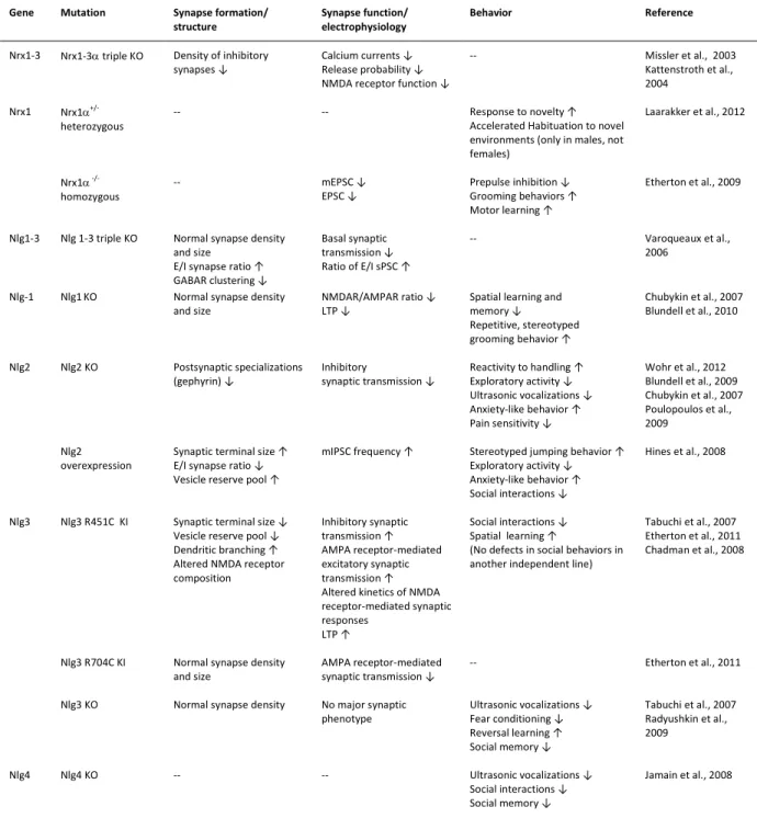
Table 1.1 Phenotypes of neuroligin and neurexin mutant mice
Gene Mutation Synapse formation/
structure
Synapse function/ electrophysiology
Behavior Reference
Nrx1-3 Nrx1-3triple KO Density of inhibitory
synapses ↓
Calcium currents ↓ Release probability ↓ NMDA receptor function ↓
-- Missler et al., 2003
Kattenstroth et al., 2004
Nrx1 Nrx1
+/-heterozygous
-- -- Response to novelty ↑
Accelerated Habituation to novel environments (only in males, not females)
Laarakker et al., 2012
Nrx1
-/-homozygous
-- mEPSC ↓
EPSC ↓
Prepulse inhibition ↓ Grooming behaviors ↑ Motor learning ↑
Etherton et al., 2009
Nlg1-3 Nlg 1-3 triple KO Normal synapse density
and size E/I synapse ratio ↑ GABAR clustering ↓
Basal synaptic transmission ↓ Ratio of E/I sPSC ↑
-- Varoqueaux et al.,
2006
Nlg-1 Nlg1KO Normal synapse density
and size
NMDAR/AMPAR ratio ↓ LTP ↓
Spatial learning and memory ↓ Repetitive, stereotyped grooming behavior ↑
Chubykin et al., 2007 Blundell et al., 2010
Nlg2 Nlg2 KO Postsynaptic specializations
(gephyrin) ↓
Inhibitory
synaptic transmission ↓
Reactivity to handling ↑ Exploratory activity ↓ Ultrasonic vocalizations ↓ Anxiety-like behavior ↑ Pain sensitivity ↓
Wohr et al., 2012 Blundell et al., 2009 Chubykin et al., 2007 Poulopoulos et al., 2009
Nlg2 overexpression
Synaptic terminal size ↑ E/I synapse ratio ↓ Vesicle reserve pool ↑
mIPSC frequency ↑ Stereotyped jumping behavior ↑
Exploratory activity ↓ Anxiety-like behavior ↑ Social interactions ↓
Hines et al., 2008
Nlg3 Nlg3 R451C KI Synaptic terminal size ↓
Vesicle reserve pool ↓ Dendritic branching ↑ Altered NMDA receptor composition Inhibitory synaptic transmission ↑ AMPA receptor-mediated excitatory synaptic transmission ↑ Altered kinetics of NMDA receptor-mediated synaptic responses
LTP ↑
Social interactions ↓ Spatial learning ↑
(No defects in social behaviors in another independent line)
Tabuchi et al., 2007 Etherton et al., 2011 Chadman et al., 2008
Nlg3 R704C KI Normal synapse density
and size
AMPA receptor-mediated synaptic transmission ↓
-- Etherton et al., 2011
Nlg3 KO Normal synapse density No major synaptic
phenotype
Ultrasonic vocalizations ↓ Fear conditioning ↓ Reversal learning ↑ Social memory ↓
Tabuchi et al., 2007 Radyushkin et al., 2009
Nlg4 Nlg4 KO -- -- Ultrasonic vocalizations ↓
Social interactions ↓ Social memory ↓
Jamain et al., 2008
CHAPTER 2
Drosophila Neuroligin 2 is Required Presynaptically and Postsynaptically for Proper Synaptic Differentiation and Synaptic Transmission
2.1 Introduction
Synapses are the fundamental units of neural networks and exhibit tightly apposed pre- and post-synaptic areas that are enriched in cell adhesion molecules (Giagtzoglou et al., 2009). A group of synaptic adhesion proteins thought to orchestrate formation of the pre- and postsynaptic structures are the Neuroligins (Nlgs) and their binding partners Neurexins (Nrxs) (Craig and Kang, 2007; Sudhof, 2008). A growing body of evidence associates these
molecules with Autism Spectrum Disorders (ASD), as mutations in human NLGs were discovered in ASD patients (Jamain et al., 2003; Szatmari et al., 2007). Nlgs are a family of transmembrane proteins with an extracellular domain that displays homology to
acetylcholinesterase (AChE) and localize to the postsynaptic membranes (Ichtchenko, 1995; Song et al., 1999). Nlgs form dimers and bind to Nrxs through this AChE-like domain. At the C-terminus, Nlgs have a PDZ (PSD-95, Dlg, and ZO-1) domain binding sequence motif which can interact with PDZ domain containing proteins (Song et al., 1999; Nourry et al., 2003) such as PSD-95 (Irie et al., 1997; Iida et al., 2004; Meyer et al., 2004).
20
numbers but defective synaptic transmission pointing to their role in synapse function (Missler et al., 2003; Varoqueaux et al., 2006), as opposed to synapse formation. To further analyze the Nlg/Nrx function, recent studies utilized Drosophila to circumvent the functional redundancy issues and address the function of these proteins in vivo (Li et al., 2007; Zeng et al., 2007; Banovic et al., 2010).
Genome analyses in Drosophila identify four Nlg-like proteins (CG31146, CG13772, CG34127, and CG34139) (Biswas et al., 2008; Banovic et al., 2010; Sun et al., 2011). We have been attempting to determine the role of CG13772 [Drosophila Neuroligin 2 (Dnlg2)], but during the final stages of preparation of this work, Sun et al., (2011) reported the
characterization of a null mutation in dnlg2. Here we report the generation of an independent null allele of dnlg2. We show that loss of Dnlg2 results in reduced synaptic development and neurotransmission. The synaptic function of Dnlg2 is only restored when Dnlg2 is expressed both pre- and post-synaptically at the NMJs, unlike what was reported (Sun et al., 2011). Furthermore, post-synaptic overexpression of Dnlg2 causes reduction in bouton growth, whereas combined pre- and post-synaptic overexpression leads to synaptic bouton
21
2.2 Experimental Procedures
Cloning of dnlg2 Full-Length cDNA
A PCR fragment was amplified from fly genomic DNA based on sequence homology with the vertebrate Neuroligin-1. The PCR fragment was radiolabeled to screen a Drosophila 0–20 hr embryonic cDNA library. Overlapping partial cDNA clones were isolated,
sequenced, and compiled as into a full-length cDNA sequence of 4195 base pairs encoding an open reading frame of 1248 amino acids. This cDNA corresponds to dnlg2. The GenBank accession number of dnlg2 sequence is AAF52450.
In Situ Hybridization
PCR amplified DNA fragments from the 3’region of dnlg2 cDNA were amplified and labeled with digoxigenin-UTP (Roche) as sense and anti-sense probes and used for in situ hybridization following standard protocols (Kearney et al., 2004).
Production and purification of Dnlg2 Antibody
22
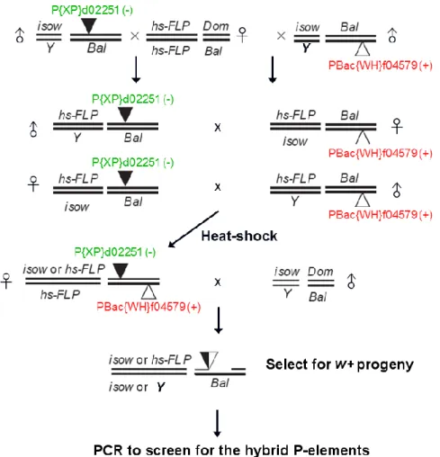
Generation of dnlg2 Mutants
dnlg2 null alleles were generated by targeted deletion using FLP-FRT recombination (Parks et al., 2004; Thibault et al., 2004). A P insertion upstream of the dnlg2 genomic locus, P{XP}d02251, and a piggyBac insertion downstream of dnlg2 locus, PBac{WH}f04579 were selected. The males from P{XP}d02251 and PBac{WH}f04579 were individually crossed to virgin females bearing FLP recombinase. Male progeny carrying both P{XP}d02251 and FLP recombinase were crossed to females carrying PBac{WH}f04579 and FLP recombinase. After 2 days of egg laying, the parents and progeny were both heat-shocked at 37°C for 1 hour. On the 3rd day, the parents were removed and the progeny were heat-shocked for 1 hour each day for 4 more consecutive days. After eclosion, mosaic virgin females were mated with yw;L/CyO males. The red-eye progeny males were individually crossed to yw;L/CyO virgin females to obtain balanced stocks which were analyzed for dnlg2 deletion by PCR. The following primers were used to verify the targeted deletion of dnlg2 locus and to determine the breakpoints of the deletion: TGCTGAGCGCAACAAGGACCA-3’,
CGGGTGAATCTCTCCCACTAA-3’, CCAAAGCTCCCGGATTTACC-3’,
5’-CTACGTAAAGACTCGGCCCCATTCAGC-3’, 5’-CTAACATCTCATCTGGGTCCTC-3’, GACCAGGAGATCAAGATCCGC-3’, CCGAGTCCAAGTCCAACTACA-3’, 5’-CGGTTTTGGAATTCTCTAGAAATCTCTTTA-3’.
23
Fly Stocks and Genetics
The same isogenized w1118 line used for outcrossing dnlg2 null allele served as the control for all analyses. P[acman]BAC CH322-173I20 (Venken et al., 2009), which carries the entire dnlg2 genomic locus, was used to generate transgenic flies using PhiC31 integrase-mediated site specific transgenesis (attP docking site at 68A4) (Bateman et al., 2006). The UAS-dnlg2 flies used in rescue experiments were provided by G. Boulianne (Sun et al., 2011). The dnrx null allele, dnrx273, was used for the genetic analyses in this study (Li et al., 2007). Df(3R)5C1 (referred in the text as Df) which uncovers the dnrx locus, has been described previously (Li et al., 2007). Gal4 lines used for Dnlg2 overexpression were: C57-Gal4 (Budnik et al., 1996) and 24B-Gal4 (Luo et al., 1994) (expressed mainly in the musculature), elav-Gal4 (expressed in all neurons) (Lin and Goodman, 1994) and tubP-Gal4 (expressed ubiquitously) (Lee and Luo, 1999). All stocks and crosses were raised at 21oC. For each set of experiments, all genotypes and crosses were transferred to fresh culture at the same time to maintain consistency. Other fly stocks were obtained from the Drosophila Stock Center, Bloomington, IN.
Immunostaining, Confocal Microscopy, and Bouton Number Quantification
24
Dnrx signal at the NMJ was detected by using the VECTASTAIN ABC system (Vector Laboratories) and Tyramide Signal Amplification (TSA, Invitrogen-Molecular Probes) (Li et al. 2007). Secondary antibodies conjugated to Alexa 488, 568, and 647
(Invitrogen-Molecular Probes) were used at 1:400. Fluorescence-conjugated anti-HRP (Jackson Immuno Labs) antibodies were used at 1:50.
Samples for each set of experiments were processed simultaneously, stained in the same tube and imaged with the same parameters using Olympus FV1000 confocal
microscope. Quantification of bouton numbers was performed at muscles 6/7 and muscle 4 of abdominal segment 3. Type Ib boutons at NMJ6/7 and at NMJ4 were visualized and
quantified by staining of body wall muscle preparations with anti-HRP and anti-Dlg. Quantification for bouton numbers was normalized to wild type.
Quantification of fluorescence intensity
25
objects” of Volocity. The areas with anti-Brp staining intensity at 10% to 100% were selected and the touching dots were separated using 0.03μm as the size reference.
Electron Microscopy and Morphometric Analysis
For ultrastructural NMJ studies, third-instar larval fillets were dissected at room temperature in ice-cold calcium free HL-3 medium (Stewart et al., 1994) containing 70 mM NaCl, 5 mM KCl, 20 mM MgCl2, 10 mM NaHCO3, 5 mM Trehalose, 5 mM HEPES, 115 mM Sucrose; pH 7.2 and subsequently fixed overnight in 4% paraformaldehyde/1% glutaraldehyde/0.1 M cacodylic acid (pH 7.2). Microwave irradiation (MWI) with the PELCO BioWave® 34700 laboratory microwave system was used for subsequent EM processing steps. After overnight fixation, the fixed fillets were additionally fixed at 640W with a cycle of 10 sec on, 20 sec off, 10 sec on, followed by 4x water rinses at 150W for 40s each, post-fixed with 1% aqueous osmium tetroxide 2x at 90W with a cycle of 2 min on, 2 min off, 2 min on under vacuum and placed on ice in between changes with additional 1 hour incubation on rotator, dehydrated in increasing ethanol concentrations 1x at 150W for 40s each, followed by propylene oxide 2x at 250W for 40s each. Samples were gradually
infiltrated with increasing resin to propylene oxide ratio up to full resin 2x at 250W for 3min each under vacuum. The samples were embedded in flat silicone mold with EMBED-812 resin and cured in the oven at 60oC.
26
SSR width was quantified as described in Budnik et al. (1996). Three to four different measurements were made from postsynaptic density (PSD) to distal SSR for each bouton. The SSR width was then calculated by averaging these measurements. To reduce the effect of bouton size, the averaged SSR width was further normalized by the diameter of the bouton (averaged SSR width / bouton diameter). The postsynaptic area was defined as the area between the PSD and the SSR. Only those active zones which clearly showed postsynaptic area were measured. (N represents the number of boutons analyzed while n is the number of active zones).
Electrophysiology
27
muscle for analysis. Miniature EJPs (mEJPs) events were collected for 5 minutes. Both EJPs and mEJPs were amplified with an Axonclamp 2B amplifier in bridge mode under the control of Clampex 8.2 (Axon Instruments Inc). All experiments were performed at room temperature (20oC–22oC).
EJPs and paired-pulse stimulation were analyzed with pClamp 9.2 software (Axon Instruments). mEJPs was analyzed using the Mini Analysis Program (Synaptosoft Inc., Decatur, GA). Evoked EJP amplitude was corrected by using nonlinear summation (Feeney et al., 1998). The quantal content of evoked release was calculated from individual muscles by ratio of the averaged EJP and averaged mEJP amplitude. Statistical analyses of EJP and mEJPs between genotypes were made using Student’s t test (SigmaPlot 10.0, Systat software Inc.).
Immunoprecipitation and Immunoblotting Analysis
The immunoprecipitation (IP) experiments were carried out as previously described (Banerjee et al., 2010). Briefly, fly heads of desired genotypes were homogenized using a glass homogenizer in a weight/volume ratio of 1:3 in ice cold lysis buffer containing 50mM HEPES (pH 7.2), 100mM NaCl, 1mM MgCl2, 1mM CaCl2 and 1% NP-40 with protease
28
immunocomplexes in 30µl of PBS/SDS buffer and resolved on SDS-PAGE for
29
2.3 Results
Generation of dnlg2 null mutants
The domain structure of Dnlg2 is similar to that of mammalian Nlg1. The extracellular domain contains an N-terminal signal peptide and an acetylcholinesterase-like (AChE) domain. This is followed by a transmembrane domain (TM) and a cytoplasmic region with a PDZ binding motif (PBM) (Fig. 2.1A). The AChE domains of Drosophila Dnlg2 and human Nlg1 (NCBI Reference Sequence: NP_055747.1) (Saus et al., 2010) share ~36% amino acid sequence identity and ~56% similarity (Fig. 2.1A). To determine the dnlg2 expression in the Drosophila we performed in situ hybridization in embryos. A dnlg2 probe recognizing the transmembrane region revealed that dnlg2 is primarily expressed in the ventral nerve cord (VNC) and the brain of stages 14-16 embryos (Fig. 2.1B). In addition, Dnlg2 expression is also observed at low levels in the embryonic musculature (data not shown).
30
Next we generated antibodies against Dnlg2 to determine its subcellular localization in the third instar larvae. Immunostaining using anti-Dnlg2 and anti-Bruchpilot (Brp), a marker for presynaptic active zones (Wagh et al., 2006; Weyhersmüller et al., 2011) indicates that Dnlg2 and Brp are localized to CNS synapses of the VNC (Fig. 2.1E). No staining was observed in dnlg2 mutants (Fig. 2.1F). To determine whether Dnlg2 is present pre- and/or post-synaptically at larval NMJs, we carried out immunostaining of 3rd instar larval musculature. Despite generating 11 antibodies against Dnlg2, we were unable to detect Dnlg2 at NMJs. Although Sun et al. (2011) reported that Dnlg2 localizes post-synaptically at the larval NMJs, we were not able to detect NMJ labeling using the anti-Dnlg2 with the protocol reported by Sun et al. (2011). We thus conclude that Dnlg2 levels at the larval NMJs are too low to be consistently detected.
31
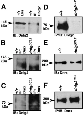
higher than that of the 145kDa band. The 145kDa molecular weight of Dnlg2 is slightly higher than that predicted from the open reading frame (~138kDa) and was not observed by Sun et al. (2011). These data show that dnlg2 is indeed a null allele.
Since our immunohistochemical analysis could not detect the presence of Dnlg2 at the wild type larval NMJ (arrows, Fig. 2.1I), we overexpressed the full length UAS-dnlg2 ubiquitously using tubP-Gal4 driver (Fig. 2.1J). Upon staining with Brp (red) and anti-Dnlg2 (green), we were able to detect anti-Dnlg2 at the NMJ synaptic boutons (Fig. 2.1J). In summary, our data indicate that Dnlg2 is a 145kDa protein and that it may undergo
proteolytic processing or degradation to form a 70kDa isoform. It can easily be detected in the synaptic-rich areas of the larval VNC, but its abundance at NMJs is probably very low.
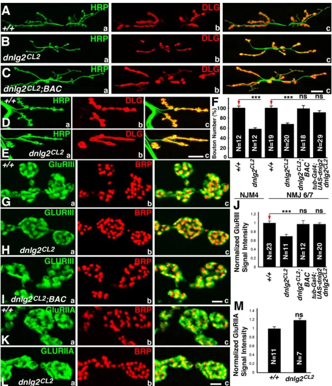
dnlg2 mutants exhibit a reduced number of boutons at larval NMJs
To determine if the NMJs were affected we performed immunostaining on the larval body walls of wild type and dnlg2 mutants using anti-HRP to identify neuronal membranes and anti-Dlg to label type I boutons (Fig. 2.2A) (Budnik et al., 1990; Lahey et al., 1994). As shown in Fig. 2.2Ba-c, in dnlg2 mutants, the number of boutons is severely reduced: they have fewer boutons at muscle 6/7 (NMJ6/7) (Fig. 2.2Ba-c) and muscle 4 (NMJ4) (Fig. 2.2Ea-c) when compared to wild type (Fig. 2.2A and D; quantified in Fig. 2.2F). This defect is caused by the loss of dnlg2 and/or CG13773 as this phenotype as well as other phenotypes (see below) are rescued with a genomic BAC (P[acman]BAC CH322-173I20; indicated by the green line in Fig.1D; Venken et al., 2009) that contains the entire genomic region of dnlg2 and CG13773 (Fig. 2.2C and 2.2F). However, CG13773 is not implicated as
wild-32
type levels in the dnlg2 excision mutants (Fig. 2.2F; also see later). The boutons in dnlg2 mutants (Fig. 2.1E) are less defined when compared to the wild type (Fig. 2.1C). The wild type synaptic boutons have a rounded to oval morphology and are separated from each other by a distinct neural process giving a beaded appearance (Fig. 2.2Ea) whereas the dnlg2 mutant boutons are not well separated (Fig. 2.2Eb). These data show that loss of Dnlg2 causes a reduction of boutons as well as an aberrant overall morphology.
To examine the distribution and localization of pre- and post-synaptic proteins at the dnlg2 mutant synapses, we performed immunostaining using anti-Brp (pre-synaptic) and anti-GluRIII, (post-synaptic) which labels one of the subunits of Drosophila glutamate receptors (Marrus et al., 2004). Although all active zones have both Brp and GluRIII punctae juxtaposed to each other (Fig. 2.2G-I), the level of GluRIII is reduced in dnlg2 mutants (Fig. 2.2Ha) compared to wild type (Fig. 2.2Ga). Quantification of the fluorescent intensity of GluRIII punctae suggests that there is a 30% decrease in dnlg2 mutants (Fig. 2.2J). This phenotype is also rescued by genomic BAC construct or by ubiquitous Dnlg2 overexpression using tubP-Gal4 driver in dnlg2 mutants (Fig. 2.2I, J; data not shown). However, staining with anti-Brp and anti-GluRIIA, another subunit of glutamate receptors, showed that there is a slight, but not statistically significant, increase in the level of GluRIIA in dnlg2 mutants (Fig. 2.2K-M). These studies suggest that Dnlg2 is required for proper synaptic development and proper postsynaptic protein assembly at the NMJs.
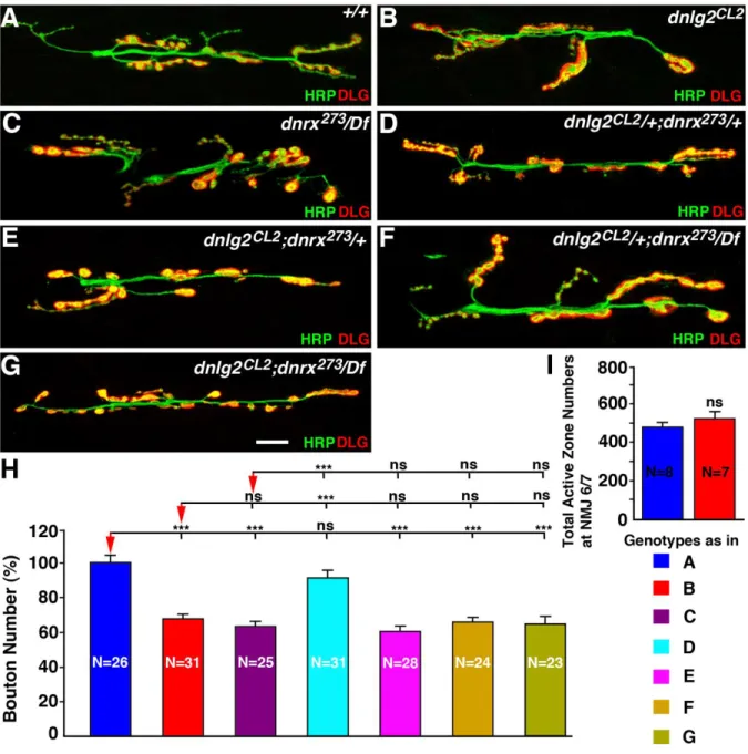
dnlg2 and dnrx affect NMJ morphology and function in a similar manner
33
presynaptic DNRX affects Dnlg1 clusters in the postsynaptic densities. However, it has been argued that dnrx and dnlg2 serve different functions at the NMJ as double mutants have a much more severe reduction in bouton number than either of the single mutants (Sun et al., 2011). To assess whether Dnlg2 and Dnrx serve similar or different functions at the NMJ synapses, we examined the morphology and the bouton numbers at the larval NMJs of dnlg2 and dnrx single and double mutants. Both dnlg2 (Fig. 2.3B) and dnrx/Df (Fig. 2.3C) single mutants are null mutations that display a significantly reduced number of boutons compared to their wild type counterpart (Fig. 2.3A, H). Larvae transheterozygotes for dnlg2+/-;dnrx +/-exhibit normal NMJ morphology (Fig. 2.3D, H) similar to the wild type (Fig. 2.3A, H). However, dnlg2-/-;dnrx+/- (Fig. 2.3E, H), dnlg2+/-;dnrx/Df (Fig. 2.3F, H) and dnlg2-/-;dnrx/Df (Fig. 2.3G, H) all display a similar reduction in bouton numbers as dnlg2 (Fig. 2.3B, H) and dnrx/Df single mutants (Fig. 2.3C, H). The differences in bouton numbers between these mutant genotypes (Fig. 2.3B-G) do not reach any statistical significance. Furthermore, the total active zone numbers as visualized by anti-Brp staining at NMJ6/7 did not show any significant difference between the wild type and dnlg2 mutants (Fig. 2.3I). In addition, whereas Sun et al. (2011) documented that dnlg2-/-;dnrx-/- are lethal, we find that our double null mutants are viable, further suggesting that loss of dnlg2 and/or dnrx do not exacerbate the phenotype of the other, consistent with the conclusion that both proteins affect the same molecular events and cause very similar phenotypes at the NMJs.
dnlg2 mutants exhibit synaptic differentiation defects at the NMJs
34
Bellen, 2004; Fouquet et al., 2009). Since dnlg2 mutants display synaptic growth defects at the NMJs (Fig. 2.2), we examined the ultrastructural features associated with the loss of Dnlg2 at synapses. We performed transmission electron microscopy (TEM) analyses on dnlg2 mutants. Cross sections of the wild type boutons show several active zones with characteristic T-bars surrounded by synaptic vesicles (Fig. 2.4A) (Mendoza-Topaz et al., 2008; Fouquet et al., 2009). A wild type synapse at a higher magnification shows an active zone (AZ), the post-synaptic area (PSA) and SSR (Fig. 2.4B). These NMJ synaptic boutons are embedded in the muscle and surrounded by specialized membrane folds, the SSR. Several defects were observed in dnlg2 mutants. dnlg2 mutant boutons exhibit an increased number of active zones in each bouton (Fig. 2.4C). Interestingly, the space between
postsynaptic density and the SSR, the PSA, is increased in dnlg2 mutants (Fig. 2.4C, D; quantified in J). In addition, we find that the width of SSR is severely reduced in dnlg2 mutants. All these phenotype are rescued by introduction of a BAC construct (P[acman]BAC CH322-173I20) that contains the genomic region of dnlg2 (Fig. 2.4E; quantified in Fig. 2.4H-K).
35
Dnlg2 and Dnrx form a molecular complex
The morphological analyses presented in the preceding sections indicate that Dnlg2 and Dnrx function together to coordinate synaptic growth at the NMJs. To test if Dnlg2 and Dnrx are present in the same molecular complex, we performed immunoprecipitations (IP)
followed by immunoblot analyses using Dnlg2 and Dnrx antibodies. When anti-Dnlg2 antibodies were used for IP in wild type adult fly head extracts, we were able to IP the 145 kDa Dnlg2 protein (Fig. 2.5A). When anti-Dnrx antibodies were used for IP in adult wild type and dnlg2 fly heads, the anti-Dnlg2 antibody detected the 145kDa Dnlg2 protein in the IP complex (Fig. 2.5B) of wild type but not dnlg2. Interestingly, in the same blot, the 70kDa Dnlg2 could not be detected in both the wild type and dnlg2 IP complex (Fig. 2.5C,
arrowhead). These results show that Dnlg2 (145kDa) and Dnrx are present in the same molecular complex. When fly head lysates from wild type and dnlg2 mutants were immunoprecipitated using anti-Dnlg2 antibodies, the Dnlg2 (70kDa) was abundantly detected in the wild type but not in the dnlg2 mutants (Fig. 2.5D). To further determine whether loss of Dnlg2 had any effect on the protein stability and levels of Dnrx, we
36
Dnlg2 is required pre- and post-synaptically for synaptic development at NMJs
Vertebrate studies have shown that Nlgs that are expressed post-synaptically interact with Nrxs expressed exclusively pre-synaptically (Song et al., 1999; Chih et al., 2005; Nam and Chen, 2005; Sudhof, 2008; Wittenmayer et al., 2009). These conclusions were
challenged as Nrxs were also observed to be expressed post-synaptically pointing to a complex mechanism of interactions between Nrxs and Nlgs in synapse function and
37
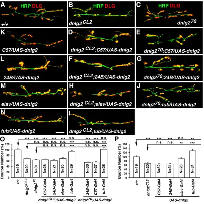
Sun et al. (2011) previously reported that Dnlg2 functions post-synaptically and that dnlg2 mutant phenotypes at the NMJs are fully rescued by post-synaptic expression of Dnlg2. However, we failed to rescue their dnlg2K070 mutants (Sun et al., 2011) by post-synaptic expression of Dnlg2 using 24B-Gal4 and C57-Gal4 (Fig. 2.6E, G, O). Together, our data indicate that pre- or post-synaptic expression alone of Dnlg2 is not sufficient to rescue dnlg2 mutant NMJ phenotypes; rather Dnlg2 is required pre- and post-synaptically for proper bouton formation.
Several vertebrate studies have shown that overexpression of Nlgs is sufficient to promote synapse formation in cultured mammalian neurons (Scheiffele et al., 2000;
Comoletti et al., 2003; Prange et al., 2004). We therefore assessed whether overexpression of Dnlg2 in the wild type animals affected normal bouton growth at NMJs. Surprisingly, post-synaptic overexpression of Dnlg2 using (C57-Gal4 and 24B-Gal4) reduced bouton numbers to levels similar to those observed in dnlg2 mutants (Fig. 2.6K and L; quantified in Fig. 2.6P). However, pre-synaptic overexpression of Dnlg2 using elav-Gal4 had no effect on bouton growth (Fig. 2.6M; quantified in Fig. 2.6P). In contrast, when Dnlg2 was overexpressed both pre- and post-synaptically using tubP-Gal4, we observe an increase in bouton growth of about 27% when compared to wild type (Fig. 2.6N; quantified in Fig. 2.6O). Hence, Dnlg2 promotes bouton formation and synaptic growth at NMJs when expressed pre- and post-synaptically during development.
Synaptic transmission is reduced in dnlg2 mutants
38
examined the consequences of loss of Dnlg2 alone as well as the combined loss of Dnlg2 and Dnrx on synaptic transmission at the NMJs. We performed our electrophysiological analyses on muscle 6 of 3rd instar larval body walls and recorded the evoked excitatory junction potentials (EJPs) in 0.5 mM [Ca2+]o at 0.2 Hz. Both dnlg2CL2 and dnlg2KO70 mutants exhibit a
reduction in EJP amplitude which is rescued by the genomic BAC construct in dnlg2CL2 (Fig. 2.7A). Under identical conditions, dnrx mutants also have reduced EJP amplitudes,
consistent with previous reports (Zeng et al., 2007; Ching et al., 2010). Interestingly, dnlg2;dnrx double mutants show a similar reduction in EJP amplitudes as dnlg2 or dnrx single mutants, again suggesting that Dnlg2 and Dnrx function together at the synapse. We observed no significant changes in mEJP amplitudes in all mutant combinations when compared to control wild type (data not shown) and dnlg2CL2;BAC-Res (Fig. 2.7B). All mutant combinations revealed severely decreased quantal contents compared to wild type (data not shown) and the genomic BAC rescue of dnlg2 mutants (Fig. 2.7C). Interestingly, the total number of active zones at the NMJs on muscle 6/7 are comparable between wild type and dnlg2 mutants (Fig. 2.3I), indicating that dnlg2 mutants have a lower release probability due to synaptic structural alterations.
39
40
2.4 Discussion
Sequence analyses of the Drosophila genome revealed 4 neuroligin genes and
mutational analyses of two of these genes dnlg1 (Banovic et al. 2010) and dnlg2 (Sun et al., 2011) revealed that Dnlg1 and Dnlg2 are required independently for synaptic growth and function. Dnlg1 functions post-synaptically and is required for proper synaptic development and differentiation (Banovic et al, 2010). Dnlg2 was also shown to function post-synaptically (Sun et al., 2011), however, some of the previously reported functions of Dnlg2 are
inconsistent with the data presented here. We report the generation of mutations in dnlg2 and characterization of the associated phenotypes. We find that loss of dnlg2 causes a
developmental defect at NMJs, with reduced bouton numbers. This phenotype is fully rescued when dnlg2 was expressed pre- and post-synaptically, indicating that Dnlg2 is required in both pre- and post-synaptic compartments for normal synaptic growth.
Ultrastructural analyses revealed that dnlg2 mutants have significantly increased numbers of active zones and postsynaptic density length. However, the postsynaptic SSR width is reduced. Electrophysiological measurements revealed that dnlg2 mutants have reduced EJP amplitude, but normal mEJP amplitude, indicating a reduced release probability. Furthermore, dnlg2 and dnrx double mutants are viable and reveal phenotypes similar to dnlg2 and dnrx single mutants, indicating that dnlg2 and dnrx likely function in the same pathway to
coordinate synaptic development and transmission. Finally, our phenotypic rescue data using the Gal4/UAS system (Brand and Perrimon, 1993) suggest that Dnlg2 is required both pre- and post-synaptically for proper NMJ bouton growth, synapse structure and
41
Although some of our results are in agreement with published data on dnlg2, many of the results reported here are in disagreement with the data presented in Sun et al. (2011). First, it was reported that post-synaptic Dnlg2 expression alone is sufficient to rescue the dnlg2 mutant phenotypes. Using the dnlg2 mutant alleles reported in Sun et al. (2011) and post-synaptic Dnlg2 expression, we were unable to rescue the bouton growth phenotypes. Second, it was reported that EJP amplitudes are much increased in dnlg2 mutants. However we find that EJP amplitudes in both dnlg2 and dnlg2KO70 mutants are decreased and that both mutants exhibit a reduction in neurotransmitter release probability. Third, our biochemical studies support the existence of a ~145 kDa molecular weight Dnlg2 that based on protein
42
lethality observed in dnlg2/dnrx double mutants reported in Sun et al. (2011) could be attributed to contributions from the genetic background.
Drosophila Neuroligins and their role at the synapse
43
serves to fine tune and refine synapse organization as is revealed by ultrastructural analysis with increased number of active zones in the remaining boutons (Fig. 2.4). In the absence of Dnlg2 and Dnrx active zone number increase and the synaptic areas are significantly
increased suggesting that the mutants fail to prune away ectopic active zones and are unable to refine densities. dnlg1 mutants on the other hand lack post-synaptic differentiation at the synapses indicating that Dnlg1 and Dnlg2 perform distinct functions during synapse differentiation (Banovic et al., 2010). Interestingly post-synaptic expression of Dnlg1 and Dnlg2 repress bouton growth, implying that postsynaptic Dnlg1 and Dnlg2 may either interact and interfere with the functions of presynaptic proteins or dilute out functions of a key post-synaptic protein/s which is involved in normal bouton growth. How a single pre-synaptic Dnrx protein interacts with post-pre-synaptic Dnlg1 and pre- and post-pre-synaptic Dnlg2 to coordinate synaptic development remains unresolved.
Pre- and post-synaptic requirements of Neuroligins
Many studies have suggested that Nlgs primarily function as postsynaptic adhesion molecules and interact with pre-synaptic Nrxs (Song et al., 1999; Scheiffele et al., 2000; Berninghausen et al., 2007). However, there may be exceptions to the post-synaptic
44
data provide evidence in support of both a pre- and postsynaptic function of Dnlg2 in
synapse formation. We show that a full complement of boutons at dnlg2 mutant NMJs is only restored when Dnlg2 is expressed both pre- and post-synaptically. Expression of Dnlg2 only pre- or post-synaptically was not sufficient to restore bouton growth. Surprisingly,
overexpression of Dnlg2 in the post-synaptic areas in the wild type animals also leads to a reduction in bouton growth, almost similar to dnlg2 mutant levels. However, overexpression of Dnlg2 pre-synaptically did not result in such phenotypes. On the other hand, when Dnlg2 is expressed both pre- and post-synaptically in the wild type larvae, there is excess bouton growth at NMJs, similar to when Dnrx is overexpressed pre-synaptically (Li et al., 2007). These data suggest that a fine balance of the Dnlg2 protein levels is critical for normal bouton growth. It is possible that high levels of post-synaptic Dnlg2 may lead to an uncontrolled or untimely interaction with pre-or post synaptic proteins, such as Dnrx and DNlg1, respectively, and hinder bouton growth at NMJs, leading to phenotypes that are similar to dnlg2 or dnrx mutants. A recent study also suggested that some neuroligin functions are neurexin-independent and that neuroligins can form complexes with other proteins at the synapses (Ko et al., 2009). This raises the possibility that pre-synaptic and post-synaptic Dnlg2 functions are dependent on formation of homophilic interactions with itself or heterophilic interactions with other synaptic proteins across the synaptic cleft to organize bouton growth at NMJs. It would be of significant interest to determine how loss of Dnlg2 leads to increased active zones and how mechanistically these functions of Dnlg2 are linked with Dnrx and other synaptic proteins.
In summary, our results show that Neuroligin functions is required pre- and
45
et al. (2008) in C. elegans suggest that Nlgs have pre- and post-synaptic functions that may be required to counter balance the functions of Nrxs or other proteins during synaptic growth and modulation. It was recently suggested that post-synaptic Nrxs counter the functions of Nlgs to ensure that synapses do not form at random places. However, in our model, antagonistic functions are unlikely given the similarity in phenotypes between the two mutants. Other synaptic adhesion molecules, such as LRRTM2 (Ko et al., 2009) and the recently identified Teneurins (Mosca et al., 2012) as new interacting partners of Dnrx and Dnlg1, respectively, further add to the complexity of trans-synaptic interactions and synapse organization. In this context, the requirement of Dnlg2 in both the pre- and post-synaptic compartments raises interesting questions about how synaptic organization is fine-tuned, and how signaling pathways regulate the expression of pre- and post-synaptic proteins during synaptic development and maturation. Deciphering the signaling role of Nrxs and Nlgs at the Drosophila synapses coupled with structure/function analyses should provide a better
46
Figure 2.1. Generation of dnlg2 mutants.
(A) Protein domain structure of Drosophila Dnlg2 and human NLG1. Similar to human NLG1, Dnlg2 is composed of a signal peptide, an acetylcholine esterase-like (AChE) domain and a transmembrane (TM) domain followed by a PDZ-domain-binding motif (PBM) at the C-terminus. The percent amino acid identity (I) and similarity (S) between Dnlg2 and NLG1 in the AChE domains are indicated. (B) In situ hybridization of wild-type embryo at stage 16 using a dnlg2 labeled anti-sense probe shows mRNA expression in the ventral nerve cord (VNC, arrowhead,) and brain lobes (BL, arrowhead). (C) Genomic structure of dnlg2 and the flanking insertions, P{XP}d02251 in the 5’-end and PBac{WH}f04579 in the 3’-end. The arrows pointing down indicate the sites of insertion. The arrow in the dnlg2 locus shows the direction of transcription. dnlg2 null mutant was generated using FRT-based recombination. The deleted genomic region is shown by the red line. A genomic BAC construct,
P[acman]BAC CH322-173I20, spanning the region shown by green line was used to rescue the deletion. (D) PCR confirmation of the targeted deletion using different primer
47
48
Figure 2.2. Synaptic bouton growth at NMJs is reduced in dnlg2 mutants.
(A-E) Confocal images of NMJ6/7 (A-C) and NMJ4 (D, E) from abdominal segment 3 of 3rd instar larvae labeled with anti-HRP (green) and anti-Dlg (red). Compared to wild type NMJ6/7 (A), dnlg2 homozygous mutants (B) show reduced NMJ expansion and fewer
49
bouton number deficits in dnlg2 mutants are rescued by BAC transgene or by ubiquitous Dnlg2 expression using tubP-Gal4. (G-I) Confocal images of synaptic boutons at segment 3 NMJ6/7 labeled with postsynaptic marker, GluRIII (green) and active zone marker, Brp (red). The alignment of pre- and postsynaptic areas appears to be unaffected in dnlg2mutants (Hc). However, the levels of GluRIII in dnlg2mutants (Ha) are significantly reduced. This
50
Figure 2.3. dnlg2 and dnrx mutants display similar NMJ developmental defects.
(A-G) Confocal images of NMJ6/7 from abdominal segment 3 in 3rd instar larvae labeled with anti-HRP (green) and anti-DLG (red). Compared to control (w1118) (A), dnlg2 mutants (B), dnrx/Df mutants (C), dnlg2;dnrx+/- (E), dnlg2+/-;dnrx (F), dnlg2;dnrx double
52
Figure 2.4. dnlg2 mutants display synapse differentiation defects with severely
disorganized postsynaptic areas.
53
Figure 2.5. Dnlg2 forms a biochemical complex with Dnrx.
54
Figure 2.6. Dnlg2 is required pre- and post-synaptically for proper synaptic growth at NMJs.
(A-J) dnlg2 cDNA transgene rescue analyses at NMJ 6/7. Compared to two dnlg2 mutants, dnlg2CL2 (B) and dnlg2KO70(C, Sun et al., 2011), expression of Dnlg2 in muscles with C57-Gal4 (D, E) or 24B-Gal4 (F, G) failed to rescue bouton number deficits in both dnlg2CL2 and dnlg2KO70 mutants. Similarly, expression of Dnlg2 in neurons using elav-Gal4 (H) also failed to rescue the NMJ phenotype. However, when Dnlg2 was expressed ubiquitously with tub-Gal4 (J, I), the NMJ phenotype in both dnlg2CL2 and dnlg2KO70 mutants was restored to wild type levels. (K-N) dnlg2 overexpression analyses in the wild type background.
55
57
Figure 2.7. Dnlg2 expression is required in pre- and post-synaptic areas for rescue of
synaptic transmission defects in dnlg2 mutants.
(A-C) Quantification of electrophysiological analyses for dnlg2 and dnrx273 single and dnlg2;dnrx double mutants at muscle 6 of the third abdominal segment. dnlg2 and dnrx273 single and dnlg2;dnrx double mutants showed reduced EJP amplitudes (A) but normal mEJP amplitude (B). All the mutants have reduced quantal contents (C). Similarly, pre- (elav-Gal4) and post-synaptic (24B- or C57-Gal4) expression of Dnlg2 in dnlg2CL2 and dnlg2KO70
58
Supplemental Figures
Figure S2.1Genetic crossing scheme for the generation of dnlg2 null mutants.
FLP-FRT-based recombination was used to generate a site-specific deletion containing dnlg2 genomic locus (Parks et al., 2004; Thibault et al., 2004). In the presence of the hs-FLP recombinase, two FRT-carrying transposable insertions, P{XP}d02251 and
59
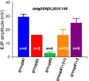
Figure S2.2 dnlg2/Df mutants display variable EJP amplitudes.
CHAPTER 3
Conclusion and Future Directions
3.1 Conclusion
In this study, we characterized the in vivo function of Drosophila neuroligin 2 and its interaction with Drosophila neurexins at the neuromuscular junction.
Multiple sequence alignments suggest that Dnlg2 is a homolog of mammalian neuroligins. At embryonic stage 16, the mRNA of Dnlg2 is localized at the central nervous system, the brain and the ventral nerve cord. Dnlg2 can be detected at the ventral nerve cord of the 3rd instar larvae, where a high density of synapses are localized, while the Dnlg2 at the NMJ bouton can only be detected when Dnlg2 is overexpressed. We identified two forms of Dnlg2, the 145kD full-length form and the 70kD cleaved form. Immunoprecipitation
analyses suggest that only the 145kD full length Dnlg2 can interact with Dnrx.
61
mutants revealed several structural defects, including increases in the length of postsynaptic densities and postsynaptic area as well as decreases in the width of the subsynaptic reticulum. These data indicate that although Dnlg2 is not required for the formation of active zones, it is responsible for proper organization of the synaptic structure.
Electrophysiological analyses show that dnlg2 mutants have reduced evoked
junctional potentials, but normal miniature junctional potentials; together, they suggest that the quantal content is decreased. Given that the total number of active zones at NMJ6/7 is normal, the reduced quantal content may result from a decrease in the release probability in response to presynaptic stimuli. Surprisingly, our rescue and overexpression analyses
indicate that Dnlg2 function at both pre- and post-synaptic terminals and that overexpression of Dnlg2 only at the postsynaptic terminal reduces bouton numbers. These results may imply that the balance of Dnlg2 at the pre- and post-synaptic terminals is important in regulating bouton growth and synaptic function.
In order to study whether Dnlg2 and Dnrx interact genetically, we generated dnlg2 and dnrx double mutants. Compared to dnlg2 and dnrx single mutants, the double mutants have very similar morphological, electrophysiological and ultrastructural phenotypes, which suggest that Dnlg2 and Dnrx cooperate in the same complex to organize NMJ synaptic development and organization.
62
underlying pathways which can in turn facilitate new diagnostic or therapeutic strategies for neural developmental disorders.
3.2 Future directions
Identifying functional domains of Dnlg2
63
endogenous Dnlg2. The function of each domain can then be assessed by testing their ability to rescue the phenotypes when expressed in a dnlg2 mutant background.
Cell culture studies tested the functions of the neuroligin domains by expressing truncated neuroligins in wild type neurons (Chih et al., 2005), although it is still not clear if the same mechanisms occur in vivo. This hypothesis can be easily tested by overexpressing different forms of truncated Dnlg2 in the wild type or the dnrx mutant background followed by NMJ phenotypic analyses. Similar studies have been performed for Dnlg1, in which the truncated Dnlg1, containing only the extracellular region, can disrupt the function of endogenous Dnlg1 in a dominant negative fashion (Banovic et al., 2010). Once all the truncations of Dnlg1, Dnlg2 and Dnrx are made, it will be interesting to test if Dnlg1, Dnlg2 and Dnrx interact with each other through their extracellular or intracellular regions by overexpressing truncated Dnlg1, Dnlg2 or Dnrx in the various combinations of dnlg1, dnlg2 and dnrx single or double mutant backgrounds.
The physiological role of the cleaved Dnlg2
64
is initiated by the activation of NMDA receptors and is dependent on synaptic activity. The resulting extracellular portion of neuroligin 1 is then released into the synaptic cleft which causes destabilization of neurexin1, reduces presynaptic release probability, and decreases synaptic transmission. In contrast, blocking this proteolytic process can lead to an increase in synaptic spine formation. The other product of the cleavage is the membrane-bound
intracellular region which is then processed by presenilin. It is possible that Dnlg2 also undergoes similar proteolytic cleavage as the mechanism to regulate synaptic development and activity. Interestingly, Drosophila also contains MMPs and therefore, the hypothesis could be tested by examining the Dnlg2 level and/or NMJ morphology when MMPs are reduced or inactive (Llano et al., 2000; Page-McCaw et al., 2003). Studying the mechanism in a model system that is easier to manipulate genetically could provide more novel insights.
In addition, the dominant negative effect of cleaved Dnlg2 may help explain the observation that postsynaptic Dnlg2 overexpression results in the reduction in bouton number in the wild type background. In this case, the enzymes responsible for this proteolytic process could be restricted to the postsynaptic membrane or the neighboring extracellular matrix because presynaptic Dnlg2 overexpression does not change the NMJ bouton number. Further biochemical and genetic analyses are required to test this hypothesis.
Potential modifiers of dnlg2
65
(Gruneberg, 1950; Hummel, 1958; Bykhovskaya et al., 2000; Nadeau, 2001). The dnlg2 mutants that are yet to be isogenized show variable EJP amplitudes (Fig. S2.2), implying that there are mutations or genetic variations in the modifier genes of Dnlg2. It is very likely that these mutations are present in the fly stocks carrying the transposable elements,
P{XP}d02251 and/or PBac{WH}f04579. At least some of the mutations appear to be dominant because the variation in EJP amplitudes was found in both dnlg2/dnlg2 and dnlg2/Df (Fig. S2.2 and data not shown). In addition, the dnlg2 mutants generated in this study are null mutants (Fig. 2.1), suggesting that these modifiers probably act downstream of Dnlg2. Therefore, performing a modifier screen will help identify the dominant enhancers or suppressors that affect the EJP amplitude of the dnlg2 mutants as well as the signaling pathways downstream of Dnlg2 (Gruneberg, 1950; Chen et al., 1998; Bykhovskaya et al., 2000; Nadeau, 2001; LaJeunesse et al., 2001; Johnston, 2002; Ward et al., 2003; Bilen and Bonini, 2007; Kaplow et al., 2007; Kucherenko et al., 2008; Ma et al., 2009; Fernandes and Rao, 2011). The modifier screen can be performed by crossing the dnlg2 mutants in various genetic backgrounds to the isogenized dnlg2 mutants followed by screening for the progeny with higher or lower EJP amplitude. The modifiers can then be mapped by sequencing the genome of the stock and comparing to the single nucleotide polymorphism maps (Johnston 2002).