Hybrid eye center localization using cascaded regression and
hand-crafted model fitting
Alex Levinshtein
1 [email protected]Edmund Phung
1 [email protected]Parham Aarabi
1,2 [email protected] 1ModiFace Inc., Toronto, Canada
2
Department of Electrical and Computer Engineering, University of Toronto, Toronto, Canada
July 5, 2018
Abstract
We propose a new cascaded regressor for eye center tection. Previous methods start from a face or an eye de-tector and use either advanced features or powerful re-gressors for eye center localization, but not both. Instead, we detect the eyes more accurately using an existing fa-cial feature alignment method. We improve the robustness of localization by using both advanced features and pow-erful regression machinery. Unlike most other methods that do not refine the regression results, we make the lo-calization more accurate by adding a robust circle fitting post-processing step. Finally, using a simple hand-crafted method for eye center localization, we show how to train the cascaded regressor without the need for manually an-notated training data. We evaluate our new approach and show that it achieves state-of-the-art performance on the BioID, GI4E, and the TalkingFace datasets. At an aver-age normalized error ofe < 0.05, the regressor trained on manually annotated data yields an accuracy of95.07%
(BioID),99.27%(GI4E), and95.68%(TalkingFace). The automatically trained regressor is nearly as good, yield-ing an accuracy of93.9%(BioID),99.27%(GI4E), and
95.46%(TalkingFace).
1
Introduction
Eye center localization in the wild is important for a vari-ety of applications, such as eye tracking, iris recognition,
and more recently augmented reality applications for the beauty industry enabling virtual try out of contact lenses. While some techniques require the use of specialized head-mounts or active illumination, such machinery is ex-pensive and is not applicable in many cases. In this paper, we focus on eye center localization using a standard cam-era. Approaches for eye center localization can be divided into two categories. The first, predominant, category con-sists of hand-crafted model fitting methods. These tech-niques employ the appearance, such as the darkness of the pupil, and/or the circular shape of the pupil and the iris for detection [3, 9, 10, 11, 16, 19, 20, 22, 24, 25]. These methods are typically accurate but often lack robustness in more challenging settings, such as low resolution or noisy images and poor illumination. More recently, a sec-ond category emerged - machine learning based methods. While there are approaches that train sliding window eye center detectors, recent success of cascaded regression for facial feature alignment [6, 18, 14, 26] has prompted the community to apply these methods for eye center localiza-tion [17, 21, 28]. These new methods have proven to be more robust, but they lack the accuracy of the model fit-ting approaches and require annotated training data which may be cumbersome to obtain.
In this paper, we propose a novel eye center detec-tion method that combines the strengths of the aforemen-tioned categories. In the literature on facial feature align-ment, there are two types of cascaded regression methods, simple cascaded linear regressors using complex features such as SIFT or HoG [4, 26] and more complex cascaded
…
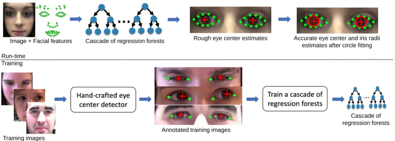
Image + Facial features Cascade of regression forests Rough eye center estimates Accurate eye center and iris radii estimates after circle fitting
Training images
Hand-crafted eye center detector
Annotated training images
Train a cascade of regression forests … Cascade of regression forests Run-time Training
Figure 1: Overview of our method. Top: At run-time an image and the detected facial features are used to regress the eye centers, which are then refined by fitting circles to irises. Bottom: Rather than training the regressor from manually annotated data, we can train the regressor on automatically generated annotations from a hand-crafted eye center detector.
regression forests using simple pairwise pixel difference features [6, 14]. Our first contribution is a new method for eye center localization that employs complex features
and complex regressors. It outperforms simple regres-sors with complex features [28] and complex regresregres-sors with simple features [17, 21]. Similar to [17, 21] our method is based on cascaded regression trees, but unlike these authors, following [6, 18, 14, 26], our features for each cascade are anchored to the current eye center esti-mates. Moreover, based on the pedestrian detection work of Dollaret al.[8] we employ more powerful gradient his-togram features rather than simple pairwise pixel differ-ences. Finally, while the aforementioned eye center re-gressors bootstrap the regression using face or eye detec-tors, given the success of facial feature alignment methods we make use of accurate eye contours to initialize the re-gressor and normalize feature locations. We show that the resulting method achieves state-of-the-art performance on BioID [1], GI4E [5], and TalkingFace [2] datasets.
The proposed cascaded regression approach is robust, but suffers from the same disadvantages of other discrim-inative regression-based methods. Namely, it is inaccu-rate and requires annotated training data. To make our approach more accurate, in our second contribution we re-fine the regressor estimate by adding a circle fitting
post-processing step. Employing robust estimation and prior knowledge of iris size facilitates sub-pixel accuracy eye center detection. We show the benefit of this refinement step by evaluating our approach on GI4E [5] and Talking-Face [2] datasets, as well as performing qualitative evalu-ation.
Finally, for our third contribution, rather than training our regressor on manually generated annotations we em-ploy a hand-crafted method to generate annotated data au-tomatically. Combining recent advances of eye center and iris detection methods, we build a new hand-crafted eye center localization method. It performs well compared to its hand-crafted peers, but is inferior to regressor-based approaches. Despite the noisy annotations generated by the hand-crafted algorithm, we show that the resulting re-gressor trained on these annotation is nearly as good as the regressor trained on manually annotated data. What is even more unexpected is that the regressor performs much better than the hand-crafted method used for training data annotation.
In summary, this paper proposes a new state-of-the-art method for eye center localization and has three main con-tributions that are shown in Figure 1. First, we present a novel cascaded regression framework for eye center lo-calization, leading to increased robustness. Second, we
add a circle fitting step, leading to eye center localization with sub-pixel accuracy. Finally, we show that by employ-ing a hand-crafted eye center detector the regressor can be trained without the need for manually annotated training data. This paper builds on the work that was presented in a preliminary form in [15].
2
Related Work
The majority of eye center localization methods are hand-crafted approaches and can be divided into shape and ap-pearance based methods. In the iris recognition literature there are also many segmentation based approaches, such as methods that employ active contours. An extensive overview is given by Hansen and Li [12]. Shape-based techniques make use of the circular or elliptical nature of the iris and pupil. Early methods attempted to de-tect irises or pupils directly by fitting circles or ellipses. Many techniques have roots in the iris recognition and are based on the integrodifferential operator [7] . Others, such as Kawaguchiet al.[13], use blob detection to extract iris candidates and use Hough transform to fit circles to these blobs. Toennieset al.[23] also employ generalized Hough transform to detect irises, but assume that every pixel is a potential edge point and cast votes proportional to gradient strength. Liet al.[16] propose the Startburst algorithm, where rays are iteratively cast from the cur-rent pupil center estimate to detect pupil boundaries and RANSAC is used for robust ellipse fitting.
Recently, some authors focused on robust eye center lo-calization without an explicit segmentation of the iris or the pupil. Typically, these are either voting or learning-based approaches. The method of Timm and Barth [22] is a popular voting based approach where pixels cast votes for the eye center based on agreement in the direction of their gradient with the direction of radial rays. A similar voting scheme is suggested by Valenti and Gevers [24], who also cast votes based on the aforementioned align-ment but rely on isophote curvatures in the intensity image to cast votes at the right distance. Skodras and Fakotakis [19] propose a similar method but use color to better dis-tinguish between the eye and the skin. Ahujaet al.[3] im-prove the voting using radius constraints, better weights, and contrast normalization.
The next set of methods are multistage approaches that
first robustly detect the eye center and then refine the es-timate using circle or ellipse fitting. ´Swirski et al.[20] propose to find the pupil using a cascade of weak classi-fiers based on Haar-like features combined with intensity-based segmentation. Subsequently, an ellipse is fit to the pupil using RANSAC. Wood and Bulling [25], as well as George and Routray [11], have a similar scheme but em-ploy a voting-based approach to get an initial eye center estimate. Fuhlet al.propose the Excuse [9] and Else [10] algorithms. Both methods use a combination of ellipse fitting with appearance-based blob detection.
While the above methods are accurate, they still lack robustness in challenging in-the-wild scenarios. The suc-cess of discriminative cascaded regression for facial fea-ture alignment prompted the use of such methods for eye center localization. [17, 21] start by detecting the face and initializing the eye center estimates using anthropo-metric relations. Subsequently, they use a cascade of re-gression forests with binary pixel difference features to estimate the eye centers. Inspired by the recent success of the SDM method for facial feature alignment Zhouet al.[28] propose a similar method for eye center localiza-tion. Unlike the original SDM work, their regressor is based on a combination of SIFT and LBP features. More-over, unlike [17, 21] who regress individual eye centers, Zhouet al.estimate a shape vector that includes both eye centers and eye contours. In line with this trend we de-velop a new regression-based eye center estimator, but additionally employ circle-based refinement and voting-based techniques to get an accurate detector that is easy to train.
3
Eye Center Localization
In this section, we describe our three main contributions in detail. We start by introducing our cascaded regres-sion framework for eye center localization (Section 3.1). Next, we show how the eye center estimate can be refined with a robust circle fitting step by fitting a circle to the iris (Section 3.2). Section 3.3 explains how to train the regres-sor without manually annotated eye center data by using a hand-crafted method for automatic annotation. Finally, in Section 3.4 we discuss our handling of closed or nearly closed eyes.
𝒄𝑅 𝒄𝐿
𝑬𝑖𝑛𝑡𝑒𝑟
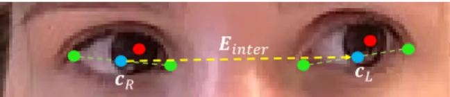
Figure 2: Normalized eye center locations. The eye cen-ters (red) coordinates are normalized such that the vector
Einter (yellow) between the two eye centerscR andcL (cyan) maps to the vector(1,0)T.
3.1
Cascaded regression framework
Inspired by the face alignment work in [6, 14], we build an eye center detector using a cascade of regression forests. Our shape is represented by a vector S = (xT
R,x T L), wherexRandxLare the coordinates of right and left eye centers respectively. Starting from an initial guessS0, we
refine the shape using a cascade of regression forests:
St+1=St+rt(I,St), (1) wherert is thet-th regressor in the cascade estimating the shape update given the imageIand the current shape estimateSt. Next, we describe our choice of image fea-tures, our regression machinery, and the mechanism for obtaining an initial shape estimateS0.
For our choice of image features, similar to Dollaret al.[8], we selected HoG features anchored to the cur-rent shape estimate. We find that using HoG is espe-cially helpful for bright eyes, where variation in appear-ance due to different lighting and image noise is more ap-parent, hurting the performance of regressors employing simple pixel difference features. Zhouet al.[28], also em-ploy advanced image features, but in contrast to them we use regression forests at each level of our cascade. Fi-nally, while [17, 21] estimate eye center positions inde-pendently, we find that due to the large amount of cor-relation between the two eyes it is beneficial to estimate both eyes jointly. In [28], the shape vector consists of eye centers and their contours. However, since it is possi-ble to change gaze direction without a change in eye con-tours, our shape vectorSincludes only the two eye center points.
To get an initial shape estimate, existing approaches use eye detectors or face detectors with anthropometric
relations to extract the eye regions. Instead, we employ a facial feature alignment method to get an initial shape estimate and anchor features. Specifically, the four eye corners are used to construct a normalized representa-tion of shapeS. We definecR andcL, to be the center points between the corners of the right and left eyes re-spectively. The vectorEinter between the two eye cen-ters is defined as the interocular vector with its magni-tudekEinterkdefined as the interocular distance. Figure 2 illustrates this geometry. The similarity transformation
T(x) maps points from image to face-normalized coor-dinates and is defined to be the transformation mapping
Einter to a unit vector aligned with theX axis with the
cRmapped to the origin. Therefore, the shape vectorS consists of two normalized eye centersxR=T(ximageR ) andxL =T(ximageL )withx
image
R andx
image
L being the eye center estimates in the image. The eye center esti-matescRandcL are also used to define the initial shape
S0= (T(cR)T, T(cL)T).
At each level of the cascade, we extract HoG features centered at the current eye center estimates.To make HoG feature extraction independent of the face size we scale the image by a factor s = Ehog
kEinterk, whereEhog is the constant interocular distance used for HoG computation. Using bilinear interpolation, we extractW ×W patches centered at the current eye center estimates sT−1(xR) andsT−1(xL), with W = 0.4Ehog. Both patches are split into4×4HoG cells with 6 oriented gradient his-togram bins per cell. The cell hishis-tograms are concate-nated and the resulting vector normalized to a unit L2
norm, yielding a96dimensional feature vector for each eye. Instead of using these features directly at the decision nodes of regression trees, we use binary HoG difference features. Specifically, at each decision node we generate a pool ofK(K= 20 in our implementation) pairwise HoG features by randomly choosing an eye, two of the96HoG dimensions, and a threshold. The binary HoG difference feature is defined as the thresholded difference between the chosen HoG components. During training, the feature that minimizes the regression error is selected.
To train the cascaded regressor, we use a dataset of an-notated images with eye corners and centers. To model the variability in eye center locations we use Principal Components Analysis. Using a simple form of Procrustes Analysis, each training shape is translated to the mean
shape and the resulting shapes are used to build a PCA basis. Subsequently, for each training image, multiple ini-tial shapesS0are sampled by generating random PCA co-efficients, centering the resulting shape at the mean, and translating both eyes by the same random amount. The random translation vector is sampled uniformly from the range−0.1,0.1
inX and−0.03,0.03
inY. The remaining parameters of the regressor are selected using cross validation. Currently, our regressor has 10 levels with200 depth-4trees per level. Each training image is oversampled50times. The regressor is trained using gra-dient boosting, similar to [14], with the learning rate pa-rameter set toν = 0.1.
3.2
Iris refinement by robust circle fitting
We refine the eye center position from the regressor by fitting a circle to the iris. Our initial circle center is taken from the regressor and the radius estimaterinitstarts with a default of0.1kEinterk. The iris is refined by fitting a circle to the iris boundaries. To that end, assuming the initial circle estimate is good enough, we extract edge points that are close to the initial circle boundary as can-didates for the iris boundary. Not all of the extracted edge points will belong to the iris boundary and our circle fit-ting method will need to handle these outliers.
Employing the eye contours once again, we start by samplingN points on the circle and removing the points that lie outside the eye mask. To avoid extracting the eye-lid edges we only consider circle samples in range±45◦
and135◦,225◦
. For each circle point sample we form a scan line centered on that point and directed toward the center of the circle. The scan line is kept short (±30%
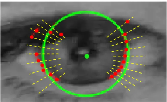
of the circle radius) to avoid extracting spurious edges. Each point on the scan line is assigned a score equal to the dot product between the gradient and outwards-facing cir-cle normal. The highest scoring point location is stored. Points for which the angle between the gradient and the normal is above25◦are not being considered. This pro-cess results in a list of edge points (red points in Figure 3).
Given the above edge points ei
N
i=1, the circle fitting
cost is defined as follows:
C(a, b, r) = N X i=1 q (eix−a)2+ (eiy−b)2−r 2 , (2)
Figure 3: Robust circle fitting for iris refinement. Starting from an initial iris estimate (green center and circle), short scan lines (yellow) perpendicular to the initial circle are used to detect candidate iris boundaries (red). A robust circle fitting method is then used to refine the estimate.
where(a, b)is the circle center andris the radius. How-ever, this cost is not robust to outliers nor are any priors for circle location and size being considered. Thus, we modify the cost to the following:
C2=w1· 1 N N X i=1 ρq(eix−a)2+ (eiy−b)2−r +w2· a−a0)2 +w2· b−b0)2 +w3· r−rdef ault)2. (3)
Note that the squared cost in the first term was con-verted to a robust cost (we choseρto be the Tukey ro-bust estimator function). The rest are prior terms, where
(a0, b0) is the center estimate from the regressor and rdef ault = 0.1kEinterk. We set the weights to w1 = 1, w2 = 0.1, w3 = 0.1and minimize the cost using the
Gauss-Newton method with iteratively re-weighted least squares. The minimization process terminates if the rela-tive change in cost is small enough or if a preset number of iterations (currently30) was exceeded. For the Tukey estimator, we start by setting its parameterC = 0.3rinit and decrease it toC= 0.1rinitafter initial convergence.
3.3
Using a hand-crafted detector for
auto-matic annotations
As mentioned in Section 2, there are a variety of hand-crafted techniques for eye center localization. Some methods work well in simple scenarios but are still falling short in more challenging cases. In this section, we con-struct our own hand-crafted method for eye center local-ization and use it to automatically generate annotations for a set of training images. The resulting annotations can be considered as noisy training data. One can imagine similar data as the output of a careless human annotator. We then train the cascaded regressor from Section 3.1 on this data. Since the output of the regressor is a weighted average of many training samples, it naturally smooths the noise in the annotations and yields better eye center estimates than the hand-crafted method used to generate the annotations. Next, we describe the approach in more detail.
Our hand-crafted eye center localization method is based on the work of Timm and Barth [22]. Since we are looking for circular structures, [22] propose finding the maximum of an eye center score functionS(c) that measures the agreement between vectors from a candidate center pointcand underlying gradient orientation:
c∗= arg max c S(c) = arg max c ( 1 N N X i=1 wc dTi gi 2 ) , (4) wherediis the normalized vector fromcto pointiandgi is the normalized image gradient ati.wcis the weight of a candidate centerc. Since the pupil is dark,wcis high for dark pixels and low otherwise. Specifically,wc = 255−
I∗(c), whereI∗is an 8-bit smoothed grayscale image. Similar to [3], we observe that an iris has a constrained size. More specifically, we find that its radius is about
20%of the eye size E, which we define as the distance between the two eye corners. Thus, we only consider pix-elsiwithin a certain range ofc. Furthermore, the iris is darker than the surrounding sclera. The resulting score function is: S(c) = 1 N X 0.3E≤kd∗ ik≤0.5E wcmax dTigi,0 , (5)
whered∗i is the unnormalized vector fromctoi.
Unlike [22] that find the global maximum of the score function in Eqn 4, we consider several local maxima of our score function as candidates for eye center locations. To constrain the search, we use a facial feature alignment method to obtain an accurate eye mask. We erode this mask to avoid the effect of eye lashes and eyelids, and find all local maxima ofS(c)in Eqn 5 within the eroded eye mask region whose value is above80%of the global maximum. Next, we refine each candidate and select the best one.
Since each candidate’s score has been accumulated over a range of radii, starting with a default iris radius of0.2E, the position and the radius of each candidate is refined. The refinement process evaluates the score func-tion in an 8-connected neighborhood around the current estimate. However, instead of summing over a range of radii as in Eqn 5, we search for a single radius that max-imizes the score. Out of all the 8-connected neighbors together with the central position, we select the location with the maximum score and update the estimate. The process stops when all the 8-connected neighbors have a lower score than the central position. Finally, after pro-cessing all eye center candidates in the above fashion, we select a single highest scoring candidate. Its location and radius estimates are then refined to sub-pixel accuracy us-ing the robust circle fittus-ing method from Section 3.2.
In the next step, we use our hand-crafted method to au-tomatically annotate training images. Given a set of im-ages, we run the facial feature alignment method and the hand-crafted eye center detector on each image. We anno-tate each image with the position of the four eye corners from the facial feature alignment method and the two iris centers from our hand-crafted detector. Finally, we train the regressor from Section 3.1 on this data. In Section 4 we show that the resulting regressor performs much bet-ter than the hand-crafted method on both training and test data, and performs nearly as well as a regressor trained on manually annotated images.
3.4
Handling closed eyes
Our algorithm has the benefit of having direct access to eye contours for estimating the amount of eye closure. To that end, we fit an ellipse to each eye’s contour and use its height to width ratiorto control our algorithm flow. For
examined, we apply both the regression and the circle fit-ting methods described in previous sections. For reliable circle refinement, a large enough portion of the iris bound-ary needs to be visible. Thus, for0.15< r≤0.3we only use the regressor’s output. Forr≤0.15we find even the regressor to be unreliable, thus the eye center is computed by averaging the central contour points on the upper and lower eyelids.
4
Evaluation
We perform quantitative and qualitative evaluation of our method and compare it to other approaches. For quan-titative evaluation we use the normalized error measure defined as:
e=1
dmax(eR, eL), (6)
whereeR,eLare the Euclidean distances between the es-timated and the correct right and left eye centers, anddis the distance between the correct eye centers. When ana-lyzing the performance, different thresholds oneare used to assess the level of accuracy. The most popular met-ric is the fraction of images for whiche ≤ 0.05, which roughly means that the eye center was estimated some-where within the pupil. In our analysis we pay closer at-tention to even finer levels of accuracy as they may be needed for some applications, such as augmented beauty or iris recognition, where the pupil/iris need to be detected very accurately.
While there are many public facial datasets available, many do not contain iris labels. Others may have iris only images or NIR images. For our analysis, we re-quire color or grayscale images containing the entire face with labeled iris centers. Therefore, we use the BioID [1], GI4E [5], and TalkingFace [2] datasets for evalua-tion. The BioID dataset consists of1521low resolution (384×286) images. Images exhibit wide variability in illumination and contain several closed or nearly closed eyes. While this dataset tests the robustness of eye cen-ter detection, its low resolution and the presence of closed eyes make it less suitable to test the fine level accuracy (finer levels than e ≤ 0.05). The GI4E and the Talk-ing Face datasets have1236and5000high resolution im-ages respectively and contain very few closed eye imim-ages.
Thus, we find these datasets to be more appropriate for fine level accuracy evaluation.
We implement our method in C/C++ using OpenCV and DLIB libraries. Our code takes4ms to detect both eye centers on images from the BioID dataset using a modern laptop computer with Xeon2.8GHz CPU, not including the face detection and face alignment time. The majority of this time is spent on image resizing and HoG feature computation using the unoptimized code in the DLIB li-brary and can be significantly sped up. Given the features, traversing the cascade is fast. For each4-level tree,3 sub-tractions and comparisons are needed to reach the leaf. The shifts (4values for the two iris center coordinates) in all the trees are added together to compute the final regres-sor output. This results in3×200×10 = 6000 subtrac-tions and comparisons, and4×200×10 = 8000floating point additions per image. The facial alignment method we use is based on [14] and is part of the DLIB library, but any approach could be used for this purpose. Simi-lar to previous methods, which rely on accurate face de-tection for eye center estimation, we require accurate eye contours for this purpose. To that end, we implemented a simple SVM-based approach for verifying alignment. Similar to previous methods, which evaluate eye center localization only on images with detected faces, we eval-uate our method only on images for which the alignment was successful. While the alignment is successful in the vast majority of cases, some detected faces do not have an accurate alignment result. After filtering out images without successful alignment we are left with1459/1521
images (95.9%) of the BioID dataset,1235/1236images of the GI4E dataset, and all the5000frames in the Talking Face dataset.
4.1
Quantitative Evaluation
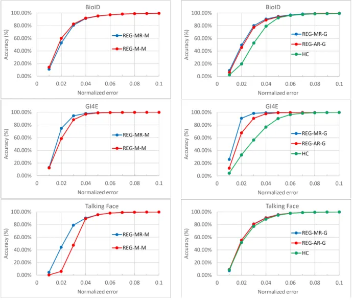
We evaluate several versions of our method. To evaluate against alternative approaches and illustrate the effect of circle refinement we evaluate a regressor trained on man-ually annotated data with (REG-MR) and without (REG-M) circle refinement. We also evaluate a regressor trained on automatically annotated data (REG-AR) and show how it compares toREG-MR, the hand crafted approach used to generate annotations (HC), and the competition. To eval-uate the regressors trained on manual annotations we use the MPIIGaze dataset [27] for training, which has10229
Refined/Unrefined comparison Manual/Auto/HC comparison
Figure 4: Quantitative evaluation. Left: Evaluation of regression-based eye center detector trained on manually annotated MPIIGaze data with (REG-MR-M) and without (REG-M-M) circle refinement. Right: Comparison of auto-matically trained regressor (REG-AR-G) to manually trained regressor (REG-MR-G) and the hand-crafted method (HC) used to generate automatic annotations.
cropped out eye images with eye corners and center an-notations. To testREG-AR, we need a dataset where the entire face is visible, thus we use the GI4E dataset with flipped images for training. Since GI4E is smaller than MPIIGaze, the regressor trained on it works marginally worse than the regressor trained on MPIIGaze, but
never-theless achieves state-of-the-art performance. We indicate the dataset used for training as a suffix to the method’s name (-M for MPIIGaze and -G for GI4E). Figure 4 left shows that for errors below 0.05, for the two high-res datasets (GI4E and Talking Face), circle refinement leads to significant boost in accuracy. This is particularly
ap-Method e <0.025 e <0.05 e <0.1 e <0.25 BioID Dataset REG-MR-M 68.13% 95.07% 99.59% 100% REG-M-M 74.3% 95.27% 99.52% 100% REG-MR-G 68.75% 94.65% 99.73% 100% REG-AR-G 64.15% 93.9% 99.79% 100% HC 36.26% 92.39% 99.38% 100%
Timm and Barth [22] 38%∗ 82.5% 93.4% 98%
Valenti and Gevers [24] 55%∗ 86.1% 91.7% 97.9%
Zhou et al. [28] 50%∗ 93.8% 99.8% 99.9% Ahuja et al. [3] N A 92.06% 97.96% 100% Markuˇs et al. [17] 61%∗ 89.9% 97.1% 99.7% GI4E Dataset REG-MR-M 88.34% 99.27% 99.92% 100% REG-M-M 77.57% 99.03% 99.92% 100% REG-AR-G 83.32% 99.27% 99.92% 100% HC 47.21% 90.2% 99.84% 100% Fuhl et al. [10] 49.8% 91.5% 97.17% 99.51%
George and Routray [11] N A 89.28% 92.3% N A
Talking Face Dataset
REG-MR-M 65.78% 95.68% 99.88% 99.98% REG-M-M 18.7% 95.62% 99.88% 99.98% REG-MR-G 71.56% 95.76% 99.86% 99.98% REG-AR-G 71.16% 95.46% 99.82% 99.98% HC 67.74% 94.86% 99.84% 99.98% ELSE [10] 59.26% 92% 98.98% 99.94% Ahuja [3] N A 94.78% 99% 99.42%
Table 1: Quantitative evaluation. Values are taken from respective papers. For [10], we used the implementation provided by the authors with eye regions from facial feature alignment. * = value estimated from authors’ graphs. Performance ofREG-MR-G on GI4E is omitted since GI4E was used for training. The three best methods in each category are marked withgreen,orange, andredrespectively.
parent on the Talking Face dataset, where the accuracy for e ≤ 0.025 increased from 18.70% to65.78%. For the BioID dataset, foregoing refinement is marginally bet-ter. This is in line with our previous observation, and is likely due to the low resolution and poor quality of the images. Our method achieves state of the art per-formance across all the three datasets. In particular, for
e≤0.05REG-MR-M(REG-M-M) achieves the accuracy of
95.07%(95.27%) on BioID,99.27%(99.03%) on GI4E, and95.68%(95.62%) on the Talking Face dataset.
Recall that the evaluation is restricted to images where
facial alignment passed verification. On GI4E and Talk-ing Face datasets combined, only one image failed verifi-cation. However, on BioID62images failed verification compared to only6images where a face was not detected. EvaluatingREG-MR-Mon all images with a detected face yields a performance of67.19% fore ≤ 0.025,94.19%
fore≤0.05,99.47%fore≤0.1, and100%fore≤0.25, which is only marginally worse than the method with fa-cial verification and still out-performs the competition. Future improvements to facial feature alignment will re-move this gap in performance. Table 1 summarizes the
results.
Next, we compare the performance of the automati-cally trained regressor (REG-AR-G) to the hand-crafted ap-proach that generated its annotations (HC), as well as to REG-MR-Gtrained on manually annotated GI4E data. The results are shown in Figure 4 right and are included in Table 1. Observe thatREG-AR-G outperforms the hand-crafted method on both the train (GI4E) and the test (BioID and Talking Face) sets. Moreover, on the test sets its performance is close toREG-MR-G.
4.2
Qualitative Evaluation
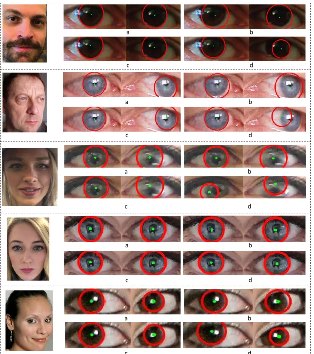
For qualitative evaluation we compare the performance ofREG-MR-G,REG-M-G,REG-AR-G, andHC. For consis-tency, all regressors were trained on GI4E. Figure 5 shows selected results. The first four examples illustrate the in-creased accuracy when using circle refinement. Further-more, the first three examples illustrate a failure ofHCon at least one eye whileREG-AR-G, which was trained using HC, is performing well. Finally, in the provided examples we observe no significant difference in quality between the manually and the automatically trained regressors, as well as their ability to cope with challenging imaging con-ditions such as poor lighting (1st and3rd images) and mo-tion blur (left eye in3rd image).
One failure mode of our approach is when the pupils are near the eye corners. This is especially true for the inner corner (left eye in the last example of Figure 5), where the proximity to the nose creates strong edges that likely confuse the HoG descriptor. The lack of training data with significant face rotation may also be a contributing factor. Training on more rotated faces, as well as using the skin color to downweigh the gradients from the nose, should help alleviate this issue.
5
Conclusions
We presented a novel method for eye center localiza-tion. State-of-the-art performance is achieved by local-izing the eyes using facial feature regression and then de-tecting eye centers using a cascade of regression forests with HoG features. Combining the regressor with a ro-bust circle fitting step for iris refinement results in both robust and accuratelocalization. Using a hand-crafted
eye center detector, the regressor can be trained automat-ically. The resulting regressor out-performs the hand-crafted method, works nearly as well as the manually trained regressor, and performs favorably compared to competing approaches.
As a result of our ongoing work in this research area, we created an internal database consisting of 726 very precisely labeled iris images. We observed that, although the number of photos were limited, fore <0.025we ob-tained better results using the methods outlined in this pa-per but trained on this highly-accurate database. As part of our future work, we aim to expand this database and observe the extent to which the accuracy can be improved.
References
[1] Bioid dataset. https://www.bioid.com/
About/BioID-Face-Database.
[2] Talking face dataset. http://
www-prima.inrialpes.fr/FGnet/data/
01-TalkingFace/talking_face.html.
[3] K. Ahuja, R. Banerjee, S. Nagar, K. Dey, and F. Barbhuiya. Eye center localization and detection using radial mapping. InICIP, pages 3121–3125, 2016.
[4] E. Antonakos, J. Alabort-i Medina, G. Tzimiropou-los, and S. P. Zafeiriou. Feature-based lucas–kanade and active appearance models. IEEE Transactions on Image Processing, 24(9):2617–2632, 2015. [5] M. Ariz, J. J. Bengoechea, A. Villanueva, and
R. Cabeza. A novel 2D/3D database with automatic face annotation for head tracking and pose estima-tion. Computer Vision and Image Understanding, 148:201–210, 2016.
[6] X. Cao, Y. Wei, F. Wen, and J. Sun. Face alignment by explicit shape regression. International Journal of Computer Vision, 107(2):177–190, 2014. [7] J. Daugman. How iris recognition works. IEEE
Transactions on circuits and systems for video tech-nology, 14(1):21–30, 2004.
[8] P. Doll´ar, Z. Tu, P. Perona, and S. Belongie. Integral channel features. InBMVC, 2009.
[9] W. Fuhl, T. K¨ubler, K. Sippel, W. Rosenstiel, and E. Kasneci. Excuse: Robust pupil detection in real-world scenarios. In International Conference on Computer Analysis of Images and Patterns, pages 39–51. Springer, 2015.
[10] W. Fuhl, T. C. Santini, T. K¨ubler, and E. Kasneci. Else: Ellipse selection for robust pupil detection in real-world environments. In Proceedings of the Ninth Biennial ACM Symposium on Eye Tracking Research & Applications, pages 123–130. ACM, 2016.
[11] A. George and A. Routray. Fast and accurate algo-rithm for eye localisation for gaze tracking in low-resolution images.IET Computer Vision, 10(7):660– 669, 2016.
[12] D. W. Hansen and Q. Ji. In the eye of the beholder: A survey of models for eyes and gaze.IEEE transac-tions on pattern analysis and machine intelligence, 32(3):478–500, 2010.
[13] T. Kawaguchi, D. Hidaka, and M. Rizon. Detection of eyes from human faces by Hough transform and separability filter. InICIP, volume 1, pages 49–52, 2000.
[14] V. Kazemi and J. Sullivan. One millisecond face alignment with an ensemble of regression trees. In Proceedings of the IEEE Conference on Computer Vision and Pattern Recognition, pages 1867–1874, 2014.
[15] A. Levinshtein, E. Phung, and P. Aarabi. Hybrid eye center localization using cascaded regression and ro-bust circle fitting. InGlobal Conference on Signal and Information Processing. IEEE, 2017.
[16] D. Li, D. Winfield, and D. J. Parkhurst. Starburst: A hybrid algorithm for video-based eye tracking com-bining feature-based and model-based approaches. InCVPRW, pages 79–79, 2005.
[17] N. Markuˇs, M. Frljak, I. S. Pandˇzi´c, J. Ahlberg, and R. Forchheimer. Eye pupil localization with an en-semble of randomized trees. Pattern recognition, 47(2):578–587, 2014.
[18] S. Ren, X. Cao, Y. Wei, and J. Sun. Face alignment at 3000 fps via regressing local binary features. In Proceedings of the IEEE Conference on Computer
Vision and Pattern Recognition, pages 1685–1692, 2014.
[19] E. Skodras and N. Fakotakis. Precise localization of eye centers in low resolution color images. Image and Vision Computing, 36:51–60, 2015.
[20] L. ´Swirski, A. Bulling, and N. Dodgson. Robust real-time pupil tracking in highly off-axis images. In Proceedings of the Symposium on Eye Tracking Research and Applications, pages 173–176. ACM, 2012.
[21] D. Tian, G. He, J. Wu, H. Chen, and Y. Jiang. An accurate eye pupil localization approach based on adaptive gradient boosting decision tree. In Visual Communications and Image Processing (VCIP), 2016, pages 1–4. IEEE, 2016.
[22] F. Timm and E. Barth. Accurate eye centre locali-sation by means of gradients.VISAPP, 11:125–130, 2011.
[23] K. Toennies, F. Behrens, and M. Aurnhammer. Fea-sibility of hough-transform-based iris localisation for real-time-application. In Pattern Recognition, 2002. Proceedings. 16th International Conference on, volume 2, pages 1053–1056. IEEE, 2002. [24] R. Valenti and T. Gevers. Accurate eye center
lo-cation through invariant isocentric patterns. IEEE transactions on pattern analysis and machine intel-ligence, 34(9):1785–1798, 2012.
[25] E. Wood and A. Bulling. Eyetab: Model-based gaze estimation on unmodified tablet computers. In Proceedings of the Symposium on Eye Tracking Research and Applications, pages 207–210. ACM, 2014.
[26] X. Xiong and F. De la Torre. Supervised descent method and its applications to face alignment. In Proceedings of the IEEE conference on computer vi-sion and pattern recognition, pages 532–539, 2013. [27] X. Zhang, Y. Sugano, M. Fritz, and A. Bulling. Appearance-based gaze estimation in the wild. In Proc. of the IEEE International Conference on Com-puter Vision and Pattern Recognition (CVPR), pages 4511–4520, June 2015.
[28] M. Zhou, X. Wang, H. Wang, J. Heo, and D. Nam. Precise eye localization with improved sdm. In ICIP, pages 4466–4470, 2015.
a b c d a b c d a b c d a b c d a b c d a b c d
Figure 5: Qualitative evaluation. For each image we show the results of (a)REG-MR-G, (b)REG-M-G, (c)REG-AR-G, and (d)HC.
![Table 1: Quantitative evaluation. Values are taken from respective papers. For [10], we used the implementation provided by the authors with eye regions from facial feature alignment](https://thumb-us.123doks.com/thumbv2/123dok_us/9352413.2417823/9.918.230.687.183.690/quantitative-evaluation-values-respective-implementation-provided-authors-alignment.webp)