Original Article
Effect of video-assisted thoracoscopic surgery on
immune function and trauma in patients
with non-small cell lung cancer
Le Han*, Hao Cheng*, Jia Liu*, Wei Gao, Hua Wang, Haiyan Xie, Xinwei Zhang, Yangrong Song
Department of Thoracic Surgery, Shaanxi Provincial Tumor Hospital, Xi’an Jiaotong University, Xi’an, Shaanxi Prov-ince, P. R. China. *Equal contributors and co-first authors.
Received April 16, 2018; Accepted May 28, 2018; Epub November 15, 2018; Published November 30, 2018
Abstract: Objective: to explore the effect of minimally invasive video-assisted thoracoscopic surgery (VATS) on
im-mune function and trauma in patients with non-small cell lung cancer (NSCLC). Methods: One hundred and
fifty-eight patients with NSCLC presented to Shaanxi Provincial Tumor Hospital between January 2016 and October 2017
were recruited as participants in this study. The patients were randomly classified into the observation group (n=83) and the control group (n=75). The patients in the observation group underwent minimally invasive VATS whereas
those in the control group received conventional thoracotomy. The patients in the two groups were compared in operation time, the number of dissected mediastinal lymph nodes, intraoperative blood loss, duration of catheter drainage, hospital stay, time to ambulation, the rates of postoperative complications, as well as the levels of serum
C-reactive protein (CRP), interleukin-6 (IL-6), tumor necrosis factor-α (TNF-α) and serum amyloid A (SAA) expression, and the levels of immune cells. Results: No significant disparities were found in the operation time and the number
of dissected mediastinal lymph nodes between the two groups (both P>0.05). Nevertheless, less intraoperative blood loss (P<0.05), shorter catheter drainage duration, hospital stay, and time to ambulation, as well as a lower rate of postoperative complications were noted in the observation group than in the control group (all P<0.001).
In-significant variations were observed in the levels of preoperative inflammatory cytokines and immune cells between the two groups (all P>0.05). Three days after surgery, CRP, IL-6, TNF-α, and SAA expression in the observation group
were remarkably lower than those in the control group (all P<0.001). Total blood lymphocyte count and the percent-ages of CD4+, CD8+, and natural killer (NK) T-cells in the observations were higher than those in the control group (all P<0.001). Conclusion: Minimally invasive VATS is superior to conventional thoracotomy in reducing the degree of trauma and suppression of immune functions in patients with NSCLCs. Hence, it is worthy of clinically extensive use.
Keywords: Non-small cell lung cancer, minimally invasive video-assisted thoracoscopic surgery, conventional thoracotomy, immune function, trauma
Introduction
Lung cancer (LC) is one of the most severe malignancies which post threat to human health. The prevalence of LC is increasing on a yearly basis, and approximately 80% of lung cancers are non-small cell lung cancers (NSCLC) [1, 2]. To date, surgery is considered as the most direct and effective technique for the treatment of NSCLCs at the early and inter-mediate stages [3]. However, surgery may cause trauma to patients. Surgical trauma has shown to give rise to acute phase reactions, exacerbate the release of pro-inflammatory
cytokines and the cytokines that inhibit cellular immune function, thereby suppressing the immune function of the body [4, 5]. For NSCLC patients, surgical trauma inhibits their immune function, which may attenuate the anti-tumor effect, and results in higher risks for local recur-rence and distant metastasis of tumor [6, 7]. Therefore, the goal of thoracic surgeons devel-oping new surgical modalities is to minimize the degree of surgical trauma while ensuring favor-able therapeutic effects.
events of long postoperative pain, much blood loss, and severe trauma [8, 9]. With advances in minimally invasive techniques, video-assist-ed thoracoscopic surgery (VATS) is increasingly favored by thoracic surgeons in the clinical set-ting, and its effectiveness, safety, and minimal invasiveness have been confirmed by evidence-based medicine [10, 11]. For patients with NSCLCs at early and intermediate stages, mini-mally invasive VATS is better than conventional thoracotomy in the rates of 5-year survival, local recurrence, and distant metastasis [12]. Currently, relevant research has been focused on validating that VATS is more minimally inva-sive than conventional thoracotomy by compar-ing the clinical data of surgical incision size,
[image:2.612.91.384.84.347.2]n=75). Inclusion criteria were patients that var-ied in age from 18 to 75 years old; patients that were pathologically diagnosed as having NSCLCs; the tumor being than 5 cm in diame-ter; CT enhancement scanning and other imag-ing studies showimag-ing no significant enlargement of mediastinal and hilar lymph nodes; patients having had no surgical contraindications. Exclusion criteria were patients having had dis-tant metastases; patients having had severe hepatic and renal dysfunction; patients having had a 50% excess of pleural adhesions and cancer cells that also involved the chest wall; patients having had a history of previous ipsilat-eral thoracotomy; patients having had convert-ed to thoracotomy after a failure of minimally
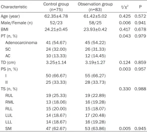
Table 1. Characteristics of patients at baseline
Characteristic Control group (n=75) Observation group (n=83) t/χ2 P
Age (year) 62.35±4.78 61.42±5.02 0.425 0.572
Male/Female (n) 52/23 58/25 0.006 0.941
BMI 24.21±0.45 23.93±0.42 0.417 0.678
PT (n, %) 0.043 0.979
Adenocarcinoma 41 (54.67) 45 (54.22)
SC 24 (32.00) 26 (31.33)
AC 10 (13.33) 12 (14.45)
TD (cm) 3.25±1.14 3.19±1.27 0.124 0.859
PS (n, %) 0.003 0.957
I 50 (66.67) 55 (66.27)
II 25 (33.33) 28 (33.73)
TS (n, %) 0.330 0.988
RUL 19 (25.33) 19 (22.89)
RML 13 (18.06) 16 (19.28)
RLL 15 (20.00) 15 (18.07)
LUL 14 (18.67) 17 (20.48)
LLL 14 (18.67) 16 (19.28)
SM 47 (62.67) 53 (63.86) 0.005 0.945
Note: BMI denotes body mass index; PT, pathologic type; SC, squamous carcinoma; AC, adenosquamous carcinoma; TD, tumor diameter; PS, pathologic staging; TS tumor site; RUL, right upper lobe; RML, right middle lobe; RLL, right lower lobe; LUL, left up-per lobe; LLL, left lower lobe; SM, smoking history.
Table 2. Intraoperative indictors of patients
Variable Case time (min)Operative No. of dissected mediastinal lymph nodes
Intraoperative blood loss (mL) Control group 75 132.47±15.36 11.23±2.17 261.47±55.36 Observation group 83 139.86±18.45 10.86±2.35 143.25±42.34
t/χ2 1.085 0.262 12.962
P 0.102 0.754 <0.001
intraoperative blood loss, and complications [13, 14]. Nevertheless, microcosmic differences in the immune function and inflammation between the two surgical techniques are rarely re- ported. Therefore, in the current study, we compared the NSCLC patients with minimally invasive VATS and those with conventional thoracotomy in the preop-erative and postoppreop-erative indicators (the levels of inflammatory cytokines, im- mune functions, and trau-ma), as well as their clinical data, with an aim to develop a safer and more effective modality for the manage-ment of NSCLC patients.
Materials and methods
Patients
[image:2.612.90.384.421.510.2]invasive VATS; patients with prior immune sys-tem disease; patients having received prior chemotherapy, radiotherapy, and other target-ed therapies before enrollment. This study was approved by the Medical Ethics Committee of Shaanxi Provincial Tumor Hospital, and all the enrolled patients submitted written informed consent.
Surgical procedures
Under general anesthesia, patients in both groups underwent surgery with double-lumen endotracheal intubation. The patients were
placed in contralateral position and ventilated at contralateral lung. Each patient in the obser-vation group underwent minimally invasive VATS. In the observation group, minimally inva-sive thoracotomy was performed through the main operation hole at a 4.0-cm incision in the fifth or fourth axillary frontline, the auxiliary operation hole at a 2.0-cm incision in the sev-enth intercostal subscapular line, and the observation hole at a 1.5-cm incision in the seventh intercostal midaxillary centerline. The incisional skin and subcutaneous tissue in the main operation hole were pulled with the
mas-Table 3. Postoperative indicators of patients
Variable Case Catheter drainage duration (d) hospital stay (d)Length of ambulation (d)Time to complications (n, %)Postoperative
Control group 75 5.16±0.49 12.23±3.17 3.05±0.78 18 (24.00)
Observation group 83 3.45±0.36 9.52±2.42 1.58±0.64 10 (12.05)
t/χ2 12.617 14.452 11.761 4.193
[image:3.612.90.525.85.161.2]P <0.001 <0.001 <0.001 0.041
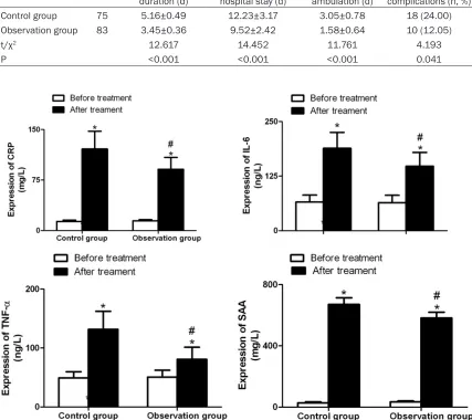
Figure 1. Comparison of expression of CRP, IL-6, TNF-α, and SAA of patients in the control group and the observation group. *P<0.001, compared within the same group before surgery; #P<0.001, compared with the control group at
the same time points. CRP denotes C-reactive protein; IL-6, interleukin-6; TNF-α, tumor necrosis factor-α; SAA serum
[image:3.612.92.519.102.482.2]toid retractor to facilitate the operation of the instrument and complete the operation. A soft rubber was used to protect the incision, instead of a rib spreader to support the rib. After the tumor location had been determined under a thoracoscope, lobectomy and lymphadenecto-my were performed. Patients in the control group were treated with conventional thoracot-omy with the fourth intercostal posterolateral incision as the surgical approach. The latissi-mus dorsi, serratus anterior and intercostal muscles were dissected. The ribs were spread with a rib spreader, and the rib at the lower bor-der of the incision was cut if necessary. The patient’s diseased lobes were removed, and conventional lymphadenectomy was per-formed. The intraoperative bronchia and blood vessels of patients in the two groups were sutured directly or with a stapler. The lungs were then conventionally inflated and exhaust-ed, and indwelled with drainage tubes, and finally the thoracic cavity was closed layer by layer. Routine anti-infective treatment was given to all patients after surgery.
Outcome measures
Primary outcomes included intraoperative indi-cators and the results of postoperative cellular immunity assays. Secondary outcomes com-prised postoperative measures, as well as serum C-reactive protein (CRP), interleukin-6 (IL-6), tumor necrosis factor-α (TNF-α), and serum amyloid A (SAA) levels.
The patients in the two groups were compared regarding the intraoperative indicators (includ-ing operation time, the number of dissected mediastinal lymph nodes, and intraoperative blood loss), postoperative indicators (including catheter drainage duration, hospital stay, time to ambulation and the rates of postoperative complications).
[image:4.612.96.517.72.378.2]serum was separated and the samples were stored at -20°C. The serum CRP, IL-6, TNF-α, and SAA levels were detected by an enzyme-linked immunosorbent assay (ELISA) using the CRP, IL-6, TNF-α, and SAA kits (R&D science, USA). The above experimental procedures were performed strictly following the instructions on the kits.
Assays of cellular immunity were conducted as follows: the levels of CD4+, CD8+, and natural
killer (NK) T-cells of all patients were detected using a Beckmann Quanta SC flow cytometer (USA) before surgery and 3 days after surgery. The number of lymphocytes in both groups was measured using an automated hematology analyzer.
Statistical analysis
All data in this study were processed with the use of the SPSS software, version 18.0. Measurement data are presented as mean ± sd; between-group comparisons at the same time points were conducted by the indepen-dent samples t-test, whereas intra-group com-parisons at different time points were made by the paired t-test. Count data were described as rates, and the Chi-square test was utilized for between-group comparisons. A P value less than 0.05 was deemed significant for the dif-ference between the data of the two groups.
Results
Patients
Patients in the two study groups were basically balanced in age, gender, body mass index (BMI), pathologic type, tumor diameter, patho-logic staging, tumor sites, and smoking (all P>0.05), so they were comparable, as shown in
Table 1.
Intraoperative parameters of patients
The operation time and the number of dissect-ed mdissect-ediastinal lymph nodes varidissect-ed insignifi-cantly between the two groups (both P>0.05). The intraoperative blood loss of the observa-tion group was remarkably less than that of the control group (P<0.05; Table 2).
Postoperative indicators of patients
The duration of catheter drainage, length of hospital stay, time to ambulation and the rates of postoperative complications in the
observa-tion group were markedly lower than those in the control group (P<0.05; Table 3).
CRP, IL-6, TNF-α and SAA expression of pa-tients
There were insignificant disparities in the CRP, IL-6, TNF-α, and SAA expression between the two groups before surgery (all P>0.05). Three days after surgery, higher serum CRP, IL-6, TNF-α, and SAA levels were observed in the two groups after surgery than before surgery (all P<0.001), and the levels in the observation group were substantially lower than those in the control group (all P<0.001; Figure 1).
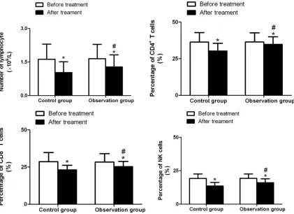
Immune cell levels of patients
The preoperative total blood lymphocyte counts, the percentages of CD4+ T, CD8+ T, and
NK cells varied insignificantly between the two groups (all P>0.05). Nevertheless, 3 days after surgery, the percentages of CD4+, CD8+, and
NK T-cells were markedly lower than those before surgery in both groups (all P<0.05). Total blood lymphocyte count, the percentages of CD4+ T cells, CD8+ T cells, and NK cells in the
observation group were remarkably higher than those in the control group (all P<0.001; Figure 2).
Discussion
Studies indicate that minimally invasive VATS is associated with stress response and sup-pressed immune function at various degrees, and closely related to the degree of surgical trauma in NSCLC patients [16]. For patients with cancers, suppression of immune function is associated with postoperative prognosis of patients. The operation time, intraoperative blood loss, the number of dissected mediasti-nal lymph nodes, and postoperative complica-tion rates are considered as important markers reflecting the degree of surgical trauma. Catheter drainage duration, length of hospital stay, and time to ambulation are regarded as crucial markers for postoperative recovery. The before-mentioned markers are related to the degree of surgical trauma. The results of this study indicate insignificant disparities in the operation time and the number of dissected mediastinal lymph nodes between the two groups, but the patients in the observation group had less intraoperative blood loss than those in the control group, as well as shorter time to ambulation, shorter catheter drainage duration and hospital stay (all P<0.05). The rea-son is that for minimally invasive VATS, it is not necessary to cut off the latissimus dorsi, ser-ratus anterior and intercostal muscles, or to spread the ribs. Hence it has small damage to tissues and cells, and can protect muscle and nerves more effectively, reduce postoperative pain, and alleviate the fears of the patients. In this way, the patients can recover more quickly and ambulate earlier. Meanwhile, the magni-fied views of the thoracoscope allow subtle anatomy, complete hemostasis, and less blood loss during the surgery. Pulmonary infection, atelectasis, and arrhythmias are common com-plications of surgery, which are also related to the degree of surgical trauma [17]. In the cur-rent study, the rate of postoperative complica-tions in the observation group was 12.05%, remarkably lower than 24.00% of the control group. This was attributed to the fact that mini-mally invasive VATS resulted in smaller injuries to the tissues in the chest wall and less postop-erative pain in patients which helped the patients take deep breaths, do exercise and cough up sputum. Earlier ambulation was also beneficial to the recovery of pulmonary func-tions. Overall, the findings demonstrate that compared with conventional thoracotomy, VATS has a significant advantage of minimal
inva-sion, which is consistent with the results report-ed previously [18, 19].
Additionally, in the current study, the severity of trauma following VATS or conventional thora-cotomy were evaluated from the perspective of the changes in the levels of inflammatory cyto-kines CRP, IL-6, TNF-α, and SAA in blood of patients. A study states that surgical trauma induces the production and response of cyto-kines, which primarily manifests as dramatical-ly elevated levels of pro-inflammatory cytokines and cytokines that inhibit cellular immunity. The effects of surgical trauma on the cytokines vary greatly, with more severe surgical trauma presenting higher levels of cytokines [20]. Both CRP and SAA are acute phase proteins, and the levels of them rapidly enhance when there is trauma in the body. IL-6 and TNF-α both can act to regulate inflammation. Studies have indicat-ed that the CRP, SAA, IL-6, and TNF-α levels are associated with surgical trauma, postoperative inflammation, and immune status [21, 22]. The result of our current study revealed higher serum CRP, IL-6, TNF-α, and SAA levels in the two groups after surgery than before surgery, suggesting that NSCLC patients are present with diverse degrees of postoperative trauma and acute phase response, and that serum CRP, IL-6, TNF-α and SAA levels in the observa-tion group were considerably lower than those in the control group 3 days after surgery (all P<0.05). Altogether, these data indicate that patients with VATS have a milder acute phase response, and it is further confirmed that VATS is more minimally invasive.
Surgical trauma inhibits the immune function of patients with NSCLCs, which is associated with the cytotoxicity of inflammatory cytokines on lymphocytes [23]. The current study indi-cates that conventional thoracotomy induces a more significant inflammatory response in patients than minimally invasive VATS, so the immune function was inhibited more severely in patients with conventional thoracotomy than those with minimally invasive VATS. Moreover, in the current study, the total blood lymphocyte count, the percentages of CD4+, CD8+, and NK
VATS has fewer impacts on the immune func-tion of patients with NSCLCs, and the immune function is recovered more quickly, which is in line with the findings reported by Ng et al. [24]. In conclusion, minimally invasive VATS shows significant advantages over conventional thora-cotomy in terms of reducing the degree of trau-ma and suppression of immune function in patients with NSCLCs. However, some limita-tions still exist in this study, for example, the differences in surgical techniques adopted by different surgeons which might affect the surgi-cal results, there is lack of patient data on post-operative adjuvant therapy, and failure to evalu-ate the long-term effects of the two surgical modalities. Therefore, prospective randomized controlled trials with larger sample sizes and long-term follow-ups are required for further validating the advantages of minimally invasive VATS for NSCLC.
Disclosure of conflict of interest
None.
Address correspondence to: Yangrong Song, Depart- ment of Thoracic Surgery, Shaanxi Provincial Tumor Hospital, Xi’an Jiaotong University, No. 309 Yanta West Road, Xi’an 710061, Shaanxi Province, P. R.
China. Tel: 85276156; Fax:
+86-029-85276156; E-mail: [email protected]
References
[1] Barone M, Cipollone G and Mucilli F. Immune
response after video-assisted thoracic surgery in non-small cell lung cancer patients. J Vis Surg 2018; 4: 25.
[2] Wang LY, Cui JJ, Guo AX and Yin JY. Clinical
ef-ficacy and safety of afatinib in the treatment of
non-small-cell lung cancer in Chinese patients. Onco Targets Ther 2018; 11: 529-538. [3] She XW, Gu YB, Xu C, Li C, Ding C, Chen J and
Zhao J. Three-dimensional (3D)-computed to-mography bronchography and angiography combined with 3D-video-assisted thoracic sur-gery (VATS) versus conventional 2D-VATS ana-tomic pulmonary segmentectomy for the treat-ment of non-small cell lung cancer. Thorac Cancer 2018; 9: 305-309.
[4] Schneider T, Hoffmann H, Dienemann H, Her-pel E, Heussel CP, Enk AH, Ring S and Mahnke K. Immune response after radiofrequency ab-lation and surgical resection in nonsmall cell lung cancer. Semin Thorac Cardiovasc Surg 2016; 28: 585-592.
[5] Jones RO, Anderson NH, Murchison JT, Brittan M, Simon EJ, Casali G, Simpson AJ and Walker WS. Innate immune responses after resection for lung cancer via video-assisted thoracoscop-ic surgery and thoracotomy. Innovations (Phila) 2014; 9: 93-103.
[6] Cata JP, Bauer M, Sokari T, Ramirez MF, Mason
D, Plautz G and Kurz A. Effects of surgery, gen-eral anesthesia, and perioperative epidural analgesia on the immune function of patients with non-small cell lung cancer. J Clin Anesth 2013; 25: 255-262.
[7] Yan X, Jiao SC, Zhang GQ, Guan Y and Wang JL. Tumor-associated immune factors are associ-ated with recurrence and metastasis in non-small cell lung cancer. Cancer Gene Ther 2017; 24: 57-63.
[8] Bendixen M, Jorgensen OD, Kronborg C, Ander-sen C and Licht PB. Postoperative pain and quality of life after lobectomy via video-assist-ed thoracoscopic surgery or anterolateral tho-racotomy for early stage lung cancer: a ran-domised controlled trial. Lancet Oncol 2016; 17: 836-844.
[9] Luo QQ, Lin H, Tan Q, Huang J and Xu L. Analy-sis of clinical application of thoracoscopic lo-bectomy for lung cancer. World J Surg Oncol 2014; 12: 157.
[10] Vannucci F and Gonzalez-Rivas D. Is VATS lo -bectomy standard of care for operable non-small cell lung cancer? Lung Cancer 2016; 100: 114-119.
[11] Whitson BA, Groth SS, Duval SJ, Swanson SJ and Maddaus MA. Surgery for early-stage non-small cell lung cancer: a systematic review of the video-assisted thoracoscopic surgery ver-sus thoracotomy approaches to lobectomy. Ann Thorac Surg 2008; 86: 2008-2016. [12] Wang S, Sun T, Sun H, Li X, Li J, Zheng X,
Mal-lampati S, Sun H, Zhou X, Zhou C, Zhang H, Cheng Z and Ma H. Survival improvement in patients with non-small cell lung cancer between 1983 and 2012: analysis of the surveillance, epidemiology, and end results database. Tumour Biol 2017; 39: 1010- 428317691677.
[13] Whitson BA, Andrade RS, Boettcher A, Bar-dales R, Kratzke RA, Dahlberg PS and Mad-daus MA. Video-assisted thoracoscopic sur-gery is more favorable than thoracotomy for resection of clinical stage I non-small cell lung cancer. Ann Thorac Surg 2007; 83: 1965-1970.
[14] Oh DS, Reddy RM, Gorrepati ML, Mehendale S
and Reed MF. Robotic-assisted, video-assisted
[15] Kim SW, Hong JM and Kim D. What is difficult
about doing video-assisted thoracic surgery (VATS)? A retrospective study comparing VATS anatomical resection and conversion to thora-cotomy for lung cancer in a university-based hospital. J Thorac Dis 2017; 9: 3825-3831. [16] Ng CS and Lau KK. Surgical trauma and
im-mune functional changes following major lung resection. Indian J Surg 2015; 77: 49-54. [17] Park JS, Kim K, Choi MS, Chang SW and Han
WS. Video-assisted thoracic surgery (VATS) lo-bectomy for pathologic stage i non-small cell lung cancer: a comparative study with thora-cotomy lobectomy. Korean J Thorac Cardiovasc Surg 2011; 44: 32-38.
[18] Yan TD, Black D, Bannon PG and McCaughan BC. Systematic review and meta-analysis of randomized and nonrandomized trials on
safe-ty and efficacy of video-assisted thoracic sur -gery lobectomy for early-stage non-small-cell lung cancer. J Clin Oncol 2009; 27: 2553-2562.
[19] Long H, Tan Q, Luo Q, Wang Z, Jiang G, Situ D, Lin Y, Su X, Liu Q and Rong T. Thoracoscopic surgery versus thoracotomy for lung cancer: short-term outcomes of a randomized trial. Ann Thorac Surg 2018; 105: 386-392.
[20] Ng CS, Wan IY and Yim AP. Impact of video-as-sisted thoracoscopic major lung resection on immune function. Asian Cardiovasc Thorac Ann 2009; 17: 426-432.
[21] Walker WS and Leaver HA. Immunologic and stress responses following video-assisted tho-racic surgery and open pulmonary lobectomy in early stage lung cancer. Thorac Surg Clin 2007; 17: 241-249.
[22] Nobata H, Suga N, Itoh A, Miura N, Kitagawa W, Morita H, Yokoi T, Banno S and Imai H. Sys-temic AA amyloidosis in a patient with lung me-tastasis from renal cell carcinoma. Amyloid 2012; 19: 197-200.
[23] Uso M, Jantus-Lewintre E, Bremnes RM, Cala-buig S, Blasco A, Pastor E, Borreda I, Molina-Pinelo S, Paz-Ares L, Guijarro R, Martorell M,
Forteza J, Camps C and Sirera R. Analysis of
the immune microenvironment in resected non-small cell lung cancer: the prognostic val-ue of different T lymphocyte markers. Oncotar-get 2016; 7: 52849-52861.
[24] Ng CS, Lee TW, Wan S, Wan IY, Sihoe AD, Arifi
AA and Yim AP. Thoracotomy is associated with
significantly more profound suppression in