With 27 figures frinted in Great Britain
A COMPARATIVE SURVEY OF THE FUNCTION,
MECHANISM AND CONTROL OF CELLULAR OSCILLATORS
BY M. J. BERRIDGE* AND P. E. RAPPf
* A.R.C. Unit of Invertebrate Chemistry and Physiology, Department of Zoology, University of Cambridge, Downing Street, Cambridge CB2 3EJ, U.K.,
t Gonville and Caius College, Cambridge CBz iTA, U.K.
SUMMARY
This review attempts to survey in a uniform manner the available evidence concerning the generation and behaviour of several well-investigated cellular oscillators. Members of two broad classifications are contrasted: (i) cyto-plasmic oscillations, where the periodic phenomena is generated by an in-stability in a metabolic pathway and (ii) membrane oscillators in which a membrane potential rhythm is generated at the membrane. Interactions between the cytoplasmic and membrane compartments are considered and the effects of these interactions on oscillatory behaviour is discussed. Because of their biological importance and the greater body of experimental results, particular attention is directed to a study of membrane potential oscillations. These systems can be approximately classified in two groups: (i) systems in which a periodic potential results from oscillatory changes in permeability and (ii) systems in which potential oscillations result from the periodic activity of an electrogenic pump.
The examples considered include the glycolytic oscillator, oscillations in vein contraction in the slime mould Physarum polycephalum, rhythmic aggre-gation in Dictyostelium discoideum, neural oscillators, the periodic potential in Purkinje fibres and the sino-atrial node and rhythmic behaviour in smooth muscle. Questions considered include the generation of periodic activity, the modulation of the oscillation by drugs and other metabolic and membrane effectors and the question of the functional role of these oscillations.
INTRODUCTION
2 l 8
1 msec I s 1 min I h I day 1 month I year
Organism Complex regulations Smooth muscle Circulation Peristalsis Respiration Heart E.E.G. Ciliated ephithelium Nervous action
I A I
I 10"3
10"2
10"' 10° 10" 10s 106 107 108
sec 10' 102 103
Period duration (s)
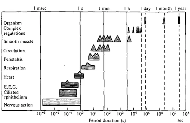
[image:2.451.64.388.63.272.2]Fig. i. Periodic activity in man. Ranges in period are indicated by horizontal bars. Predominant periods are indicated by triangular peaks. (Taken from Golenhofen (1970) as modified from Hildebrandt (1967).)
Table 1. A summary Tissue or cell type Dictyostelium discoideum
Physarum polycephalum
Acetabularia
Macrophages L-cells
Aplysia burster cells
/?-cells
Anterior pituitary
Smooth muscle Sino-atrial node
of the nature and the propos*
Nature of oscillation Periodic release of cAMP
and contractility Shuttle-streaming Periodic action potentials Membrane potential
hyperpolarizations Membrane potential
hyperpolarizations Bursts of action potentials
Bursts of action potentials Action potentials
Slow wave potential changes Action potentials
Proposed function Aggregation and differentiation
Distribution of materials and chemotaxis
May provide positional information during regeneration
Contractility and possibly chemotaxis Contractility
Release of neurohormone Release of insulin Release of hormone
Pacemaker activity for myogenic rhythm Cardiac contraction
cyclic AMP signals are important during differentiation in the slime mould Dictyo-stelium discoideum. The expression of a surface glycoprotein involved in cell adhesion may also depend upon pulses of cyclic AMP (see Gerisch et al., p. 45). However, there are examples where the spontaneous activity has no obvious function and may simply reflect the dynamic nature of cellular control mechanisms. As an understanding of the mechanisms responsible for driving such rhythmical activity begins to appear, it is of interest to consider whether or not there is a common mechanism underlying all such oscillatory behaviour. In this article we attempt to summarize some of the major points which emerged during the course of this meeting on Cellular Oscillators. Much of the information appears in a more detailed form in the preceding articles to which frequent reference will be made. Details of other oscillating systems, which were not specifically dealt with during the meeting, will also be described.
A major problem in trying to unravel the mechanisms responsible for oscillatory activity is to detect the basic instability responsible for generating the rhythm. Since a wide range of cellular processes will be entrained to the basic oscillator, it is always difficult to isolate those processes directly responsible for oscillatory activity. One way of trying to understand the basic oscillator is to identify the input and output properties of the oscillatory system (Fig. 2). A characteristic feature of most oscillatory cells is that the periodicity of the rhythm can be altered by a variety of external signals. A detailed analysis of the mode of action of such signals may help to detect some of the processes involved in generating the oscillation. In the heart, for example, the ability of adrenaline to accelerate pacemaker activity in the sino-atrial node and in Purkinje fibres is apparently mediated by cyclic AMP, which acts by modifying some of the key processes of the membrane oscillator. By uncovering the nature of this cyclic AMP-sensitive process, it may be possible to identify a key component of the oscillator. A knowledge of the output signal can also provide important clues about the nature of the oscillator. A simple example to illustrate this point is a typical myogenic system such as smooth muscle where rhythmical contractions are presumably driven by oscillations in the intracellular level of calcium. Therefore, the oscillator must have a component which is connected in some way with the mechanisms responsible for generating such calcium signals. Identification of the nature of these input and output signals can thus provide valuable insights into the cellular mechanisms responsible for generating oscillatory activity.
There appear to be two distinct kinds of cellular oscillators, those based in the surface membrane and cytoplasmic oscillators originating from inside the cell (Fig. 2). As pointed out by Tsien (p. 209), these two oscillators are not mutually exclusive and it is likely that in some cells they may co-exist and even interact with each other. Such interactions are all the more likely when the two oscillators possess some common component or intermediate as shown in Fig. 2. One component which is common to many membrane and cytoplasmic oscillators is calcium. Calcium occupies such a central position because its intracellular level is determined by processes located both in the surface membrane as well as within the cell. It is not too surprising, therefore, to find that this important second messenger features prominently in many cellular oscillators.
2 2 0
Input for frequency modulation
Output
Output
Fig. 2. The location and relationships of cellular oscillators. A membrane oscillator composed of a variable number of components (a-d) is responsible for generating a rhythmical output usually in the form of fluctuations in membrane potential. A chemical output may also be generated by various cytoplasmic oscillators (d-g). The two oscillators might be linked to each other by sharing a common component (d). The frequency of these oscillators can be adjusted by a variety of input signals which interact with specific components of the oscillators.
example, the glycolytic oscillator, which is most commonly studied in yeast cell extracts, is a biochemical process occurring in the cytoplasm. In contrast the rhythmical trains of action potentials generated by neural or cardiac pacemaker cells originate from processes mainly restricted to the membrane. However, the organization of the cell as a functional unit is such that membrane and cytoplasmic oscillatory mechanisms can never be entirely independent of each other. Even in those cells where a membrane oscillator predominates, interactions between membrane events and internal bio-chemical processes are an important aspect of most oscillatory systems and are often an essential feature of many of the mechanisms responsible for frequency modulation. The main properties of some cytoplasmic and membrane oscillators will be described first before considering how they may function in a variety of specific examples.
CYTOPLASMIC OSCILLATORS
most interesting aspects of these metabolic oscillations is that they display frequencies within the second to minute range which thus makes them possible candidates to drive some of the cellular oscillations which will be described later.
The glycolytic oscillator
Glycolysis has provided a classical system for studying metabolic oscillations (see Hess, p. 7). Most of the observations have been performed on cell-free extracts of yeast (Boiteux & Hess, 1974), skeletal muscle (Tornheim & Lowenstein, 1974, 1975), cardiac muscle (Frenkel, 1968) and Ehrlich ascites tumour cells (Ibsen & Schiller, 1967, 1971). However, the phenomenon may not be confined to cell extracts because oscillations have been recorded from intact yeast cells (Chance et al. 1973). The possibility that the glycolytic pathway might oscillate within intact cells has important implications and it is not too surprising, therefore, to find that the glycolytic oscillator has been invoked to explain several cellular oscillators. For example, Sachsenmaier & Hansen (1973) have proposed that such a metabolic oscillator might drive rhythmical contractile activity in Physarum. Components of the glycolytic oscillator may also play a role in inducing the slow potential waves in /?-cells (Matthews & O'Connor, p. 75) and in molluscan burster cells (Chaplain, see p. 113).
The main features of the glycolytic oscillator are summarized in Fig. 3. Phospho-fructokinase (PFK) is the key enzyme whose activity is sensitive to allosteric control by various components and products of the overall glycolytic pathway. In particular, the enzyme is very sensitive to adenine nucleotides in that it is inhibited by ATP but activated by ADP and AMP. The enzyme is also activated by its substrate fructose-6-phosphate (F6P). When the various glycolytic intermediates in the pathway are measured at different times, they are found to oscillate (Fig. 4). Some intermediates oscillate in phase, whereas others oscillate as much as 1800 out of phase (Fig. 3^, 4). By analysing such phase relationships it is possible to construct the sequence of events which occur during an oscillatory cycle. The two extremes in the activity of PFK are shown in Fig. 3(6, c). When PFK has been active it builds up the level of fructose-1,6-bisphosphate (FDP), which provides substrate for the rest of the cycle leading to an increase of ATP which then rises towards a peak at the expense of ADP and AMP (Fig. 3d, 4). However, as ADP, AMP and F6P fall, the activation of PFK declines and is further inhibited by the increase in ATP. As PFK switches off, the production of FDP declines as does ATP thus leading to an accumulation of ADP and AMP. Additionally, a decline in the activity of PFK will result in a build up of its substrate F6P. As these intermediates accumulate they once again switch the enzyme back to an active state (Fig. 3 c) and the cycle will repeat itself. The existence of a single control point at PFK seems to be sufficient to explain oscillatory activity in extracts from skeletal muscle and beef heart (Frenkel, 1968; Tornheim & Lowestein, 1975). How-ever, the phase relationships in yeast cells are much more complex and indicate that an additional control point exists at pyruvate kinase (see Hess, p. 10). One of the interesting features of this glycolytic oscillator is its sensitivity to the rate of substrate entry into the pathway. For example, increasing the concentration of glucose over a fairly wide range increases the frequency of the oscillation (Boiteux & Hess, 1974) and fiis feature may help in trying to assess the possible significance of the glycolytic Oscillator in various cells.
While the existence of glycolytic oscillations has been demonstrated in several
222
Glucose (a)
223
500
/JM
jUM.
350
-60
Fig.
1 20 ' 40
Time (min) Time (min)
4. Oscillations in the concentrations (expressed as /iM) of various glycolytic components in extract of skeletal muscle. (Taken from Tornheim & Lowenstein, 1974.)
cell-free extracts, there is little evidence on whether or not they exist in intact cells apart from the study on yeast cells mentioned earlier. It is unlikely that such oscilla-tions can exist in cells which have active aerobic respiration where a continuous supply of ATP would tend to suppress glycolytic oscillations by damping out the oscillations in the adenine nucleotide cycle. On the other hand, oscillations in glycolysis might induce oscillations in mitochondrial metabolism through a periodic input of pyruvate. The other problem to consider is the way in which the glycolytic oscillator might be linked to various effector systems. Any fluctuation in the level of ATP would certainly have repercussions for a number of processes. Protons may represent another impor-tant output from the oscillator because each time a molecule of F6P is converted to FDP a hydrogen ion is released (Fig. 3 a). Chaplain (see p. 113) has proposed that fluctuations in the level of ATP and hydrogen ions might be important in regulating ion permeability in burster neurones. In insulin-secreting /?-cells there appears to be an interesting relationship between glycolysis and membrane permeability (Dean, Matthews & Sakamoto, 1975). The enzymes glyceraldehyde-3-phosphate dehydro-|^ase (GDH) and phosphoglycerate kinase (PGK) seem to be particularly important
in that the flux of metabolites through these enzymes somehow alters potassiufl conductance leading to membrane depolarization (see Matthews & O'Connor, p. 757. In addition to effecting ionic permeabilities, variations in intracellular pH could alter contractile activity as will be described presently (p. 244).
It is clear from this brief survey that oscillations in the glycolytic pathway are potentially capable of generating a variety of output signals which could drive a range of oscillatory phenomena. However, an unequivocal demonstration of the existence of glycolytic oscillations in intact cells other than yeast cells has not appeared. Attempts to monitor fluctuations in the NAD/NADH ratio in Physarum were un-successful due to changes in cell geometry which occurred during each contraction (Sachsenmaier & Hansen, 1973). More sophisticated techniques for monitoring intracellular metabolism will have to be devised in order to assess the contribution of the glycolytic oscillator to other cellular oscillations.
Mitochondrial oscillations
Isolated mitochondria display a range of oscillatory activity (Fig. 5) (Boiteux & Hess, 1974; Goldbeter & Caplan, 1976). The oscillations in NADH fluorescence are par-ticularly significant because spectrophotometric analysis of intact slime mould (see Fig. 17 on p. 246) and smooth muscle (see Connor, p. 164) have revealed similar fluctuations in the redox state of NAD and cytochrome b respectively. Since most of the NAD and cytochrome b is concentrated in mitochondria, it is reasonable to specu-late that they may also oscilspecu-late within the intact cell. Mitochondrial respiration is sensitive to both external and internal controls which makes it difficult to assess whether the oscillations originate from within or are driven from outside by a periodic input such as pyruvate originating from glycolytic oscillations as described in the previous section. The release of calcium from mitochondria may also be regulated by the level of phosphoenolpyruvate (PEP) (Roos, Crompton & Carafoli, 1978), which suggests another possible way in which mitochondrial function might be entrained to glycolytic oscillations.
225
NADH fluorescence
H* uptake
K+uptake
Swelling
l A
\\
/
j
A
J \
A
/
A
/ \
Viv
A
/ V
M min)H \ /A
A
/ V
\ rA
1 \
\ r
/ Redu / ction M-equival. g proteinV
,0,
oLS. [image:9.451.80.333.48.230.2]L
Fig. 5. Oscillation of various properties of isolated mitochondria. (Taken from Boiteux & Hess, 1974.)
protons across the mitochondrial membrane (Meech & Thomas, 1977). The pH returns to normal presumably as hydrogen ions were extruded from the cell. These studies on mitochondria suggest that they could play an important role not only in contributing to oscillations in internal calcium but they may also be responsible for establishing oscillations in intracellular pH. Cell suspensions of the slime mould Dictyostelium discoideum display oscillations in hydrogen release which lag slightly behind the peaks in light scattering (Malchow, Nanjundiah & Gerisch, 1978). The precise source of the hydrogen ions which are being extruded from the cell has not been established, but if they reflect the existence of an intracellular pH oscillation, this could have some interesting implications for contractile systems, especially those in non-muscle cells as described later (p. 244).
Cyclic nucleotide-calcium interactions
226 M. J. BERRIDGE AND P. E. RAPP
Signal S iBn a l
AC
Membrane Ca1
* phosphorylalion pump
'. ATP ADP
Cyclic AMP
PDE O
Calmodulin
Ca-Calmodulin complex
Cyclic GMP
PDE
5'-AMP 5-GMP
Fig. 6. The role of calmodulin in cyclic nucleotide—calcium interactions. Cyclic AMP, cyclic GMP and calcium form a triumvarate of second messengers which are all linked together through a variety of interactions. Many of the effects of calcium on the cyclic nucleotides are mediated through a specific receptor protein calmodulin.Guanylate cyclase exists in both a soluble (GC.) and membrane-bound (GCm) form. For simplicity, the effects of the cyclic nucleotides
on calcium homeostasis have been omitted. AC, adenylate cyclase; PDE, phosphodiesterase.
calcium (Wolff, Brostom & Brostom, 1977). At low concentrations, calcium is stimula-tory but becomes inhibistimula-tory at high levels of calcium (Fig. 6). Studies on intact cells have also indicated that high levels of calcium may inhibit adenylate cyclase (Butcher, 1975; Campbell & Siddle, 1976). Calmodulin thus occupies a pivotal position between the cyclic nucleotides and calcium.
Another important function of calcium which seems to require the participation of calmodulin is membrane phosphorylation. Schulman & Greengard (1978) and Greengard (1978) have shown that the calcium-calmodulin complex seems to be responsible for phosphorylating specific proteins of synaptosomal membranes. The functions of these proteins have not been established. Perhaps the ability of calcium to increase potassium conductance, which is such a key feature of many cellular oscilla-tors, might be mediated through such a phosphorylation reaction. Another possible outcome of such phosphorylation might be the activation of calcium extrusion. The calcium-calmodulin complex activates the surface calcium pump of red blood cells (Gopinath & Vincenzi, 1977; Jarrett & Penniston, 1977). If such a feedback mechan-ism is widespread in other cell types it could also be important in oscillatory systems because any increase in the level of calcium will automatically activate the surface pumps which extrude calcium (Fig. 6).
The activation of guanylate cyclase is complicated by the existence of soluble and paniculate or membrane-bound forms of the enzyme (Fig. 6, GCm and GCg), which
may be affected differently by calcium (Mittal & Murad, 1977). Cyclic GMP has proved somewhat of an enigma because, despite the fact that its concentration rises significantly during the activation of many different cells, its precise function is still unknown. It has been suggested that cyclic GMP might act in smooth muscle by inhibiting the entry of calcium (Schultz, Schultz & Schultz, 1977). Interactions between cyclic GMP and calcium may also be important in photoreceptors (Lipton, Rasmussen & Dowling, 1977).
This ability of cyclic nucleotides to modulate the level of calcium has been studied more extensively in the case of cyclic AMP. There are numerous reports in the litera-ture to suggest that some of the affects of cyclic AMP may depend on its ability to modulate the movement of calcium across both surface and internal membranes (Fig. 6) (Berridge, 1975; Rasmussen, Jensen & Goodman, 1976; Rasmussen & Good-man, 1977; Putney, Weiss, Leslie and Marier, 1977; Fitzpatrick and Szentivanyi, 1977). The biochemical basis of many of these feedback interactions operating between cyclic AMP and calcium have yet to be determined so it is difficult to construct precise control loops. However, on the basis of available evidence it is possible to organize some of these second messenger interactions in the form of classical feedback control loops which could generate oscillations under appropriate conditions (Durham, 1974; Rapp & Berridge, 1977). Feedback interactions involving cyclic AMP feature significantly in many of the models designed to account for cyclic AMP oscillations generated by the slime mould Dictyostelium discoideum (see p. 248).
Apart from the possible direct contribution of these various second messengers to the generation of oscillations, the way in which they interact with each other may also be an important component of some of the mechanisms responsible for modulating the frequency of oscijlatory activity. It will be evident from the following sections that an important featurt of rhythmical activity in many cells is an oscillation in the intra-cellular level of calcium. Since both cyclic AMP and cyclic GMP seem to be capable of adjusting the level of calcium, it is not too surprising to find that such second mes-senger interactions have been implicated in modulating the frequency of various cellular oscillators (see subsequent sections for details).
Calcium-induced calcium release
0 J "
A
pCa 7-65 \ 7-50
B
pCa 7-65 | 7-40
X
l_L_L_i
c
pCa 7j65 4 7-0
p C a J_M55 J 650 f 5-50
10s
Fig. 7. Cyclical contractions of a skinned cardiac cell. Note how the amplitude and the frequency of the oscillations increased as the external concentration of calcium was increased. (Taken from Fabiato & Fabiato, 1975.)
release of calcium occurs in intact cells it could be responsible for driving various oscillatory mechanisms. Such calcium-induced calcium release has already been incorporated into models to explain oscillatory behaviour in Physarum (Wohlfarth-Bottermann, see p. 23), Purkinje fibres (Tsien et ah, see p. 211), in macrophages and L-cells (Nelson & Henkart, see p. 57) and in sympathetic ganglion cells (Kuba & Nishi, 1976; see p. 242).
Membrane oscillators
229
A RL2a: spontaneous - 1 8 0 —
B UM300a: spontaneous - 1 3 0
n o
-0
- 5 0
- 1 0 0
- 1 5 0
0 1
1 l
1 11111111 n it
IN A
-\ l^DIIHDiHIUlli
2
1 1 1
1 l l l l k 1 I I I
\WWW\I
3
I 1
\\W\\\
l
I
1/1/1/1/V1/1/
r(h)
0
- 5 0
- 1 0 0
[image:13.451.46.379.64.427.2]- 1 5 0
Fig. 8. Spontaneous membrane potentials produced by Neurospora crassa (A and B) and by
Acetabularia crenulata (C). (A and B taken from Slayman, Long & Gradmann, 1976; C taken
from Gradmann, 1976.)
or in a feedback pathway between ion pump and metabolic cycle'. Some evidence for the idea that pump activity is closely linked with metabolism was derived from the fact that the level of NADH seems to oscillate in phase with the slow waves (Connor et al. 1976).
A close correlation between metabolism and pump activity has also been observed in fungal and algal cells. In Neurospora crassa the mycelium can display several different types of oscillatory activity. Some of the higher-frequency spontaneous oscillations are shown in Fig. 8 A and B. These potential oscillations in Neurospora seem to originate from the periodic activity of an electrogenic hydrogen pump (Gradmann & Slayman,
enhance oscillatory frequency. The so-called 'metabolic' action potentials in the algal cell Acetabularia (Fig. 8C) are driven by periodic changes in an electrogenic chloride pump (Novak & Bentrup, 1972). Gradmann (1976) considers that these action poten-tials are driven by a metabolic event which could be a fluctuation in ATP which de-clines during depolarization but increases during repolarization. As mentioned earlier, these oscillations in membrane potential could provide positional information during regeneration (Novak & Bentrup, 1972). The role of electrical fields in the morphogen-esis of Acetabularia is described by Goodwin & Pateromichelakis (1979).
In all the examples of membrane oscillators which are driven by periodic pump activity there appears to be a close dependence on metabolism which is not too sur-prising since the pump requires a constant input of energy. However, in none of the cases described so far is there a clear indication of whether or not the potential oscillations are actually driven by fluctuations in energy metabolism or how they might be coupled to metabolism except in the case of Acetabularia where ATP may be an intermediary.
Metabolism seems to be of less importance in the second main group of oscillators which are driven by periodic fluctuations in ionic permeability. Despite the fact that the nature of the ionic channels responsible for oscillatory activity varies considerably between different cell types, certain generalizations are beginning to emerge. For example, the depolarizing and hyperpolarizing phases of most of the oscillators seems to depend on an interplay between at least two separate channels. The membrane is depolarized by an inward flow of current carried either by sodium or calcium. This depolarizing phase then gives way to a hyperpolarizing phase due to the onset of an outward current usually carried by potassium. An interplay between fluctuations in an inwardly directed flow of calcium and an outward flow of potassium is a characteristic feature of many membrane oscillators (Table 2). The decay of this outward potassium current usually exposes the inward current mechanisms which once again depolarize the membrane thus completing the cycle. In order for such oscillators to operate over an extended time span the ion gradients necessary for the flow of current through these various channels must be maintained through active ion pumps. Except in the examples described earlier, voltage changes due to pump activity do not figure significantly in those membrane oscillators where potential fluctuations are the result of changes in ion permeability. In some of the membrane oscillators, the inward and outward current mechanisms are related to each other through calcium. During depolarization, calcium enters from outside or is released from internal reservoirs leading to a build up of internal calcium which then interacts with the outward current mechanism by switching on a calcium-dependent potassium conductance. The ability of calcium to switch on potassium conductance is widespread (Meech, 1978; Putney, 1979) and is particularly important in most of the membrane oscillators described in the following sections (Table 2).
231
Table 2. A summary of those membrane oscillators where potential fluctuations have been
attributed to alterations between an inward depolarizing current (usually carried by calcium and/'or sodium) and an outward hyperpolarizing current carried by potassium
Tissue Anterior pituitary
/S-cell
Adrenocortical cells Molluscan burster cells L cells
Macrophages Sino-atrial node
Cardiac Purkinje fibres
Sympathetic ganglion cells
References
Kidokoro (1975); Poulsen & Williams (1976); Taraskevich & Douglas Inward current Outward current
Ca"+ K+
Ca8+ K+(Ca8+-dependent) Matthews & O'Connor (see p . 75); Atwater et al. (1979)
Ca8+ K+ Matthews & Saffran (1973)
Ca8 +/Na+ K+(Ca2+-dependent) Meech (see p . 93)
? K+* Nelson & Henkert (see p. 49) ? K+*
Ca1+/Na+ K+(i
p is Ca2+ Brown, Noble & DiFrancesco
dependent) (see p. 175) Ca8+/Na+ K+(g
Kl and gKa Isenberg (1977)
are Ca2+-dependent)
Na+ or Caa+ K+» Kuba & Nishi (1976)
• It has been proposed that these potassium currents are activated by calcium but direct evidence is lacking.
necessary to extrude calcium following each depolarizing phase. If metabolism oscil-lates by one of the mechanisms discussed earlier, then it is easy to see how oscillations in the supply of energy might be translated into oscillations in calcium and hence membrane potential. Such a mechanism may be found in L cells (see Nelson & Henkart, p. 49), where oscillations in intracellular calcium are thought to arise through a process of calcium-induced calcium release from the endoplasmic reticulum. This calcium oscillation may then feedback onto the membrane to produce the characteristic oscillations in transmembrane potential (see fig. 2 on p. 220). Calcium is thus inti-mately connected with both cytoplasmic and membrane oscillators and will feature significantly in the following descriptions of a variety of cellular oscillators.
OSCILLATIONS IN SECRETORY CELLS
A wide variety of secretory cells display oscillations which are mainly restricted to regular fluctuations in membrane potential. In many cases, these membrane potential oscillations seem to be a reflection of the intracellular events connected with the role of calcium in stimulus-secretion coupling. For example, in /?-cells and in the molluscan burster cell, the membrane oscillations are intimately connected with the mechanisms responsible for calcium entry whereas in other secretory cells the membrane oscilla-tions seem to be a consequence of changes in calcium concentration, perhaps resulting from a cytoplasmic oscillator.
P-cells
Glucagon Glucose
r\
ACATP
rv Ca
2
*'
Insulin
G6P
Y
'••[•'].•-'
Pyruvate
Cyclic
AMP
1
o
Calcium' store,
Fig. 9. A summary of the control mechanisms responsible for insulin secretion from pancreatic /?-cells. AC, Adenylate cyclase.
calcium which is then responsible for triggering the release of insulin by exocytosis (Fig. 9). In order to stimulate the entry of calcium, glucose must be metabolized by the glycolytic pathway (Dean et al. 1975). Studies with glycolytic intermediates and inhibitors seem to indicate that the enzymes glyceraldehyde-3-phosphate dehydro-genase (GDH) and phosphoglycerate kinase (PGK) (see Fig. 3 in Matthews & O'Connor, p. 78) are of critical importance in linking glycolysis to the membrane events responsible for calcium entry. As metabolites pass through these enzymes there is a decrease in potassium conductance (Fig. 9). The addition of glucose to /?-cells causes a marked decrease in the flux of radioactive potassium from prelabelled cells (Henquin, 1978).
glucose are responsible for the pacemaker depolarization which initiates each burst as the potential reaches the threshold for the voltage-dependent calcium channels re-sponsible for spike activity. The ionic mechanisms rere-sponsible for repolarization have not been established. Matthews and O'Connor consider that these calcium channels may close due to a build up of calcium near the membrane which switches off further entry. They also consider that there may be a separate potassium conduc-tance mechanism responsible for terminating the fast spikes.
As spike activity continues during the crest of the wave there presumably is a gradual increase in the intracellular level of calcium which will trigger the release of insulin. The accumulation of intracellular calcium during the burst serves another important function in that it is probably responsible for once again switching on the potassium conductance which terminates the burst as the membrane hyperpolarizes below the threshold for spike activity (Atwater et al. 1979). Presumably the subsequent removal of calcium will cause these potassium channels to close thus initiating the depolariza-tion to switch on the next burst. In the absence of voltage-clamp informadepolariza-tion it is difficult to decide whether the glucose-sensitive potassium conductance is synonymous with this proposed calcium-dependent potassium conductance. Alternatively, there might be separate potassium conductances and glucose may act to inhibit a pacemaker potassium conductance which causes the membrane to enter a potential domain where the slow wave oscillations can be established. A related phenomenon has been ob-served in cardiac atrial cells which can be induced to display pacemaker activity by applying a steady current to slightly depolarize the membrane (Brown & Noble, 1969).
An important consequence of the yS-cell oscillations is that they must be accompanied by oscillations in the intracellular level of calcium which must rise periodically during each burst (Atwater et al. 1979). As this increase in calcium during the course of a burst is thought to be responsible for repolarization, the subsequent depolarization will depend on how fast this calcium is removed. Since the intracellular level of calcium seems to be sensitive to cyclic AMP (Charles et al. 1975; Sehlin, 1976), it is conceivable that the latter might have some role to play in these oscillations. Charles et al. (1975) have speculated that cyclic AMP may provide a positive-feed forward signal for secretion. Glucose was found to raise the level of cyclic AMP which, in turn, may act by releasing internal calcium (Fig. 9). Interactions between cyclic AMP and calcium may thus have some role to play in the intricate control mechanisms which generate oscillatory activity and regulate the release of insulin.
In addition to responding to glucose, the cell is also sensitive to glucagon and the neurotransmitter acetylcholine. Gagerman et al. (1978) have shown that like glucagon, acetylcholine seems to act by potentiating the action of glucose. Acetylcholine can also modulate the bursting pattern and may provide another tool for trying to analyse the relationship between oscillatory activity and insulin secretion.
Calliphora salivary glands
234
40
30
10 mV
S. 20
Fig. io. Oscillations in transepithelial potential recorded across the salivary gland of the blowfly Calliphora erythrocephala. The regular downward deflexions are the result of passing constant current pulses across the gland to record changes in resistance which are represented as changes in potential on the lower diagram. Note that the negative peaks of the oscillation correspond with marked decreases in resistance.
235
Effect of S-HT
2 X 1 0 " ' M
(6) Effect of CaJ*
10 HIM
I 5 mV
1 min
(e) Effect of IBMX
I m « IBMX = 2-5 X 1 0 " ' M 5-HT
1 mM
Fig. 11. The effect of various treatments designed to alter the intracellular levels of cyclic AMP and calcium on the frequency of transepithelial potential oscillations across the salivary gland of
Calliphora. (a) The effect of varying the concentration of 5-HT between 2 and 5 x icr" M.
(6) The effect of lowering the levels of calcium from 10 to o-i mM. During the course of this experiment the gland was perfused with 25 x io"0 M 5-HT. (c) The effect of 1 mM isobutyl-methylxanthine (IBMX) on the frequency produced by a gland during continuous treatment with 25 x 10"' M 5-HT.
Berridge, 1973; Berridge, Lindley & Prince, 1975). The second messenger responsible for activating the potassium pump has not been established as current evidence indicates it could either be calcium, cyclic AMP or both. Some of the uncertainty arises from the fact that there appears to be an interaction between these two second messengers whereby cyclic AMP seems to stimulate the release of internal calcium (Prince et al. 1972) in much the same way as just described for the /?-cell. The increase in pump activity observed during the action of cyclic AMP could thus arise either from a direct action of this cyclic nucleotide or indirectly due to a release of internal calcium. Such second messenger interactions might form the basis of oscillations in transepithelial potential which occur under certain conditions.
maximally (icr8 M), the transepithelial potential is found to oscillate (Fig. 10). These oscillations in transepithelial potential occur in phase with oscillations in resistance measured by passing constant current pulses across the gland (Fig. 10). Since a decrease in resistance is attributable to the calcium-dependent change in chloride conductance, it is reasonable to assume that these potential oscillations in Calliphora reflect an underlying oscillation in the intracellular level of calcium. Rapp & Berridge (1977) have proposed that such calcium oscillations might be generated through feedback interactions operating between cyclic AMP and calcium. However, the precise nature of the oscillator remains to be determined. Some evidence for the participation of cyclic AMP and calcium has been obtained by studying the effect of varying the level of these two second messengers on the frequency of the potential oscillations.
All treatments which are expected to raise the level of either cyclic AMP or calcium accelerate the oscillator. 5-HT acts to increase the level of both cyclic AMP and calcium. At low 5-HT concentrations (2 x io~9 M), the oscillations have a low fre-quency which accelerates considerably as the level of 5-HT is increased to 5 x io~9 M (Fig. 11 a). Recent experiments have shown that 5-HT increases the entry of calcium across the plasma membrane through a coupling process which involves the hydrolysis of phosphatidylinositol (Berridge & Fain, 1979; Fain & Berridge, 1979). The amount of phosphatidylinositol hydrolysed increases as the dose of 5-HT rises above io~9 M and is closely correlated with a parallel increase in the rate of calcium entry. The observation that 5-HT accelerates the oscillator (Fig. 11 a) may thus depend on this ability to increase the rate at which calcium enters the cell. Some evidence for the importance of calcium can be obtained by varying the level of external calcium. At low calcium concentrations there is a significant decrease in frequency (Fig. 11 b). Since 5-HT also seems to act by raising the level of cyclic AMP (Berridge, 1970; Prince et al. 1972), the latter may also play a role in regulating the oscillator. The phosphodiesterase inhibitor isobutyl methylxanthine (IBMX), which probably acts to increase the intracellular level of cyclic AMP, was also found to accelerate the oscillator (Fig. 11 c). Note that all the treatments in Fig. 11 which resulted in increases in frequency were always associated with decreases in amplitude. The fact that the frequency of the oscillator can be adjusted by treatments designed to alter the intra-cellular levels of either calcium or cyclic AMP seems to implicate these two second messengers as important components of the oscillator.
Nerve cells
237
Ca2+/Na*
Ca2 +/Na+ K +
00 „ (iv)'
&
Fig. 12. A summary of the ionic mechanisms responsible for bursting activity in molluscan neurones. The diagram indicates the state of the ionic channels responsible for the various inward and outward currents which produce the changes in membrane potential (Em) during
the course of a typical burst cycle. The thickness of the arrows provides an approximate indication of how much current is Mowing through each channel at different stages during the burst. The changes in the intracellular level of calcium (based on measurements made by Stinnakre & Tauc, 1973, and Thomas & Gorman, 1977) are illustrated below the potential trace. The build-up of intracellular calcium helps to terminate the burst by switching on the Ca2+ -dependent potassium channel as shown by the dashed lines. The four conductances depicted in the diagram are: (i) Ca2 +/Na+ channels responsible for the rapid upstroke of the action potential (i'in.f»,t). (ii) Caa +/Na+ channels responsible! for the slow regenerative inward current
(iiQ-tiow) which underlies pacemaker activity, (iii) One of the potassium channels responsible for repolarization during the action potentials, (iv) Ca2+-dependent potassium channel which contributes to pacemaker activity. See text for further details of the stages (a)—(/).
Chaplain (see p. 124) has also pointed out that microneurones which might be re-sponsible for memory storage in vertebrates also display spontaneous activity often in the form of slow waves. The existence of such intrinsic excitability may be an essential feature of the nervous system and it becomes of considerable interest to understand the cellular basis of this rhythmicity. Most of our information on the ionic basis of neuronal rhythmicity has come from studies of molluscan pacemaker neurones.
Molluscan pacemaker neurones
A number of neurones within molluscan ganglia generate slow membrane potential oscillations which trigger bursts of action potentials on the crests of waves. When these neurones are isolated from their synaptic input, this endogenous bursting rhythm is remarkably constant and will persist for many hours with a constant fre-quency. Under normal conditions, however, the frequency of this rhythm becomes •regular as it is modulated by synaptic input. As in many other oscillating systems, The way in which the rhythm is modulated can provide valuable clues as to the nature
A number of authors have proposed a membrane model to explain bursting activity (Junge & Stevens, 1973; Wilson & Wachtel, 1974; Barker & Gainer, 1975; Smith, Barker & Gainer, 1975; Eckert & Lux, 1976; Johnston, 1976). A detailed description of this membrane model and the evidence for the ionic components responsible for bursting is presented by Meech (see p. 93). An intriguing feature of this model is its basic similarity to the membrane models put forward to account for cardiac pacemaker activity (see subsequent sections). The potential oscillations in bursting neurones depend on cyclic variations between an inward current (which depolarizes the membrane) and an outward current (which hyperpolarizes the mem-brane). Voltage-clamp experiments have revealed that these fluctuating inward and outward currents can be separated into at least five separate components (see Meech, p. 93). There are two mixed inward currents carried by either sodium or calcium and at least three outward potassium currents. The fluctuations in membrane potential are due solely to changes in ionic permeability with little or no contribution from the active pump mechanisms. The latter, however, play a crucial role in maintaining the ionic gradients responsible for driving current through the various channels as they open and close during the course of a burst cycle. The properties of the channels which carry these currents and their contribution to the burst are summarized in
Fig. 12(0-/):
(a) Before dealing with the channels directly responsible for the pacemaker wave it is convenient to describe the currents responsible for the action potentials which occur on the crests of the wave. As the potential depolarizes due to the development of the slow inward current (Fig. 12, ii) it reaches threshold for the initiation of spike activity. The upstroke of the spike results from the opening of a voltage-dependent channel (^inrast)tnat c a n carry both sodium and calcium (Fig. 12, i).
(b) The membrane repolarizes after each action potential due to rapid inactivation of *Infast together with the activation of a delayed
voltage-dependent outward potassium current (Fig. 12, iii). However, the membrane does not hyperpolarize completely and the potential rapidly returns back towards the threshold for the next action potential where the sequence of events just described in (a) and (b) are repeated. The voltage-dependent potassium channel (Fig. 12, iii) gradually inactivates during the burst and the process of repolarization is probably taken over by the calcium-dependent potassium current (Fig. 12, iv), which slowly develops as the calcium con-centration begins to rise. The reason why the membrane remains depolarized during the burst is because of the existence of the slow inward current (zmsiow) (Fig. 12, ii) mainly carried by calcium (Eckert & Lux, 1976), which persists during prolonged depolarization. As we shall see later, the gradual development of iin slow provides the background current which depolarizes the membrane as the potassium current wanes during the pacemaker wave. (c) In the middle or the burst (i.e. at the crest of the slow wave) the inward current carried by *)nsiow just balances the developing outward potassium
article on page 95) the amount of calcium entering will be larger during the final spikes of the burst. This increase in the intracellular level of calcium during a burst has been measured directly using aequorin (Stinnakre & Tauc, 1973) and arsenazo III (Thomas & Gorman, 1977; see Fig. 4 of Meech's article on page 98).
(d) This calcium-dependent potassium conductance continues to develop until it reaches the point where it hyperpolarizes the membrane and the potential falls below the threshold for action potentials thus terminating the burst. This developing potassium current coinciding with an inactivation of the slow inward current causing the 'distinctive "sweep" which drives the cell towards the bottom of the cycle' (Wilson & Wachtel, 1974).
(e) At the point of maximum hyperpolarization, there is a large outward current through these calcium-dependent potassium channels which are now fully open.
(/) The membrane does not remain hyperpolarized because the cell begins to lower the calcium concentration by active transport mechanisms which will thus reduce this calcium-dependent potassium conductance to initiate the so-called pacemaker depolarization. In addition to the decrease in potassium conductance, an increase in the slow regenerative inward current (z"in slow) contributes to the pacemaker depolarization. This inward current which inactivates slowly is particularly important because it is responsible for the negative slope region which is present in the steady state current-voltage (I-V) curve (Wilson & Wachtel, 1974; Smith et al. 1975). The I-V curve is N-shaped (Fig. 13) with the region of negative slope spanning the normal range of the pacemaker potential (i.e. — 60 to — 30 mV). Therefore, as the potential starts to depolarize the inward current begins to develop in a regenerative manner which causes the depolarization to accelerate towards the threshold for the initiation of action potentials (Wilson & Wachtel, 1974). This slow inward current inactivates slowly and is thus responsible for keeping the membrane depolarized during the burst and is only overcome when the build up of internal calcium switches on the potassium current which completes the cycle by hyperpolarizing the membrane.
B
mV,
-100 -80 -60 -40./-20 10
nA 0
-10
- 2 0
- 3 0
Fig. 13. Current-voltage curves from burster cells in Aplysia. (A) Cell L3 before (•) and during
the action of acetylcholine (O), which produces long-lasting inhibition. (B) Cell RI6 before (•)
and during the action of dopamine (O), which also produces prolonged inhibition. (Taken from Wilson & Wachtel, Copyright 1978 by the American Association for the Advancement of Science.)
curve, it was found that this region of the curve was lost during application of acetylcholine to neurone L3 or dopamine to R15 of Aplysia (Fig. 13) (Wilson & Wachtel,
1978). The sensitivity of the potassium channel to calcium suggests that fluctuations in the latter could influence the timing of the burst cycle and thus constitutes an important interface between the membrane oscillator and cellular metabolism.
So far the oscillator has been described in terms of membrane events which raises the obvious question of whether the oscillator is entirely confined to the membrane or whether it is driven in some way by an underlying internal metabolic oscillator. The completeness of the membrane model seems to argue against the necessity of having a metabolic oscillator. However, it is still possible that the changes in inward and outward currents which underly pacemaker activity might be regulated by fluctuations in some key metabolic intermediates. Chaplain (see p. 113) has suggested that a glyco-lytic oscillator may be closely linked to the membrane oscillator. When added to burster cells, a range of key glycolytic intermediates have a profound effect on the bursting pattern (Chaplain, 1976). Some of the most active intermediates such as fructose-6-phosphate (F6P) activate phosphofructokinase (PFK) whereas others (citrate, 3-phosphoglycerate) activate fructose-1,6-bisphosphatase (FDPase). These two enzymes, especially PFK, are responsible for initiating oscillations in glycolysis as described earlier (see page 221). Chaplain considers that PFK and FDPase may be linked together in the form of a substrate cycle which generates periodic fluctuations in the level of hydrogen ions and ATP in the immediate vicinity of the membrane. One of the problems is to establish directly whether such a glycolytic oscillator exists and how it might be linked to membrane events. Chaplain has proposed that the main link between this metabolic oscillator and the membrane is mediated through hydro-gen ions altering the slow inward current. However, there is experimental evidence to indicate that variations of pH are more likely to alter potassium conductance (Meech, see p. 107). Clearly, further evidence is necessary to test this interesting suggestion that bursting activity might be linked in some way to glycolysis.
of intracellular calcium during a burst is derived from calcium entering mainly through \he I'iniut channels which open phasically during each action potential. However, there are clear indications that the existence of action potentials are not essential for maintaining the pacemaker wave. If action potentials are blocked using tetrodotoxin (TTX) and calcium-free media, the pacemaker wave persists even though there are no action potentials on the wave crests (Junge & Stephens, 1973; Strumwasser, 1974; Barker & Gainer, 1975). Under such conditions the slow wave can persist for many hours (see fig. 14 in Meech's contribution to this volume). So-called calcium-free media apparently have enough calcium to maintain the basic rhythm because if the medium also contains the chelator EGTA to reduce calcium to very low levels, then the slow wave is abolished.
Junge & Stephens (1973) have also suggested that some calcium might be released from internal reservoirs during the depolarizing phase and this may also help to main-tain the rhythm when the external calcium concentration is low. The possible inter-vention of internal calcium in oscillatory activity is discussed more fully in the next section on sympathetic ganglion cells.
The other second messengers, cyclic AMP and cyclic GMP, appear to mediate some of the hormonal effects on burster cells (see Levitan et al., on p. 144, for details). Increasing the intracellular level of cyclic nucleotides either by direct addition of various derivatives or by using phosphodiesterase inhibitors seems to slow down the overall pacemaker wave by greatly prolonging the depolarizing phase (see Fig. 13 of Levitan et al. on page 146). This means that there is an enormous increase in the number of action potentials during each burst. Therefore, cyclic nucleotides seem to act by preventing repolarization. Since a build up of calcium seems to be important in switching on the calcium signal responsible for repolarization, it would appear that cyclic nucleotides may act by somehow preventing the calcium level from rising. There is some indirect evidence for such a mechanism in that calcium antagonists such as D600 and lanthanum which prevent calcium entry produce long-lasting bursts (Barker & Gainer, 1975) which are very similar to those seen during the action of cyclic nucleotides. It is not clear which of the cyclic nucleotides might be mediating these effects. When the neurones are treated with the non-metabolizable GTP analogue Gpp(NH)p there is a specific increase in the level of cyclic AMP resulting in a long-lasting hyperpolarization in contrast to the prolonged depolarization produced when both cyclic AMP and cyclic GMP act together (Levitan et al., on p. 149). Such observations raise the possible existence of complicated interactions operating between these second messengers including calcium. Rapp & Berridge (1977) have speculated on the possible role of second messenger interactions in oscillatory phenomena. Some of these cyclic nucleotide effects might be mediated by altering calcium homeostasis.
Sympathetic ganglion cells
72 > ^ > » > ^ ^ T ^> W ^ > V > > V
-1-8
0-9
045
0
1 min
Fig. 14. The effect of varying the external calcium concentration on the frequency of t spontaneous hyperpolarizations produced by sympathetic ganglion cells of the bullfrog duri treatment with 3 mM caffeine. The figures refer to calcium concentration in mM. (Taken frc Kuba & Nishi, 1976.)
spontaneous hyperpolarizing responses which have been described in macrophages (Gallin et al. 1975; see also Nelson & Henkart on p. 49). As in macrophages, the spontaneous hyperpolarizations in the ganglion cells results from a large decrease in resistance due to an increase in potassium permeability. Kuba & Nishi (1976) have proposed that these fluctuations in potassium conductance are driven by periodic changes in the intracellular level of calcium. As the calcium level is lowered the frequency declines and disappears in the absence of external calcium (Fig. 14). It is not clear why caffeine should induce this rhythmical activity. Since the addition of dibutyryl cyclic AMP had no effect on the rhythm it would appear that caffeine is not acting as a phosphodiesterase inhibitor to raise the intracellular level of cyclic AMP. Kuba & Nishi (1976) favour the view that caffeine somehow liberates calcium either from the membrane or from some intracellular reservoir. The rhythmicity is thought to arise from the periodic release of internal calcium through a calcium-induced calcium release mechanism resembling that described for the sarcoplasmic reticulum of skinned muscle fibres (see p. 227). It is of some interest, therefore, to find that the endoplasmic reticulum of squid giant axons is capable of sequestering calcium in much the same way as does the sarcoplasmic reticulum of muscle cells (Henkart, Reese & Brinley, 1978). The junctions between the plasma membrane and endoplasmic reticulum are also remarkably similar to the corresponding triad junctions in skeletal muscle (Henkart, Landis & Reese, 1976).
Anterior pituitary and adrenal cortical cells
Spontaneous activity has been recorded in a number of other secretory cells under a variety of experimental conditons. In some cases the significance of such oscillations are not immediately apparent since they appear only under non-physiological con-ditions. For example, if adrenocortical cells are exposed to adrenocorticotrophic hormone (ACTH) in a potassium-free solution, they begin to produce spontaneous action potentials with periods ranging from 2 to 12 s (Matthews & Saffran, 1973). The ionic basis of these action potentials was not studied in detail. They were not blocked by tetrodotoxin which suggests that the depolarizing phase may be driven by calcium. Similar fluctuations, which have been described in various cells of the anterior pitui-tary, may be of physiological significance since they occur under normal conditions and their firing rate can also be modulated by the hypophysiotropic hormonsi (Kidokoro, 1975; Poulsen & Williams, 1976; Taraskevich & Douglas, 1977, 1978).
J - 5 0 mV
0-2 nA
[image:27.451.114.341.42.179.2]10s
Fig. 15. Spontaneous oscillations in the membrane potential of the anterior pituitary are shown in trace (a). Note that the rhythm accelerates when the membrane was depolarized but slows down when hyperpolarized. The currents necessary to alter membrane potential are illustrated in trace (6). (Taken from Poulsen & Williams, 1976.)
which also display spontaneous hyperpolarizations (Poulsen & Williams, 1976) re-sembling those described earlier in sympathetic ganglion cells (Fig. 15). The frequency of these hyperpolarizing responses is increased if the membrane is depolarized by injecting current (Fig. 15) or by raising the extracellular concentration of potassium. Other pituitary cells, particularly those involved in the secretion of growth hormone and prolactin, produce spontaneous calcium action potentials (Kidokoro, 1975; Taraskevich & Douglas, 1977, 1978). The frequency of these action potentials was also increased by passing a depolarizing current and in certain cells the frequency was also accelerated by the hypophysiotropic thyrotropin releasing hormone (Taraskevich & Douglas, 1977). On the other hand, dopamine and noradrenaline were found to decrease the spontaneous firing of the prolactin-secreting cells of fish (Taraskevich & Douglas, 1978). The rhythmical activity found in the prolactin-secreting cells is particularly interesting because the release of hormone from these cells seems to occur spontaneously. A clonal cell line isolated from the anterior pituitary of rats secretes both growth hormone and prolactin continuously (Kidokoro, 1975). This continuous release of hormone seems to be triggered by these spontaneous calcium-dependent action potentials.
The mechanisms responsible for this spontaneous electrical activity have not been established. Apart from a direct involvement of calcium in stimulus-secretion coupling, there are numerous reports that both cyclic AMP and cyclic GMP may also play some role in initiating secretion (Berridge, 1975). Interactions between this triumvarate of second messengers could play some role either in initiating spontaneous activity or in mediating the modulatory action of the catecholamines or the hypophysiotropic hormones. Some support for the latter comes from the observation that dopamine, which inhibits spontaneous firing in prolactin-secreting cells, has been found to inhibit adenylate cyclase activity (DeCamilli, Macconi & Spada, 1979).
OSCILLATIONS IN CONTRACTILE CELLS
driven by rhythmical fluctuations in the intracellular level of calcium which is responsible for triggering the interaction between actin and myosin. Even the non-^ muscle cells such as Dictyostelium, Physarum and various tissue culture cells seem to employ actomyosin which implies that periodic contractile activity is likely to be driven by regular fluctuations in the intracellular level of calcium. However, the actomyosin system in these non-muscle cells shows some important differences from that in muscle cells in that it appears to be much more labile. In particular, the subunits of actin may constantly associate and dissociate during contractile activity. If this cyclic transformation between globular (G) and filamentous (F) actin is important in oscillating cells, it suggests another form of control which might be mediated through hydrogen ions. Studies on the acrosome reaction of echinoderm spermatozoa have revealed that this G to F transformation of actin might be regulated by intracellular pH since the extrusion of hydrogen ions was essential for polymerization (Tilney et al. 1978). In most of these contractile cells, however, most attention has been focused on how the oscillations in intracellular calcium are generated.
Dictyostelium discoideum
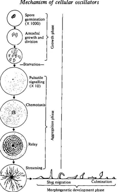
The life-cycle of the slime mould Dictyostelium discoideum has been described in detail by Bonner (1967). In an excellent review, Newell (1977&) has surveyed recent biochemical studies of this organism. The life-cycle has three distinct periods: a unicellular growth phase, an aggregation phase and a multicellular phase (Fig. 16). Unicellular amoeboid cells feed on bacteria and divide by binary fission. If the supply of bacteria is exhausted, the amoebae aggregate to form a multicellular stage. Starva-tion is the direct stimulus for the transiStarva-tion from the unicellular stage to the aggregative stage (Rickenberg et al. 1975; Marin, 1976). The response to deprivation of bacteria is not immediately obsovable as amoebae first pass through a preaggregative inter-phase of several hours. During aggregation 200-100000 cells converge on a centre from distances as far as 20 mm.
Of present interest is the observation that cellular movement to centres during aggregation is not continuous but rather periodic; individual cells move rhythmically. Aggregation can produce whorls and streams of cells, or, if the cell density is high enough, concentric rings or spirals of cells (Arndt, 1937; Bonner, 1944; Shaffer, 1957; Gerisch, 1968). The period of rhythmic cell movement is not constant during aggrega-tion. At the start of aggregation the period is approximately 10 min. It decreases rapidly to 5 min and then decreases slowly to 2-5 min (Gerish, 1965; Durston,
1974 a, b).
245
Spore germination (X 1000)
Amoebal growth and division
—Starvation—
Pulsatile" signalling
( X 1 0 )
Chemotaxis
Relay
S t r e a m i n g
-•s
I
[image:29.451.110.346.52.431.2]Slug migration Culmination Morphogenetic development phase
Fig. 16. A summary of the life-history of the slime mould, Dictyostelium discoideum. The oscillatory activity described in this article concerns the pulsatile signalling system which operates early in the aggregation phase. (Taken from Newell, 1977a.)
246 M. J. BERRIDGE AND P. E. RAPP
10 15 Time (min)
20
Light scattering"
Cytochrome 6
Adenylate cyclase and intracellular cyclic AMP concentration
Intracellular cyclic GMP concentration
PH
25
Fig. 17. The phase relationship of various components of the oscillator in Dictyostelium
dis-coideum. The curves have been normalized for the purpose of comparison ((a) and (6) redrawn
from Gerisch & Hess, 1974; (c) and (d) redrawn from Gerisch et al. 1977; (e) redrawn from Malchow, Nanjundiah & Gerisch, 1978.)
Cyclic AMP oscillations seem to fulfil an important function in the control of developmental events. There is evidence that cyclic AMP pulses (but not a continuous cyclic AMP signal) may be required for cell differentiation from the growth to aggregation competent stage (see Gerisch et al., p. 43). Darmon et al. (1975) found that aggregateless mutants subjected to periodic cyclic AMP pulses are able to complete development but did not respond to a continuous supply of cyclic AMP. Oscillations in the levels of cyclic AMP may thus be important not only for aggregation but also for initiating the developmental programme.
cell shape and agglutination (Gerisch & Hess, 1974). Since calcium is almost certainly involved in the reactions necessary for cell movement, it is reasonable to assume that these light-scattering changes in Dictyostelium are probably associated with periodic changes in the intracellular level of calcium. Mockrin & Spudich (1976) found that actomyosin from Dictyostelium was sensitive to calcium. Not only is calcium important in generating mechanical force but it may also be important in directional control during chemotaxis. Nuccitelli, Poo & Jaffe (1977) found that a calcium current was related to the direction of movement in the giant amoeba Chaos chaos. Oscillations in intracellular calcium thus appear to be one important output signal from the oscillator.
Another important output from the oscillator is a periodic release of cyclic AMP. In phase with the changes in light scattering there is a marked change in the activity of adenylate cyclase (Klein et al. 1977; Roos et al. 1977), resulting in large fluctuations in the intracellular level of cyclic AMP some of which is released to the medium as a chemotactic signal (Gerisch et al. 1977; see also Gerisch et al, p. 33). In addition, there are also cyclic GMP oscillations whose peaks slightly preceed those for cyclic AMP (Fig. 17) (Wurster et al. 1977). These second messenger oscillations are accompanied by oscillations in the redox state of cytochrome b (Hess & Gerisch, 1974) and also in the rate at which hydrogen ions are extruded to produce fluctuations in the pH of the medium (Malchow, Nanjundiah & Gerisch, 1978). With so many oscillating components, it is difficult to assess their relationships either to each other or to the basic oscillator.
One way of trying to determine their direct involvement in generating periodic activity is to find out whether they can interfere with the oscillator. On the basis of such studies it is clear that cyclic AMP is a key intermediate because it can induce marked phase shifts in light scattering and hydrogen-ion extrusion when added at specific points during the oscillatory cycle (Hess & Gerisch, 1974; Malchow, Nan-jundiah & Gerisch, 1978; see also Gerisch et al, p. 41). Therefore, cyclic AMP must be of central importance because not only can it alter the oscillator as an input signal, but it is also one of the major output signals from the oscillator. Further evidence in favour of a central role for cyclic AMP comes from studying the changes which occur during the chemotactic response to this cyclic nucleotide. As the amoebae become aggregation competent, they develop surface receptors capable of detecting levels of cyclic AMP as low as io~8 M (Mato et al. 1975; Gerisch & Malchow, 1976). When cyclic AMP is added to amoebae, it induces most of the changes shown in Fig. 17. Cyclic AMP causes a decrease in light scattering and when cyclic AMP is applied to individual amoebae they contract and protrude pseudopods in the direction of the source (Gerisch & Malchow, 1976). In addition to inducing these changes in motility, cyclic AMP also stimulates both adenylate and guanylate cyclases leading to an increase in the intracellular level of both cyclic AMP and cyclic GMP (Wurster et al. 1977; Mato et al. 1977). Cyclic AMP is also capable of stimulating a release of protons (Malchow, Nanjundiah, Wurster, Eckstein & Gerisch, 1978). Since all these events seem to be linked to cyclic AMP during chemotaxis, it is reasonable to suppose that similar relationships may exist during the generation of endogenous oscillations of cyclic AMP.
AMP is its transient nature. For example, when cyclic AMP is applied to Dictyostelium the level of cyclic GMP rises rapidly but soon returns to its basal value despite the continuous presence of external cyclic AMP (see Gerisch et ai, p. 36). Receptor binding studies clearly reveal that this apparent desensitization is not due to a change in receptor affinity but seems to result from a complex series of intracellular events. The nature of these intracellular events are probably intimately connected with the mechanisms responsible for generating oscillatory activity.
A number of models have been proposed to explain how the cells of Dictyostelium produce an endogenous oscillation of cyclic AMP through a periodic activation of adenylate cyclase. In one model put forward by Cohen (1977) the activation of adenylate cyclase is thought to be under allosteric control through an unidentified variable which somehow reflects energy metabolism. The nature of this variable is not defined although various possibilities are put forward such as the ATP/AMP ratio or the level of cytoplasmic calcium (Cohen, 1977). The fact that the activity of adeny-late cyclase is linked to metabolism is certainly consistent with the fact that there are oscillations in the redox state of cytochrome b (Fig. 17). The possible contribution of a glycolytic oscillator can apparently be ruled out by the fact that Geller & Brenner (1978) were unable to detect oscillations in ATP, GTP, several amino acids, isocitrate, a-ketoglutarate or in the glycolytic intermediates glucose-1-phosphate and glucose-6-phosphate. However, they note that their assay would not detect low amplitude oscillations. Further reason for excluding the possible involvement of the glycolytic oscillator stems from the fact that the enzyme PFK in Dictyostelium is not an allosteric enzyme.
Goldbeter (1974, 1975) published a more specific mathematical model of the autono-mous oscillation in cyclic AMP that depends on the biochemical results of Rosso-mondo & Sussman (1972, 1973). Their experiments indicated that 5'-AMP activated adenylate cyclase and that cyclic AMP activated ATP pyrophosphohydrolase. The activation curves of both enzymes were sigmoidal. This produces the cross-activation control circuit shown in Fig. 18 A. Goldbeter's mathematical realization of this scheme contains three variables: ATP, cyclic AMP and 5'AMP. The enzymes are assumed to be allosteric dimers that obey Monod-Wyman-Changeaux concerted transition kinetics. For appropriate parameters, the corresponding system of three first order nonlinear differential equations possess an attracting periodic solution. Dynamically this model is very similar to Goldbeter's previous model for glycolytic oscillations (Goldbeter & Lefever, 1972). The feedback activation of ATP pyrophosphohydrolase is not essential for producing oscillations. A reduced system that does not contain that enzyme still admits periodic solutions.
249
nal
ft
i i t VCyclic AMP v
V
Cyclic , GMP
[image:33.451.107.335.45.474.2]1 1
Fig. 18. A summary of some models which have been proposed to account for the periodic activation of adenylate cyclase (AC). All diagrams have been redrawn to facilitate comparison. (A) A cross-activation model (adapted from Goldbeter, 1975). (B) Goldbeter-Segel model (re-drawn from Goldbeter & Segel, 1977). (C) The possible involvement of cyclic GMP in the activation of adenylate cyclase (modified from Fig. 14 in Gerisch et al. 1977). ATPase, ATP pyrophosphohydrolase; PDE, phosphodiesterase; R, receptor; GC, guanylate cyclase.
requirement for oscillation in bulk cellular ATP. Experiments on brain adenylate cyclase have given some support for this possibility (Reporter, 1972; Skolnick & Daly, 1975; Lindl, Heinl-Sawaya & Cramer, 1975). Though the requirement for oscillations in ATP is not, on the basis of experimental evidence now available, a critical failure of the Goldbeter model, other problems remain.
Newell (19776) has noted that the concentrations required for activation of adeny-late cyclase by 5'-AMP and ATP pyrophosphohydrolase by cyclic AMP are much greater than the normal concentrations of these compounds. According to Rosso-mondo & Sussman (1973), half maximal activation of ATP pyrophosphohydrolase requires a cyclic AMP concentration of about 16 mM. Roos et al. (1977) report a maximum intracellular concentration of 20 //.M.
The feedback activation of adenylate cyclase by 5'-AMP (Fig. 18A), which was reported by Rossomondo & Sussman (1972, 1973), is essential to the Goldbeter model. Other investigators have been unable to reproduce this observation. Roos & Gerisch (1976) found that adenylate cyclase activity was not significantly changed by 1 mM 5'-AMP. Klein (1976) reported that 5'-AMP actually inhibits the enzyme. Unless the present contradiction can be resolved in favour of the Rossomondo and Sussman result, the Goldbeter model must be regarded as unconfirmed.
The original Goldbeter model could not account for the control of oscillations by extracellular cyclic AMP or for the relay of signals (Hess, Goldbeter & Lefever, 1978). This problem is explored in a subsequent model (Goldbeter & Segel, 1977; Goldbeter, Erneux & Segel, 1978). This model is also a three variable positive feedback loop. The variables are ATP, internal cyclic AMP and external cyclic AMP (Fig. 18 B). The activation of adenylate cyclase by external cyclic AMP (Roos & Gerisch, 1976) provides the destabilizing control that can produce oscillations. Again it is assumed that adenylate cyclase is an allosteric dimer of the Monod-Wyman-Changeux type and again this model is dynamically equivalent to the Goldbeter model of the glycolytic oscillator and to the previous Goldbeter Dictyostelium model for the reduced system lacking ATP pyrophosphohydrolase. For correct choices of parameters, the differential equations have a periodic solution. For a small domain of the subcritical parameter space, the equations can display relay behaviour in response to pulses of external cyclic AMP. By a careful choice of parameters it