Original Article
Clinicopathologic insight of synchronous
primary urologic cancers
Jie Qin1*, Bo Fan1*, Xiaochi Chen1, Xiunan Li1, Xiaogang Wang1, Xishuang Song1, Xiancheng Li2
1Department of Urology, The First Affiliated Hospital of Dalian Medical University, Dalian 116011, Liaoning
Prov-ince, China; 2Department of Urology, The Second Affiliated Hospital of Dalian Medical University, Dalian 116023,
Liaoning Province, China. *Equal contributors and co-first authors.
Received December 23, 2015; Accepted May 18, 2016; Epub June 15, 2016; Published June 30, 2016
Abstract: Objective: This study aimed to reveal the clinical and histopathologic characteristics of patients with syn-chronous primary urologic cancers (SPUC). Methods: A retrospective chart review was performed in a large group of patients with genitourinary cancer in our hospital from 1980 to 2010, and patients with SPUC were mined to conduct the clinical and pathologic characteristics of SPUC. Results: In this work, among 2107 patients with genito-urinary cancer, a total of 127 cases were SPUC, with a prevalence rate of 6.0%. While only 102 SPUC patients were enrolled because 25 of them miss follow-up. A total of 204 patients with single primary cancer randomly selected from our hospital were set as control group. Presence of SPUC was associated with family history of cancer, smoking history and drinking history. Moreover, SPUC presented a less differentiated degree relative to control group, and the proportion of tumors in stage T2 and T3 was higher than that in control group. The survival rates were 29.4% and 54.9%, respectively, in study group and control group. Furthermore, the mean interval between diagnosis and death in study group (1.35 years) was significant shorter than that in control group (3.26 years). Conclusion: There were significant differences in several clinical and pathologic characteristics between SPUC and single primary cancer. SPUC was more aggressive with a higher mortality relative to single primary cancer, more attention should be paid to the diagnosis and treatment of SPUC.
Keywords: Synchronous primary urologic cancers, genitourinary cancer, clinical presentation, prognosis
Introduction
Multiple primary cancers (MPC) were defined
as two or more cancers that each cancer must
present a definite malignant lesion and do not
have any subordinate relationships [1]. Subse-
quently, Moertel et al. [2] proposed a new defi -nition, i.e., MPC diagnosed at the same time or
within six months were classified to be synchro -nous multiple primary cancers (SMPC), other-wise were considered to be metachronous MPC. Owing to the increased longevity and the advanced diagnostic and surgical techniques, an increasing number of SMPC have been dis-covered recently [3-6], attracting much atten-tion of physicians and investigators.
Multiple documentations have demonstrated that genitourinary system is inclined to develop multiple malignant neoplasm [7-9]. Palou et al. [10] indicated that 46% of synchronous upper
urinary tract tumor and superficial bladder
tumor were invasive, despite of uncommon occurrence. Thus, the urologist and other spe-cialist should pay more attention to evaluating patients for the initial presenting symptoms of tumors [11]. Although synchronous primary
uro-logic cancer (SPUC) is recognized as a signifi -cant entity on clinical and molecular level, its clinical and pathological features and
progno-sis are still controversial. The difficulties of
assessing the characteristics of SPUC might be partly related to its relative low prevalence. Previous studies mainly centered on individual cases with SPUC [7, 12, 13] or just focused on multiple primary urologic cancers [14-16]. Statistical researches of SPUC only have been reported in a few studies with a varied inci-dence rate decades ago [17, 18].
depth, the various clinicopathological charac-teristics of patients with SPUC in a large cohort of patients with long-term follow-up data. This work might lead to a better understanding of the pathogenesis of SPUC, and help us develop more effective methods for the prevention and treatment of neoplastic disease.
Methods
In our study, among 2107 patients who were diagnosed with genitourinary cancer and
received surgical treatment at First Affiliated
Hospital of Dalian Medical University from 1980 to 2010, a total of 127 patients showed SPUC. While, there were 25 SPUC patients who missed the follow-up. Finally, only 102 SPUC
patients were enrolled in our study. The flow
[image:2.612.91.525.71.364.2]diagram of the study population was shown in Figure 1. Of these 102 SPUC patients, the first
primary malignance was diagnosed as bladder cancer, and second primary malignances were, respectively, diagnosed as upper urinary tract cancer (n=71), renal cell cancer (n=22), pros-tatic cancer (n=6), penile cancer (n=2) and tes-ticular cancer (n=1). Synchronous cancers in this study met the criteria of synchronous mul-tiple primary cancers described by Moertel et
13.0 (SPSS Inc, Chicago, IL, USA). Categorical
data were analyzed by χ2 test. Continuous vari-ables were expressed as median or mean ± standard deviation (SD) and were compared using Student’s t-test. Kaplan-Meier method was used to calculate the survival rate of patients and Log-rank was used to assess
sta-tistical significance. A difference with P < 0.05 was considered statistically significant.
Result
Among the 2107 patients diagnosed with uro-logic cancer in our hospital from 1980 to 2010, the prevalence rate of SPUC was 6.0% (127/2107). A total of 102 SPUC patients were enrolled in our study.
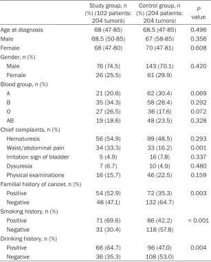
The basic characteristics of the study popu- lation were shown in Table 1. Among the
study population, no significant differences
were existed in age, gender and blood groups
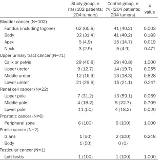
In renal cell cancers, 50% occurred at lower pole in SPUC group while only 18.2% in control
group (P < 0.05). Most of renal cell cancers
(59.1%) occurred at upper pole in control group.
No significant difference existed in anatomical
distribution of other cancers between two groups.
The pathological categories of SPUC were sum-marized in Table 3. Generally, there was no
sig-nificant difference in the pathological patterns
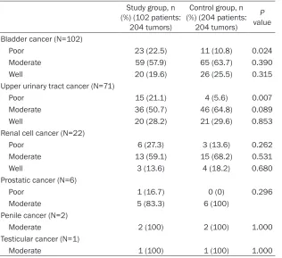
in all cancers between two groups (P > 0.05). The differentiation degrees of SPUC were listed in Table 4. In SPUC group, 22.5% of bladder cancers and 21.1% of upper urinary tract can-cers were pathologically diagnosed to be poorly
differentiated, which were significantly higher
than that in control group (10.8% and 5.6%,
[image:3.612.92.399.84.464.2]respectively; P < 0.05). While, no significant dif -ference was found in differentiation degrees of Table 1. Basic characteristics of the study population
Study group, n (%) (102 patients:
204 tumors)
Control group, n (%) (204 patients:
204 tumors)
P value Age at diagnosis 68 (47-85) 68.5 (47-85) 0.496 Male 68.5 (50-85) 67 (58-85) 0.356 Female 68 (47-80) 70 (47-81) 0.608 Gender, n (%)
Male 76 (74.5) 143 (70.1) 0.420 Female 26 (25.5) 61 (29.9)
Blood group, n (%)
A 21 (20.6) 62 (30.4) 0.069 B 35 (34.3) 58 (28.4) 0.292 O 27 (26.5) 36 (17.6) 0.072 AB 19 (18.6) 48 (23.5) 0.328 Chief complaints, n (%)
Hematuresis 56 (54.9) 99 (48.5) 0.293 Waist/abdominal pain 34 (33.3) 33 (16.2) 0.001 Irritation sign of bladder 5 (4.9) 16 (7.8) 0.337 Dysuresia 7 (6.7) 10 (4.9) 0.480 Physical examinations 16 (15.7) 46 (22.5) 0.159 Familial history of cancer, n (%)
Positive 54 (52.9) 72 (35.3) 0.003 Negative 48 (47.1) 132 (64.7)
Smoking history, n (%)
Positive 71 (69.6) 86 (42.2) < 0.001 Negative 31 (30.4) 118 (57.8)
Drinking history, n (%)
Positive 66 (64.7) 96 (47.0) 0.004 Negative 36 (35.3) 108 (53.0)
between SPUC group and control group (P > 0.05). While, patients with smoking and drink-ing history, and family history of cancer had a
significant tendency to suffer from SPUC (P <
0.05). Among the chief complaints, waist/abdo- minal pain showed a higher proportion in SPUC group relative to
that in control group (P <
0.05).
The anatomical distribu-tion of SPUC was shown in Table 2. Compared with patients with single bladder cancer, 60.8% of bladder cancers in SPUC group tend to occur at the fundus of bladder, including
tri-gone, which was signifi -cantly different with control group with the
percentage of 40.2 (P <
0.05). Moreover, only 4.9% of bladder cancers occurred at the apex of bladder in SPUC group compared with 14.7% in
Table 2. Anatomical distribution of synchronous primary urologic cancers
Study group, n (%) (102 patients:
204 tumors)
Control group, n (%) (204 patients:
204 tumors)
P
value Bladder cancer (N=102)
Fundus (including trigone) 62 (60.8) 41 (40.2) 0.003 Body 32 (31.4) 41 (40.2) 0.189 Apex 5 (4.9) 15 (14.7) 0.019 Neck 3 (2.9) 5 (4.9) 0.471 Upper urinary tract cancer (N=71)
Calix or pelvis 29 (40.8) 29 (40.8) 1.000 Upper ureter 9 (12.7) 14 (19.7) 0.255 Middle ureter 12 (16.9) 13 (18.3) 0.828 Lower ureter 21 (29.6) 15 (21.1) 0.247 Renal cell cancer (N=22)
Upper pole 7 (31.2) 13 (59.1) 0.069 Middle pole 4 (18.2) 5 (22.7) 0.709 Lower pole 11 (50) 4 (18.2) 0.026 Prostatic cancer (N=6)
Peripheral zone 6 (100) 6 (100) 1.000 Penile cancer (N=2)
Glans 1 (50) 2 (100) 0.248 Body 1 (50) 0 (0)
Testicular cancer (N=1)
Left testis 1 (100) 1 (100) 1.000
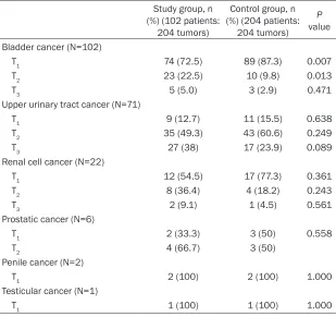
other cancers between two groups (P > 0.05). Interestingly, compared with control group, the ratio of poor differentiation in each cancer, except for penile cancer and testicular cancer, was obviously increased in SPUC group. The tumor stages of SPUC were summarized in Table 5. In general, the percentage of each cancer in stage T2 and T3 became greater in SPUC group than that in control group. While,
no statistical significance existed between two
groups (P > 0.05), except for bladder cancers. Compared with control group, the proportion
of bladder cancers in stage T2 was signifi-cantly higher in SPUC group, and signifisignifi-cantly lower in stage T1 in SPUC group (P < 0.05).
Moreover, in two groups, the majority of blad-der cancers and renal cell cancers were in stage T1, while the majority of upper urinary tract cancers were in stage T2 and T3.
Treatment methods of SPUC were listed in Table 6. Most of the patients with bladder can-cers received transurethral resection in both groups (70.6% in SPUC group and 84.3% in
control group), there was significant difference between two groups (P < 0.05). The proportion
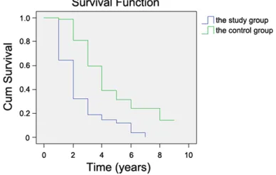
vived in SPUC group, while 54.9% survived in control group. The average interval between the time of diagnosis and the time of death or alive was 1.35 years in SPUC group and 3.26 years in control group, respectively. Kaplan-Meier estimates for cumulative event-free rate of patients with SPUC and single primary can-cers were presented in Figure 2. There was a
significant difference in survival rate between
two groups; the SPUC group had an obviously
lower survival rate than control group (P <
0.05). Discussion
According to our results, we found that family history of cancer, smoking history and drinking
history all had influences on the occurrence of
SPUC. Besides, there were differences in ana-tomical distribution, pathological patterns, dif-ferentiation degrees, tumor stages and treat-ment methods between SPUC group and con-trol group to some extent.
The presence of SMPC was once regarded as rare, but is a very frequent situation currently. However, reports on SPUC are scarce, most are of patients with bladder cancers who received partial cystectomy in
SPUC group was signifi -cantly higher than that
in control group (P <
sur-Table 3. Pathological patterns of synchronous primary urologic cancers
Study group, n (%) (102 patients:
204 tumors)
Control group, n (%) (204 patients:
204 tumors)
P
value Bladder cancer (N=102)
Transitional cell carcinoma 95 (93.1) 100 (98.0) 0.088 Squamous carcinoma 5 (4.9) 1 (1.0) 0.097 Adenocarcinoma 2 (2.0) 1 (1.0) 0.561 Upper urinary tract cancer (N=71)
Transitional cell carcinoma 65 (91.5) 70 (98.6) 0.053 Squamous carcinoma 2 (2.8) 0 (0) 0.154 Adenocarcinoma 4 (5.6) 1 (1.4) 0.172 Renal cell cancer (N=22)
Clear cell carcinoma 19 (86.4) 20 (91.0) 0.635 Papillary adenocarcinoma 2 (9.1) 1 (4.5) 0.550 Chromophobe cell carcinoma 1 (4.5) 1 (4.5) 1.000 Prostatic cancer (N=6)
Adenocarcinoma 6 (100) 6 (100) 1.000 Penile cancer (N=2)
Squamous carcinoma 2 (100) 2 (100) 1.000 Testicular cancer (N=1)
[image:5.612.91.399.84.358.2]Spermatocytoma 1 (100) 1 (100) 1.000
Table 4. Differentiation degrees of synchronous primary urologic can-cers
Study group, n (%) (102 patients:
204 tumors)
Control group, n (%) (204 patients:
204 tumors)
P
value Bladder cancer (N=102)
Poor 23 (22.5) 11 (10.8) 0.024 Moderate 59 (57.9) 65 (63.7) 0.390 Well 20 (19.6) 26 (25.5) 0.315 Upper urinary tract cancer (N=71)
Poor 15 (21.1) 4 (5.6) 0.007 Moderate 36 (50.7) 46 (64.8) 0.089 Well 20 (28.2) 21 (29.6) 0.853 Renal cell cancer (N=22)
Poor 6 (27.3) 3 (13.6) 0.262 Moderate 13 (59.1) 15 (68.2) 0.531 Well 3 (13.6) 4 (18.2) 0.680 Prostatic cancer (N=6)
Poor 1 (16.7) 0 (0) 0.296 Moderate 5 (83.3) 6 (100)
Penile cancer (N=2)
Moderate 2 (100) 2 (100) 1.000 Testicular cancer (N=1)
Moderate 1 (100) 1 (100) 1.000
case reports or case series focused on MPC.
Previously, Wegner [17] reported that the prev- was 9:1, while the ratio was 5:1 among patients with multiple primary urologic cancers. In our alence of MPC among patients with urologic cancer was 3.3% in Berlin (1969 to 1988), of which the incidence of synchronous cancer was 23.4% in female and 48.3% in male patients. Another report by Nakata et al. [18] pre-sented that, of 765 patients with urologic cancer in Japan, 12.3% had MPC (1972-1995), of which 11% in female and 27% in male pa-
tients were identified as
synchronous cancers. In our study, the preva-lence of SPUC was 6% in China (1980-2010). This difference in fre-quency might be due partly to the aging po- pulation and the ad- vances in medical tech-nologies, especially in diagnostic techniques and cancer treatment modalities.
In our survey, no sig-
nificant difference was
[image:5.612.92.408.403.692.2]Table 5. Tumor stages of synchronous primary urologic cancers
Study group, n (%) (102 patients:
204 tumors)
Control group, n (%) (204 patients:
204 tumors)
P
value Bladder cancer (N=102)
T1 74 (72.5) 89 (87.3) 0.007 T2 23 (22.5) 10 (9.8) 0.013 T3 5 (5.0) 3 (2.9) 0.471
Upper urinary tract cancer (N=71)
T1 9 (12.7) 11 (15.5) 0.638 T2 35 (49.3) 43 (60.6) 0.249 T3 27 (38) 17 (23.9) 0.089
Renal cell cancer (N=22)
T1 12 (54.5) 17 (77.3) 0.361 T2 8 (36.4) 4 (18.2) 0.243 T3 2 (9.1) 1 (4.5) 0.561
Prostatic cancer (N=6)
T1 2 (33.3) 3 (50) 0.558 T2 4 (66.7) 3 (50)
Penile cancer (N=2)
T1 2 (100) 2 (100) 1.000 Testicular cancer (N=1)
T1 1 (100) 1 (100) 1.000
work, the ratio of men to women was about 3:1 in SPUC group, this might due to the decreased
occurrence in male-specific cancers including
prostatic, penile and testicular cancer in SPUC group. Compared with a study of Nakata et al.
[18], there was no significant difference in our
paper between SPUC group and single primary cancer group in the distribution of ABO blood group. Nevertheless the percentage of type B blood and type O blood in SPUC group were larger than that in control group, indicating that patients with type B blood and type O blood might have more tendencies to suffer SPUC. With regard to the clinical symptoms of patients
with SPUC, there was no significant difference
in the chief complaints of hematuresis, irrita-tion sign of bladder and dysuresia between two groups, except for waist or abdominal pain. The overall percentage of patients with waist or
abdominal pain in SPUC group was significantly
higher than that in control group. This may indi-cate that patients with waist or abdominal pain should be paid more attention to the risk of SPUC.
Family history of cancer has been reported to
influence on the formation and progression of
multiple malignancies in many documents. In
USA, Uccella et al. [20] found that endometrial cancer patients with a family history of here- ditary nonpolyposis co- lorectal cancer-related cancers had a great ten-dency of developing co- lorectal cancer within 5 years after endome- trial cancer treatment. In Asian, Bai et al. [21] showed that 68.8% patients with synchro-nous upper gastrointes-tinal malignancies had a family history of cancer. In our study, we noted that a positive family history of cancer was also more prevalent in SPUC group (52.9%),
which was significantly
higher than that in con-trol group (35.3%). The results showed that family history of cancer
had a significant effect on the occurrence of
SPUC, thus regular physical examination was recommended for people who had family his-tory of cancer.
Numerous investigators have noted a high inci-dence of secondary primary cancer in patients with continuation of smoking and drinking hab-its. Although smoking is a very strong risk fac-tor for various kinds of cancers [22, 23], a study of Setiawan et al. demonstrated that tobacco use was an independent risk factor for renal cell cancer in males and females [24]. Moreover, Wynder et al. [25] proved that continued con-sumption of tobacco increased the risk of developing a secondary primary cancer after the diagnosis of the index cancer. Similarly, Druesne-Pecollo et al. [26] indicated that in upper aerodigestive tract (UADT) cancer survi-vors, alcohol drinking patients had a more than 2-fold increased risk of second UADT cancers than that not drinking alcohol. In our study, we found that 69.6% patients in SPUC group had a history of tobacco use and 64.7% patients had
risk of second primary malignancies and affect-ed the survival rates adversely, it may be vital for patients to limit the tobacco and alcohol use after the primary urologic cancers treatment to expect better prognoses by preventing addi-tional secondary urologic cancers.
Previously, renal cell carcinoma and prostatic cancer have frequently been reported to be associated with primary bladder cancer [12, 27, 28]. While, only 21.6% of patients were syn-chronous bladder and renal cell cancers, and 6% of patients were synchronous bladder and prostatic cancers in the present study. A large percentage of patients were synchronous
blad-der and upper urinary tract cancers, which accounted for 69.6% in SPUC group. This may attribute to the similar origin between upper urothelium and bladder. In both two groups, the predilection site of bladder cancers was at the fundus of bladder, however, it should be point-ed that the proportion in SPUC group was
sig-nificantly higher than that in control group. Another significant difference in anatomical
distribution of cancers between two groups was renal cell cancer. The majority of renal cell cancers located at lower pole in SPUC group while at upper pole in the control group. Concerning the pathological patterns, no
sig-nificant difference was found between two
groups.
In this study, the degree of tumor tion was also measured [29]. The differentia-tion degree of cancer usually divided from very well differentiation to very poorly differentia-tion. Usually, a tumor whose structure was well differentiated will probably had a biological behavior relatively close to normal, i.e., not very aggressively malignant. In our study, the pro-portion of poorly differentiated bladder cancer
and upper urinary tract cancer were significant -ly higher in SPUC group than that in control group. Besides, accurate staging of cancer is also critical for patient management. The pri-Table 6. Treatment methods of synchronous primary urologic cancers
Study group, n (%) (102 patients:
204 tumors)
Control group, n (%) (204 patients:
204 tumors)
P
value Bladder cancer (N=102)
Transurethral resection 72 (70.6) 86 (84.3) 0.019 Partial cystectomy 25 (24.5) 12 (11.8) 0.018 Radical cystectomy 5 (4.9) 4 (3.9) 0.733 Upper urinary tract cancer (N=71)
Radical nephroureterectomy 45 (63.4) 54 (76.1) 0.100 Radical nephroureterectomy plus bladder cuff shape excision 26 (36.6) 17 (23.9)
Renal cell cancer (N=22)
Nephron sparing surgery 10 (45.5) 16 (72.7) 0.066 Radical nephrectomy 12 (54.5) 6 (27.3)
Prostatic cancer (N=6)
Radical prostatectomy 2 (33.3) 3 (50) 0.558 Transurethral resection plus double testicular castration 4 (66.7) 3 (50)
Penile cancer (N=2)
Partial resection 2 (100) 2 (100) 1.000 Testicular cancer (N=1)
[image:7.612.87.522.85.345.2]Inguinal probe plus radical orchiectomy 1 (100) 1 (100) 1.000
[image:7.612.92.286.372.495.2]mary tumors were staged according to the 2002 TNM system (T1: limited to the lamina propria, T2: invading the muscularis, T3: inva-sion beyond the muscularis, T4: invading other organ structures) [29]. In this work, the per-centage of cancers in stage T2 and T3 was higher in SPUC group than that in control group,
while no significant difference existed except
for bladder cancers. Moreover, according to our report, there was a remarkably lower 5-year survival rate in SPUC group. This might
attri-bute to two reasons: first, SPUC exhibited more
aggressive tumors because they were diag-nosed as a much poor differentiation and a higher tumor stage in a large proportion; sec-ond, a national screening program for SPUC is still inadequate in China.
To our best knowledge, the present report was the largest case series on SPUC at present. However, there were also some limitations in the present study. First, this is a monocentric study only based on our hospital and lack of information of patients managed at outside institutions. Second, although we have collect-ed all patients with synchronous cancers in our hospital, the sample size is still relatively small. Third, the information was provided to the phy-sicians relying on statistical considerations and did not specify how to individualize patients’ management. Thus parts of routine procedure, including abdominal computed tomography, cystoscope, digital rectal examination, and serum PSA, should be undertaken for bladder cancer patients and futurer studies need to fur-ther reveal the pathophysiology of SPUC. Acknowledgements
This work was supported by a grant from Chinese National Science Foundation (No. 81572505) and the Science and Technology Research Plan by Liaoning Province, China (No. 2011225013).
Disclosure of conflict of interest
None.
Address correspondence to: Xiancheng Li, Depart- ment of Urology, The Second Affiliated Hospital of Dalian Medical University, Zhongshan Road 467, Shahekou District, Dalian 116023, Liaoning Pro- vince, China. Tel: 86-0411-84671291; Fax: 86-04- 11-84671291-5177; E-mail: lixiancheng0918@163. com
References
[1] Cho YS, Lee JA, Kim SB, Gong SJ, Kim JH, Youn SM, Kim ET. A case of synchronous double pri-mary cancer of the penis and urinary bladder. Cancer Res Treat 2010; 42: 53.
[2] Moertel CG, Dockerty MB, Baggenstoss AH. Multiple primary malignant neoplasms. I. Introduction and presentation of data. Cancer 1961; 14: 221-230.
[3] Ramasamy M, Manoharan G, Narayanasamy SN. Synchronous primary malignancy of head and neck-a case report. Int J Res Med Sci 2015; 3: 1792-1794.
[4] Gutiérrez-Palomino L, Romo-de Los Reyes J, Pareja-Megía M, García-Mejido J. [Triple syn-chronous primary gynaecological tumours. A case report]. Cir Cir 2016; 84: 69-72.
[5] Solmaz U, Karatasli V, Mat E, Dereli L, Hasdemir P, Ekin A, Gezer C, Sayhan S, Sanci M, Guvenal T. Synchronous primary endometrial and ovar-ian cancers: a multicenter review of 63 cases. Tumori 2015; [Epub ahead of print].
[6] Babacan T, Dag S, Sarici F, Dilli I, Turkbeyler IH, Altundag K. The synchronous primary carcinomas of the rectum and thymus. 2015; Uhod-Uluslararasi Hematoloji-Onkoloji Dergisi, Turkey: 143-144.
[7] Smith MT, Taylor FD, Gianakopoulus WP, Brown RR. Two separate synchronous primary genito-urinary tumors. Rev Urol 2012; 14: 104-107. [8] Mucciardi G, Galì A, D’Amico C, Muscarà G,
Barresi V, Magno C. Transitional cell carcinoma of the renal pelvis with synchronous ipsilateral papillary renal cell carcinoma: case report and review. Urology Case Reports 2015; 3: 93-5. [9] Mohammed A, Al-Zahrani A, Mansour M,
Ghanem H, El Saify A, Hani EK. Triple primary carcinomas: prostatic adenocarcinoma, blad-der urethral carcinoma and papillary thyroid carcinoma: a case report. Am J Cancer Case Rep 2015; 3: 24-28.
[10] Palou J, Rodriguez-Rubio F, Huguet J, Segarra J, Ribal MJ, Alcaraz A and Villavicencio H. Multivariate analysis of clinical parameters of synchronous primary superficial bladder can-cer and upper urinary tract tumor. J Urol 2005; 174: 859-861.
[11] Mydlo JH and Gerstein M. Patients with uro-logic cancer and other nonurouro-logic malignan-cies: analysis of a sample and review of the literature. Urology 2001; 58: 864-869. [12] Tiwari P, Tripathi A, Bansal P, Vijay M, Gupta A,
Kundu AK. Synchronous primary cancers of urinary bladder and kidney and prostate. Saudi J Kidney Dis Transpl 2012; 23: 786-789. [13] Shetty GS, Bhalla P, Desai SM, Wagle PK and
synchro-nous primary malignancies. Indian J Surg 2013; 75: 290-292.
[14] Koyama K, Furukawa Y, Tanaka H. Multiple pri-mary malignant neoplasms in urologic pa-tients. Scand J Urol Nephrol 1995; 29: 483-490.
[15] Mydlo JH, Agins JA, Donohoe J and Grob BM. A review of urologic cancer patients with multiple primary malignancies. World J Urol 2001; 19: 240-243.
[16] Mukaiyama Y, Suzuki M, Morikawa T, Mori Y, Takeshima Y, Fujimura T, Fukuhara H, Nakagawa T, Nishimatsu H and Kume H. Multiple primary malignant neoplasms of the glottis, renal pelvis, oral floor, urinary bladder, prostate, and esophagus in a Japanese male patient: a case report. World J Surg Oncol 2014; 12: 294-294.
[17] Wegner HE. Multiple primary cancers in uro-logic patients. Audit of 19-year experience in Berlin and review of the literature. Urology 1992; 39: 231-236.
[18] Nakata S, Kato Y, Sato J, Mayuzumi T, Kumasaka F, Shimizu T. Analysis of multiple primary cancers in patients with urologic can-cer. Int J Clin Oncol 1997; 2: 40-46.
[19] Inci O, Kaya E, Alagol B, Atakan IH, Aydin S, Ereselli H. Multiple primary malignant neo-plasms in urologic patients. Int Urol Nephrol 2004; 36: 1-4.
[20] Uccella S, Cha SS, Melton LJ 3rd, Bergstralh EJ, Boardman LA, Keeney GL, Podratz KC, Ciancio FF, Mariani A. Risk factors for develop-ing multiple malignancies in patients with en-dometrial cancer. Int J Gynecol Cancer 2011; 21: 896-901.
[21] Bai Y, Zou DW and Li ZS. Clinical presentation, endoscopic features, treatment and prognosis of synchronous upper gastrointestinal malig-nancies. J Dig Dis 2012; 13: 19-23.
[22] Doll R and Peto R. The causes of cancer: quan-titative estimates of avoidable risks of cancer in the United States today. J Natl Cancer Inst 1981; 66: 1191-1308.
[23] Wynder EL and Gori GB. Contribution of the en-vironment to cancer incidence: an epidemio-logic exercise. J Natl Cancer Inst 1977; 58: 825-832.
[24] Setiawan VW, Stram DO, Nomura AM, Kolonel LN and Henderson BE. Risk factors for renal cell cancer: the multiethnic cohort. Am J Epidemiol 2007; 166: 932-940.
[25] Wynder EL, Mushinski MH, Spivak JC. Tobacco and alcohol consumption in relation to the de-velopment of multiple primary cancers. Cancer 1977; 40: 1872-1878.
[26] Druesne-Pecollo N, Keita Y, Touvier M, Chan DS, Norat T, Hercberg S and Latino-Martel P. Alcohol drinking and second primary cancer risk in patients with upper aerodigestive tract cancers: a systematic review and meta-analy-sis of observational studies. Cancer Epidemiol Biomarkers Prev 2014; 23: 324-331.
[27] McAchran SE, Williams DH and MacLennan GT. Renal cell carcinoma metastasis to the bladder. J Urol 2010; 184: 726-727.
[28] Lee SH, Chang PL, Chen SM, Sun GH, Chen CL, Shen BY, Wu YS and Tsui KH. Synchronous pri-mary carcinomas of the bladder and prostate. Asian J Androl 2006; 8: 357-359.