Original Article
MiR-34a and miR-34c are involved in the pathogenesis
of type-2 diabetes by modulating the cell
cycle of pancreatic beta-cell
Hongyu Liu*, Mengmeng Jin*, Hui Tian, Jian Li, Nan Li, Shuangtong Yan
Department of Geriatric Endocrinology, Chinese PLA General Hospital, Beijing, China. *Equal contributors.
Received January 12, 2016; Accepted March 23, 2016; Epub May 1, 2016; Published May 15, 2016
Abstract: Beta-cell dysfunction is one of the main characters of type 2 diabetes; however the mechanism under this
phenomenon is not well understood. It has been reported disturbed miRNA profile is related to the pathogenesis of diabetes and miR-34a overexpression is related to impaired beta cell proliferation in rats. In this study, we first
detected the level of miR-34a, b and c in the serum of T2DM patients and found a higher level of miR-34a and c. Subsequently, the potential targets of miR-34 were predicted using bioinformatics tools. Using dual-luciferase
assay and immunoblotting, we confirmed that CCNE2 is a direct target of miR-34a and c, and the target region is conserved in mice. Finally, we identified that miR-34a and c can induce G0/G1 arrest and apoptosis in mouse
pancreatic beta-cells. Thus, our data collectively suggest that higher expression of miR-34a and miR-34c may be related to the pathogenesis of T2DM, and the serum level of miR-34a and miR-34c has a potential to be used as a biomarker for T2DM diagnosis.
Keywords: Type 2 diabetes, miRNA, CCNE2, cell cycle, apoptosis
Introduction
The number of patients suffered from diabetes is continuously increasing all over the world and among the peoples with diabetes, more than 90% have type 2 diabetes (T2DM). T2DM is characterized by insulin resistance and β-cell dysfunction. Till now, a number of studies have consistently shown that β-cell function is dimin-ished in people with T2DM [1, 2]. Further study has shown that β-cell apoptosis is increased in patients with T2DM, whereas neither β-cell rep-lication nor neogenesis is decreased, suggest-ing that increased β-cell loss is the main cause of β-cell dysfunction [3]. Research from rats also provides the evidence that ageing induced β-cell dysfunction which was associated with altered miRNAs expression profile, is involved in the pathogenesis of diabetes [4].
MiRNA is a group of endogenous, short non-coding RNAs, which regulates genes expres-sion through targeting the 3’UTR of mRNA. MiRNAs have been found in various organisms, and many of them are evolutionary conserved.
Meanwhile, it is estimated that more than a half of all human protein-coding genes are poten-tially regulated by miRNAs [5]. Recently, dis-turbed miRNAs profiles in serum and monocyte have been found in the patients with T2DM [6, 7]. Meanwhile, single-nucleotide polymorph- isms in the coding regions of some miRNAs have been found to be related to the suscepti-bility to T2DM [8].
In this study, we first detected the expression of 3 miR-34 family members in the serum sam-ples of patients suffered with type 2 diabetes. Subsequently, predicted and confirmed the two targets of miR-34. The biological functions of miR-34 were also examined.
Materials and methods
Subjects
Chinese PLA General Hospital, China. 65 he- althy controls with similar ethnical and social background, neither suffering from T2DM nor other important medical disorders served as controls. Informed consents were obtained from all the participants. All study protocols and designs were approved by the ethics com-mittees of the Chinese PLA General Hospital.
Blood samples were collected from each par-ticipant in EDTA-anticoagulated tubes. Portion of blood sample was processed for plasma separation which was stored at -80°C until used.
RNA isolation and qRT-PCR
Total RNA was extracted from 100 μl serum by using Trizol LS Reagent (Invitrogen, Carlsbad, CA, USA) according to the manufacturer’s instructions. The expression level of miRNAs was detected by TaqMan miRNA RT-Real Time PCR. Single-stranded cDNA was synthesized by using TaqMan MicroRNA Reverse Transcription Kit (Applied Biosystems, Foster City, CA, USA) and then amplified by using TaqMan Universal PCR Master Mix (Applied Biosystems, Foster City, CA, USA) together with miRNA-specific TaqMan MGB probes (Applied Biosystems, Foster City, CA, USA). The serum miR-16 level was quantified for normalization. Each sample in each group was measured in triplicate and the experiment was repeated at least three times for the detection of miRNAs.
Cell culture
HEK293T and HeLa cells were cultured in Dulbecco’s Modified Eagle Medium containing 10% fetal bovine serum (Hyclone, Logan, UT, USA), 100 IU/ml penicillin and 10 mg/mL strep-tomycin. All cells were maintained at 37°C under an atmosphere of 5% CO2.
Islet isolation and cell culture
Islets were isolated by collagenase digestion and cultured in RPMI 1640 [10]. Dissociated islet cells were obtained by trypsin digestion.
Cell cycle analysis
Cells were seeded onto 12-well plates (1 × 10⁵
per well). After transfection for 48 hours, cells were harvested and fixed in 70% ethanol and stored overnight at 4°C. For analysis, 1 mL of
freshly prepared PBS staining solution [200 μg/mL RNase A, 20 mg/mL propidium iodide, and 0.1% Triton X-100] was added to the cells. DNA content was analyzed the same day by flow cytometry. ModFit Cell Cycle Analysis so- ftware was used to determine the cell cycle phase.
Cell apoptosis assay
Flow cytometry (FCM) was performed 48 h post-transfection in 6-well plates. The cells were resuspended in binding buffer containing Annexin V-fluorescein isothiocyanate and prop-idium iodide (PI) according to the manufactur-er’s instructions (KeyGen Biotech Co., Ltd., Nanjing, China). The samples were analyzed using a Beckman Coulter Flow Cytometer (Be- ckman Coulter, Fullerton, CA, USA) and experi-ments were performed in triplicate.
Dual luciferase assay
A segment of 1862 bp CCNE2 3’UTR segment was cloned into downstream of firefly luciferase coding region in pmirGLO plasmid (Promega, Madison, WI, USA) to generate luciferase reporter vector. For luciferase reporter assays, HEK293T cells were seeded in 48-well plates. miRNAs mimics or antagonists and luciferase reporter vector were co-transfected by using lipofectamine 2000 (Invitrogen, Carlsbad, CA USA). Two days later, cells were harvested and assayed with the Dual-Luciferase Assay kit (Promega, Madison, WI USA). Each treatment was performed in triplicate in three indepen-dent experiments. The results were expressed as relative luciferase activity (Firefly LUC/ Renilla LUC).
Western blotting
gy Inc., Santa Cruz, CA, USA) for 2 h at 37°C. The specific protein-antibody complex was detected by using horseradish peroxidase con-jugated goat anti-rabbit or rabbit anti-mouse IgG. Detection by the chemiluminescence reac-tion was carried using the ECL kit (Pierce, Appleton, WI, USA). The β-actin signal was used as a loading control.
Statistical analysis
All the results were analyzed by using SPSS Statistical Package version 16. The data of two groups were analyzed by student’s t-test and
the correlation analysis was processed by χ2-analysis. P < 0.05 was considered
statisti-cally significant.
Results
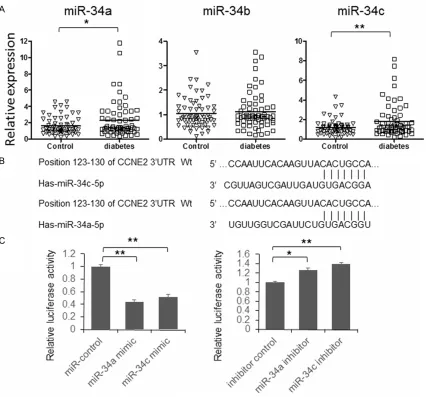
In this study, we first detected the expression of miR-34 family members in the serum of patients suffered from T2DM. As shown in
[image:3.612.95.521.70.467.2]Figure 1A, miR-34a and miR-34c have signifi-cant higher expression in patients’ serum sam-ples. To further explore the role of miR-34a and miR-34c during the pathogenesis of T2DM, the direct targets of them were predicted using Figure 1. miR-34a and c overexpressed in serum of T2DM patients. A. Serum total RNA was isolated using Trizol and the expression of miR-34a, b and c was detected by qRT-PCR. The results were analyzed by student’s t-test and
P<0.05 was considered statistically significant. B. The schematic diagram of interaction between miR-34a/c and
3’UTR of CCNE2. C. Dual-luciferase assay to examine the interaction between miR-34a/c and CCNE2 mRNA. The
online bioinformatics tool: TargetScan. As shown in Fig- ure 1B, there is an 8-mer pre-dicted target region in the 3’UTR of CCNE2 messenger RNA. So we cloned the full length of 1862 bp 3’UTR of CCNE2 into pmirGLO vector, before the stop codon of fire-fly luciferase. Subsequently, the interaction between miR-34a/c and CCNE2 3’UTR was examined by dual-luciferase assay. As shown in Figure 1C
(left panel), the relative lucif-erase activity was significant-ly repressed by miR-34a and miR-34c mimics and up-regu-lated by miR-34a or miR-34c inhibitor. When 3 nucleotides in the predicted target region were mutated (Figure 2), the luciferase activity was redu- ced in the cells transfected with miR-34a or miR-34c mi- mic. These results indicated that miR-34a and miR-34c can repress the expression of firefly luciferase expression by targeting 3’UTR of CCNE2. To further examine whether endogenous CCNE2 was re- pressed by 34a and miR-34c or not, HeLa cells were transfected with miR-34a or miR-34c, with sequence sc- rambled short RNA as control. The cells were lysed 48 hours after transfection and the ex- pression of CCNE2 was exam-ined by immunoblotting. As shown in Figure 3A, the ex- pression of CCNE2 is signifi-cantly repressed by miR-34a and miR-34c. Meanwhile, the inhibitor of 34a or miR-34c can up-regulate CCNE2 expression.
[image:4.612.92.375.74.281.2]By searching the results of conservation analysis betw- een several species on Tar- Figure 2. Examine the miR-34a/c target region of CCNE2 mRNA. A.
Sche-matic diagram of mutant reporter vector construction. Mutated nucleotides were shown as red characters. B. miR-34a or c co-transfected with mutated reporter vector into 293T cells. The luciferase activity was detected 48 hours after transfection. The relative miR-34a and c levels were detected by qRT-PCR.
Figure 3. miR-34a and c repress CCNE2 expression in human and mouse. A. HeLa cells were transfected with miR-34a/c mimic or inhibitor. The
expres-sion of CCNE2 was detected by immunoblotting. GAPDH was used as loading
[image:4.612.93.374.376.635.2]getScan, we find that the miR-34a and miR-34c target region in CCNE2 3’UTR is conserved between human and mouse (Figure 3B). So we detected whether overexpressed miR-34a and miR-34c can repress mouse CCNE2 expression in mouse pancreatic beta-cells. Primary mouse beta-cells were transfected with miR-34a/c mimic or inhibitor using lipofectamine 3000, the expression of CCNE2 was detected 48 hours after transfection. As shown in Figure 3C, the expression of CCNE2 was repressed by 34a/c mimics and up-regulated by miR-34a/c inhibitors.
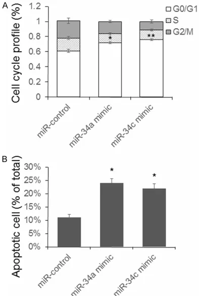
To explore the biological function of overex-pressed miR-34a and miR-34c in beta-cells, the cell cycle of mouse beta-cells was detected after miR-34a and miR-34c mimic transfection. As shown in Figure 4A, a significant G0/G1 arrest has been found in the cells transfected with miR-34a and miR-34c. Since miR-34a has
been reported to be related to beta cell apopto-sis [4], we test the effect of 34a and miR-34c on beta-cell apoptosis. As shown in Figure 4, the number of apoptotic cells raised in the miR-34a and miR-34c group.
Discussion
Beta-cell dysfunction is one of the main charac-ters of type 2 diabetes, however the mecha-nism under this phenomenon is not well under-stood. It has been reported disturbed miRNA profile is related to the pathogenesis of diabe-tes and miR-34a overexpression is related to impaired beta cell proliferation in rats [4, 11]. In this study, we first detected the level of miR-34a, b and c in the serum of T2DM patients and found a higher level of miR-34a and c. Subsequently, the potential targets of miR-34 were predicted using bioinformatics tools. Using dual-luciferase assay and immunoblot-ting, we confirmed that CCNE2 is a direct target of miR-34a and c, and the target region is con-served in mice. Finally, we identified that miR-34a and c can induce G0/G1 arrest and apop-tosis in mouse pancreatic beta-cells. However, the reason of why miR-34a and c is up-regulat-ed is still unknown. Maybe, some factors as genetic variation and miRNA processing distur-bance are involved in this process. In addition, the expression of miR-34 family members and CCNE2 were also need to be detected in the clinical tissue samples to confirm our conclu- sion.
In conclusion, our data collectively suggest that higher expression of miR-34a and miR-34c may be related to the pathogenesis of T2DM, and the serum level of miR-34a and miR-34c has a potential to be used as a biomarker for T2DM diagnosis.
Acknowledgements
This study was supported by Chinese PLA General Hospital Scientific Innovation Nursery Funding Program (12KMM30).
Disclosure of conflict of interest
None.
Address correspondence to: Dr. Shuangtong Yan,
Department of Geriatric Endocrinology, Chinese PLA General Hospital, 28 Fuxing Road, Beijing 100853, Figure 4. miR-34a and c induce beta-cell G0/G1
phase cell cycle arrest and apoptosis. Mouse pan-creatic beta-cells were transfected with miR-34a or c
for 48 hours. Cells were fixed and stained, and then, cell cycle or cell apoptosis were determined by flow
cytometry. Results were analyzed by student’s test
and P<0.05 was considered statistically significant.
[image:5.612.90.287.72.364.2]China. Tel: 010-66876325; +86-10-66876325; Fax: +86-10-66876325; E-mail: [email protected] References
[1] Jensen CC, Cnop M, Hull RL, Fujimoto WY, Kahn SE; American Diabetes Association
GENNID Study Group. Beta-cell function is a
major contributor to oral glucose tolerance in high-risk relatives of four ethnic groups in the U.S. Diabetes 2002; 51: 2170-8.
[2] Defronzo RA. Banting Lecture. From the trium-virate to the ominous octet: a new paradigm for the treatment of type 2 diabetes mellitus. Diabetes 2009; 58: 773-95.
[3] Butler AE, Janson J, Bonner-Weir S, Ritzel R,
Rizza RA, Butler PC. Beta-cell deficit and in -creased beta-cell apoptosis in humans with type 2 diabetes. Diabetes 2003; 52: 102-10. [4] Tugay K, Guay C, Marques AC, Allagnat F, Locke
JM, Harries LW, Rutter GA, Regazzi R. Role of
microRNAs in the age-associated decline of pancreatic beta cell function in rat islets. Diabetologia 2016; 59: 161-9.
[5] Friedman RC, Farh KK, Burge CB, Bartel DP. Most mammalian mRNAs are conserved
tar-gets of microRNAs. Genome Res 2009; 19:
92-105.
[6] Baldeón R L, Weigelt K, de Wit H, Ozcan B, van Oudenaren A, Sempértegui F, Sijbrands E,
Grosse L, van Zonneveld AJ, Drexhage HA,
Leenen PJ. Type 2 Diabetes Monocyte Micro- RNA and mRNA Expression: Dyslipidemia As- sociates with Increased Differentiation-Related
Genes but Not Inflammatory Activation. PLoS
One 2015; 10: e0129421.
[7] Baldeón RL, Weigelt K, de Wit H, Ozcan B, van Oudenaren A, Sempértegui F, Sijbrands E,
Grosse L, Freire W, Drexhage HA, Leenen PJ.
Decreased serum level of miR-146a as sign of
chronic inflammation in type 2 diabetic pa -tients. PLoS One 2014; 9: e115209.
[8] Wang TT, Chen YJ, Sun LL, Zhang SJ, Zhou ZY,
Qiao H. Affection of single-nucleotide polymor-phisms in miR-27a, miR-124a, and miR-146a on susceptibility to type 2 diabetes mellitus in Chinese Han people. Chin Med J (Engl) 2015; 128: 533-9.
[9] American Diabetes Association. Diagnosis and
classification of diabetes mellitus. Diabetes
Care 2011; 34 Suppl 1: S62-9.
[10] Gotoh M, Maki T, Satomi S, Porter J,
Bonner-Weir S, O’Hara CJ, Monaco AP. Reproducible high yield of rat islets by stationary in vitro di-gestion following pancreatic ductal or portal venous collagenase injection. Transplantation 1987; 43: 725-30.