Original Article
PLAC4 mRNA SNP in non-invasive prenatal testing of
Down syndrome
Yujie Wang1*, Jianheng Bao2*, Lijun Zhang1, Yongmei Li3, Yu Zhang1, Lirong Yin1
1Department of Obstetrics and Gynecology, The Second Hospital of Tianjin Medical University, Tianjin, People’s Republic of China; 2Second Department of Hepatopancreatobiliary Surgery, Tianjin Nankai Hospital, Tianjin, People’s Republic of China; 3Institute of Basic Medicine, Tianjin Medical University, Tianjin, People’s Republic of China. *Equal contributors
Received April 14, 2017; Accepted June 19, 2017;Epub July 1, 2017; Published July 15, 2017
Abstract: The purpose of this study was to explore the mRNA expression level of PLAC4 in the maternal plasma and the clinical value of its single nucleotide polymorphism (SNP) rs8130833 in non-invasive prenatal testing (NIPT) of Down syndrome. 40 pregnant women were collected in Tianjin Medical University General Hospital from January 2014 to December 2015. Amniotic puncture karyotype analysis was adapted to determine whether the fetuses with Down syndrome (DS) or not. 20 fetuses were diagnosed with Down syndrome and recorded as the DS group, and 20 fetuses were normal and recorded as the control group. Quantitative reverse transcription-PCR (qRT-PCR) was used to detect the mRNA expression level of PLAC4 both in the whole blood and plasma of pregnant women. A/G polymorphism of rs8130833 was analyzed by pyrosequencing method using the cell-free fetal RNA (cff RNA) in maternal circulation. The mRNA expression level of PLAC4 in DS group was higher than the control group, but the difference was not statistically significant (P > 0.05). A/G polymorphism of rs8130833 was about 2:1 in DS group, and it was nearly 1:1 in control group. The PLAC4 mRNA SNP (rs8130833) has a certain value of research and ap-plication in NIPT of DS.
Keywords: Placenta-specific 4 (PLAC4), single nucleotide polymorphism (SNP), non-invasive prenatal testing (NIPT), Down syndrome (DS), pyrosequencing
Introduction
Non-invasive prenatal testing (NIPT), or non-invasive prenatal testing (NIPT), is a prenatal diagnosis with none or insignificant level of invasiveness, and has substantial medical importance [1]. With the development of cell-free fetal DNA (cff DNA) in maternal circu- lation and the emergence of high-throughput sequencing, NIPT can detect fetal chromo- somal abnormalities [2, 3]. Currently, NIPT has been studied, as a powerful tool, in the ge- netic diagnoses of many diseases, such as le- thal skeletal dysplasia, congenital adrenal hy- perplasia and methylmalonic academia [4-6]. Down syndrome (DS), also known as trisomy 21, is a genetic disorder caused by a third copy of chromosome 21 [7]. DS was first found and characterized by English physician John Langdon Down in 1862 [8]. Globally, DS occurs about 1 in per 1000 births and results in
NIPT of DS in the fetus [17]. Single-nucleotide polymorphism (SNP) arrays allow for detection of consanguinity and most cases of uniparental disomy and provide a higher sensitivity to detect low-level mosaic aneuploidies [18]. In this article, PLAC4 and the SNP rs8130833 were detected of the cell-free fetal RNA (cff RNA) in maternal plasma, in order to preliminar-ily explore the clinical value of them in the NIPT of DS.
Materials and methods
Cases collection and grouping
40 pregnant women were collected in Tianjin Medical University General Hospital from Ja- nuary 2014 to December 2015. 20 cases were diagnosed with DS fetuses by amniotic puncture karyotype analysis, and recorded as the DS group. The other 20 normal cases were diagnosed by the same method and recorded as the control group. Pregnant women in the two groups were singleton pregnancy, no medi-cal and surgimedi-cal diseases, no smoking history, and all of them provided written informed con-sent for the collection of samples and subse-quent analysis. Our research protocol was ap-proved by the ethical committee (Tianjin Medical University General Hospital).
Sample processing and cff RNA isolation
10 ml peripheral venous blood sample was drawn from every pregnant woman, and col-lected into ethylene diamine tetraacetie acid (EDTA) containing tubes within 30 min. The blood samples were centrifuged at 3000 r/min for 10 minutes at 4°C. The supernatant was placed in the frozen storage tube with RNA inactivated enzyme, and stored at -80°C. The cff RNA from the whole blood and the plasma was respectively extracted with the QIAamp RNA Blood Mini Kit (QIAGEN, Germany).
Quantitative reverse transcription-PCR
The cff RNA was subjected to reverse transcrip-tion by Sensiscript RT Kit (QIAGEN, Germany). The final cDNA was obtained, and stored at -20°C. Afterwards, Quantitative real-time PCR was performed to determine the cff RNA level of PLAC4 with an ABI Prism 7000 Sequence Detection System (Applied Biosystems, Ame- rica). The primers set of PLAC4 were designed with PyroMark Assay Design Software 2.0, and the primer sequences were 5’-TGC AAC ACC ATT
TGG GTT AAA TA-3’, and 3’-CCA TGT TTA GGC CAG ATA TAT TCG-5’. They were synthesized by Beijing Genomics Institute (BGI, Shenzhen, China). Moreover, the reaction was set up in a reaction volume of 50 µL, including 0.2 μl Taq DNA polymerase (5 U/μl), 1 μl dNTP, 2 μl primer mix, 2 μl Template, 10 μl buffer (5×) and 34.8 μl H2O. The reaction conditions: 95°C for 3 min and 40 cycles of denaturation at 94°C for 30 s, annealing at 52°C for 30 s and extension at 72°C for 1 min.
Pyrosequencing
The PCR products were immobilized on stre- ptavidin-coated sepharose beads. 2 μl reac- tion binding bead, 40 μl PLAC4 PCR products and 38 ul binding buffer were fully mixed for 10 min in the 96-well plates (15-25°C). Next, the mixture was removed by aspirating the beads with a vacuum pump, and the beads were treated for approximately 5 s with 75% ethanol, 5 s with sodium hydroxide, and 5 s with washing buffer. Then the vacuum pump was closed, and the plate was heated at 85°C for 2 min to make nucleic acid denaturation. The plate was then removed from the plate holder and the samples were allowed to cool to room temperature (15-25°C) for at least 5 min, which made primer and template annealing hybrid. Afterwards, the reagents, including en- zyme and substrate mixtures, and nucleotides were added to the cartridge. The cartridge and the reaction plate were put on the pyrosequenc-ing detector (PyroMark Q96, QIAGEN) to se- quence. Finally, the A/G ratio of the SNP site (rs8130833) was analyzed with the AQ model of PyroMark Q96 software.
Statistical analysis
The SPSS 22.0 statistical software (Chicago, IL, USA) was applied for statistical analysis. All experiments were performed in triplicate, and data were expressed as the means ± standard. For comparison between parameters of 2 groups, Fisher’s F-test and student’s t-test were used. P < 0.05 was considered as a sig-nificant difference.
Results
The mRNA expression level of PLAC4
Table 1. The mRNA expression level of PLAC4 in the maternal whole blood
Group Case PLAC4 mRNA F value P value DS 5 184.85±45.84 0.62 0.45 control 5 182.69±37.65
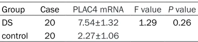
Table 2. The mRNA expression level of PLAC4 in the maternal plasma
Group Case PLAC4 mRNA F value P value DS 20 7.54±1.32 1.29 0.26 control 20 2.27±1.06
and 2. The mRNA expression level of PLAC4 in DS group (184.85±45.84) was slightly higher than the control group (182.69±37.65) in the whole blood. Similarly, the mRNA expres-sion level of PLAC4 in DS group (7.54±1.32) was higher than the control group (2.27±1.06) in maternal plasma, but it was still no signifi-cant difference (P > 0.05).
A/G polymorphism of PLAC4 mRNA SNP
A/G polymorphism of PLAC4 mRNA SNP rs- 8130833 was detected using pyrosequenc- ing method. A/G polymorphism of rs8130833 was about 2:1 in DS group (Figure 1A), while it was approximately 1:1 in control group (Figure 1B). Besides, the site was found to be homo- zygote in 5 cases, containing 2 DS cases and 3 control cases, and the A/G ratios of them were nearly 1:0 (Figure 1C).
Discussion
Currently, DS was no cure, and screening might be the most effective preventive mea-sures. The methods of prenatal diagnosis included measuring nuchal translucency in addition to blood tests for free or total beta human chorionic gonadotrophin (β-hCG) and pregnancy-associated plasma protein A (PAPP- A), detecting the maternal serum alpha-fetopro-tein, unconjugated estriol, hCG, and inhibin-A, and DNA analysis of cell-free fetal DNA from the mother [15, 19]. The different screening techniques with varying levels of accuracy were from 67% to 99.9% [20-22]. All of them had some limits, and they might cause pregnancy loss in the non-sick fetus or other related com-plications. Moreover, some tests could be car-ried out only in the mid trimester of pregnancy.
At that time, if the fetus was confirmed with DS, induced labor might be the only method to ter-minate the pregnancy. Besides, amniocentesis or chorionic villous sampling was still the “gold standard” to confirm the diagnosis, which was easy to cause infection [15]. Therefore, it was necessary to establish a non-invasive, rapid and effective prenatal diagnosis of fetal chro-mosomal diseases in the early pregnancy. NIPT avoided or reduced the need for invasive diagnostic procedures, which was safer and more effective [1]. Besides, advantages of NIPT included the rapid turnaround, relatively low cost, and simplicity of the procedure for preg-nant women [23]. Hence, NIPT might be a highly promising solution for above problems. However, current NIPT mainly detected the fetal DNA from the maternal cff DNA, which the accuracy rate was low and easy to be affected by gender. Detection of cff RNA in maternal circulation did not depend on sex of the fetus, and had the incomparable advantage. PLAC4 mRNA, a chromosome-21 transcript, was a vir-tual preference for fetal-specific makers, which rapidly disappeared from maternal circulation after delivery and cannot be detected in the plasma of nonpregnant women [17]. In 2000, Poo et al. [24] found that the fetal specific mRNA encoded by placenta-derived PLAC4 emerged in maternal plasma, which provided a possibility for the application of the free fetal mRNA in noninvasive prenatal DS screening. Furthermore, SNP was a kind of widely gene mutation in human genome with highly genetic stability and absolutely quantitative advantage [25]. It had been widely used in the detection of genetic diseases, such as the construction of genetic map, the evaluation of biological diver-sity, etc. Tsui et al. [26] reported that the quan-tification of the total PLAC4 mRNA concentra-tion could be used in a synergistic manner with the RNA-SNP allelic ratio approach to diagnose DS. Moreover, one study indicated that the sensitivity and specificity of mRNA-SNP PLAC4 detection were 100% and 89.7% respectively in the DS diagnosis [26].
[image:3.612.91.287.183.225.2]Figure 1. A/G polymorphism of PLAC4 mRNA SNP rs8130833.
from fetus in maternal plasma was much high-er than that in the whole blood of pregnant woman. YMD Lo [27] showed that a heterozy-gous euploid fetus should yield equal propor-tions of each allele, giving an allelic ratio of 1:1, and a heterozygous triploid fetus would yield the allelic ratio 1:2 or 2:1. Results of A/G
hetero-zygous fetus could be used to detect DS with the mRNA-SNP method. In the follow-up study, we would increase samples, combined detect-ing multiple SNP sites, to improve the coverage rate. Further exploration of the research and application values would be performed in the DS NIPT with the mRNA-SNP method.
Acknowledgements
This work was supported by the program of Application Foundation and Advanced Tech- nology Research of Tianjin (12JCYBJC18200).
Disclosure of conflict of interest
None.
Address correspondence to: Dr. Lirong Yin, Department of Obstetrics and Gynecology, The Second Hospital of Tianjin Medical University, 23 Pingjiang Road, He Xi District, Tianjin 300211, People’s Republic of China. Tel: + 86 136 2200 7236; E-mail: [email protected]
References
[1] Papageorgiou EA and Patsalis PC. Non-inva- sive prenatal diagnosis of aneuploidies: new technologies and clinical applications. Ge- nome Med 2011; 4: 46.
[2] Kelly SE and Farrimond HR. Non-invasive pre-natal genetic testing: a study of public atti-tudes. Public Health Genomics 2011; 15: 73-81.
[3] Chen S, Ge H, Wang X, Pan X, Yao X, Li X, Zhang C, Chen F, Jiang F, Li P, Jiang H, Zheng H, Zhang L, Zhao L, Wang W, Li S, Wang J, Wang J, Yang H, Li Y, Zhang X. Haplotype-assisted accurate non-invasive fetal whole genome recovery through maternal plasma sequencing. Ge- nome Med 2013; 5: 18.
[4] Dan S, Yuan Y, Wang Y, Chen C, Gao C, Yu S, Liu Y, Song W, Asan, Zhu H, Yang L, Deng H, Su Y, Yi X. Non-invasive prenatal diagnosis of lethal skeletal dysplasia by targeted capture se-quencing of maternal plasma. PLoS One 2016; 11: e0159355.
[5] Ma D, Ge H, Li X, Jiang T, Chen F, Zhang Y, Hu P, Chen S, Zhang J, Ji X, Xu X, Jiang H, Chen M, Wang W, Xu Z. Haplotype-based approach for noninvasive prenatal diagnosis of congenital adrenal hyperplasia by maternal plasma DNA sequencing. Gene 2014; 544: 252-258. [6] Gu W, Koh W, Blumenfeld YJ, Elsayed YY,
Hudgins L, Hintz SR and Quake SR. Nonin- vasive prenatal diagnosis in a fetus at risk for
methylmalonic acidemia. Genet Med 2014; 16: 564-567.
[7] Patterson D. Molecular genetic analysis of Down syndrome. Hum Genet 2009; 126: 195-214.
[8] Spandagou I. Down syndrome and social change: the fragile nature of progress. Australian Review of Public Affairs 2012. [9] Weijerman ME and de Winter JP. Clinical
prac-tice. The care of children with Down syndrome. Eur J Pediatr 2010; 169: 1445-1452.
Weinstock MA, Weintraub R, Wilkinson JD, Woolf AD, Wulf S, Yeh PH, Yip P, Zabetian A, Zheng ZJ, Lopez AD, Murray CJ, AlMazroa MA, Memish ZA. Global and regional mortality from 235 causes of death for 20 age groups in 1990 and 2010: a systematic analysis for the global burden of disease study 2010. Lancet 2012; 380: 2095-2128.
[11] Học GTY, Học TLY, Kinh NKBĐ, Em BNỞT, Tế
Sức Khoẻ Y. Pediatric epilepsy diagnosis and therapy-part 9. Tailieu Vn.
[12] Hook EB. The consequences of chromosome imbalance: principles, mechanisms, and mod-els. American Journal of Human Genetics 1987; 41.
[13] Hickey F, Hickey E and Summar KL. Medical update for children with down syndrome for the pediatrician and family practitioner. Adv Pediatr 2012; 59: 137-157.
[14] Ferencz C, Rubin JD, Mccarter RJ, Brenner JI, Neill CA, Perry LW, Hepner SI and Downing JW. Congenital heart disease: prevalence at live-birth. The Baltimore-Washington infant study. Am J Epidemiol 1985; 121: 31-36.
[15] ACOG Committee on Practice Bulletins. ACOG practice bulletin No. 77: screening for fetal chromosomal abnormalities. Obstet Gynecol 2007; 109: 217-227.
[16] Chiu RW, Chan KC, Gao Y, Lau VY, Zheng W, Leung TY, Foo CH, Xie B, Tsui NB, Lun FM, Zee BC, Lau TK, Cantor CR, Lo YM. Noninvasive prenatal diagnosis of fetal chromosomal aneu-ploidy by massively parallel genomic sequenc-ing of DNA in maternal plasma. Proc Natl Acad Sci U S A 2008; 105: 20458-20463.
[17] Lo YM, Tsui NB, Chiu RW, Lau TK, Leung TN, Heung MM, Gerovassili A, Jin Y, Nicolaides KH, Cantor CR, Ding C. Plasma placental RNA al-lelic ratio permits noninvasive prenatal chro-mosomal aneuploidy detection. Nat Med 2007; 13: 218-223.
[18] Schaaf CP, Wiszniewska J and Beaudet AL. Copy number and SNP arrays in clinical diag-nostics. Annu Rev Genomics Hum Genet 2011; 12: 25-51.
[19] Puszyk WM, Crea F, Old RW. Noninvasive pre-natal diagnosis of fetal aneuploidy using cell-free fetal nucleic acids in maternal blood: promises and unanswered questions. Prenat Diagn 2008; 28: 1-6.
[20] Canick J. Prenatal screening for trisomy 21: recent advances and guidelines. Clin Chem Lab Med 2012; 50: 1003-1008.
[21] Mersy E, Smits LJ, van Winden LA, de Die-Smulders CE; South-East Netherlands NIPT Consortium, Paulussen AD, Macville MV, Coumans AB, Frints SG. Noninvasive detection of fetal trisomy 21: systematic review and re-port of quality and outcomes of diagnostic ac-curacy studies performed between 1997 and 2012. Hum Reprod Update 2013; 19: 318-329.
[22] Bianchi DW, Parker RL, Wentworth J, Madankumar R, Saffer C, Das AF, Craig JA, Chudova DI, Devers PL, Jones KW, Oliver K, Rava RP, Sehnert AJ; CARE Study Group. DNA sequencing versus standard prenatal aneu-ploidy screening. N Engl J Med 2014; 370: 799-808.
[23] Zeevi DA, Altarescu G, Weinbergshukron A, Zahdeh F, Dinur T, Chicco G, Herskovitz Y, Renbaum P, Elstein D, Levylahad E, Rolfs A, Zimran A. Proof-of-principle rapid noninvasive prenatal diagnosis of autosomal recessive founder mutations. J Clin Invest 2015; 125: 3757-3765.
[24] Poon LL, Leung TN, Lau TK and Lo YM. Presence of fetal RNA in maternal plasma. Clin Chem 2000; 46: 1832-1834.
[25] Garber K. More SNPs on the way. Science 1998; 281: 1788.
[26] Tsui NB, Akolekar R, Chiu RW, Chow KC, Leung TY, Lau TK, Nicolaides KH, Lo YM. Synergy of total PLAC4 RNA concentration and measure-ment of the RNA single-nucleotide polymor-phism allelic ratio for the noninvasive prenatal detection of trisomy 21. Clin Chem 2010; 56: 73-81.