Development of Protein-Catalyzed Capture (PCC)
Agents with Application to the Specific Targeting of
the E17K Point Mutation of Akt1
Thesis By:
Kaycie Marie Deyle
In Partial Fulfillment of the Requirements
for the Degree of
Doctor of Philosophy
California Institute of Technology
Pasadena, CA
2014
“There is no passion to be found playing small - in settling for a life that is less than the one you are capable of living.” – Nelson Mandela
Abstract
This thesis describes the expansion and improvement of the iterative in situ click chemistry OBOC peptide library screening technology. Previous work provided a proof-of-concept demonstration that this technique was advantageous for the production of protein-catalyzed capture (PCC) agents that could be used as drop-in replacements for antibodies in a variety of applications. Chapter 2 describes the technology development that was undertaken to optimize this screening process and make it readily available for a wide variety of targets. This optimization is what has allowed for the explosive growth of the PCC agent project over the past few years.
Acknowledgements
I must start by thanking my family, without whom I would not be where I am today. My parents, Ed and Diane, have supported me in every way possible since the day I was born. You both have raised me and the world’s best brothers (both pretty incredible people), Matt and Shawn, to be like yourselves – strong, confident, intelligent, and hard-working. With the tools with which you have equipped me for this life, anything is possible. The closeness of my family astounds me, and I have the honor of saying that some of my best friends were destined to be a part of my life from birth. To my immediately family, but also my extended family, including my grandparents (Clarence and Mary Ann Badar), and my vast network of aunts, uncles, and cousins, thank you for always being there and for helping to mold me into the person I am today.
To my husband, Casey, who has been there for every day of this journey, cheering me on through the best days while reminding me that there is more to life during the worst. Major thanks for being a constant source of support and laughter, and for ensuring that we were always well-stocked with ice cream and wine.
Next, I must thank my advisor, Jim Heath, for his well-timed guidance throughout the course of my six years in his lab. I greatly appreciate the ability to explore and learn on my own and at my own pace, being roped in only when necessary. Thank you for showing me that nothing in science is impossible if you can think about it the right way, and for ensuring that we always had more than enough funding to do the best possible work.
turning her into a scientist, and to Dr. LeBlond, thank you for instilling in me a love for organic chemistry and science in general. To LeRoy, your constant support throughout all of the years in which I have known you has meant more to me than you will ever know.
I must next thank my colleagues in and around the Heath group. I have had the sincere pleasure these past six years to work with the hands-down smartest, most capable and genuinely nicest lab mates I have ever seen assembled. You have all enriched my Caltech experience beyond measure and have encouraged me every single day to be better than I ever thought possible. A special thanks to Blake Farrow, Jessica Pfeilsticker, Aiko Umeda, Steve Millward, Heather Agnew and Bert Lai for being such capable and absolutely amazing people to work with, not to mention some of my best friends. I would get my hands dirty again in a lab with any of you at any time. To the rest of the capture agents group: Rosemary Rohde, JingXin Liang, Arundhati Nag, Samir Das, Ryan Henning, Joey Varghese, and Mary Beth Yu, thank you for being incredible lab mates and really helping to push our technology to the limits. And to all of the Heath group members past and present, for our interactions both personal and professional, thank you for being there to both enhance my Caltech experience and keep me sane: Alex Sutherland, Min Xue, Jing Yu, Jing Zhou, Wei Wei, Jun Wang, Kevin Kan, Habib Ahmed, Udi Vermesh, Ophir Vermesh, Slobadon Mitrovik, Ann Cheung, Kiwook Huang, John Nagarah, and Jen-Kan Yu. I have met some of the best people I hope to ever have the pleasure of calling friends in this lab.
and experimental input over the years. And Mona Shahgholi for always be willing to chat, and her advice and assistance with MS techniques and experiments.
There is sometimes a life outside of lab, and mine was far richer for the people that were in it. To Tim Mui, thank you for being the world’s greatest roommate, running buddy and sounding board. To Kristina Daeffler, Chris Daeffler, Renee Thomas, Catrina Pheeney, and Matthew Van Wingerden, for always being up for a good game – be it board or football – or a good bottle of wine. Both of these are better with friends. To the Robinsons – Diane, Brent, Katherine and Brenna – for opening up your lives and your home (and grill!) to Casey and me, whenever we wanted or needed the company. Your support has been indescribable these past few years, and we consider you our extended family. To my twin, Joe Zewe, for being never more than a phone call away, and for always somehow knowing exactly what to say. I raise my phone-shot of Jack to you, sir! To my soul mate and maid-of-honor, Jessica Nichol, for…everything. Your support, whether by phone, email, gchat or random cards and care packages has certainly seen me through my fair share of tough times. Thank you for being one of the most caring and considerate people I have ever met. And to Marc, whom I still miss as a lab partner to this day, Whoomp! There it is!
Table of Contents
Abstract ... iv
Acknowledgements... v
List of Figures and Tables ... xii
Chapter 1 ... 15
1.1 Protein-Catalyzed Click (PCC) Peptide Capture Agents for Biomarker Detection and Therapeutics ... 16
1.2 Epitope Targeting Strategies ... 18
1.3 In Situ Click Screening Using Azide-Containing Phage Display Libraries ... 21
1.4 References ... 22
Chapter 2 ... 25
2.1 Introduction ... 26
2.1.1 Iterative In Situ Click Chemistry for Protein-Catalyzed Capture (PCC) Agent Development... 26
2.1.2 Prostate Specific Antigen (PSA) ... 27
2.2 Materials and Methods ... 29
2.2.1 Standard Materials ... 29
2.2.2 Peptide Library Construction ... 29
2.2.3 Bulk Peptide Synthesis ... 30
2.2.4 Typical Screening Protocol for Fluorescent Dye-labeled Protein Target Detection ……….30
2.2.5 Typical Screening Protocol for Antibody Signal Amplification Target Only Screens ……….31
2.2.6 Typical Screening Protocol for an Anti-Screen ... 32
2.2.7 Typical Target Screening Procedure During a Multi-Step Screen (Figure 2-3) ... 33
2.2.8 Typical Screening Protocol for a Preclear ... 33
2.2.9 Typical Screening Protocol for a Click Product Screen ... 34
2.2.10 Peptide Sequencing Strategies ... 34
2.3 Results and Discussion ... 36
2.3.1 Screening via Fluorescent Dye-labeled Protein Target Detection ... 36
2.3.2 Screening via Antibody Signal Amplification Target Only Screens ... 39
2.3.3 Introduction of an Anti-screen ... 40
2.3.5 Introduction of a Preclear ... 45
2.3.6 Use of Alkyne Versus Azide Libraries ... 46
2.3.7 Typical Flow of Screening ... 48
2.4 Conclusions ... 49
2.5 Acknowledgements ... 50
2.6 References ... 50
Chapter 3 ... 51
3.1 Introduction ... 52
3.1.1 The E17K Mutation in the Pleckstrin Homology Domain of Akt1 ... 52
3.1.2 A General Strategy for Targeting Single Amino Acid Point Mutations in Proteins 53 3.2 Materials and Methods ... 54
3.2.1 Akt1 PH Domain Expressions ... 54
3.2.2 Design and Synthesis of Epitope-Targeting Anchor/Target Peptide ... 56
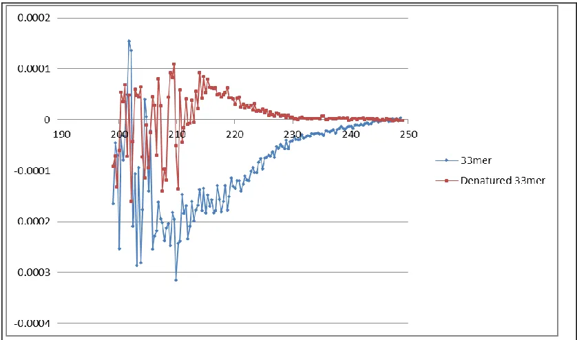
3.2.3 CD Spectroscopy of 33-mer Target Peptide Epitope ... 56
3.2.4 Screen for Initial Anchor Ligand Peptide... 57
3.2.5 Hit Library Bead Sequence Analysis ... 59
3.2.6 Streptavidin-Agarose Immunoprecipitation (Pull-down) Assays for Binding Affinity 59 3.2.7 Point ELISAs with Anchor Ligand and 33-mer Epitope (Epitope Targeting Verification)... 61
3.2.8 HPLC-Detected Immunoprecipitation (Pull-down) Assays (Epitope Targeting Verification)... 62
3.2.9 Ligand-Directed Tosyl Labeling Experiments ... 62
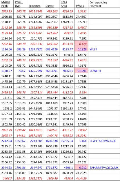
3.2.10 Details of the MALDI-TOF Analysis of Tryptic Peptide Fragments ... 64
3.2.11 Images of Anchor Ligand in HEK-293T Cells Expressing PH Domains ... 67
3.3 Results and Discussion ... 69
3.3.1 In situ Click Epitope-Targeted Screening Strategy for E17K PH Domain-Specific Ligand 69 3.3.2 CD Spectroscopy of 33-mer Target Peptide Epitope ... 72
3.3.3 Verification of the Epitope Targeting Strategy ... 73
3.3.4 Ligand-Directed Labeling Experiment to Confirm Epitope Targeting and Ligand Selectivity ... 76
3.3.5 In Cell Imaging ... 79
3.5 Acknowledgements ... 81
3.6 References ... 82
Chapter 4 ... 83
4.1 Introduction ... 84
4.2 Materials and Methods ... 84
4.2.1 Screen for Biligand Peptide ... 84
4.2.2 Streptavidin-Agarose Immunoprecipitation (Pull-down) Assays to Test Biligand Candidates (Figure 4-6) ... 88
4.2.3 Screen for Triligand Peptide ... 90
4.2.4 Full ELISA Curves for Ligands ... 91
4.2.5 Point ELISA Assays for Triligand Binding to Akt1 and Akt2 Wildtype and E17K Mutant Proteins ... 92
4.2.6 PIP3 Agarose Inhibition Assays ... 93
4.3 Results and Discussion ... 94
4.3.1 Biligand Development ... 94
4.3.2 Triligand Development ... 97
4.3.3 Inhibition Assays ... 100
4.4 Conclusions ... 101
4.5 Acknowledgements ... 101
4.6 References ... 101
Chapter 5 ... 102
5.1 Introduction ... 103
5.1.1 Azide-Containing Phage Display Libraries ... 103
5.1.2 Mirror-Image Phage Display ... 104
5.1.3 G6PD Capture for Malaria Eradication... 104
5.2 Materials and Methods ... 106
5.2.1 Preparation of Plasmid for Incorporation of Azidophenylalanine and Amp Resistant Gene ... 106
5.2.2 Test of Azidophenylalanine Incorporation into a Protein in E.coli ... 110
5.2.3 Test of Azidophenylalanine Incorporation into M13KE Phage ... 110
5.2.4 Synthesis of M13KE Azidophenylalanine-Terminated 7-mer Random Library .... 111
5.2.5 Design and Synthesis of G6PD Target and Scrambled Target for Screening ... 113
5.2.6 Optimized Phage Library Target Screening Conditions... 115
5.2.8 Incorporation of Azidophenylalanine into Phage Libraries ... 118
5.2.9 Optimized Phage Library Click Screening Conditions... 119
5.3 Results and Discussion ... 119
5.3.1 Test of Azidophenylalanine Incorporation into a Protein in E.coli ... 119
5.3.2 Test of Azidophenylalanine Incorporation into M13KE Phage ... 120
5.3.3 Phage Library Screening Conditions and Results ... 121
5.3.4 Focused Library Screening ... 122
5.4 Conclusions ... 123
5.5 Acknowledgements ... 124
5.6 References ... 124
Appendix A ... 125
A.1 Regular Use ... 126
A.1.1 Loading a Bead ... 126
A.1.2 Solvents ... 127
A.1.3 Ordering ... 128
A.1.4 Contacting AB ... 128
A.1.5 Idle machine ... 129
A.1.6 Settings ... 129
A.2 Troubleshooting ... 131
A.2.1 Computer Errors, Freezing, or Not Saving Spectra ... 131
A.2.2 Machine Bottle Runs Dry ... 132
A.2.3 HPLC Bottle Runs Dry ... 132
A.2.4 Baseline Errors – Dip at front of Spectra ... 133
A.2.5 Baseline Errors – Stretched Out Amino Acid Standards ... 135
A.2.6 Pressure Errors – Change Bottle Seals ... 135
List of Figures and Tables
Chapter 2:
Figure 2-1: Iterative In Situ Click Screening Core Technology. ... 28
Figure 2-2: OBOC Peptide Library ... 29
Figure 2-3: Typical Antibody-Detected Target Screen.. ... 33
Figure 2-4: Image of Hit Beads on GenePix microarray scanner. ... 36
Figure 2-5: Histogram of Position X2 in PSA Screens. ... 37
Table 2-1: Screens from Sample Target Screen Using Fluorescent Protein Detection. ... 38
Figure 2-6: Image of Hit Bead Developed with BCIP/NBT.. ... 39
Table 2-2: Screens from Sample Antibody Amplification Screen Using BCIP/NBT Protein Detection. ... 40
Figure 2-7: SPR Data from Akt Biligand. Data from Steve Millward’s Akt biligand capture agent ... 41
Figure 2-8: Sample Anti-Screen Step. ... 43
Table 2-3: PSA Screening Statistics. ... 44
Table 2-4: PSA Hit Bead Sequences from Product Screen. ... 44
Figure 2-9: Sample Product Screening Step. ... 45
Figure 2-10: Sample Preclear Screening Step. ... 46
Chapter 3:
Figure 3-1: PH Domain Binding Pocket Changes upon E17K Mutation: ... 52Figure 3-2: Screening Strategy for Anchor Ligand Determination ... 59
Figure 3-3: Biotin – PEG5 – yleaf – Pra Anchor ligand: ... 61
Table 3-1: Excel Table of Tryptic Fragment Analysis ... 66
Figure 3-5: yleaf – PEG5 – TAT – Cy5: ... 67
Figure 3-6: Design of Screening Target Epitope: ... 69
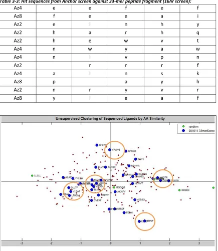
Table 3-3: Hit sequences from Anchor screen against 33-mer peptide fragment (16hr) ... 71
Figure 3-7:Clustering of Anchor Sequence Ligands by AA Similarity: ... 71
Figure 3-8:Streptavidin-Agarose Pulldown Assays for Anchor Ligand Binding Affinity: ... 72
Figure 3-9:CD Spectra of 33mer Epitope Fragment used in screening:. ... 73
Figure 3-10: HPLC-detected Immunoprecipitation Assay for Epitope Targeting Verification.. .. 75
Figure 3-11: ELISA Assay Verification of Epitope Targeting. ... 75
Figure 3-12: Ligand-Directed Labeling Diagram. ... 76
Figure 3-13: Fluorescent Gel Image to Confirm Cy5 Labeling ... 76
Figure 3-14: Images of Labeled and Unlabeled MALDI-TOF Spectra of Unlabeled (top) and Labeled (bottom) Proteins. ... 77
Figure 3-16: Trypsin Digested Sequence of PH Domain Protein.. ... 78
Figure 3-17: Compiled Crystal Structure of Fully Labeled Protein. ... 79
Figure 3-18: Images of Anchor Ligand in GFP-tagged WT and E17K PH Domain. ... 80
Chapter 4
Figure 4-1: Biotin – PEG5 – yleaf – Pra: ... 85
Figure 4-2: Screening Strategy for Biligand Determination: ... 88
Figure 4-3: Lys(N3) - yleaf - Tz - yksy - PEG5 – Biotin: ... 89
Figure 4-4: Screening Strategy for Triligand Determination: ... 91
Figure 4-5: Clustering of Biligand Sequence Ligands by AA Similarity: ... 95
Table 4-1: Hit Sequences from Biligand Screen ... 96
Figure 4-6: Pulldown Assay Results for Biligand Candidates: ... 96
Table 4-2: Hit Sequences from Triligand Screen ... 97
Figure 4-7: ELISA assays for affinity and selectivity of triligand candidates. ... 97
Figure 4-8: Structure of final triligand: ivdae – Pra – Lys (N3) – yleaf – Pra – Lys (N3) - yksy ... 98
Figure 4-9: Full ELISA curves of Anchor, Biligand, and Triligand. ... 99
Figure 4-10: Point ELISA of Triligand binding to Akt1 and Akt2. ... 99
Figure 4-11: PH Domain membrane binding in the presence of each ligand. ... 100
Figure 4-12: Expanded Inhibition Assay. ... 100
Figure 5-1: Azido - phenyl alanine. ... 103
Figure 5-2: Mirror-image phage display technique3. ... 104
Chapter 5
Table 5-1: Primers for AmpR Switch. ... 107Figure 5-3: pAC-DHPheRS-6TRN Plasmid. ... 108
Figure 5-4: Distribution of random nucleotides. ... 112
Figure 5-5: Distribution of amino acids. ... 113
Figure 5-6: Location of G6PD Mutations in protein. ... 113
Figure 5-7: Amino Acid Sequence of Exons 1 and 2 of G6PD.. ... 114
Figure 5-8: Crystal Structure of G6PD (1QKI). ... 115
Table 5-3: Primers for Colony PCR of Inserts.... 117
Figure 5-9: Formula for estimation of phage concentration. ... 118
Figure 5-10: Phage Click Screening Strategy... 119
Figure 5-11: Test of azide incorporation in e.coli. ... 120
Figure 5-12: Click-It Kit Visuablization of Azide in pIII coat protein. ... 121
Figure 5-13: Gel Image of Colony PCR. ... 122
Table 5-4: Hits from 13 Insert-Containing Phages, Figure 5-13 ... 123
Appendix A:
Table A-1: Solvent Compositions……….. 128Table A-2: Parts and Chemicals Commonly Ordered from Applied Biosystems ... 128
Table A-3: Parts and Chemicals Commonly Ordered from Sigma Aldrich ... 128
Table A-4: PulsedLiquid cLC Method ... 129
Figure A-2: Normal1 cLC Gradient ... 131
Figure A-3: Procise Screen for Backflushing a Line... 133
Figure A-4: Dip at front of spectra indicative of bad pump seal ... 135
Chapter 1
1.1
Protein-Catalyzed Click (PCC) Peptide Capture Agents
for Biomarker Detection and Therapeutics
Detecting cancer-associated biomarkers is a necessary step on the road to personalized medicine, as emerging therapeutics require the identification of specific patient populations that will respond to targeted therapies1. Methods for protein biomarker detection are highly desirable for rapidly screening changes in protein mutation status, monitoring patient treatment2, and simple point-of-care diagnostics3. Techniques that rely on detecting or monitoring protein levels mainly use antibodies for the capture and measurement of these proteins4. Antibodies, however, are biological reagents that are inherently unstable, vary from batch to batch, can exhibit high levels of cross-reactivity with other antibodies, and are expensive to produce5. Diagnostic assays are frequently prohibitively limited in both cost and stability due to the restrictions of the gold-standard antibody detection agents.
The Heath group has sought to alleviate the issue of peptide capture agent instability by relying exclusively on the use of unnatural amino acids. Because biological libraries are not conducive to this type of work, we have instead adopted a peptide screening method utilizing One-Bead, One-Compound (OBOC)8 chemically synthesized libraries on 90μm polystyrene beads. This technique trivializes the inclusion of any unnatural amino acid or structure that can be chemically synthesized, allowing for the use of biologically stable D - amino acids and azide-alkyne click chemistry handles in the library9.
The Sharpless group showed that the typical azide - alkyne click catalyst, Cu(I)10, speeds up the reaction but is only barely necessary for it to occur, and demonstrated the ability to replace this catalyst with the surface of a protein. They took advantage of this to assemble small molecule inhibitors for proteins by breaking up known inhibitors into two components and assembling two libraries – each one comprised of pieces similar to its original half of the inhibitor. One of these libraries of molecules was appended with a click handle, the other library with the opposite click handle. When two click reactants bound tightly to the protein surface and in close enough proximity to each other, the long dwell time of these reagents allowed for the click to occur without the use of Cu(I)11. In this way, they were able to bring the two libraries, which consisted of variations on the original inhibitor, together and use the surface of the protein to assemble the best possible small molecule inhibitor.
number of protein targets, and have been shown to exhibit a selectivity and affinity similar to those of monoclonal antibodies. They also can be readily integrated into all standard protein assay formats.
Chapter 2 of this thesis describes the technology development process that was undertaken to optimize the screening stages for the production of high-affinity ligands to targets of interest. Optimizing the in-depth screening procedure has allowed for the rapid expansion of this project in the past few years. This detailed in situ azide-alkyne click screening technology is now regularly used to develop peptide affinity agents that mimic the performance of antibodies 9-15. These affinity agents that maintain the stability of small molecules can be made to replace
biological reagents9,12,15, lowering the cost and increasing the robustness of detection assays13,14.
1.2
Epitope Targeting Strategies
medications immediately after they are given. In a diagnostic setting, such binders can be used to assay for the mutant protein within diseased tissues, and thus potentially provide clinical guidance for treatment decisions3.
A more ambitious application is the development of drugs that can selectively inhibit mutant proteins, and thus avoid those toxic side-effects that stem from the inhibition of the wild-type (WT) variants17 that reside in non-diseased tissues. Patients on therapies targeted very specifically to the mutations characteristic of their disease could show significant improvements without the toxic side-effects that stem from of the inhibition of the healthy, wild-type versions of these proteins17. A relevant example is compound CO-1686, which is an a epidermal growth factor receptor (EGFR) inhibitor specific for the T790M point mutation associated with certain non-small cell lung carcinomas. That drug, which is currently in clinical trials, is designed to minimize the toxicities (such as skin rash) that can appear when WT EGFR is targeted, since WT EGFR is expressed throughout the healthy tissues in the body18.
A challenge of drug targeting a single point mutation is that the mutation may not be directly associated with a binding pocket. The presence of a binding pocket is traditionally required for small molecule inhibitor development as is serves as a thermodynamic sink that can attract binders. This requirement does not hold for antibodies and, in fact, several examples of monoclonal antibodies directed against epitopes containing single amino acid mutations do exist19,2,20. However, antibodies do not readily enter the living cells that can harbor the mutated proteins21,22, and so, mutation-selective antibodies are typically only used as diagnostic reagents for staining fixed cells or tissues.
cell-penetrant inhibitors5. Our approach is inspired by the technique for developing an epitope-targeted monoclonal antibody (mAb). Such mAbs are made by injecting a small portion of the protein of interest containing the mutation (the epitope) into an animal and screening for an immune response that has the desired selectivity2,20,19. This approach can yield an antibody that exhibits focused binding to the specific designated area of the protein surface.
An all-chemical strategy for targeting PCC agent development against epitopes near phosphorylates sites was developed recently15. For that approach, an approximately 30-amino fragment representing the phosphorylated epitope of interest was synthesized, and a metalloorganic Zn-chelator was utilized to bind to the phosphate group and present an azide near that site. That epitope was then screened against a large (1 million element) one-bead-one-compound (OBOC) library of 5-mer alkyne-presenting peptides. Hits were defined as those compounds that bound to the synthesized epitope, and that were coupled to that epitope through a triazole linkage. PCC Agents with high selectivity for the epitope and the full protein, and with affinities as low as 19nM, were developed.
rendering it inactive and demonstrating the ability of these PCC agents to serve as targeted therapeutics.
1.3
In Situ
Click Screening Using Azide-Containing Phage
Display Libraries
Peptide screening technology has expanded incredibly in the past ten years since the inception of the PCC agent project. Using the protein-catalyzed click screens described above, PCC agents have been developed against only small chunks, or “epitopes” of proteins15, and various PCC agents that have shown to be unique inhibitors and activators of Akt kinase23,15, molecular imaging agents24, detection agents for anthrax14, suitable as third world detection agents for HIV13, as well as the single amino acid point mutation specific E17K agents.
The OBOC libraries have their drawbacks, however. The physical size of the library limits the number of total sequences that can be screened. A full library usually contains up to 106 members – only a portion of which are screened. The library screening and hit picking methods are exceptionally time-consuming and labor-intensive, hindering rapid peptide discovery. The sequencing of OBOC libraries is also done by either Edman degradation or MALDI TOF/TOF, rendering the sequencing process expensive, time-consuming, and reliant on expert knowledge. Many of these drawbacks are also a huge barrier to entry in this field, limiting the labs that would be able to assist in the advancement of the science. PCC agents could be produced significantly faster and cheaper with library display technology that would combine the advantages of the OBOC product screening techniques and library design with the rapid screening and sequencing of genetically displayed libraries.
M13 phage26. The Methanococcus jannaschii amber suppressor tRNATyr (MjtRNA) and the mutant M.jannaschii tyrosyl-tRNA synthetase (MjTyrRS) DNA can be contained in one plasmid that can be used to express these amber suppression tools in E.coli. In this system, the mutant synthetase is used to attach the unnatural amino acid azidophenylalanine to the tRNA in vivo, allowing for its incorporation into proteins. This tRNA recognizes the amber stop codon and should insert the amino acid in only that location, creating a new amino acid/tRNA combination that can be encoded into proteins.
Chapter 5 discusses the ongoing development of a screening technology that combines the in situ click screen advantages of the OBOC process with the rapid screening of large libraries characteristic of biological display systems. For this project, a phage display library containing azidophenylalanine for use in in situ click chemistry screening has been made and is being used to develop a PCC agent. These phage libraries can be screened in place of the OBOC peptide libraries described in previous chapters for the more rapid development of PCC agents.
1.4
References
1. Hanash, S. M.; Baik, C. S.; Kallioniemi, O., Emerging molecular biomarkers--blood-based strategies to detect and monitor cancer. Nature reviews. Clinical oncology 2011, 8 (3), 142-50. 2. Yu, J.; Kane, S.; Wu, J.; Benedettini, E.; Li, D.; Reeves, C.; Innocenti, G.; Wetzel, R.; Crosby, K.; Becker, A.; Ferrante, M.; Cheung, W. C.; Hong, X.; Chirieac, L. R.; Sholl, L. M.; Haack, H.; Smith, B. L.; Polakiewicz, R. D.; Tan, Y.; Gu, T.-L.; Loda, M.; Zhou, X.; Comb, M. J., Mutation-Specific Antibodies for the Detection of EGFR Mutations in Non–Small-Cell Lung Cancer. Clinical Cancer Research 2009, 15 (9), 3023-3028.
3. Rusling, J. F.; Kumar, C. V.; Gutkind, J. S.; Patel, V., Measurement of biomarker proteins for point-of-care early detection and monitoring of cancer. Analyst 2010, 135 (10), 2496-2511. 4. Borrebaeck, C. A., Antibodies in diagnostics - from immunoassays to protein chips. Immunology today 2000, 21 (8), 379-82.
5. Kodadek, T.; Reddy, M. M.; Olivos, H. J.; Bachhawat-Sikder, K.; Alluri, P. G., Synthetic molecules as antibody replacements. Accounts of chemical research 2004, 37 (9), 711-8. 6. Edwards, P. J.; LaPlante, S. R., Peptides as Leads for Drug Discovery. In Peptide Drug Discovery and Development, Wiley-VCH Verlag GmbH & Co. KGaA: 2011; pp 1-55.
8. Lam, K. S.; Lebl, M.; Krchňák, V., The “One-Bead-One-Compound” Combinatorial Library Method. Chemical Reviews 1997, 97 (2), 411-448.
9. Agnew, H. D.; Rohde, R. D.; Millward, S. W.; Nag, A.; Yeo, W.-S.; Hein, J. E.; Pitram, S. M.; Tariq, A. A.; Burns, V. M.; Krom, R. J.; Fokin, V. V.; Sharpless, K. B.; Heath, J. R., Iterative In Situ Click Chemistry Creates Antibody-like Protein-Capture Agents. Angewandte Chemie
International Edition 2009, 48 (27), 4944-4948.
10. Kolb, H. C.; Finn, M. G.; Sharpless, K. B., Click Chemistry: Diverse Chemical Function from a Few Good Reactions. Angewandte Chemie International Edition 2001, 40 (11), 2004-2021. 11. Lewis, W. G.; Green, L. G.; Grynszpan, F.; Radić, Z.; Carlier, P. R.; Taylor, P.; Finn, M. G.; Sharpless, K. B., Click Chemistry In Situ: Acetylcholinesterase as a Reaction Vessel for the Selective Assembly of a Femtomolar Inhibitor from an Array of Building Blocks. Angewandte Chemie International Edition 2002, 41 (6), 1053-1057.
12. Millward, S. W.; Henning, R. K.; Kwong, G. A.; Pitram, S.; Agnew, H. D.; Deyle, K. M.; Nag, A.; Hein, J.; Lee, S. S.; Lim, J.; Pfeilsticker, J. A.; Sharpless, K. B.; Heath, J. R., Iterative in situ click chemistry assembles a branched capture agent and allosteric inhibitor for Akt1. Journal of the American Chemical Society 2011, 133 (45), 18280-8.
13. Pfeilsticker, J. A.; Umeda, A.; Farrow, B.; Hsueh, C. L.; Deyle, K. M.; Kim, J. T.; Lai, B. T.; Heath, J. R., A Cocktail of Thermally Stable, Chemically Synthesized Capture Agents for the Efficient Detection of Anti-Gp41 Antibodies from Human Sera. PLoS ONE 2013, 8 (10), e76224. 14. Farrow, B.; Hong, S. A.; Romero, E. C.; Lai, B.; Coppock, M. B.; Deyle, K. M.; Finch, A. S.; Stratis-Cullum, D. N.; Agnew, H. D.; Yang, S.; Heath, J. R., A Chemically Synthesized Capture Agent Enables the Selective, Sensitive, and Robust Electrochemical Detection of Anthrax Protective Antigen. ACS Nano 2013, 7 (10), 9452-9460.
15. Nag, A.; Das, S.; Yu, M. B.; Deyle, K. M.; Millward, S. W.; Heath, J. R., A Chemical Epitope-Targeting Strategy for Protein Capture Agents: The Serine 474 Epitope of the Kinase Akt2. Angewandte Chemie International Edition 2013, 52 (52), 13975-13979.
16. Mardis, E. R., A decade's perspective on DNA sequencing technology. Nature 2011, 470 (7333), 198-203.
17. Chong, C. R.; Janne, P. A., The quest to overcome resistance to EGFR-targeted therapies in cancer. Nat Med 2013, 19 (11), 1389-1400.
18. Tjin Tham Sjin, R.; Lee, K.; Walter, A. O.; Dubrovskiy, A.; Sheets, M.; St Martin, T.; Labenski, M. T.; Zhu, Z.; Tester, R.; Karp, R.; Medikonda, A.; Chaturvedi, P.; Ren, Y.; Haringsma, H.; Etter, J.; Raponi, M.; Simmons, A. D.; Harding, T. C.; Niu, D.; Nacht, M.; Westlin, W. F.; Petter, R. C.; Allen, A.; Singh, J., In vitro and In vivo Characterization of Irreversible Mutant-Selective EGFR Inhibitors that are Wild-type Sparing. Molecular Cancer Therapeutics 2014.
19. Capper, D.; Zentgraf, H.; Balss, J.; Hartmann, C.; von Deimling, A., Monoclonal antibody specific for IDH1 R132H mutation. Acta neuropathologica 2009, 118 (5), 599-601.
20. Capper, D.; Preusser, M.; Habel, A.; Sahm, F.; Ackermann, U.; Schindler, G.; Pusch, S.; Mechtersheimer, G.; Zentgraf, H.; von Deimling, A., Assessment of BRAF V600E mutation status by immunohistochemistry with a mutation-specific monoclonal antibody. Acta neuropathologica
2011, 122 (1), 11-9.
21. Marschall, A. L. J.; Frenzel, A.; Schirrmann, T.; Schüngel, M.; Dubel, S., Targeting antibodies to the cytoplasm. mAbs 2011, 3 (1), 3-16.
22. Rondon, I. J.; Marasco; A., W., Intracellular AntibodiesS (Intrabodies) For Gene Therapy Of Infectious Diseases. Annual Review of Microbiology 1997, 51 (1), 257-283.
Chemistry Assembles a Branched Capture Agent and Allosteric Inhibitor for Akt1. Journal of the American Chemical Society 2011, 133 (45), 18280-18288.
24. Millward, S. W.; Agnew, H. D.; Pitram, S.; Lai, B. T.; Rohde, R. D.; Hardman, N., Protein catalysed capture agents for molecular imaging. Internation Hospital Equipment and Solutions
2013.
25. Wang, L.; Xie, J.; Schultz, P. G., Expanding the Genetic Code. Annual Review of Biophysics and Biomolecular Structure 2006, 35 (1), 225-249.
Chapter 2
Evolution of the OBOC Peptide Library
2.1
Introduction
2.1.1
Iterative In Situ Click Chemistry for Protein-Catalyzed Capture (PCC) Agent
Development
Previous work in the Heath lab demonstrated that the flexibility of chemically synthesized One-Bead, One-Compound (OBOC) peptide libraries could be combined with the selective power of the in situ click process to develop multi-peptide ligand capture agents that can serve as drop-in antibody replacements drop-in assays1. These peptide ligands can be made in large quantities entirely by robots, making the scale-up cheap and robust. They are also highly stable agents that can be used in a variety of assays, removing the need for the gold-standard antibodies in a variety of protein detection techniques2,3.
The technology as presented by Agnew, et al1 provided a solid foundation for the construction of these PCC agents, but the methods, discussed in section 2.3.1, were time-consuming and labor-intensive, making rapid ligand discovery very difficult. After this BCAii proof-of-concept PCC agent was completed, the next stage of technology development required an optimization of the techniques involved in order to increase the robustness and output of the overall process. This chapter describes the transformation of the OBOC iterative in situ click technology into an efficient and robust technique.
2.1.2
Prostate Specific Antigen (PSA)
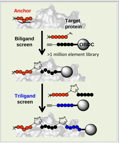
Figure 2-1: Iterative In Situ Click Screening Core Technology.
An anchor ligand that binds to the protein target can be appended with a click handle. In the presence of the protein and a OBOC library appended with the opposite click handle, the anchor can click onto the library to form a biligand. The click only occurs when the anchor and library bead are held long enough on the protein surface, so the protein selects ligands with high affinities and selectivities. This process can be repeated as many times as necessary.
Target
protein
Biligand
screen
Triligand
screen
OBOC
>1 million element library
X
X
X
X
Anchor
2.2
Materials and Methods
2.2.1
Standard Materials
All amino acids were purchased from Aapptec as the FMOC carboxylic acid with the standard TFA side-chain protecting groups. HATU (2-(7-Aza-1H-benzotriazole-1-yl)-1,1,3,3-tetramethyluronium hexafluorophosphate) and PEG5 (Fmoc-NH-PEG5-CH2CH2COOH, Fmoc-18-amino-4,7,10,13,16-pentaoxaoctadecanoic acid) were purchased from ChemPep. DIEA (diisoproylethylamine), TES (triethylsilane), and TFA (trifluoroacetic acid) were purchased from Sigma. TentaGel beads were purchased as 90μm S-NH2 beads, 0.29mmol/g, 2.86x10⁶ beads/g from Rapp Polymere (Germany), and Rink Amide resin was purchased from Anaspec.
2.2.2
Peptide Library Construction
Peptides and peptide libraries were synthesized by hand until the summer of 2009, when they were then synthesized on a Titan 357 split-and-mix automated peptide synthesizer (Aapptec) via standard FMOC SPPS coupling chemistry5 using 90μm TentaGel S-NH2 beads. Libraries
contain 18 D-stereoisomers of the natural amino acids, minus cysteine and methionine (unless otherwise stated), at each of five randomized positions and an azide or alkyne in situ click handle. At least a five-fold excess of beads is used when synthesizing libraries to ensure efficient oversampling of each sequence. Amino acid side-chains are protected by TFA labile protecting groups that are removed all at once following library synthesis.
Figure 2-2: OBOC Peptide Library
2.2.3
Bulk Peptide Synthesis
Bulk synthesis of peptide sequences was performed using standard FMOC SPPS peptide chemistry on either the Titan 357 automated peptide synthesizer (AAPPTEC) or a Liberty 1 microwave peptide synthesizer (CEM Corporation). The typical scale was 300mg on Rink Amide Resin, unless otherwise noted. Peptides were cleaved from the beads with side-chains deprotected using a 95:5:5 ratio of TFA: H2O: TES. The peptides were purified on a prep-scale Dionex U3000 HPLC with a reverse-phase C18 column (Phenomenex).
2.2.4
Typical Screening Protocol for Fluorescent Dye-labeled Protein Target
Detection
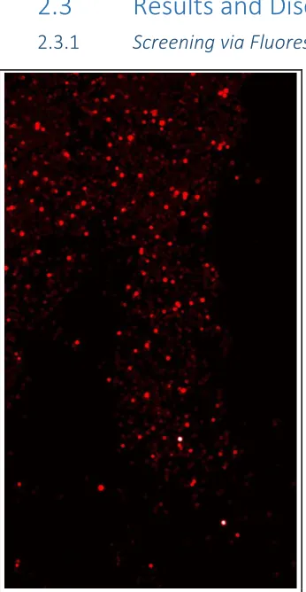
Pro 5.1 microarray scanner at 635nm to view beads containing bound fluorescent protein target. The dye saturated the color signal of the GenePix, and the hit “beads” that were considered appeared white in a sea of red, due to the background auto fluorescence of the TentaGel library (Figure 2-4). These hit beads were then removed from the microscope slides using a needle, stripped of protein with 7.5M pH = 2.0 Guanadine-HCl buffer, rinsed in water, and sequenced via Edman degradation on an Applied Biosystems Procise CLC 494 system.
2.2.5
Typical Screening Protocol for Antibody Signal Amplification Target Only
Screens
2.2.6
Typical Screening Protocol for an Anti-Screen
The library beads (typically 250-500mg) swelled in 1xTBS were blocked 2 hours to overnight in 5% milk in 1xTBS, washed three times with 1x TBS, then incubated with an off-target protein in 0.5% milk in 1xTBS for one hour on the shaking arm at room temperature. The beads were washed three times with 1x TBS, then incubated with the anti-off-target protein - alkaline phosphatase conjugated antibody in 0.5% milk for one hour at room temperature. The antibody used here must be the same antibody used in the target screen in order to ensure that the library members that bind to this antibody are removed and not mistaken for hits. The library resin was then washed three times with high salt buffer and let shake for one hour in high salt at room temperature before being washed three times with BCIP buffer (100mM Tris-Cl, 150mM NaCl, 1mM MgCl2, pH = 9.0) and developed by adding 15mL BCIP buffer plus 13μL BCIP and 26μL NBT. The beads that turned purple bound to both mutant and wildtype protein or to the detection antibodies, and were discarded. The beads that remained clear after this step were picked and washed with guanidine-HCl to remove any bound proteins.
2.2.7
Typical Target Screening Procedure During a Multi-Step Screen (Figure 2-3)
The library beads were blocked in 5% milk in 1x TBS for two hours to overnight. They were then washed three times with 1x TBS. The target protein and anchor peptide or small molecule targeting agent6 were pre-incubated in 3-5mL of 0.5% milk in an approximately a 10:1 ratio, ensuring the same concentration of anchor peptide used in the preclear. This solution was added to the blocked library beads and incubated for either 5 hours or overnight to allow an in situ click reaction to occur. In the morning, the beads were washed three times with 1x TBS, thenincubated with the same dilution of an anti-target alkaline phosphatase conjugated antibody that was used in the anti-screen in 0.5% milk for one hour. The beads were then washed three times with a high salt TBS, then incubated on the shaking arm for one hour with the high salt buffer. They were then washed three times with BCIP buffer and developed as previously. Hit beads turned purple and were removed and washed in NMP for four hours to decolorize, then guanidine-HCl to denature and remove and remaining protein.
2.2.8
Typical Screening Protocol for a Preclear
phosphatase was then incubated with the library for 1 hour after it was washed three times in 1xTBS. The beads were washed with a high-salt TBS buffer three times, then were left to shake in high salt buffer for one hour. The beads were then washed three times with BCIP and developed as for the anti-screen. After one hour, the purple beads were removed by pipette and discarded. The remaining beads were incubated in NMP 4 hours to remove trace purple precipitate from the BCIP/NBT reaction, then were washed five times with methanol, five times with water, five times with TBS and blocked overnight in 5% milk.
2.2.9
Typical Screening Protocol for a Click Product Screen
The beads that pass through the target and anti-screen were washed three times with 1x TBS. They were then incubated with a 1:10,000 dilution of either streptavidin – alkaline phosphatase conjugate or anti-biotin antibody (whichever was used in the preclear) in 0.5% milk for one hour. The beads were washed three times with high salt TBS then let shake for one hour with high salt buffer before being washed three times with BCIP buffer and developed as previously. The beads that turned purple contained the anchor peptide covalently bound to the bead and had formed a protein-catalyzed in situ click reaction. These beads were collected and stripped with guanidine-HCl for one hour, washed ten times with water, and sequenced via Edman degradation.
2.2.10
Peptide Sequencing Strategies
next cycle. Meanwhile, the PTC amino acid is then analyzed via HPLC, and the peak is compared to standards of all of the PTC-amino acids in order to determine the residue. One cycle per amino acid residue is performed and analyzed, providing the sequence of the peptide on the hit library bead7. This method is slow, but highly accurate and has been automated by Applied Biosystems into the Procise CLC 494 Automated Edman Degradation machine used by Caltech.
Hit peptide sequences can also be determined through MALDI-TOF/TOF MS. For this method, the library must be specially made. The peptide must be attached to the library through a methionine amino acid, and no other methionine can be present in the library. The isobaric amino acids, isoleucine and leucine, lysine and glutamine, are doped by anther amino acid in order to properly call the sequence by mass. Glutamine is doped with a 6% molar equivalent of glycine, and isoleucine is doped with a 7% molar equivalent of alanine. While reading the mass of these amino acids on the MALDI, any residue that has one of these amino acids can be distinguished by the presence or absence of the small satellite parent mass corresponding to the same sequence plus glycine or alanine8.
2.3
Results and Discussion
2.3.1
Screening via Fluorescent Dye-labeled Protein Target Detection
The initial OBOC peptide screening strategies developed by Heather Agnew1 relied on a fluorescent dye-labeled protein in order to detect hit binding. The target protein of interest was labeled with a dye, and any library beads that bound to the target were detected on a GenePix microarray reader. As seen in Figure 2-4, the TentaGel library beads also auto-fluoresce, meaning that all screens conducted in this fashion were highly subjective, and the hit quantity depended entirely on the gain settings of the microarray. AlexaFluor-647 was also the only dye that was used, as the beads auto fluoresce the least in the range of this dye. These hits were mostly picked using a light microscope, meaning that the images from the microarray had to be used as a “map” to guide the bead picker to the correct clear bead on a slide of thousands. This process was highly inefficient, requiring up to an hour to pick each individual hit bead. These picked hits were always imaged again on the GenePix to ensure that each bead that had been selected was a highly fluorescent bead, indicating that the correct one had been chosen based on the map. It was possible to use a COPAS automatic bead sorter to separate out the hit beads, though one was not available at Caltech.
The sequences from a typical fluorescent target screen are shown in Table 2-1. The hits were generally dominated by the positively charged residues, arginine and lysine. This overwhelming charged signal is most likely due to the overall (-3) charge on the AlexaFluor 647 Figure 2-4: Image of Hit Beads on GenePix
Microarray Scanner. The bright white beads are saturating the fluorescence and are
considered “hits” above the background
[image:36.612.105.278.79.413.2]dye,9 which is attracting the positively charged amino acid sequences and creating a significant level of noise in the final hits. Most screens had to be run many times in order to find enough quality hit sequences, meaning ones that did not contain almost exclusively arginine and lysine residues, because of this high background. Generally, a hit that contained 3 or more positively charged amino acids was considered background and removed from the pool. One screen rarely yielded more than a handful of hits that appeared to be binding to the surface of the protein and not just to the dye.
Focused screens were also used in order to hone in on target-binding peptide sequences. The focused libraries used in these screens were designed based on histograms of the amino acids that were seen at each library position, meaning X1 -> X5 as seen in Figure 2-2, after the removal of the dye label background sequences. As can be seen in Figure 2-5, in this particular PSA screen, there were only six amino acids that were seen at position 2, so only these six amino acids were built into the focused library at position 2. This reduction in total amino acids present in each position allowed for the synthesis of a much smaller library that could be oversampled in each screen to permit a more thorough sampling of the sequence space. Only about 100mg of beads were usually screened, but 100mg could frequently oversample the sequence space of a focused library, compared to that of naïve libraries where less than half of the space was sampled. Due to this increase in sequence space sampling, focused libraries were generally extended by one or two amino acid positions in the hopes that a slightly longer peptide would have a higher affinity Figure 2-5: Histogram of Position X2 in PSA Screens. The hits from
multiple PSA screens were pooled and analyzed. This sample chart shows the frequency of an amino acid at position X2 in the library, and is used to synthesize the focused library.
0 2 4 6
i h w q a t
Fr e q u e n cy Amino Acid
and selectivity for the protein target. The screening was then repeated with the focused libraries, and the same process for analyzing hits was repeated until the peptide sequences converged in sequence homology and produced a peptide ligand that showed near μM affinity for the protein target.
[image:38.612.128.522.361.618.2]This convergence frequently required the use of two to three separate focused libraries with accompanying screening and sequencing. The overall time required to determine one peptide ligand that bound to the target protein of interest could easily take more than six months. These ligands also regularly bound in the range of low μM affinities, which are generally considered to be fairly weak binders.
Table 2-1: Screens from Sample Target Screen Using Fluorescent Protein Detection. This screen
was performed against PSA protein labeled with AlexaFluor 647 dye. Note the high prevalence of “r” and “k”
positively charged amino acids. See Figure 2-2 for a visualization of the X amino acid positions on bead.
X1 X2 X3 X4 X5
y r r r r
r i f r r
r f l r a
r r k r f
m r r w r
r r r w p
r r w i r
r r r f l
r l r w r
r f r i r
l s r r r
r r r y t
r r m r w
r r k p r
f y r r r
r k w l w
2.3.2
Screening via Antibody Signal Amplification Target Only Screens
Detecting hit peptides via fluorescence was a very time-consuming process in which the high noise from the overwhelming
presence of positively charged amino acids meant that very little meaningful output was obtained. For this reason, a new method of screening was developed using a tag-less protein to switch the screening focus from the charged dye label back to
the target. This technique relied on anti-target antibodies conjugated to alkaline phosphatase, which is an enzyme that can form a dark purple precipitate in the presence of its BCIP/NBT substrate. This meant that any “hit” now showed up as a very dark purple bead in a sea of clear. The label-less detection technique, therefore, provided the additional benefit of a colorimetric readout of a hit, allowing for the much easier separation of these beads from the rest of the library.
time, as all of the hits could be picked in the time it used to take to pick one. With this increase in both sampled sequence space and in the overall signal to noise seen in the sequences, hit quality and screening speed improved dramatically in a much shorter overall time.
2.3.3
Introduction of an Anti-screen
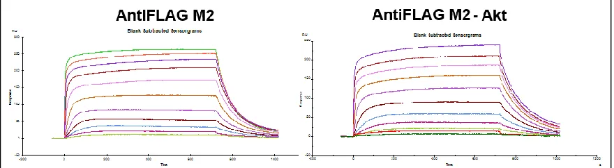
The antibody development technique dramatically improved the quality of hit peptides by visual inspection (Table 2-1 versus Table 2-2), but also introduced a hidden source of noise into the screens. The presence of several different antibodies and a new detection agent in the screen itself provided more “off-target” sources of library binding. This was conclusively demonstrated by Steve Millward while screening for an Akt capture agent. He developed a biligand using the standard in situ click chemistry technique with antibody development, and proceeded to test the affinity of this ligand via SPR. The SPR was set up to immobilize an anti-FLAG antibody (the same used in screening) to the flow cell in order to capture the much less stable Akt protein that might
Table 2-2: Screens from Sample Antibody Amplification Screen Using BCIP/NBT Protein
Detection. Screen was performed against unlabeled (PSA), detected with PS2 mouse mAb anti-PSA antibody
and anti-mouse-AP secondary antibody with BCIP/NBT readout.
X1 X2 X3 X4 X5
n g m e d
e t q m d
w t d e m
s e d d t
a n d e e
n y d p e
G n m d d
e d v l i
f e n d a
e i n e l
v e f G e
e h d a y
d e t a t
i w n m e
y d d s l
d d e a G
[image:40.612.119.517.194.449.2]not survive the required EDC/NHS coupling step. A blank flow cell of only Anti-FLAG antibody without Akt was used as a chip blank. The data from these SPRs is seen in Figure 2-7. The sensorgram on the right shows binding to the Akt, as to be expected, but the sensorgram from the blank flow cell on the left shows an identical signal. In conjunction with data (not shown) from the anchor ligand that has almost no binding to the anti-FLAG flow cell, we can conclude that the biligand is actually binding to the anti-FLAG antibody, present in both of those flow cells, and not to the desired Akt target protein. It is only logical that we would see “hits” of peptide sequences that bind to these antibodies, because the presence of the detection antibody bound to a library bead would show BCIP precipitation exactly like the presence of the target protein. A new screening step was needed that would remove the signal seen from the binding of these other proteins used in the screening process.
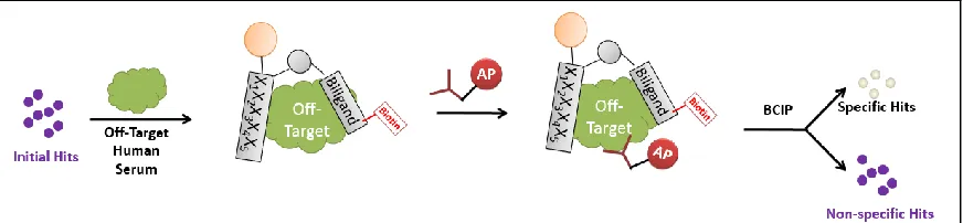
Around this time, there was interest in developing capture agents for proteins containing post-translational modifications, such as phosphorylations or glycosylations. It was hypothesized that hits specific for a post-translational modification could be discovered by screening against the protein target containing the modification, then anti-screening against the protein target with Figure 2-7: SPR Data from Akt Biligand. Data from Steve Millward’s Akt Biligand Capture Agent. An anti-FLAG antibody was immobilized onto an SPR chip via standard EDC/NHS coupling techniques. It was used to capture a FLAG-tagged Akt protein for testing. As seen from the figure on the left, the capture agent bound equally as well to the flow cell immobilized with only Anti-FLAG antibody, supposed to be the chip blank, as to the one on the right that also
[image:41.612.105.545.383.504.2]the post-translational modification removed, since everything else in the screen would be identical (Figure 2-8). These screens entailed first “target screening,” as per usual antibody detection screens, to find all of the hit beads that have an affinity for the target. These beads were then be stripped of their purple color and bound proteins and incubated with the off-target protein that had the post-translational modification removed. Any purple hits from the anti-screen were thrown out as not specific for the modification, since they demonstrated binding in a screen that did not contain the site of interest. This new screening step has the added benefit of removing all of the hits that also have an affinity for the antibodies or developing solution that was used in the screen. An anti-screen like this would have prevented the development of a biligand with an affinity for the anti-FLAG antibody, as these hits would have been detected in both the target and the anti-target screen, and would have been discarded.
The anti-screen is an important step that is now incorporated into each screen that is run in the lab, and is responsible for a significant reduction in background hits. For example, an anti-screen that was run for the PSA protein eliminated 91% of the hit beads from the target anti-screen, indicating that approximately 91% of what was previously considered to be a target hit was just background. For visualization purposes (Table 2-3), this means that a screen run with 250mg of beads went from 167 hits down to 15 after this step. This cut down on not only sequencing and hit analysis/testing time, but also eliminated the time that was usually spent trying to tease out signal from noise. Focused screens were also no longer necessary, as that step was designed to help enrich for signal, eliminating a significant chunk of time necessary for developing a capture agent.
incubated with anywhere from 1% - 25% human serum to remove even the marginally sticky peptides from the pool of potential candidates.
2.3.4
Introduction of a Click Product Screen
The in situ screening process has an inherent screening advantage that had not yet been exploited. A covalently-linked product is formed on the surface of the bead during the screen that can be detected separately from target binding. This means that in addition to probing the library for beads that bind to the target, the library can be searched additionally for the presence of the in situ click product – a completely complementary screen.
[image:43.612.108.544.126.227.2]to the library will be removed and not detected by either the streptavidin conjugated to alkaline phosphatase or an anti-biotin antibody. These detection agents will bind to the biotin label on the anchor that will only be present after a covalent reaction has occurred, and can therefore detect which library members have formed a click product (Figure 2-9).
Continuing the comparison with the PSA screens from above, only 7 of the 15 remaining beads after the anti-screen showed the presence of a click product. The other 8 beads could very easily have been hits that would be a different anchor ligand – a peptide ligand that is binding specifically to the target protein, but is not close enough to the original anchor for a click to form. The sequences from these hits, shown in Table 2-4, are very nearly identical peptides, and contrast sharply with the previously identified hits from the anti-screen in Table 2-2. This indicates that the sequences are more than likely all binding very strongly to the exact same location and in close proximity to the anchor ligand, allowing for the formation of the click product.
The product screen is an elegant step in the screening process that allows for the very specific narrowing of the sequence space. It has become such a huge part of the success of the OBOC capture agent development process that naïve anchor screens, which inherently cannot Table 2-3: PSA Screening Statistics. These hit bead statistics
are taken from a screen against PSA. The percent column indicates the percent of beads that passed from one stage of the screen to the next.
Beads Percent Start 375,500
Target Screen 167 0.04%
Anti-screen 15 9%
Product Screen 7 47%
Table 2-4: PSA Hit Bead Sequences from Product Screen. The product hits shown in Table 2-3 were sequenced. There is an enormous sequence homology, meaning that the same part of the target is being targeted. The end of the last sequence and the 7th hit were lost due to machine error.
X1 X2 X3 X4 X5
Y G w r e
Y d w r q
L G w r e
e G w r e
a d w r q
include product screens, have been completely eliminated. This switch to all in situ click screens has greatly increased both the specificity and affinity of the original anchor ligands, dramatically improving the quality of the final PCC agent. Details of the rationale and results from these more targeted screens can be seen in Chapter 3.
2.3.5
Introduction of a Preclear
explain why the biligands showed no binding to the protein – the anchor could no longer even bind to the target with another ligand, potentially blocking those binding sites. It also explains why the secondary ligands showed no affinity for PSA. They were not ligands that bound to the target, and wouldn’t have an affinity for it.
To counter this effect, a new screening step was added at the beginning of the process to remove all of the library peptides that bound to the anchor ligand before the anchor ligand even saw the target protein (Figure 2-10). These screens still detect the biotin label on the anchor ligand, and the detection with streptavidin or anti-biotin in this “preclear” step eliminates the need to use these detection agents in the anti-screen. The preclear screens generally remove 1-10% of the library beads, depending on the library, and also reduce the percentage of beads that need to be removed in the anti-screen.
2.3.6
Use of Alkyne Versus Azide Libraries
residues were probably not cleaving from the beads, and the MALDI TOF/TOF was unable to identify a parent peak that contained the fixed alkyne amino acid. The alkyne-containing amino acid was the N-terminal residue, the first residue that needed to cleave via Edman, and anything modifying this amino acid would affect the cleavage. It was hypothesized that the BCIP/NBT developing solution was modifying these amino acids, which was confirmed by the use of C-terminal alkyne libraries. Even after undergoing four screening steps, the libraries still sequenced correctly using Edman degradation up to the alkyne amino acid. These same library hits, though, were not able to be sequenced using MALDI-TOF/TOF. Because the TOF/TOF would be greatly affected by an unknown change to an amino acid, it was assumed that the alkyne was somehow being modified during these screening steps. For this reason, azide-containing libraries are now always used when undergoing more than three screening steps, unless a C-terminal alkyne library with Edman degradation sequencing is appropriate.
2.3.7
Typical Flow of Screening
With a multi-stage screening process now in place, the some of the steps need to be conducted in a certain order to achieve the correct results. The first step is the preclear. This occurs before the anchor ligand sees the protein target, and has a chance to form legitimate clicked-hit peptides on bead. These screens look for anything that binds to streptavidin, alkaline-phosphatase, BCIP/NBT, and the anchor peptides. Usually, a screen begins with 300-500mg of library beads, and 1-10% are removed. Typically, any bead that has turned even the lightest shade of purple is removed in order to reduce the overall background as much as possible. This means that any bead that passes through this stage of the screening process has remained clear.
The next step is the target and click-catalyzed screen. The beads that remained clear in the preclear are incubated with the target of interest and the anchor ligand overnight for a click reaction to occur. These beads are then probed for the presence of target. Any bead bound to target will turn purple, and passes through to the next stage of screening. Even though the on-bead click has occurred during this screen, probing for the click product occurs at a later stage.
The hits from the target screen are then decolorized and incubated with an off-target protein or proteins. Any library bead that binds and turns purple in this screen demonstrates off-target interactions with other proteins, and is removed from the pool. At the end of this screen, only beads that remain entirely clear are kept. Even slight purple can indicate undesirable interactions and background binding, and are removed from the pool of hits.
clear-purple-clear-purple pattern of hit detection also ensures that the beads are behaving properly at each stage in the process.
Screens following this pattern now have several produced high-affinity ligands that are very selective to their target of interest. This methodology has an incredibly high success rate that is only getting better as the process continues to grow and develop.
2.4
Conclusions
2.5
Acknowledgements
The work described in this chapter was done in conjunction with Heather Agnew and Steve Millward. The initial OBOC screening techniques used were developed by Heather, and the colorimetric screening development was done by Steve. The rest of the work described herein was performed in conjunction with both Heather and Steve, as well as Arundhati Nag and Rosemary Rohde.
2.6
References
1. Agnew, H. D.; Rohde, R. D.; Millward, S. W.; Nag, A.; Yeo, W.-S.; Hein, J. E.; Pitram, S. M.; Tariq, A. A.; Burns, V. M.; Krom, R. J.; Fokin, V. V.; Sharpless, K. B.; Heath, J. R., Iterative In Situ Click Chemistry Creates Antibody-like Protein-Capture Agents. Angewandte Chemie
International Edition 2009, 48 (27), 4944-4948.
2. Farrow, B.; Hong, S. A.; Romero, E. C.; Lai, B.; Coppock, M. B.; Deyle, K. M.; Finch, A. S.; Stratis-Cullum, D. N.; Agnew, H. D.; Yang, S.; Heath, J. R., A Chemically Synthesized Capture Agent Enables the Selective, Sensitive, and Robust Electrochemical Detection of Anthrax Protective Antigen. ACS Nano 2013, 7 (10), 9452-9460.
3. Pfeilsticker, J. A.; Umeda, A.; Farrow, B.; Hsueh, C. L.; Deyle, K. M.; Kim, J. T.; Lai, B. T.; Heath, J. R., A Cocktail of Thermally Stable, Chemically Synthesized Capture Agents for the Efficient Detection of Anti-Gp41 Antibodies from Human Sera. PLoS ONE 2013, 8 (10), e76224. 4. Stenman, U.-H.; Leinonen, J.; Zhang, W.-M.; Finne, P., Prostate-specific antigen. Seminars in Cancer Biology 1999, 9 (2), 83-93.
5. Coin, I.; Beyermann, M.; Bienert, M., Solid-phase peptide synthesis: from standard procedures to the synthesis of difficult sequences. Nature protocols 2007, 2 (12), 3247-56. 6. Nag, A.; Das, S.; Yu, M. B.; Deyle, K. M.; Millward, S. W.; Heath, J. R., A Chemical Epitope-Targeting Strategy for Protein Capture Agents: The Serine 474 Epitope of the Kinase Akt2. Angewandte Chemie International Edition 2013, 52 (52), 13975-13979.
7. Procise Protein Sequencing System.
http://tools.lifetechnologies.com/content/sfs/manuals/cms_041125.pdf (accessed May 1). 8. Lee, S. S.; Lim, J.; Tan, S.; Cha, J.; Yeo, S. Y.; Agnew, H. D.; Heath, J. R., Accurate MALDI-TOF/TOF sequencing of one-bead-one-compound peptide libraries with application to the identification of multiligand protein affinity agents using in situ click chemistry screening. Anal Chem 2010, 82 (2), 672-9.
9. Sobek, D. J.; Aquino, C.; Schlapbach, D. R., Analyzing the Properties of Fluorescent Dyes Used for Labeling DNA in Microarray Experiments. In BioFiles, Simga-Aldrich: Online, 2011; Vol. 6.3.
Chapter 3
Development of a PCC Agent Selective for the
3.1
Introduction
3.1.1
The E17K Mutation in the Pleckstrin Homology Domain of Akt1
Akt1 kinase plays a critical role in the PI3K signaling pathway,1 the activation of which is closely linked to tumor development and cancer cell survival2. The phosphorylation of
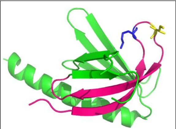
regulatory amino acids (Ser474 and Thr308) on Akt occur through the localization of Akt to the cell membrane through its membrane-binding Pleckstrin Homology Domain (PH Domain). These phosphorylations activate the Akt protein, which can then activate many other downstream signaling pathways2. The recently discovered E17K mutation in the PH Domain of Akt1 results in an increased affinity for the phosphatidylinositol-3,4,5-trisphosphate (PtdIns(3,4,5)P3, or PIP3) substrate at the cell membrane (Figure 3-1)3. This switch from a negatively charged glutamic acid to a positively charged lysine amino acid in the PIP3 binding pocket causes this mutant protein to have a four times higher affinity for the PIP3 substrate. This increased affinity causes the Akt1 to be bound to the cell membrane, and hence activated four times longer than in healthy, wildtype cells. Consequently, this deregulated recruitment of Akt1 to the cell membrane causes constitutive activation of the PI3K pathway, which has been shown to be sufficient to induce leukemia in mice3. The oncogenic properties of the driving E17K single point mutation make it a target for specific detection and inhibition.
3.1.2
A General Strategy for Targeting Single Amino Acid Point Mutations in
Proteins
Targeting single amino acid point mutations in proteins is becoming a necessary step in the era of personalized medicine, and methods for the detection of these mutant protein biomarkers are highly desirable for guiding treatment decisions4. Thus, there is a need for an approach to identify small molecules that can be generally targeted against epitopes containing single amino acid point mutations, and can also potentially be developed into cell-penetrant inhibitors. Previously, a strategy was developed for targeting the phospho-epitopes by chemically synthesizing the surrounding chunk of protein and focusing the site of the in situ click screen by attaching an azide click handle to a phosphate chelating group.5 This method has been generalized by directly substituting an alkyne click handle into the chemically synthesized peptide epitope. For this work, the peptide represents the epitope of Akt1 containing the E17K mutation, an attractive target due to the oncogenic nature of this mutation3. That target is subjected to an in situ click screen against an OBOC peptide library of 5-mers (comprehensive in 18 amino acids),
each terminated in an azide presenting amino acid.