Original citation:
Facet Injection Study team, (2014) Diagnostic assessment manual. University of Warwick, Warwick Medical School, Clinical Trials Unit.
Permanent WRAP URL:
http://wrap.warwick.ac.uk/78982
Copyright and reuse:
The Warwick Research Archive Portal (WRAP) makes this work by researchers of the University of Warwick available open access under the following conditions. Copyright © and all moral rights to the version of the paper presented here belong to the individual author(s) and/or other copyright owners. To the extent reasonable and practicable the material made available in WRAP has been checked for eligibility before being made available.
Copies of full items can be used for personal research or study, educational, or not-for-profit purposes without prior permission or charge. Provided that the authors, title and full bibliographic details are credited, a hyperlink and/or URL is given for the original metadata page and the content is not changed in any way.
Publisher’s statement:
Facet Injection Study – Diagnostic Manual © 2014
A note on versions:
Diagnostic Assessment Manual
Version 1.0 Date:12-12-2014 Recruiting Site: [enter site name ]
Contact details and further information
This manual has been developed by Dr Mindy Cairns and members of the FIS team.
If you require further information or support during the trial please contact (email
preferable):
Dr Mindy Cairns (Post Doctorate Research Fellow & Chartered
Physiotherapist)
Telephone:
01707 285 288
E-mail:
[email protected]
Online support and Discussion Forum:
http://fis-project.ucoz.co.uk/If you have any questions, queries or would just like to share experiences throughout
the programme, then please do use the Discussions Forum. The online Discussion
Forum is checked regularly by Harbinder and Mindy and a response to postings, if
required, will be given within 48 hours.
Some direct observation of carrying out the diagnostic assessments by participating
physiotherapists may be monitored.
General enquiries
: Facet Injection Study email:
[email protected]
Acknowledgements
Contents
Introduction ... 4
Diagnostic criteria ... 4
Standardisation ... 4
1. Increased pain unilaterally or bilaterally, on lumbar para-spinal palpation ... 5
2. Increased low back pain on one or more active movements ... 5
Extension (more than flexion)... 5
Rotation ... 5
Extension/side flexion ... 5
Extension/rotation ... Error! Bookmark not defined. 3. Radicular symptoms ... 7
4. Absence of sacro-iliac pain ... 8
SIJ Clinical Prediction Rule ... 8
Sacro-iliac joint pain provocation testing ... 9
Additional data collection – exploratory only ... 13
Laslett’s clinical prediction rule for facet joint pain ... 13
No exacerbation on rising from flexion ... 14
Appendix 1: Sequencing of diagnostic testing ... 15
Appendix 2: Combined movements ... 16
Appendix 3: Procedures undertaken within Facet Injection Study Diagnostic Assessment visit ... 17
References: ... 21
Table 1: Diagnostic criteria for trial ... 4
Table 2: SIJ pain-provocation tests for exclusion criteria ... 9
Table 3: Components in Laslett’s Clinical Prediction Rule ... 14
Figure 1: Testing position for extension side flexion and extension rotation ... 6
Figure 2: Extension side flexion ... 6
Figure 3: Extension rotation ... 7
Figure 4. Radicular pain ... 8
Figure 5: Distraction test: hand positioning and alternate hand positioning ... 10
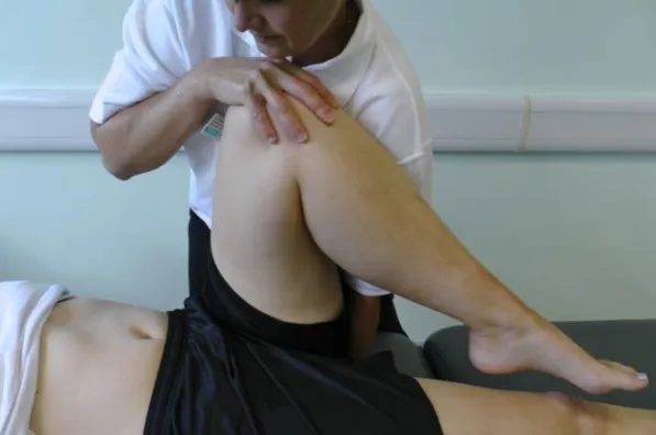
Figure 6: Thigh thrust (PPP) ... 11
Figure 7: Gaenslen’s test ... 12
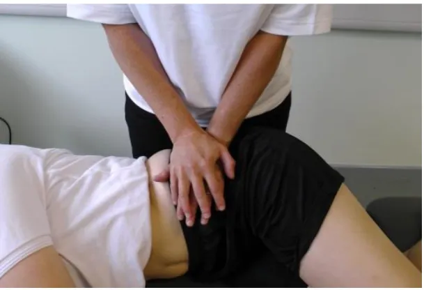
Figure 8: The compression test ... 12
Introduction
There is considerable diagnostic uncertainly about how to identify people with pain of facet joint origin amongst the wider chronic low back pain population. Therefore the diagnostic criteria used in this trial have been drawn from the available evidence base and following consensus gained from a range of experts and clinicians.
Diagnostic criteria
A summary of the diagnostic criteria is shown in Table 1. Criterion 1 and 2 cover the issue of presence of pain on palpation or symptom reproduction on movement testing. The second two criteria relate to the absence of symptoms, namely radicular symptoms and sacro-iliac pain. Details of testing procedures are provided under the relevant headings. The sequencing of testing is shown in appendix 1.
Standardisation
In order to maximize reliability and reproducibility of application between recruitment centres, and ensure maximum validity of tests, it is vital that tests are undertaken in a standardized manner. Any specific handling or positioning requirements for tests is detailed in the relevant sections. It is particularly important with combined movement testing that all components of the movement are maintained to ensure accuracy, however, as with all tests, ensure that participants’ comfort is maintained and that patients are only taken as far into range that is clinically appropriate. Therapists should rely on their clinical reasoning and judgement when performing diagnostic tests.
Table 1: Diagnostic criteria for trial
1. Increased pain unilaterally or bilaterally, on lumbar para-spinal palpation
AND
2. Increased low back pain on one or more of the following;
extension (more than flexion)
rotation
extension/side flexion*
extension/rotation* AND
3. No radicular symptoms (defined as pain radiating below the knee or
objective neurological signs above the knee#) AND
4. No sacro-iliac joint pain elicited using a pain provocation test.
*Both tests representative of regular compression patterns (Edwards, 1999)
1. Increased pain unilaterally or bilaterally, on lumbar para-spinal palpation
This is defined as pain when manually palpating over the articular pillar of the vertebral column (Maitland et al., 2005) i.e. not centrally over the spinous process. As it is known that reliability of correctly identifying specific spinal level is low (Billis et al., 2003); a positive finding is purely the presence of pain.
2. Increased low back pain on one or more active movements
In this criterion, it is necessary for at least one active movement to increase a participants’ back pain.
Extension (more than flexion)
Testing lumbar spine extension should be undertaken in standing. Before extension is tested, flexion in standing should be tested to ensure a comparator movement. Any
deviation in movement or shift should be noted and response from rising from flexion should also be recorded (See “No exacerbation on rising from flexion”).
Rotation
Lumbar spine rotation should be tested in standing and the quality of movement and approximate range noted.
Extension/side flexion
The tests of extension/side flexion and extension/rotation are examples of regular compression patterns (Edwards, 1999) and initial, small scale study has indicated that a regular compression pattern may be useful in identifying patients who will respond favourably to facet joint pain (Challinor et al., 2013) .
Both extension side flexion and extension rotation are undertaken in standing with the patient standing against a plinth positioned at approximately level with the ASISs to provide support (See Figure 1).
Figure 1: Testing position for extension side flexion and extension rotation
[image:7.595.77.271.422.715.2]
Figure 3: Extension rotation
This test is undertaken in standing and the patient is taken passively into full extension which is maintained whilst full passive rotation is applied (Maitland et al., 2005) (See Figure 3)
Care should be taken to ensure that all components of the movement are maintained during the combination to ensure no component is lost. As with all tests, ensure that participants’ comfort is maintained and that patients are only taken as far into range that is clinically appropriate.
The extension/rotation test also has high sensitivity (100%) for 95% pain reduction after a screening facet joint block if the ER tests are negative bilaterally (Laslett et al., 2006).
3. Radicular symptoms
For the trial, radicular symptoms are defined as pain radiating below the knee (See Figure 4) or objective neurological signs using a ‘contracted’ neurological examination (McCarthy, 2010). If participants report symptoms above the knee that may be consistent with a high lumbar radiculopathy or neural pain a ‘contracted’ neurological integrity/ conduction
Figure 4. Extent of pain permissible before patient excluded (‘radicular pain’)
4. Absence of sacro-iliac pain
Diagnosis of pain arising from the SIJ remains a controversial area but it is generally accepted that approximately 13% of patients with persistent LBP have the pain generator confirmed as the SIJ.
SIJ pain-provocation tests are designed to mechanically stress the SIJ in order to produce pain and thus indicate the SIJ as a potential pain generator. They have been demonstrated to be reasonably reliable if performed in a highly standardized manner and using sufficient force to stress the SIJ.
SIJ Clinical Prediction Rule
Sacro-iliac joint pain provocation testing
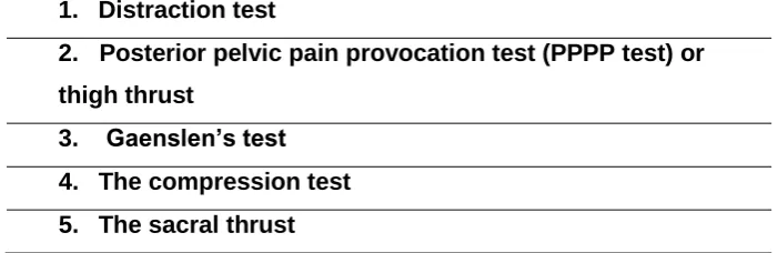
[image:10.595.105.456.316.430.2]Five SIJ pain-provocation tests are going to be used in this trial, with 3 or more positive tests being classed as indicative of SIJ pain. The five tests are listed in and described in the following sections and are illustrated in Figure 5 to Figure 9. In order to be classified as a ‘positive’ test it is necessary for the test to reproduce the participants’ pain, rather than a different pain. All tests should be performed if clinically possible; even if the first three are positive.
Table 2: SIJ pain-provocation tests for exclusion criteria
1. Distraction test
2. Posterior pelvic pain provocation test (PPPP test) or
thigh thrust
3. Gaenslen’s test
4. The compression test
Figure 5: Distraction test: hand positioning and alternate hand positioning
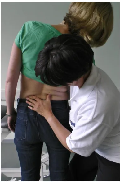
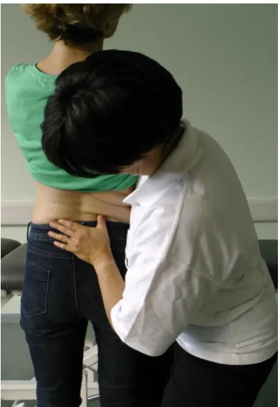
Figure 6: Thigh thrust (PPP)
Figure 7: Gaenslen’s test
(Testing the left SIJ in posterior rotation and the right SIJ in anterior rotation). The pelvis is stressed with a torsion force by a superior/posterior force applied to the right knee and a posteriorly directed force applied to the left knee.
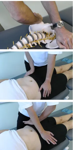
Figure 8: The compression test
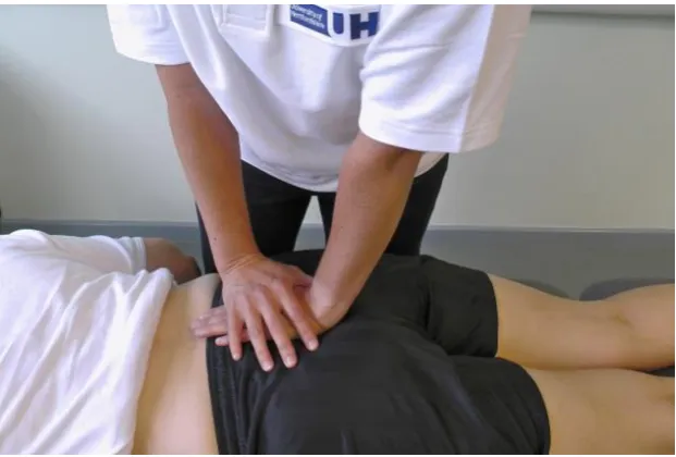
[image:13.595.74.379.377.585.2]Figure 9: The sacral thrust
A vertically directed force is applied to the midline of the sacrum at the apex of the curve of the sacrum, directed anteriorly, producing a posterior shearing force at the SIJs with the sacrum nutated
Additional data collection – exploratory only
Certain symptom presentations or tests which are either in common clinical use or have some predictive validity in identifying potential facet joint pain were presented as to
Consensus Conference but not agreed. Therefore whilst not forming part of the diagnostic procedure for entry to the trail, as part of the feasibility study we intend to collect this information for explanatory purposes.
Laslett’s clinical prediction rule for facet joint pain
Table 3: Components in Laslett’s Clinical Prediction Rule
Age> 50
Symptoms best when walking
Symptoms best when sitting
Onset of pain paraspinal
Positive extension/rotation test*
*a component of the entry criteria therefore will already have been tested
No exacerbation on rising from flexion
One of the criterion that was suggested by Consensus Conference was one of Revel’s criteria; ‘no increased pain on rising from flexion’ (Revel et al., 1998). This has not been included in this diagnostic criteria as there no study has demonstrated an appropriate prognostic value for facet joint pain (Laslett et al., 2006, Laslett et al., 2004). However this information will be collected for exploratory purposes only during the pilot study. The information should be collected when testing active movements (See
Appendix 1: Sequencing of diagnostic testing
Subjective questioning
Physical testing
1.Radicular pain?
4. SIJ tests
5. Palpation
2. Lsp flexion
2.Pain on rising
(Extra)
3. Active movts
Ext, Rotation
E/SF, E/Rot
3. Ext vs Flex
Age> 50
Symptoms best when walking Symptoms best when sitting Onset of pain paraspinal Positive Ext/rotation test*
Appendix 2: Combined movements
Flex LSF RSF Shade painful quadrant Ext Flex LSF RSF Ext Flex LSF RSF ExtRegular compression: Right Left Primary Combination
Regular stretch: Right Left Primary Combination
Extension to half expected range ‘Prime movement’
Extension, left side flexion ‘Prime combination’
Low back
Severe
Prime movement; flexion
Prime combination: L SF/Flexion
Low back
Severe
Bolder arrow= most provocative movement (Prime movement)
Prime combination’
Single headed arrow = second movement applied of primary combination
Place a judgement of
severity of pain next to
Appendix 3: Procedures undertaken within Facet Injection Study
Diagnostic Assessment visit
Prior to the diagnostic assessment visit appointment by the patient, diagnostic assessment physiotherapist will undertake review of clinical record for the patient in relation to their attendance of the study assessment visit and potential study enrolment; ie study screening questionnaire/expression of interest, referral letter/clinical notes, reconfirm documented inclusion/exclusion, clinical history, concomitant medications.
1. During the appointment with the patient, physiotherapist will ensure the patient has read the Participant Information Sheet, and carefully explain the study to ensure the patient is fully informed and has read and understood the participant information sheet, with the opportunity to ask any questions.
2. Undertake clinical history - back pain history, back pain treatments and date of last treatment if known, troublesomeness, other medical history, participants current status of general health, concomitant medication
3. Undertake clinical assessment to confirm patient meets clinical diagnostic criteria for facet joint pain. All the following assessments are to be undertaken
confirmed/completed to ensure eligibility assessment is completed for the purpose of inclusion into the study:
No radicular symptoms (defined as pain radiating below the knee or objective neurological signs above the knee using a ‘contracted’ neurological examination) No sacro-iliac joint pain elicited using the follow pain provocation tests (3 or more
positive tests)
- Distraction test, PPPP test or thigh thrust, Gaenslen’s test, compression test, sacral thrust
Increase in pain, unilaterally or bilaterally, on lumbar para-spinal palpation
Increased low back pain confirmed on one or more of the following elements* - Extension (more than flexion)
- Rotation
- extension/side flexion - extension/rotation
*All the elements – extension, rotation, extension/side flexion, extension/rotation – are to be performed. If an element is for some reasons not performed, there will be a requirement within the worksheet/CRF to document the reason why, for example ‘patient unable fully extend for full passive rotation’.
4. In addition to the above eligibility assessments, additional symptom presentation or tests to be performed include the following:
a. Symptoms best on walking b. Symptoms best when sitting c. Onset of pain paraspinal
d. No exacerbation on rising from flexion
back to the patient’s GP or referring source. Specific causes of back pain include
malignancy, fracture, infection, possible ankylosing spondylitis, Cauda equina compression, radicular pain suitable for surgery and will determine the patient not eligible for continuation in the facet injection study.
If a participant expresses suicidal thoughts or signs of severe depression, we would advise you to follow your standard procedure within your Trust which may include a referral to the mental health team or a Psychologist. If a participant is referred to a mental health team or Psychologist, they are still in the Facet Injection Study and we would expect them to continue with the Best Usual Care Package if possible, unless it is a clinical decision in the best interests of the participant to withdraw him/her from the trial.
5. Complete participant diagnostic assessment worksheet and eligibility CRF (visit A) Eligibility of patient
Once diagnostic assessment for probably facet joint pain is completed, the physiotherapist must determine if the patient is eligible for the study or not.
Eligible
I. Physiotherapist confirms eligibility to the patient and obtains patient’s signature and date of consent on the Facet Injection Study ‘Participant Consent Form’ (Note: ensure
patient completes all details on form) and cross check the form
II. Physiotherapist (as delegated by the Principal Investigator) also signs and dates the consent form after the patient.
III. After informed consent fully signed, patient will complete Baseline Questionnaire. In parallel whilst patient is completing the Questionnaire, the diagnostic assessment physiotherapist will arrange to :
a. complete the ‘Patient Enrolment’ form
b. complete the ‘Participant Contact Details’ form
c. contact WCTU Enrolment & Randomisation service for unique participant ID number
d. add participant trial ID number to all participant documents
e. schedule Best Usual Care Treatment Session 1 for patient, as well as scheduling dates for potential FJI and BUC Treatment sessions 2-6; document appointments in patient letter
f. writes appointment dates and times in ‘FJI and BUC appointment’ letter for patient g. mail out GP letter relating to the participant enrolled into the study
h. complete diagnostic assessment CRF/worksheet i. label Baseline Questionnaire with participant ID number j. complete screening and enrolment log
k. inform your site investigator team (PI and physiotherapists) of patient enrolment
Non Eligible patients
I. Physiotherapist informs the patient that following their clinical assessment they are not eligible to participate in the study. Provide reasons if asked. Further inform the patient that he/she will continue within the referral system for standard treatment of care, as originally referred (as stated in the Participant Information Sheet).
assessment data (as stated in the Participant Information Sheet) and if the patient remains agreeable, the patient is required to sign the ‘Consent Form – Eligibility Assessment Data’
Management of diagnostic assessment documentation
Eligible /enrolled participants:
- Screening Questionnaire/EOI; original for WCTU, copy investigator site file
- Worksheets / CRF pages; original for WCTU, copy patient notes, copy investigator site file
- Consent form ; original investigator site file, copy to patient, copy in notes
- Enrolment Form; original investigator site file, copy (faxed to) WCTU
- Participant Contact Details Form ; original investigator site file, copy (faxed to) WCTU
- Appointment letter ; to participant
- GP letter ; original to participant’s GP, copy to patient notes
Non eligible – not enrolled:
If ‘Eligibility Assessment Data Consent Form’ signed by participant:
o Screening Questionnaire/EOI : Original investigator site file ; copy to WCTU o Worksheets / CRF pages; Original patient notes, copy WCTU, copy investigator
site file
o Consent form ; original investigator site file, copy to patient, copy in notes
If consent not provided for eligibility assessment data:
o Screening Questionnaire/EOI : Original investigator site file.
References:
BILLIS, E. V., FOSTER, N. E. & WRIGHT, C. C. 2003. Reproducibility and repeatability: errors of three groups of physiotherapists in locating spinal levels by palpation. Man Ther, 8, 223-32. CHALLINOR, H., HOURIGAN, P. G., POWELL, R. & CONN, D. Does A Regular Compression Combined
Movement Test Diagnose Lumbar Facet Joint Pain? Society for Back Pain Research, 2013 London.
EDWARDS, B. 1999. Manual of Combined Movements, Butterworth-Heinenmann. FULLER, G. 2004. Neurological Examination Made Easy, Churchill Livingstone.
LASLETT, M., MCDONALD, B., APRILL, C. N., TROPP, H. & OBERG, B. 2006. Clinical predictors of screening lumbar zygapophyseal joint blocks: development of clinical prediction rules. Spine J, 6, 370-9.
LASLETT, M., OBERG, B., APRILL, C. N. & MCDONALD, B. 2004. Zygapophysial joint blocks in chronic low back pain: a test of Revel's model as a screening test. BMC Musculoskelet Disord, 5, 43. MAITLAND, G., HENGEVELD, E., BANKS, K. & ENGLISH, L. (eds.) 2005. Maitland's Vertebral
Manipulation: Elsevier.
MCCARTHY, C. 2010. Neurological assessment. In: MCCARTHY, C. (ed.) Combined movement Theory.
Rotational Mobilization of the Vertebral Column. Churchill Livingstone.
REVEL, M., POIRAUDEAU, S., AULELEY, G. R., PAYAN, C., DENKE, A., NGUYEN, M., CHEVROT, A. & FERMANIAN, J. 1998. Capacity of the clinical picture to characterize low back pain relieved by facet joint anesthesia. Proposed criteria to identify patients with painful facet joints.