ISSN Print: 2164-2788
DOI: 10.4236/ojmi.2018.83007 Sep. 6, 2018 54 Open Journal of Medical Imaging
AXS Image Ondition of Beam Is Small Angle
X-Ray Scattering Imaging of Soft Tissue by
Using Laue Diffraction Optical System
Ge Jin
1*, Kenichi Okamura
1, Yoshifumi Suzuki
1, Yong Sun
1, Yoshinori Chikaura
1, Masami Ando
21Department of Applied Science for Integrated System Engineering, Kyushu Institute of Technology, Kitakyushu,Japan 2Research Institute for Science and Technology, Tokyo University of Science, Noda, Japan
Abstract
We performed a feasibility study of small angle X-ray scattering imaging un-der the condition of X-ray bright field imaging by Laue crystal diffraction op-tics of X-ray dark-field imaging that works as an angular analysis. Collagen in chicken tibia containing abundant soft fibrous tissue was chosen as a speci-men. In traditional small angle X-ray scattering optical system, we can derive the structure information of sample by calculating q value which is available from a scattering pattern. Thus it is usually necessary to conduct a 2D scan in order to obtain scattering image. In this paper it is described by a method by which not only small angle X-ray scattering imaging is available directly but also bright-field imaging and dark-field imaging can be obtained at the same time. As the first step, the feasibility of the imaging method should be con-firmed by taking pictures of the samples with known periodic length. The preliminary test showed that the collgan’s lattice spacing d is 65.1 nm that was also taken photos by scanning electron microscopy. By rotating Laue an-gular analyzer by 112 arcseconds small angle X-ray scattering image appeared in bright-field.
Keywords
XDFI, SAXS, Synchrotron Radiation, Soft Tissue
1. Introduction
When the X-rays pass through the object, three kinds of interactions mainly oc-cur such as absorption, refraction, scattering (small angle scattering and Comp-ton scattering). Since the discovery of X-ray by Roentgen, absorption contrast How to cite this paper: Jin, G., Okamura,
K., Suzuki, Y., Sun, Y., Chikaura, Y. and Ando, M. (2018) AXS Image Ondition of Beam Is Small Angle X-Ray Scattering Imaging of Soft Tissue by Using Laue Dif-fraction Optical System. Open Journal of Medical Imaging, 8, 54-63.
https://doi.org/10.4236/ojmi.2018.83007
Received: February 26, 2018 Accepted: September 3, 2018 Published: September 6, 2018
Copyright © 2018 by authors and Scientific Research Publishing Inc. This work is licensed under the Creative Commons Attribution International License (CC BY 4.0).
DOI: 10.4236/ojmi.2018.83007 55 Open Journal of Medical Imaging imaging [1] [2] based on absorption principle has playing an important role in so many fields in the past hundred years. In recent years a lot of attention from medical imaging and material characterization has been paid to refraction phase-contrast imaging [3].
Refraction phase contrast imaging has two advantages over absorption con-trast: it can provide high contrast in soft tissue including articular cartilage and it can relatively suppress radiation dose. This X-ray imaging technology is ap-plicable to medical diagnosis, performance evaluation of biomaterials and so on. Articular cartilage, ligament, cancer tissue and atherosclerotic plaque in blood cell that has very low X-ray absorption contrast so that one can not easily see these. There is drawback of X-ray absorption contrast in muscle and cancer tis-sue imaging so that these provide a very weak contrast. In order to solve this problem usually X-ray energy has been reduced substantially that might induce higher radiation dose. On the other hand articular cartilage whose main com-ponent is Ca is relatively low Ca concentration but this tissue is surrounded by high Ca concentration trabeculars so that there is no clear technique to visualize this under X-ray absorption contrast.
We are developing X-ray optical system called X-ray dark-field imaging (XDFI) [4]. This comprises two optical components: monochromator-collimator (MC) that provides with highly collimated X-ray beam with divergence of
7
2.5 10× − rad and Laue angular analyzer (LAA) that has angular acceptance of
6
3.85 10× − rad. This system that is a function of over all the radiation source,
the degree of parallelity of the beam incident that can be made by adoption of MC onto specimen, thickness of LAA and the pixel size of CCD camera has reached till now the spatial resolution of approximately 10 μm. The divergence of outgoing beam is at order of 10−7 rad. So far XDFI has got success of viewing a
variety of internal organs such as breast cancer, articular cartilages of finger, knee and shoulder, eye ball and blood cells such as iliac artery and coronary ar-tery without staining with contrast agent in pathological way [5] [6].
This has brought us an image of scattering quite different from absorption based images.
On the other hand, biological object usually contains long-period nanome-ter-scale structures. Due to the low crystallinity of this long period structure and a low X-ray absorptivity leading a low intensity, it becomes difficult to obtain a scattered image. In traditional small angle X-ray scattering (SAXS) optical sys-tem, e.g. Bonse-Hart system [7] and Kratky system [8], we can derive the struc-ture information of sample by calculating q value which is available from a scat-tering pattern. Thus it is usually necessary to conduct a 2D scan in order to ob-tain scattering image. A small angle X-ray scattering topography [9] [10] that was proposed by Yoneda and Chikaura first in 1981 was used for visualizing in-ternal periodic structure distribution of some biomaterials by mapping differ-ences of small angle scattering.
DOI: 10.4236/ojmi.2018.83007 56 Open Journal of Medical Imaging imaging by rotating the Laue angular analyzer to exact scattering part by XDFI optical system. Not only SAXS imaging could be obtained directly, but also ab-sorption contrast imaging, BFI imaging and DFI imaging the four kinds of im-aging can be obtained and compared with each other. In the application aspect, at the first the feasibility of the imaging method should be confirmed by taking pictures of the periodic length known samples. It is expected that revealing the genesis of soft tissue lesions by visualization of subtle structures and becoming a useful tool for medical imaging in future.
2. Experiment and Principle
2.1. Preliminary Experiment
Preliminary test was performed at BL14B at Photon Factory before SAXS expe-riment at BL14C using XDFI optics. This was done in order to measure d-value spacing of fibers of collagen specimen and to estimate exposure time of the col-lagen specimen by the SAXS intensity. The angular position of the X-ray image of collagen with spacing of 65 nm should meet the angular information of SAXS. This system uses synchrotron X-ray produced at a 5 T vertical wiggler installed in the 2.5 GeV storage ring at Photon Factory, KEK in Tsukuba, Japan.
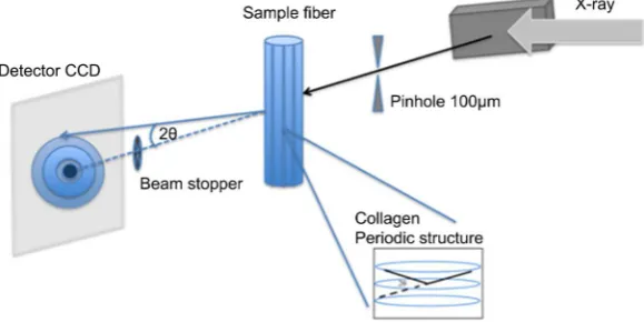
SAXS image corresponds to that taken at angular position calculated by the spacing 65 nm. Precision measurement of the spacing is very crucial; unless oth-erwise we cannot attain reasonable SAXS intensity. In order to obtain high inci-dent intensity the asymmetric factor was reversely to condensate the beam in-tensity by making the beam size smaller so that the irradiated area has become 100 μm instead of 5 mm as shown in Figure 1. Energy was adopted at 17.5 keV. Camera distance was set up to 850 mm. An Al needle of injector around 1mm diameter was used as beam stopper. Camera resolution was 7.4 pixel/μm with FOV 24 mm × 36 mm.
2.2. SAXS Imaging Experiment
[image:3.595.228.518.547.692.2]The experiment was carried out at BL-14C station where optics is double-crystal
DOI: 10.4236/ojmi.2018.83007 57 Open Journal of Medical Imaging monochromater 220 and mainly doing the bigger horizontal medical imaging station. Beam intensity of this station is approximately 108 photons/mm2/s at
33 keV. Vertically polarized white X-ray radiation at short wavelengths occurs from the super conducting wiggler that has a 5 T horizontal magnetic field. The size of the experimental hutch is 3.7 m (L) × 3.7 m (W) × 2.9 m (H), and height from the middle line of X-ray beams in the hutch is about 1360 mm. Beam size is V: 38 mm, H: 8 mm by design.
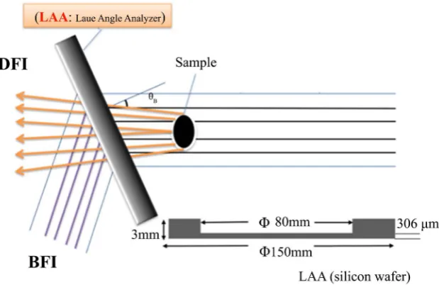
The X-ray optical system called X-ray dark-field is shown in Figure 2. There is an asymmetrically cut Bragg-case monochromator-collimator (MC), a sam-ple rotational stage, LAA, and an X-ray CCD camera. As X-ray beam that has already been monochromated by a double-crystal monochromator up stream, called pre-monochromator, in the optical hutch is incident on MC with the in-cident grazing angle of θB−α where θB is the Bragg angle and 𝛼𝛼 is the angle between the crystal surface and the diffracting planes. Due to this process, the MC expands the size of incident X-ray beam by a factor of
(
)
(
)
1b=sin θB+α sin θB −α . Also the divergence of the outgoing beam be-comes smaller compared to the intrinsic one by a factor b.
[image:4.595.219.529.505.706.2]X-ray is irradiating a sample, the phase shift happens at internal boundary of tissue so that the traveling direction changes. Utilizing this principle, the X-ray phase contrast occurs. It becomes possible to visualize the structure even regard to X-rays for the low absorption part such as living tissues. The X-ray phase contrast imaging is a method that detects the X-ray refraction of substance by utilizing the diffraction of the LAA which is installed after the sample. The dif-fraction occurs when the incident angle is at θB formed between the back-ground beam and the crystal lattice plane. After beam pass through LAA it will split into two waves such as forward diffraction wave and diffraction wave and dark field imaging and bright field imaging are obtained when put a sample into optic as shown in Figure 2.
DOI: 10.4236/ojmi.2018.83007 58 Open Journal of Medical Imaging We used a chicken’s leg (collagen d ≈ 65 nm) available from market to make into a specimen by size 1 mm × 7 mm after drying. Collagen and silver docosa-noate with a chemical form of C22H43AgO2 (d ≈ 5.8 nm) usually were used
as a standard sample with calibrated q value q 2 4sin d
π θ
λ
= =
in small-angle
scattering experiment. Camera distance L = 750 mm and the X-ray energy E = 17.5 keV were adopted. Under this condition, it was calculated that collagen corresponding to the Bragg angle is 112" by the Bragg’s law. Adjusting the optical system at w=2Λsinb
(
θ θ− B−Δθ0)
λ=0, where Λ=λχcosθB P χΓ is thePendellosung fringe distance, P the polarization factor, λ the X-ray
wave-length, 2
Γ r Fe G/ VC
χ = − λ π the polarizability, where re is the classical radius of
electron, FG the crystal structure form factor, VC the volume of unit cell, θ the
angle that deviated from the Bragg angle θB and Δθ0, θ θ= B− ∆θ0, correc-tion of the Bragg angle due to refraccorrec-tion expressed as Δθ0 =2 1
(
−n)
sin 2θBshown in Figure 3. Signal to noise ratio at DFI, S N+ N DFI is much smaller than that at BFI, S N+ N BFI so that one cannot expect a high quality of image using DFI. In order to have higher S/N image a BFI image without specimen at the angular position where small-angle scattering image was taken was subtracted from the sample image. Then we only attempted in BFI at position of b (outside of figure) where in the case of collagen 11,200 pulses.
[image:5.595.212.538.471.702.2]The horizontal axis unit is arcsecond, and 100 pulse equals 1 arcsecond. When the X-rays pass through the object, three kinds of interactions mainly occur such as absorption, refraction, scattering (small angle scattering and Compton scat-tering). According to the above three physical phenomena, the beams which be-long to different angle’s order can be distinguished by rotating LAA. In the
DOI: 10.4236/ojmi.2018.83007 59 Open Journal of Medical Imaging case of X-ray, the refraction index is defined by n= −1 i
β δ
− . The real part δ relate to refraction angle is 10−5 radian which almost belongs 0.01% degreerange. The small angle scattered beam belongs 1% degree to several degrees in the order of the angle range. Refraction beam with a relatively small angle can be analyzed and visualized by LAA, then the range of small angle scattering that is hundred times larger than it should be able to be analyzed. Many living organ-isms comprise collagen, and collagen has very low sensitivity in the absorption contrast image. And collagen is usually composed of a large number of light elements and periodic structure, therefore it is considered to be suitable for photographing both refraction phase contrast imaging and small-angle scatter-ing imagscatter-ing as a sample. To establish the imagscatter-ing technique of small-angle scat-tering image, at the first stage, we did an experiment by using XDFI optics with spatial resolution of 8 μm. Exposure time for refraction beam was around 50 seconds while that for scattering beam was 60 min.
3. Result
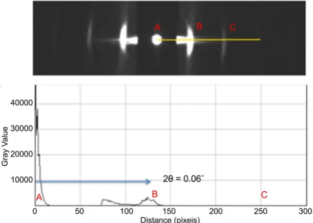
The result of the preliminary test in Figure 4 showed that a position is direct beam. B is the first order diffraction and C is the second order diffraction. From A to B there are almost 125 pixels. The lattice spacing d is 65.1 nm. This means by rotating LAA by 112 arcseconds SAXS image should appear. The SAXS in-tensity is expected to be approximately 1/120,000 of the incident X-ray inin-tensity. This has made us possible to estimate the exposure time of SAXS image so that it is about 10 min. As shown in Figure 5 dark-field imaging, bright-field imaging, SAXS imaging 112 arcseconds apart from the just Bragg angle were confirmed.
[image:6.595.220.526.485.703.2]X-ray contrast image of collagen at chicken tibia appeared when the periodic-ity matched the X-ray Bragg condition.
DOI: 10.4236/ojmi.2018.83007 60 Open Journal of Medical Imaging
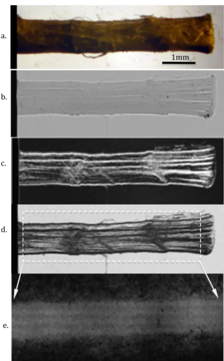
Figure 5. (Color online) The result of various kinds of imaging. (a) Optical microscope; (b) Absorption imaging; (c) DFI imaging; (d) BFI imaging; (e) SAXS imaging.
4. Discussion
It has become clear that SAXS image can be taken under the XDFI optical sys-tem. In order to increase the X-ray intensity delivered to specimen that is relat-ing to shortenrelat-ing of exposure time one could introduce a bent crystal or asym-metric crystal with b1. Under this condition due to angular distribution of crystallite of the specimen and its physical size the above consideration can work.
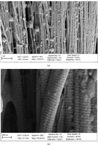
In order to obtain internal structure of collagen to confirm the results of small-angle scattering image, we took electron microscopic images of scanning electron microscopy (SEM). And the result of 65 nm periodic length shown in SEM image is corresponding to the diffraction pattern in preliminary test and SAXS imaging in Figure 5(e).
DOI: 10.4236/ojmi.2018.83007 61 Open Journal of Medical Imaging (a)
[image:8.595.209.538.67.554.2](b)
Figure 6. SEM images of chicken collagen’s structure by different magnification.
CCD camera that has pixel size of 7.4 μm cannot observe the nanometer order of collagen structure itself. This means that the contrast observed in X-ray im-aging in Figure 5(e) provides us information of local distribution of periodicity of collagen around 65 nm.
X-ray absorption contrast shows difference of absorption at local point of ma-terial, while that of refraction contrast is dependent on local electron density distribution.
DOI: 10.4236/ojmi.2018.83007 62 Open Journal of Medical Imaging taken just at the peak position of the RC, the intensity is already strong enough to show the periodic structure. On the other hand, even if the images were taken along RC with different periods position, due to the resolution, an obvious change of image in brightness reflecting the distribution of different periodic positions at different part of the sample cannot be expected under current con-ditions. A precision sample turntable for scanning the peak of the small angle scattering and a CCD camera of which the field of view does not have to be large but higher resolution is needed in the future experiment.
5. Conclusions
We have successfully achieved a novel technique of viewing inside collagen of chicken tibia by use of SAXS. The achievement tells us that BFI of XDFI can provide us with SAXS image. In view of exposure time and angular resolution in SAXS image we could have advanced this technique based on the system devel-oped by Yoneda and Chikaura.
With approach by taking three images such as phase-contrast one, absorption one and SAXS this system proposed here can be a powerful tool to analyze structure of a specimen. Nevertheless there is still a space to improve the quality of X-ray images. For instance one can introduce vacuum tube for X-ray path to reduce scattering source and reduce beam quality towards the spherical beam rather than a parallel beam.
In the next step we could proceed onto a more complicated unknown struc-ture of biomaterial. Thus we can expect that each X-ray image can tell different components of material.
As a conclusion we could foresee application of SAXS image to pathological diagnosis to see 3-dimensional distribution of soft tissue of biomaterial and na-nomaterial.
Conflicts of Interest
The authors declare no conflicts of interest regarding the publication of this pa-per.
References
[1] Momose, A. (2005) Recent Advances in X-Ray Phase Imaging. Japanese Journal of Applied Physics, 44, 6355-6367. https://doi.org/10.1143/JJAP.44.6355
[2] Takeda, T., Momose, A., Yu, Q., Wu, J., Hirano, K. and Itai, Y. (2000) Phase-Contrast X-Ray Imaging with a Large Monolithic X-Ray Interferometer. Journal of Synchro-tron Radiation, 7, 280-282. https://doi.org/10.1107/S0909049500004295
[3] Chapman, D., Thomlinson, W., Johnston, R.E., Washburn, D., Pisano, E., Gmür, N. et al. (1997) Diffraction Enhanced X-Ray Imaging. Physics in Medicine & Biology, 42, 2015-2025. https://doi.org/10.1088/0031-9155/42/11/001
DOI: 10.4236/ojmi.2018.83007 63 Open Journal of Medical Imaging [5] Ando, M., Sunaguchi, N., Wu, Y., Do, S., Sung, Y., Louissaint, A., Yuasa, T., Ichiha-ra, S. and Gupta, R. (2013) X-Ray Phase Contrast Imaging in the Dark Field: Im-plementation and Evaluation Using Excised Tissue Specimens. European Radiology, 23, 3021-3029.
[6] Ando, M., Maksimenko, A., Yuasa, T., Hashimoto, E., Yamasaki, K., Ohbayashi, C., Sugiyama, H., Hyodo, K., Kimura, T., Esumi, H., Akatsuka, T., Li, G., Xian, D., Ue-no, E., Bando, H., Ichihara, S., Endo, T., Moriyama, N. and NishiUe-no, H. (2006) 2D and 3D Visualization of Ductal Carcinoma in Situ (DCIS) Due to X-Ray Refraction Contrast. Bioimages,14, 1-8.
[7] Bonse, U. and Hart, M. (1966) Small-Angle X-Ray Scattering. Gordon and Breach, New York.
[8] Glatter, G. and Kratky, O. (1982) Small Angle X-Ray Scattering. Academic Press, London.
[9] Yoneda, Y. and Chikaura, Y. (1981) Polycrystal Scattering Topography, Scattering Tomography and Their Perspective Fields of Application. Japanese Journal of Ap-plied Physics, 37, 412-418.