Percutaneous Collagen Induction Therapy: An
Alternative Treatment for Scars, Wrinkles, and
Skin Laxity
Matthias C. Aust, M.D. Des Fernandes, M.D. Perikles Kolokythas, M.D. Hilton M. Kaplan, M.D. Peter M. Vogt, M.D. Hannover, Germany; Cape Town, South Africa; and Los Angeles, Calif.Background: Skin laxity, rhytides, and photoaging are generally treated by ablative procedures that injure or destroy the epidermis and its basement membrane, at least in the beginning, and subsequently lead to fibrosis of the papillary dermis. The ideal treatment would be to preserve the epidermis and promote normal collagen and elastin formation in the dermis. Percutaneous collagen induction takes us closer to this ideal.
Methods: The authors performed a retrospective analysis of 480 patients in South Africa and Germany with fine wrinkles, lax skin, scarring, and stretch marks treated with percutaneous collagen induction using the Medical Roll-CIT to produce tighter, smoother skin. Most patients had only one treatment, but some have had as many as four treatments. Patients were prepared with topical vitamin A and C cosmetic creams for a minimum of 4 weeks preoperatively.
Results: On average, patients in Germany rated their improvement between 60 and 80 percent better than before the treatment. Histologic examination was carried out in 20 patients and showed a considerable increase in collagen and elastin deposition at 6 months postoperatively. The epidermis demonstrated 40 percent thickening of stratum spinosum and normal rete ridges at 1 year postoperatively.
Conclusions: Percutaneous collagen induction was started in 1997 and has proved to be a simple and fast method for safely treating wrinkles and scars. As opposed to ablative laser treatments, the epidermis remains intact and is not damaged. For this reason, the procedure can be repeated safely and is also suited to regions where laser treatments and deep peels cannot be performed. (Plast. Reconstr. Surg.121: 1421, 2008.)
A
s the demand for less invasive, highly effec-tive cosmetic procedures grows, plastic sur-geons must explore and develop new treat-ment options. Patients request skin treattreat-ments to rejuvenate photoaging, abnormal pigmentation or vascularity, textural problems, rhytides, and lax-ity caused by chronological aging. We have nu-merous methods today to tighten skin, such as laser resurfacing and deep peeling.1–5 However, these treatments are ablative and tighten the skin From the Klinik fu¨r Plastische, Hand- und Wiederherstel-lungschirurgie, Medizinische Hochschule Hannover; De-partment of Plastic and Reconstructive Surgery, Groote Schuur Hospital; and Department of Biomedical Engineer-ing, University of Southern California.Received for publication November 17, 2006; accepted Feb-ruary 28, 2007.
Copyright ©2008 by the American Society of Plastic Surgeons DOI: 10.1097/01.prs.0000304612.72899.02
Disclosures:Dr. Des Fernandes is employed as the senior medical consultant for Environ Skin Care Pty. Ltd. (South Africa), whose products were used by all the patients in this report. He does not own any equity in Environ Skin Care Pty. Ltd. The Roll-Cit products are made by Vivida Closed Corpo-ration. Dr. Des Fernandes is a shareholder in Vivida Closed Corporation and is employed by Vivida as a medical consultant. Dr. Matthias Aust is the med-ical consultant for Care Concept, distributors for Environ Skin Care Products and Roll-Cit in Ger-many. Dr. Hilton Kaplan is the medical advisor to DermoGenesis, USA, a U.S. distributor of Environ. Prof. Vogt and Dr. Kolokythas have no sources of funds supporting the work and no financial interest in any of the products, devices, or drugs mentioned in this article.
or lighten scars because generally they destroy the epidermis and, very importantly, its basement membrane, which is replaced by an epidermis that no longer has dermal papillae and, at least in the beginning, is thinner than before.6 – 8The destruc-tion of the epidermis initiates an inflammatory response that stimulates fibroblasts to produce thick bundles of scar collagen in parallel orienta-tion rather than the normal lattice network of collagen found in normal skin.9,10 The skin be-comes more sensitive to photodamage and may also develop dyschromias.8
As far as scars are concerned, these treatments all work on the same principle as dermabrasion: the level of the normal skin is taken down closer to the level of the offending scar, which does not have normal epidermis.11–13 The ideal treatment would be to do exactly the opposite: by instead improving the scar quality and building up the scarred tissue to the level of the normal skin. On account of the complications of resurfacing lasers and deep peels, other modalities such as fraction-ated laser, radiofrequency heat, superficial repet-itive peels, and intense pulsed light have become more popular.14
Furthermore, patients are now demanding less aggressive treatments. This is borne out by statistics from the American Society of Plastic Surgeons that demonstrate a 43 percent increase in minimally in-vasive surgical interventions between 2000 and 2005, compared with only a 16 percent increase for inva-sive cosmetic surgical interventions.15 Over time, minimally invasive surgical procedures will outnum-ber traditional open surgical procedures, with the market for related minimally invasive surgical de-vices set to soar from its current size in Europe of $779 million to $1164 million in 201115 (in U.S. dollars).
We have learned in recent years that trans-forming growth factor (TGF)-plays an enormous role in the first 48 hours of scar formation. Whereas TGF-1 and TGF-2 promote scar colla-gen, TGF-3 seems to promote regeneration and scarless wound healing with a normal collagen lattice.10,16,17 The ideal treatment of wrinkles and scars should be to promote regeneration rather than cicatrization, and this could offer our pa-tients the result that they are hoping for.
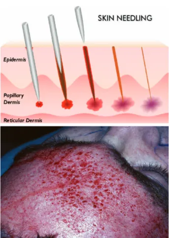
We believe that percutaneous collagen induc-tion brings us closer to this ideal of regenerainduc-tion, even though we still have far to go. Percutaneous collagen induction creates thousands of micro-clefts through the epidermis into the papillary dermis. These tiny wounds in the papillary dermis create a virtually confluent zone of superficial
bleeding that is a powerful stimulus to initiate the normal process of wound healing. Because it is nonablative, percutaneous collagen induction can be performed on the face and body, on all skin types, without concern for aesthetic unit bound-aries, and, very importantly, without predisposing the patient to hyperpigmentation. For percutane-ous collagen induction, we have used the Environ Medical Roll-CIT (Vivida-SA cc, Cape Town, South Africa), which is a sterile plastic cylinder with nee-dles protruding between 1 and 3 mm from the surface to roll vigorously over the skin (Fig. 1).
PATIENTS AND METHODS
This is a retrospective analysis of patients op-erated on between 1997 and 2006 in South Africa and Germany. These patients underwent percu-taneous collagen induction for fine wrinkles, lax skin, scarring, and stretch marks. The skin of all 480 patients was prepared preoperatively for at least 1 month with vitamin A (retinyl palmitate) cream and vitamin C cream [ascorbyl tetra-isopalmitate (Environ Original and C-Boost; En-viron Skin Care, Pty. Ltd., Cape Town, South Af-rica)] applied topically twice daily.
Demographic Data
The demographic data of 480 patients are listed in Table 1. Four hundred of the patients were women and 80 were men (female-to-male ratio, 5:1). The average age was 49 ⫾ 15.5 years (range, 18 to 74 years). The average time to sur-gery (duration from consultation) was 3 ⫾ 15.5 months (range, 1 to 12 months). This is relevant because it represents the duration over which
pa-Fig. 1. The Environ Medical Roll-CIT (made by Vivida-SA cc, Re-naissance Body Science Institute, Cape Town, South Africa).
tients can pretreat their skin with vitamins A and C. Twenty-eight of the 480 patients had been treated previously with chemical peeling or derm-abrasion elsewhere.
Method
Orentreich and Fernandes independently de-scribed “subcision” or dermal needling.18,19 This involves pricking the skin and then scarifying the dermis with a needle to build up connective tissue under scars and wrinkles. However, this technique could not be used over large surface areas. Cam-irand used a tattoo gun to treat scars with “needle abrasion.”20 The fundamental similarity of these different techniques is that the needles disrupt the old collagen structures that connect the scar with the upper dermis. The associated trauma induces the normal wound-healing inflammatory cascade, and the scar collagen that is broken down is re-placed with new collagen under the epidermis. Based on these principles, Fernandes developed a new technology, percutaneous collagen induc-tion, to initiate the natural posttraumatic inflam-matory cascade by rolling needles vertically, hor-izontally, and diagonally with pressure over the treated area21(Figs. 2 and 3). These needle pricks lead to thousands of microwounds in the dermis. For percutaneous collagen induction, the skin was anesthetized with a topical anesthetic cream or
with regional nerve blocks and/or infiltration of local anesthetic and conscious sedation (where large or sensitive areas are being treated), or gen-eral anesthesia.
Histology
Histology was carried out with 3-mm punch bi-opsies, to compare preoperative collagen and elastin to that obtained postoperatively, both qualitatively and quantitatively, using van Gieson, elastica, and hematoxylin and eosin stains (Figs. 4 and 5).
Indications for Percutaneous Collagen Induction
Patients with indications for percutaneous col-lagen induction were divided into three groups: Table 1. Demographic Data
Group Wrinkles Scars Lax Skin Average
No. (n⫽480) 350 72 58
Female/male 300/50 55/17 45/13 5/1
Age, years 51.9⫾12.1 41.7⫾10.3 49.7⫾10.1 49⫾15.5 Time from consultation to
operation, months 2.9⫾1.3 3.1⫾1.4 3.2⫾1.6 3⫾15.5
Fig. 2. The operative procedure.
Fig. 3. (Above) Schematic image of the procedure. (Below) Intra-operative view of a needled forehead.
fine wrinkles (face); scarring (acne, varicella, burns); and stretch marks (striae) or lax skin (face, abdomen, arms, legs, and thighs).
Group I: Wrinkles
Group I consisted of patients with fine wrin-kles (n ⫽350) (Figs. 6 and 7).
Group II: Scarring
Group II consisted of patients with acne or burn scars (n ⫽72) (Fig. 8).
Group III: Lax Skin/Striae
Group III consisted of patients with stretch marks or lax skin of the abdomen or arms (n⫽58) (Figs. 9 and 10).
Postoperative Care
Immediately after percutaneous collagen in-duction, the area is swollen, with superficial bruising. After the initial bleeding stops within a few minutes, there is a serous ooze, which stops within the first few hours after the operation. To absorb the bleeding and serous ooze, the treated area should be covered with cool, damp swabs that are periodically replaced for the first 2 hours after the operation. The skin is finally washed with a tea tree oil– based cleanser and the vitamin A and C regimen starts immediately. The patient is instructed to wash the face thoroughly again when they return home.
Patients usually do not require postoperative analgesia if the procedure has been performed under local anesthesia. When the procedure has been performed under general anesthesia without any local/topical anesthetic, the patient may
com-plain of burning for the first hour postoperatively, so it is wise to administer an analgesic at the end. The edema can be quite significant when needling has been performed but starts resolving from the second day postoperatively, and by the fourth or fifth day there is usually only mild erythema remaining. Pa-tients are usually able to return to normal daily life by the seventh day. Topical vitamin A and C both maximize initial release of growth factors and stim-ulate collagen production.22–26
Advantages and Disadvantages
Major advantages of percutaneous collagen in-duction are that the patients have no open wound and consequently require only a short healing phase. Because the epidermis and stratum corneum are only clefted and are never removed, there is no exposure to air and no risk of photosensitivity or any postinflammatory hyperpigmentation or hypopig-mentation. Disadvantages that the authors see are blood exposure of the surgeon, the need for com-plete anesthesia of the skin when performing nee-dling, unsightly swelling and bruising for the first 4 to 7 days, and that the final result takes longer than with laser resurfacing (new collagen continues to be laid down for approximately 3 months).19,21
Statistical Analysis
Statistical analysis was performed using the chi-square test. Significance was accepted at a level ofp ⱕ0.05.
Fig. 4. (Left) Preoperative histologic photomicrograph of the abdomen. (Right) Histologic photomicrograph of the abdomen obtained 1 year postoperatively. Hematoxylin and eosin staining shows that, at 1 year postopera-tively, the stratum corneum is normal and the epidermis shows no signs of any abnormality and is of normal thickness, with regular rete peg formation.
RESULTS
Postoperatively, all treated patients were able to return to normal daily life after 1 week. No patient required postoperative analgesia, and all continued to apply vitamins A and C twice daily on an ongoing basis.
Histology
Van Gieson staining showed a considerable increase in collagen deposition at 6 months post-operatively. The collagen also appears to have been laid down in a normal lattice pattern, rather than in parallel bundles as seen in scar tissue. Similarly, elastica staining showed an increase in elastin at 6 months postoperatively. Hematoxylin and eosin staining demonstrated a normal stratum
corneum, thickened epidermis (40 percent thick-ening of the stratum granulosum), and normal rete ridges at 1 year postoperatively (Figs. 4, left, and 5,below).
Patient Satisfaction
In 50 patients treated in Germany (15 cases with scars and stretch marks and 35 with wrinkles), patient satisfaction was evaluated prospectively be-fore percutaneous collagen induction and 12 months postoperatively by the patient themselves using a visual analogue scale (0⫽absolutely dis-satisfied and 10⫽completely satisfied). In group I (wrinkles), the average preoperative score was 4.5 (range, 2 to 5), which improved significantly to 8.5 (range, 7 to 10) postoperatively. Group II (scarring) also improved significantly from 3.0 (range, 1 to 5) to 7.5 (range, 7 to 8); and so did group III (lax skin/striae), from 3.5 (range, 2 to 5) to 8.0 (range, 7 to 9) (Table 2).
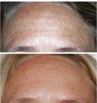
In the 15 cases with scars and stretch marks treated in Germany, the Vancouver Scar Scale was compared with two reliable, objective, and universal methods for assessing scars: the Van-couver Scar Scale, and the Patient and Observer Scar Assessment Scales. The Vancouver Scar Scale has been described to provide a descriptive termi-nology for the comparison of scars and assessing the results of treatments.27–29It considers four parame-ters: vascularity, height (thickness), pliability, and pigmentation. Each variable is scored for severity Fig. 6. A 47-year-old woman with wrinkles of the forehead pre-operatively (above) and 1 year postoperatively (below).
Fig. 5. (Above) Preoperative histologic photomicrograph of the face. (Below) Histologic photomicrograph of the face obtained 6 months postoperatively. Van Gieson stain shows that, 6 months after needling, there is a considerable increase in collagen and elastin deposition, and the collagen appears not to have been laid down in parallel bundles but is rather in the normal lattice pattern.
between 0 (normal skin) and 4 (most severe) to give a total score of between 0 and 16 (where 0 reflects normal skin).27–29The assessments were carried out by two independent observers who regularly treat scarred patients.
The Patient and Observer Scar Assessment Scales consists of two scales: the patient scale, which considers six parameters (color, pliability, thickness, relief, itching, and pain); and the observer scale, which considers five parameters (vasculariza-tion, pliability, pigmenta(vasculariza-tion, thickness, and relief). Each parameter is scored from 0 to 10, where 10 reflects the worst severity. At 12 months after per-cutaneous collagen induction, each patient and the two independent observers completed the observer and the patient scales for that patient’s scars.
In these 15 patients with scarring, the aver-age preoperative Vancouver Scar Scale score was 7.5 ⫾11.5 (range, 4 to 11), which improved to 4.8 ⫾ 15.5 (range, 1 to 6) at 1 year after per-cutaneous collagen induction therapy. The
Pa-tient and Observer Scar Assessment Scale scores improved on average from 27⫾13.5 (range, 14 to 38) preoperatively to 19⫾11.5 (range, 14 to 25) at 1 year after percutaneous collagen induc-tion therapy (Table 3). Postoperatively, no patients experienced any photosensitivity or developed any postinflammatory hyperpigmentation or hypopigmen-tation.
Limitations and Complications
Limitations include the inadequate pretreat-ment with vitamin A of some patients, any active skin processes (e.g., active acne), and unrealistic expectations of some patients. Some patients came for a second or third treatment to improve the outcome, but as far as the authors know, no treated patient underwent any open surgical pro-cedure because the percutaneous collagen induc-tion did not meet their expectainduc-tion.
Two patients developed herpes simplex infec-tion after a full-face needling. These infecinfec-tions Fig. 7. A 41-year-old woman with wrinkles of the face preoperatively (left) and 9 months postoperatively (right).
were treated successfully with acyclovir. Some pa-tients reported swelling and bruising for up to 7 to 10 days. No patients reported scarring, hypopig-mentation or hyperpighypopig-mentation, or photosensi-tivity postoperatively.
DISCUSSION
Rationale for Using Topical Vitamins A and C
The necessity for using vitamins A and C for percutaneous collagen induction has been well described by Fernandes.19 Vitamin A, a retinoic acid, is an essential vitamin (actually a hormone) for skin. It expresses its influence on 400 to 1000 genes that control proliferation and differentia-tion of all the major cells in the epidermis and dermis.30 –38It may control the release of TGF-3 in preference to TGF-1 and TGF-2 because, in general, retinoic acid seems to favor the develop-ment of a regenerative lattice-patterned collagen network rather than the parallel deposition of scar collagen found with cicatrization.39,40 Retinyl es-ters are the main form of vitamin A in the skin,19,41,42and for these reasons, we have elected to apply vitamin A in its ester forms (retinyl
palmi-tate and retinyl acepalmi-tate), with little use of retinol or retinoic acid directly.
Vitamin C is also essential for the production of normal collagen.19,41Percutaneous collagen
in-Fig. 9. A 51-year-old woman with lax skin abdomen preopera-tively (above) and 1 year postoperatively (below).
Fig. 10. A 39-year-old woman with striae abdominales after three pregnancies preoperatively (above) and 6 months postop-eratively (below).
Table 2. Patient Satisfaction: Visual Analogue Scale
Group
Wrinkles Scars Lax Skin Preoperative score 4.5 3.0 3.5 Postoperative score 8.5 7.5 8.5 p ⱕ0.005 ⱕ0.005 ⱕ0.005 Table 3. Patient Satisfaction: VSS and POSAS Scores*
VSS POSAS
No. 72 72
Preoperative score 7.5 27 Postoperative score 4.8 19
p ⱕ0.005 ⱕ0.005
*VSS, Vancouver Scar Scale; POSAS, Patient and Observer Scar As-sessment Scales.
duction and vitamin A switch on the fibroblasts to produce collagen and therefore increase the need for vitamin C.
Normal Stimulation of Collagen Production
Percutaneous collagen induction aims to stimulate collagen production by using the nor-mal chemical cascade that ensues after any trauma. There are three phases in the body’s wound-healing process, which follow each other in a predictable fashion. This has been well de-scribed inThe Biology of the Skinby Falabella and Falanga.43 Platelets and eventually neutrophils release growth factors such as TGF-␣, TGF-, platelet-derived growth factor, connective tissue activating protein III, connective tissue growth factor, and others that work in concert to in-crease the production of intercellular matrix.
Monocytes then also produce growth factors to increase the production of collagen III, elas-tin, glycosaminoglycans, and so forth. Approx-imately 5 days after skin injury, a fibronectin matrix forms with an alignment of the fibroblasts that de-termines the deposition of collagen. Eventually, col-lagen III is converted into colcol-lagen I, which remains for 5 to 7 years. With this conversion, the collagen tightens naturally over a few months. Percutaneous collagen induction causes even further tightening of lax skin and smoothening of scars and wrinkles sev-eral weeks or even months after the injury.44
During the usual conditions of wound heal-ing, scar tissue is formed with minimal regen-eration of normal tissue. We hypothesize that the controlled wound milieu created during percutaneous collagen induction, by minimiz-ing the usual stresses such as exposure to air, infection, mechanical tension, and so forth, may take us closer to regenerative healing. We also believe that our results support our hypothesis.
CONCLUSIONS
Finally, we should question why we destroy the epidermis to achieve smooth skin. The epi-dermis is a complex, highly specialized protec-tive layer, even though it is only approximately 0.2 mm thick. We should only destroy the epi-dermis for medical reasons, never for aesthetic ones. The first step to a healthier skin is to restore the natural levels of photosensitive vita-mins (e.g., vitavita-mins A and B12), antioxidants (e.g., vitamins C and E), and carotenoids, which all become depleted after exposure of the skin to light each day.45,46
Although structural changes to the face and body may be achieved with surgery (e.g., face lifts), true rejuvenation also depends largely on youthful appearing skin. Percutaneous collagen induction offers an antiaging modality to reju-venate and improve the appearance of old skin. We can now improve our patients’ skin from the inside out and not just from the surface. Expe-rience has shown that percutaneous collagen induction works optimally when combined with a scientific skin care program to restore a youth-ful appearance. In addition, percutaneous col-lagen induction has proven to be very effective in minimizing acne and burn scars, by promot-ing the replacement of scar collagen with nor-mal collagen and the reduction of depressed and contracted scars.
Since the introduction of percutaneous colla-gen induction therapy in 1997, it has evolved into a simple and fast method for safely treating wrin-kles and scars and producing smoothness. As op-posed to ablative laser treatments, the epidermis remains intact and is not damaged. For this rea-son, the procedure can be repeated safely and is also suited to regions where laser treatments and deep peels cannot be performed.
Matthias C. Aust, M.D. Klinik fu¨ r Plastische, Hand- und Wiederherstellungschirurgie Hochschule Hannover Carl-Neuberg Strae 1 Hannover 30625, Germany [email protected] REFERENCES
1. Manolins, E. N. In vivo effect of carbon dioxide laser-skin resurfacing and mechanical abrasion on the skin’s microbial flora in an animal model.Dermatol. Surg.32: 359, 2006. 2. Atkins, D. Skin rejuvenation in facial surgery.Facial Plast.
Surg.22: 129, 2006.
3. Landau, M. Advances in deep chemical peels.Dermatol. Nurs. 17: 438, 2005.
4. Hegedus, F. Non-surgical treatment modalities of facial pho-todamage: Practical knowledge for the oral and maxillofacial professional.Int. J. Oral Maxillofac. Surg.35: 389, 2006. 5. Fulton, J. E. Chemical peels: Their place within the range
of resurfacing techniques. Am. J. Clin. Dermatol. 5: 179, 2004.
6. Roy, D. Ablative facial resurfacing.Dermatol. Clin.23: 549, 2005.
7. Ross, E. V. Comparison of carbon dioxide laser, erbium: YAG laser, dermabrasion, and dermatome: A study of thermal damage, wound contraction, and wound healing in a live pig model. Implications for skin resurfacing.J. Am. Acad. Der-matol.42: 92, 2000.
8. Bernstein, L. J. The short- and long-term side effects of carbon dioxide laser resurfacing.Dermatol. Surg.23: 519, 1997.
9. Laws, R. A. Alabaster skin after carbon dioxide laser resur-facing with histologic correlation. Dermatol. Surg. 24: 633, 1998.
10. Rawlins, J. M. Quantifying collagen type in mature burn scars: A novel approach using histology and digital image analysis.J. Burn Care Res.27: 60, 2006.
11. Kauvar, A. N. Histology of laser resurfacing.Dermatol. Clin. 15: 459, 1997.
12. Kang, D. H. Laser resurfacing of smallpox scars.Plast. Re-constr. Surg.116: 259, 2005.
13. Ayhan, S. Combined chemical peeling and dermabrasion for deep acne and posttraumatic scars as well as aging face.Plast. Reconstr. Surg.102: 1238, 1998.
14. Sadick, N. S. Combination radiofrequency and light ener-gies: Electro-optical synergy technology in esthetic medicine. Dermatol. Surg.31: 1211, 2005.
15. Data from the Medical Devices News Article: August 3, 2005. Available at: www.medicalnews.com. Accessed September 2005.
16. Ferguson, M. W. Scar-free healing: From embryonic mech-anisms to adult therapeutic intervention.Philos. Trans. R. Soc. Lond. B Biol. Sci.359: 839, 2004.
17. Bandyopadhyay, B. A “traffic control” role for TGFbeta3: Orchestrating dermal and epidermal cell motility during wound healing.J. Cell Biol.172: 1093, 2006.
18. Orentreich, D. S. Subcutaneous incisionless (subcision) sur-gery for the correction of depressed scars and wrinkles. Der-matol. Surg.6: 543, 1995.
19. Fernandes, D. Percutaneous collagen induction: An alter-native to laser resurfacing.Aesthetic Surg. J.22: 315, 2002. 20. Camirand, A. Needle dermabrasion.Aesthetic Plast. Surg.21:
48, 1997.
21. Fernandes, D. Skin needling as an alternative to laser. Pre-sented at the International Confederation for Plastic, Re-constructive, and Aesthetic Surgery Conference, San Fran-cisco, Calif., June 26 –30, 1999.
22. Yan, J. Levels of retinyl palmitate and retinol in the stratum corneum, epidermis, and dermis of female SKH-1 mice top-ically treated with retinyl palmitate.Toxicol. Ind. Health22: 181, 2006.
23. Sorg, O. Metabolism of topical retinaldehyde. Dermatology 199(Suppl. 1): 13, 1999.
24. Ito, Y. L. Liquid-gel partition chromatography of vitamin A compounds: Formation of retinoic acid from retinyl acetate in vivo.J. Lipid Res.15: 517, 1974.
25. Sass, J. O. Metabolism of topical retinaldehyde and retinol by mouse skin in vivo: Predominant formation of retinyl esters and identification of 14-hydroxy-4, 14-retro-retinol.Exp. Der-matol.5: 267, 1996.
26. MacKay, D. Nutritional support for wound healing.Altern. Med. Rev.8: 359, 2003.
27. Baryza, M. J. The Vancouver Scar Scale: An administration tool and its interrater reliability. J. Burn Care Rehabil. 16: 535, 1995.
28. Sullivan, T. Rating the burn scar.J. Burn Care Rehabil.11: 256, 1990.
29. Draaijers, J. The Patient and Observer Scar Assessment Scale: A reliable and feasible tool for scar evaluation.Plast. Reconstr. Surg.113: 1960, 2004.
30. Bernard, F. X. Comparison of gene expression profiles in human keratinocyte mono-layer cultures, reconstituted epi-dermis and normal human skin: Transcriptional effects of retinoid treatments in reconstituted human epidermis.Exp. Dermatol.11: 59, 2002.
31. Rosdahl, I. Vitamin A metabolism and mRNA expression of retinoid-binding protein and receptor genes in human epi-dermal melanocytes and melanoma cells.Melanoma Res.7: 267, 1997.
32. Fisher, C. Retinoid receptors and keratinocytes. Crit. Rev. Oral Biol. Med.6: 284, 1995.
33. Johnstone, C. C. The physiological basics of wound healing. Nurs. Stand.19: 59, 2005.
34. Lynch, S. E. Growth factors in wound healing: Single and synergistic effects on partial thickness porcine skin wounds. J. Clin. Invest.84: 640, 1989.
35. Tran, K. T. Extracellular matrix signaling through growth factor receptors during wound healing.Wound Repair Regen. 12: 262, 2004.
36. Ruszczak, Z. Effect of collagen matrices on dermal wound healing.Adv. Drug Deliv. Rev.55: 1595, 2003.
37. Faler, B. J. Transforming growth factor-beta and wound heal-ing.Perspect. Vasc. Surg. Endovasc. Ther.18: 55, 2006. 38. Martin, P. Inflammatory cells during wound repair: The
good, the bad and the ugly. Trends Cell Biol. 15: 599, 2005.
39. Fenske, N. A. Structural and functional changes of normal aging skin.J. Am. Acad. Dermatol.15: 571, 1986.
40. Tejero-Trujeque, R. Understanding the final stages of wound contraction.J. Wound Care10: 259, 2001.
41. Nusgens, B. V. Topically applied vitamin C enhances the mRNA level of collagens I and III, their processing enzymes and tissue inhibitor of matrix metalloproteinase 1 in the human dermis.J. Invest. Dermatol.116: 853, 2001.
42. Palma, S. D. Potential use of ascorbic acid-based surfactants as skin penetration enhancers.Drug Dev. Ind. Pharm.32: 821, 2006.
43. Falabella, A. F., and Falanga, V. Wound healing. In R. K. Feinkel and D. T. Woodley (Eds.),The Biology of the Skin.New York: Parthenon, 2000. Pp. 281–299.
44. Fenske, N. A. Structural and functional changes of normal aging skin.J. Am. Acad. Dermatol.15: 571, 1986.
45. Ghyselinck, N. B. Genetic dissection of retinoic acid function in epidermis physiology. Ann. Dermatol. Venereol. 129: 793, 2002.
46. Chapellier, B. Physiological and retinoid-induced prolifera-tions of epidermis basal keratinocytes are differently con-trolled.E.M.B.O. J.21: 3402, 2002.