Received 3 November 2009/Returned for modification 15 December 2009/Accepted 19 January 2010
Intracellular pathogens such asMycobacterium tuberculosishave adapted to a life inside host cells, in which they utilize host nutrients to replicate and spread. Ineffective methods for the evaluation of growth of intracellular pathogens in their true environment pose an obstacle for basic research and drug screening. Here we present the validation of a luminometry-based method for the analysis of intramacrophage growth ofM. tuberculosis. The method, which is performed in a medium-throughput format, can easily be adapted for studies of other intracellular pathogens and cell types. The use of host cells in drug-screening assays dedicated to find antimicrobials effective against intracellular pathogens permits the discovery of not only novel antibiotics but also compounds with immunomodulatory and virulence-impairing activities, which may be future alternatives or complements to antibiotics.
One of the major health issues today, in a global perspective, is the high prevalence of tuberculosis. The causative agent,
Mycobacterium tuberculosis, infects one-third of the global
pop-ulation, mainly in developing countries, and causes 1 to 2 million deaths annually. The spread of drug-resistant strains and the increasing synergy with human immunodeficiency virus further emphasize the urgent need for efficient tools to manage this global emergency (13).
M. tuberculosisis an intracellular pathogen which primarily
targets alveolar macrophages of the host. Several recent re-ports have shown that there is a transcriptional shift of M.
tuberculosisgenes upon uptake by host macrophages, an event
that may alter the susceptibility to drugs (10, 15, 19). There-fore, potential drugs identified in screens using broth cultures may prove to be ineffective for bacteria inside cells (7). Impor-tantly, novel drug-screening methods, where the host cells of intracellular pathogens are included, will allow for the discov-ery of substances that act differently from standard antibiotics, e.g., virulence blockers that interfere with the capacity of the bacterium to cause infection and immunomodulators that en-hance the capacity of the host cell to kill the invader.
The determination of the number of viable bacteria, which is very central to studies of the interaction of intracellular patho-gens with the host cell, is generally performed using the labor-intensive method known as CFU plating. The method is based on the culturing of defined volumes of serial dilutions of lysates from infected cells on agar plates for subsequent analysis of the number of CFU. The slow growth ofM. tuberculosis (2 to 3 weeks until visible colonies appear) and the laborious handling and evaluation of the plates pose a major bottleneck for
effec-tive analysis of intracellular growth of the bacterium. We have previously reported on the use of a virulent strain ofM.
tuber-culosis, H37Rv, harboring the pSMT1 plasmid carrying the
genes forVibrio harveyiluciferase, for analysis of the growth of
M. tuberculosisandMycobacterium aviumsubsp.
paratubercu-losisin mouse spleen, liver, and lungs (9, 16, 20). The flash
luminescence emitted upon the addition of the substrate forV.
harveyiluciferase,n-decanal, is dependent on the presence of a
cofactor, reduced flavin mononucleotide (FMNH2), which is present only in living bacteria. In addition, the substrate for bacterial luciferase is very inexpensive compared to the firefly luciferase substrate luciferin, which has been used by others (6). In this work, we present the validation of the use of luciferase-expressingM. tuberculosisfor the analysis of intra-macrophage growth of the bacterium in a medium-throughput format. We also show how the viability of the host cells can be easily assessed in a large number of samples by a simple pro-cedure. Effective methods for the evaluation of the success of intracellular pathogens in their true environment will be im-portant for future research on host-pathogen interactions and will enable the screening of compound libraries for the discov-ery of drugs that impair intracellular growth of pathogens in a cost- and labor-effective manner.
MATERIALS AND METHODS
Cells and bacteria.We used primary human monocyte-derived macrophages (hMDMs) prepared as previously described (21) and seeded them in 96-well
plates (Sarstedt) in medium free of antibiotics (105
cells/well) 1 day before infection. To achieve nonopsonic conditions, the medium was replaced with serum-free medium immediately before infection. The previously developed
virulentM. tuberculosisstrain H37Rv (American Type Culture Collection)
har-boring a pSMT1 plasmid (17) carrying the gene forVibrio harveyiluciferase, the
avirulent counterpart H37Ra, and the vaccine strainMycobacterium bovis
bacil-lus Calmette-Gue´rin (BCG) (ATCC) were grown in Middlebrook 7H9 broth
supplemented with Tween 80 and oleic acid-albumin-dextrose-catalase (OADC)
(Becton Dickinson) for 2 to 3 weeks at 37°C with 100g/ml hygromycin for
selection before being reinoculated in fresh broth and incubated for 7 days to reach early log phase. Cultures of H37Rv-lux do not differ from cultures of the
* Corresponding author. Mailing address: Division of Medical Mi-crobiology, Lab 1, Level 12, Faculty of Health Sciences, Linko¨ping University, 58185 Linko¨ping, Sweden. Phone: 13-224779. Fax: 46-13-2247889. E-mail: [email protected].
† These authors contributed equally.
䌤Published ahead of print on 27 January 2010.
513
on August 17, 2020 by guest
http://cvi.asm.org/
original H37Rv strain with regard to colony morphology, generation time, and the pattern of intracellular growth, assessed by CFU plating as for the parental strain. To test the susceptibility of H37Rv-lux to standard antimycobacterial drugs, we used Bactec MGIT 960 as previously described (2). In brief, dilution series of rifampin, streptomycin, and isoniazid were prepared in MGIT tubes, as was a control with no antibiotics present. H37Rv-lux from culture was then added to every antibiotic-containing tube and a 1:100 dilution of H37Rv-lux culture to the control tube. Tubes were incubated in a Bactec MGIT 960, and the growth of H37Rv-lux with antibiotics was compared to that of the 1:100 dilution control.
Experimental infection.The bacterial suspension was centrifuged twice at
3,000⫻gfor 10 min in phosphate-buffered saline (PBS) with 0.05% Tween 80,
the pellet was resuspended in plain Dulbecco’s modified Eagle medium (DMEM), and a single-bacillus suspension was obtained by two sets of 10 pas-sages through a sterile syringe equipped with a 27-gauge needle. The
concen-tration was determined by using optical density at 600 nm (OD600) as a function
of CFU/ml obtained from a standard curve developed by plating three 100-l
portions of bacterial suspension on three individual agar plates from four dif-ferent OD values. The bacteria were added to hMDMs in 96-well plates at a multiplicity of infection (MOI) of 10. The preparations were incubated at 37°C for 1 h, and the medium was changed to serum-containing but antibiotic-free DMEM (pulse-chase approach) before incubation for the indicated times. For
evaluating the effect of antimycobacterial drugs, streptomycin (100g/ml),
iso-niazid (10g/ml), or rifampin (10g/ml) was added after the 1-h pulse-chase
and was present for the remainder of the experiment. For the amikacin
exper-iment, amikacin was added after the pulse-chase at a concentration of 200g/ml
for 2 h before the medium was changed, and the amikacin concentration was
lowered to 20g/ml for the remainder of the experiment.
Adaptation of CFU plating assay to the 96-well format.hMDMs were sub-jected to hypotonic lysis in sterile water without additives in order to avoid the possible interference of chemical substances with the subsequent growth of bacteria. Complete lysis as verified by microscopy was achieved after 10 min of incubation, and the cell-associated fraction was subjected to serial dilutions and
plating of defined volumes (5l) of the different dilutions on an individual
square of a 100-mm Middlebrook 7H10 agar plate on which an six-by-six grid was
printed. The grid made it possible to plate 36 individual 5-l samples on the same
plate, allowing the handling of large numbers of samples. The plates were sealed with Parafilm and incubated for 2 weeks before evaluation of the number of colonies. All samples were analyzed in triplicate.
Analysis of bacterial growth by luminometry.To allow the measurement of flash luminescence in the biosafety level 3 (BSL3) facility, a Modulus Microplate Multimode Reader (Turner BioSystems, Sunnyvale, CA) was used. The instru-ment was equipped with luminescence, fluorescence, and absorbance functions and an injector module allowing the measurement of flash luminescence in 96-well plates. During measurements, the instrument was placed inside a bio-safety cabinet in the BSL3 facility to avoid spread of aerosols arising during
injection. A 25-l portion of the medium supernatant was transferred to white
96-well plates (Sarstedt) containing 200l of pure water before cells in each well
were subjected to hypotonic lysis by addition of 70l of pure water and
incu-bation at room temperature for 10 min. The bottom of every well was scraped
before 25l of the cell-associated fraction of bacteria was transferred in the
same manner to white 96-well plates. To both the supernatant and cell-associated
fractions, substrate (1% decanal [Sigma]) was added by the instrument injector before luminescence was measured for 1 s (3 s postinjection). The procedure was performed in triplicates for every time point to compensate for experimental variation. When comparing the different strains of mycobacteria and the effects of antibiotics, the data for the supernatant and cell-associated fractions were standardized to compensate for dilution factors before being added together. The fold increase was calculated by dividing the luminescence value on each day
by the uptake of bacteria (day 0 value⫽1).
Measurement of cell viability.In order to assess the proportions of viable cells at the different times of infection, the infection procedure was carried out with hMDMs seeded on black 96-well plates (Sarstedt). The cells were washed three
times with PBS and incubated with 4M calcein (Invitrogen) diluted in PBS for
30 min. Fluorescence was measured in the Modulus Microplate Reader using a 490-nm filter. Methanol-treated cells were used as negative controls.
Statistical analysis.To ensure that our data were normally distributed, the Kolmogorov-Smirnov test was used. A one-way repeated-measures analysis of variance (ANOVA) was performed to evaluate the growth of H37Rv-lux as
reported by the two different methods. APvalue of⬍0.05 was considered
significant.
RESULTS AND DISCUSSION
Luminometry-based versus viable-count-based assessment of intracellular mycobacterial growth.hMDMs from different donors were infected with H37Rv-lux in several independent experiments (n ⫽ 6) at an MOI of 10 using a pulse-chase approach. The phagocytosis (uptake) resulting from the infec-tion was 0.5 bacterium/cell as determined by luminometry (not shown). After 0 to 3 days postinfection, the cells were lysed and the cell-associated fraction of bacteria was subjected to lumi-nometry by automated addition of the luciferase substrate (Fig. 1A) (the relative luminescence intensity is given in arbitrary light units [ALU]) or serially diluted and plated on solid me-dium for determination of CFU numbers (Fig. 1B). When determining the difference in relative bacterial growth, there was a 4.2-fold increase in bacterial growth between days 0 and 3 as determined by the luminometry assay and a 2.6-fold in-crease with CFU plating. This growth was statistically signifi-cant for both luminometry (P⫽0.0156) and CFU plating (P⫽
0.0103). The ALU data correlate well with microscopy data, where the number of intracellular green fluorescent protein (GFP)-expressing H37Rv cells was evaluated at every time point (not shown), indicating that the signal from the cell-associated fraction is mainly from intracellular bacteria.
From the mean value for every time point, the coefficient of variation between the experiments was calculated by dividing
FIG. 1. Growth of H37Rv-lux in cells from day 0 to 3. Cells from six different donors were infected at an MOI of 10 and incubated for up to 3 days. At days 0 to 3 the cells were lysed, and the number of intracellular H37Rv-lux cells was determined by both luminometry (A) and CFU plating (B). Both the luminometry and CFU plating show a significant increase in bacterial numbers (P⬍0.05). Error bars show standard errors of the means (SEM).
on August 17, 2020 by guest
http://cvi.asm.org/
the standard deviation by the mean (interassay variability). Furthermore, the coefficient of variation was calculated within the triplicates of every experiment and time point (intra-assay variability). The variabilities of the luminometry-based and CFU plating-based methods are summarized in Table 1, and the coefficient of variation is presented as a percentage. The intra-assay variability obtained with the luminometry was lower (1.06 to 13.99% at day 2) than the variability obtained with CFU plating (18.8 to 68.27% at day 2). The high interassay variability obtained using both luminometry and CFU plating (⬎30%) may be due to different capacities of the cells from individual donors to control infection as well as to differences in bacterial fitness between the different bacterial inoculates.
Sensitivity and range of the luminometry-based assay. To determine the sensitivity and the range of detection of the luminometry-based assay, a bacterial culture was prepared as described above and the concentration was determined by op-tical density. The suspension was diluted 1:1 in 16 steps ranging from ⬎1 ⫻ 107 to ⬍1 ⫻ 104, and the luminescence of the
samples was determined (Fig. 2A). From Fig. 2A, 1 ALU was determined to correspond to approximately two bacteria (y⫽
0.5647x). The linear relationship between the number of bac-teria and ALU was stable down to 5,000 bacbac-teria. Further dilutions rendered detectable but nonlinear signals (not shown). CFU plating was more sensitive, allowing the observer to detect a very small number of bacteria, but had a narrow range (at above 200 CFU, separate colonies were indistin-guishable) (Fig. 2B), requiring dilution of the initial sample
down to this range. Furthermore, single colonies were difficult to discriminate due to aggregates (not shown).
Cell-associated versus extracellular growth of H37Rv-lux.
To determine the ratio of bacterial growth inside the cells to that in the medium, we examined the possibility of measuring bacteria in the medium using the luminometric approach. A defined volume of supernatant from the samples to be tested was transferred to a luminescence plate before the rest of the medium was discarded, and the cells were subjected to lysis. Supernatant and cell-associated fractions were collected at days 0 to 3 and analyzed with respect to luminescence. The ALU values obtained were recalculated to correspond to the total amount of luminescence in the individual wells, and Fig. 3 shows the ratios of intracellular to free bacteria. Amikacin used at low doses has been shown to impair extracellular, but not intracellular, growth ofM. tuberculosis(18). However, as shown in Fig. 3, we observed an effect of amikacin on both the cell-associated fraction and the bacteria in the medium super-natant. Amikacin has been reported to accumulate in lyso-somes (12), and aminoglycoside antibiotics may have effects on enzymes and membrane fusion in cells (3, 5). Since our setup involved long-term experiments withM. tuberculosis-infected macrophages, we decided not to use amikacin but instead
FIG. 2. Correlation between bacterial number and luminescence intensity/CFU. (A) The graph shows the relationship between bacterial number (as determined by OD) and the luminescence signal measured by luminometry. A dilution series (1:1) shows the linearity from⬍1⫻107
bacteria down to⬎1⫻104bacteria (inset graph). One arbitrary unit corresponds to approximately two bacteria. (B) The different bacterial
dilutions from panel A were also plated on Middlebrook 7H10 agar and incubated for 2 weeks before the number of CFU was counted. The graph shows that the bacterial number (as determined by OD) and CFU have a linear relationship down to⬃100 bacteria.
H37Rv-lux over 3 days in the presence or absence of amikacin. Defined volumes of medium supernatant and cell-associated fractions were transferred to a luminescence plate and analyzed using luminometry. The measured values were then recalculated to correspond to the total volume of the sample.
on August 17, 2020 by guest
http://cvi.asm.org/
consistently measured both the cell-associated and supernatant fractions.
Detecting changes in mycobacterial growth. To establish that the method was able to detect changes in the growth of H37Rv-lux, hMDMs were infected by the pulse-chase ap-proach, followed by addition of antimycobacterial drugs (100 g/ml streptomycin, 10g/ml isoniazid, or 10g/ml rifampin) before cell-associated and supernatant fractions were analyzed by luminometry at days 0, 1, and 3. Because the initial bacterial load varied between donors due to differences in phagocytosis, the values from days 0, 1, and 3 were normalized to the initial phagocytosis (day 0 values). Figure 4 shows that the presence of antibiotics impairs the growth of H37Rv-lux, and for drug-screening purposes it is possible, from similar experiments including antibiotics, to determine a cutoff value, where the growth can be considered impaired. The pSMT1 plasmid har-boring the luxAB gene also carries a hygromycin resistance gene for growth selection. To determine that H37Rv-lux is suitable for drug-screening purposes and that hygromycin re-sistance did not induce overlapping rere-sistance, we analyzed the MIC values for the three different antibiotics used in Fig. 4. The MIC values for H37Rv-lux were determined using Bactec MGIT 960 and serial dilution and were 0.5g/ml for strepto-mycin, 0.032g/ml for isoniazid, and 0.064g/ml for rifampin. These levels were very similar to those previously reported for H37Rv, which is highly susceptible to the first-line drugs against tuberculosis (8). To further test the ability of the method to detect different kinetics of mycobacterial growth, the avirulent H37Ra strain and the BCG strain carrying the pSMT1 plasmid (H37Ra-lux and BCG-lux) were prepared as described above and allowed to infect macrophages. Growth was monitored by luminometry of both cell-associated and supernatant fractions at days 0 to 3. With luminometry, both H37Ra and BCG showed impaired growth compared to H37Rv (Fig. 5), indicating that the assay is capable of detecting differences in growth kinetics.
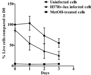
Analysis of cell viability.The replication ofM. tuberculosis
within the human macrophage eventually causes cell death
(14). This makes the testing of cell viability in the individual wells a very important issue for any method used for measuring intracellular growth ofM. tuberculosis. In addition, the mea-surement of cell viability is central for drug-screening pur-poses, since a cytotoxic substance is not a good drug candidate. To evaluate the number of living hMDMs in our experimental setup, we used calcein as a fluorescent probe of cell viability. The method is based on the conversion of nonfluorescent, cell-permeative calcein acetoxymethyl to highly fluorescent cal-cein by living cells. Cells from five different donors were either infected, treated with methanol for 10 min, or left untreated in black 96-well plates. As shown in Fig. 6, both infected and uninfected cells lost viability during the course of the experi-ment; however, the infected hMDMs died more rapidly (50% of infected cells were dead at day 1, while uninfected cells remained unaffected). At day 3, fewer than 20% of the infected
FIG. 4. Antibiotics impair growth ofM. tuberculosisin the lumi-nometry-based assay. hMDMs were incubated with streptomycin (Strep), isoniazid (INH), or rifampin (RIF) at the indicated concen-trations during the infection or were left untreated. The infection was followed by luminometry at days 0, 1, and 3 for both the cell-associated and supernatant fractions. The figure shows the fold increase of the number of bacteria compared to that at day 0, i.e., uptake of bacteria (n⫽4). Error bars show SEM.
FIG. 5. H37Ra-lux and BCG-lux display impaired growth kinetics compared to H37Rv-lux. hMDMs were infected at an MOI of 10 with either BCG-lux or H37Ra-lux, and the number of bacteria in each well was analyzed by luminometry over 3 days of incubation. The growth of both H37Ra-lux and BCG-lux was impaired compared to that of H37Rv-lux. The figure shows the fold increase of the number of bac-teria compared to that at day 0, i.e., uptake of bacbac-teria (n⫽3). Error bars show SEM.
FIG. 6. Comparison of cell death levels in noninfected and M. tuberculosisH37Rv-lux infected hMDMs. The viability of the cells was evaluated at every given time point (n⫽5) by adding 4M calcein and measuring fluorescence after 30 min. The graph shows the percentage of viable cells in H37Rv-lux infected, untreated, and methanol (Me-tOH)-treated hMDMs compared to the day 0 value for uninfected hMDMs. Error bars show SEM.
on August 17, 2020 by guest
http://cvi.asm.org/
line THP-1 could replace the hMDMs in the luciferase assay and found that the inter- and intra-assay variabilities using THP-1 cells were similar to the values obtained with hMDMs (not shown).
By including the host cell in drug screens aiming at identi-fying antimicrobial agents against intracellular pathogens, drug candidates acting via enhancement of cellular functions or by preventing the microorganism from establishing intracellular infection can be found. This may be crucial for the identifica-tion of antimicrobial drugs that act differently from classical antibiotics, since the phenotype of an infecting microorganism can differ remarkably from that of one growing in artificial medium. Also, it is possible that microorganisms are less prone to develop resistance against virulence blockers and immuno-modulators (11).
Scaling up an assay is often associated with problems, and the variability obtained with the CFU plating method used in the 96-well format would probably be lower using larger sam-ple volumes for dilution and plating of the bacteria. Neverthe-less, the adaptation of a protocol to assays allowing the analysis of a larger number of samples is a prerequisite for efficient analysis and is of interest not only for basic investigation but also for industrial approaches such as drug screens. The lucif-erase-based method described here meets these requirements, and in addition, it solves some major problems with CFU plating of mycobacteria, where colonies do not appear until 2 to 3 week after plating and the aggregated morphology of the colonies makes it difficult to evaluate the number of colonies. In conclusion, the luminometry-based method described here is a faster, less laborious, and more exact method than classical CFU plating. The described method can be used for drug-screening purposes and adapted to any intracellular pathogen, which may be transformed with plasmids carrying
theVibrio harveyiluciferase genes under the control of a
suit-able promoter. The opportunity to identify antimicrobial agents that are effectively bactericidal in their true environ-ment makes the inclusion of host cells in drug-screening assays an attractive approach for the development of effective thera-pies against intracellular pathogens.
ACKNOWLEDGMENTS
We are grateful to Kristina Orselius for excellent technical assis-tance, to Michaela Nordvall for MIC analyses, and to Douglas Young (Imperial College, London, United Kingdom) for providing the pSMT1 plasmid.
This project was funded by the Bill & Melinda Gates Foundation through the Grand Challenges Exploration Initiative (M.L.) and by grants from the Swedish Research Council (2003-5994 and 2007-2673 [M.L.] and 2005-6020 and 2008-3101 [O.S.]), the Ekhaga Foundation
rifampin. Diagn. Microbiol. Infect. Dis.28:153–156.
3.Carlier, M. B., G. Laurent, P. J. Claes, H. J. Vanderhaeghe, and P. M. Tulkens.1983. Inhibition of lysosomal phospholipases by aminoglycoside antibiotics: in vitro comparative studies. Antimicrob. Agents Chemother.
23:440–449.
4.Collins, A. R.2002. In vitro detection of apoptosis in monocytes/macro-phages infected with human coronavirus. Clin. Diagn. Lab. Immunol.
9:1392–1395.
5.Dean, R. T., W. Jessup, and C. R. Roberts.1984. Effects of exogenous amines on mammalian cells, with particular reference to membrane flow. Biochem.
J.217:27–40.
6.Deb, D. K., K. K. Srivastava, R. Srivastava, and B. S. Srivastava.2000. Bioluminescent Mycobacterium aurum expressing firefly luciferase for rapid and high throughput screening of antimycobacterial drugs in vitro and in
infected macrophages. Biochem. Biophys. Res. Commun.279:457–461.
7.Dick, T.2009. In vitro-in vivo disconnect in TB drug discovery, p. 56.In
Tuberculosis: biology, pathology and therapy. Keystone Symposia Global Health Series, 25 to 30 January, Keystone, CO.
8.Donald, P. R., and A. H. Diacon.2008. The early bactericidal activity of
anti-tuberculosis drugs: a literature review. Tuberculosis (Edinb.)88(Suppl.
1):S75–S83.
9.D’Souza, S., V. Rosseels, O. Denis, A. Tanghe, N. De Smet, F. Jurion, K. Palfliet, N. Castiglioni, A. Vanonckelen, C. Wheeler, and K. Huygen.2002. Improved tuberculosis DNA vaccines by formulation in cationic lipids.
In-fect. Immun.70:3681–3688.
10.Fontan, P., V. Aris, S. Ghanny, P. Soteropoulos, and I. Smith.2008. Global transcriptional profile of Mycobacterium tuberculosis during THP-1 human
macrophage infection. Infect. Immun.76:717–725.
11.Keyser, P., M. Elofsson, S. Rosell, and H. Wolf-Watz.2008. Virulence block-ers as alternatives to antibiotics: type III secretion inhibitors against
Gram-negative bacteria. J. Intern. Med.264:17–29.
12.Maurin, M., and D. Raoult.1994. Phagolysosomal alkalinization and
intra-cellular killing of Staphylococcus aureus by amikacin. J. Infect. Dis.169:330–
336.
13.Nathan, C.2009. Taming tuberculosis: a challenge for science and society.
Cell Host Microbe5:220–224.
14.Porcelli, S. A., and W. R. Jacobs, Jr.2008. Tuberculosis: unsealing the
apoptotic envelope. Nat. Immunol.9:1101–1102.
15.Rohde, K. H., R. B. Abramovitch, and D. G. Russell.2007. Mycobacterium tuberculosis invasion of macrophages: linking bacterial gene expression to
environmental cues. Cell Host Microbe2:352–364.
16.Rosseels, V., V. Roupie, D. Zinniel, R. G. Barletta, and K. Huygen.2006. Development of luminescent Mycobacterium avium subsp. paratuberculosis
for rapid screening of vaccine candidates in mice. Infect. Immun.74:3684–
3686.
17.Snewin, V. A., M. P. Gares, P. O. Gaora, Z. Hasan, I. N. Brown, and D. B. Young.1999. Assessment of immunity to mycobacterial infection with
lucif-erase reporter constructs. Infect. Immun.67:4586–4593.
18.Stamm, L. M., J. H. Morisaki, L. Y. Gao, R. L. Jeng, K. L. McDonald, R. Roth, S. Takeshita, J. Heuser, M. D. Welch, and E. J. Brown.2003. Myco-bacterium marinum escapes from phagosomes and is propelled by
actin-based motility. J. Exp. Med.198:1361–1368.
19.Tailleux, L., S. J. Waddell, M. Pelizzola, A. Mortellaro, M. Withers, A. Tanne, P. R. Castagnoli, B. Gicquel, N. G. Stoker, P. D. Butcher, M. Foti, and O. Neyrolles.2008. Probing host pathogen cross-talk by transcriptional profiling of both Mycobacterium tuberculosis and infected human dendritic
cells and macrophages. PLoS One3:e1403.
20.Tanghe, A., S. D’Souza, V. Rosseels, O. Denis, T. H. Ottenhoff, W. Dalemans, C. Wheeler, and K. Huygen.2001. Improved immunogenicity and protective efficacy of a tuberculosis DNA vaccine encoding Ag85 by protein boosting.
Infect. Immun.69:3041–3047.
21.Welin, A., M. E. Winberg, H. Abdalla, E. Sarndahl, B. Rasmusson, O. Stendahl, and M. Lerm.2008. Incorporation of Mycobacterium tuberculosis lipoarabinomannan into macrophage membrane rafts is a prerequisite for
the phagosomal maturation block. Infect. Immun.76:2882–2887.