Electronic Thesis and Dissertation Repository
August 2015
Engineered Cx26 Variant Established Functional Heterotypic
Engineered Cx26 Variant Established Functional Heterotypic
Cx26/Cx43 and Cx26/Cx40 Gap Junction Channels
Cx26/Cx43 and Cx26/Cx40 Gap Junction Channels
Levent Berk Karademir
The University of Western Ontario
Supervisor Donglin Bai
The University of Western Ontario Graduate Program in Neuroscience
A thesis submitted in partial fulfillment of the requirements for the degree in Master of Science © Levent Berk Karademir 2015
Follow this and additional works at: https://ir.lib.uwo.ca/etd
Part of the Cellular and Molecular Physiology Commons
Recommended Citation Recommended Citation
Karademir, Levent Berk, "Engineered Cx26 Variant Established Functional Heterotypic Cx26/Cx43 and Cx26/Cx40 Gap Junction Channels" (2015). Electronic Thesis and Dissertation Repository. 3083.
https://ir.lib.uwo.ca/etd/3083
This Dissertation/Thesis is brought to you for free and open access by Scholarship@Western. It has been accepted for inclusion in Electronic Thesis and Dissertation Repository by an authorized administrator of
(Thesis format: Integrated Article)
by
Levent Berk Karademir
Graduate Program in Neuroscience
A thesis submitted in partial fulfillment of the requirements for the degree of
Masters of Science
The School of Graduate and Postdoctoral Studies The University of Western Ontario
London, Ontario, Canada
ii
Abstract
Two hexameric connexin hemichannels dock together to from a whole gap junction (GJ)
channel. The mechanisms of docking specificity in forming homotypic and heterotypic
GJ channels are not fully clear. To reveal the key differences between Cx26 and Cx43 (or
Cx40) in their docking residues, we aligned and analyzed ten well studied connexin
sequences. Five of them are docking compatible with Cx26 and the rest (including Cx43
and Cx40) are not. According to Cx26 atomic structure at the docking interface, we
identified two putative docking residues on the second extracellular domain (E2) that are
well conserved within docking compatible connexins, but drastically different between
docking incompatible connexins. Switching both of these residues in Cx26 into the
corresponding residues in the docking incompatible connexins (K168V-N176H)
established morphological and functional heterotypic GJs with Cx43 (or Cx40),
indicating these two residues are important for docking incompatibility of these and
likely other related connexins.
Key words: Gap junction channel, heterotypic docking compatibility, patch clamp,
iii
Dedication
I’d like to dedicate this to my family and friends, both here and abroad, who have
iv
Co-Authorship Statement
Electrophysiological work was accomplished by Levent Berk Karademir, with
Honghong Chen providing major assistance in the collection of morphological and
localization data for homotypic and heterotypic gap junction combinations. Benny Yue
assisted in minor electrophysiological data collection, specifically
v
Acknowledgments
I would like to thank my supervisor Dr. Donglin Bai for giving me the
opportunity to work in his lab and for providing me with guidance when I needed it most.
I greatly appreciate his patience and support throughout this whole process. I would also
like to thank my advisory committee members, Dr. Arthur Brown and Dr. Stan Leung,
for their insight and assistance throughout the maturation of my project. Furthermore, I
would like to thank all of my lab members for the great times these past two years – it’s
vi
Table of Contents
Abstract ... ii
Dedication ... iii
Co-Authorship Statement... iv
Acknowledgments... v
Table of Contents ... vi
List of Figures ... ix
Abbreviations ... x
Chapter 1 – Introduction
... 11.1 Gap junction channels ... 1
1.2 Connexins ... 4
1.3 Connexin 26 ... 7
1.3.1 Localization and physiological functions ... 7
1.3.2 Determination of crystal structure ... 8
1.4 Connexin 43 ... 11
1.5 Heterotypic Gap Junction Docking ... 13
1.5.1 E2-E2 Interactions ... 16
1.5.2 E2 Domain in Docking ... 20
1.6 Rationale and Hypothesis ... 21
1.7 Objectives ... 21
vii
Chapter 2 – Manuscript
... 272.1 Abstract ... 28
2.2 Introduction ... 29
2.3 Methods... 33
2.3.1 Construction of Cx26 mutants ... 33
2.3.2 Cell culture and transient transfections ... 34
2.3.3 Electrophysiological recordings... 34
2.3.4 Data analysis ... 36
2.4 Results ... 37
2.4.1 Designing Cx26 variants to establish docking with Cx43 and Cx40... 37
2.4.2 Homotypic Cx26 single and double mutants show reduced gap junction channel function when compared to wildtype Cx26 ... 39
2.4.3 Cx26 K168V-N176H, but not L168V or N176H, formed functional heterotypic gap junction channels with Cx43 ... 42
2.4.4 Cx26 K168V-N176H, but not the single mutants, formed functional heterotypic gap junction channels with Cx40 ... 46
2.4.5 Heterotypic Cx26 K168V-N176H/Cx43 and K168V-N176H/Cx40 displayed different asymmetric Vj - gating properties. ... 50
2.4.6 Initial Gj rectification of K168V-N176H/Cx43 channels were also observed at unitary channel currents ... 54
2.5 Discussion ... 58
2.5.1 Structural insights of docking in members of Group2 connexins... 58
2.5.2 Docking residues in Cx26 and their equivalent residues in other connexins are mutational hotspots for disease-linked connexin mutants . 60 2.5.3 Physiological and pathological implications of docking between Cx26/Cx43 ... 61
viii
2.6 References ... 65
Chapter 3
-
Discussion
... 693.1 Overall Study ... 69
3.2 Human Cx26/Cx43 and Cx26/Cx40 channels do not show high conductance .... 70
3.3 K168 and N176 influences heterotypic docking in Cx26 ... 70
3.4 Physiological and pathological role of Cx26 and Cx43 interaction ... 73
3.5 Potential physiological role of connexin incompatibility and compartmentalization ... 75
3.6 Limitations and future directions ... 78
3.7 Summary ... 79
3.8 References ... 81
Curriculum Vitae ... 85
ix
List of Figures
Figure 1.1 Various GJ channel compositions and general topology of a single connexin subunit. ... 6
Figure 1.2 Homology model for Cx26/Cx32 homomeric heterotypic gap junction channel and E2 docking interface interactions. ... 10
Figure 1.3 Heterotypic compatibility of ten selected connexins... 15
Figure 1.4 Sequence analyses of E1 and E2 domains. ... 19
Figure 2.1 Morphological and functional analysis of homotypic Cx26 single and double mutants. ... 41
Figure 2.2 Morphological and functional status of heterotypic Cx26 mutant/Cx43
channels... 45
Figure 2.3 Morphological and functional status of heterotypic Cx26 mutant/Cx40
channels... 49
Figure 2.4 Macroscopic current analysis of heterotypic K168V-N176H/Cx43 and
K168V-N176H/Cx43 channels. ... 53
x
Abbreviations
CL Cytoplasmic loop
CT Carboxyl-terminus
Cx Connexin
DIC Differential interference contrast
DMEM Dulbecco’s modified Eagle’s medium
E1 The first extracellular loop
E2 The second extracellular loop
ECF Extracellular fluid
EGFP Enhanced green fluorescent protein
GJ Gap junction
Gj Junctional conductance
Gj,ini Initial transjunctional conductance
HB Hydrogen bond
ICF Intracellular fluid
Ij Macroscopic junctional current
ij Junctional current of single channel
ms Millisecond
xi
nS Nanosiemens
NT Amino terminus
pIRES Plasmid containing internal ribosome entry site
pS Picosiemens
RFP Red fluorescent protein
SEM Standard error of the mean
TM Transmembrane domain
Vj Transjunctional voltage
+Vj Positive transjunctional voltage
-Vj Negative transjunctional voltage
Vm Transmembrane voltage
γγγγj Unitary channel conductance
Chapter 1
–
Introduction
1
Introduction
1.1
Gap junction channels
Cellular communication allows for maintenance of homeostasis in tissues and
organs in multicellular organisms, and facilitates for quick response by cells to changes in
environmental conditions. Gap junctions (GJ), which are clusters of intercellular
membrane channels, are key players in cellular communication and serve a crucial role in
many physiological processes. The formation of these channels require the membranes of
two adjacent cells to be in close proximity of each other, leaving a 2-4 nm gap (Bruzzone
et al., 1996). GJs link the cytoplasm of two cells and facilitate the sharing of ions (K+, Cl
-and Na+), secondary messengers (cAMP), small metabolites (glucose), and small
interfering RNAs of up to 1 kDa in size (Loewenstein, 1981; Valiunas et al., 2005).
GJ communication contributes in many crucial processes ranging from
development and differentiation, to apoptosis and the maintenance of cell homeostasis
(White & Paul, 1999). There can also be detrimental effects due to GJ mediated
communication. A dying cell compromised by disease or injury can elicit a “bystander
effect”, where GJs allow the passage of metabolites from dying cells to otherwise
unaffected healthy neighboring cells. This transfer of substances can lead to the
promotion of cell death in the otherwise unaffected cells (Bi et al., 1993). GJs serve
another unique purpose in certain cell types. Due to the electrically excitable nature of
currents and the electrical synchronization between cell groups (Spray & Burt, 1990). In
the brain, GJs facilitate electrical signaling between neurons, and act as a point of passage
for metabolites and signaling molecules between glial cells to help support the
neurovascular structures (Giaume & Theis, 2010; Pereda, 2014).
There are a number of mechanisms that can modulate and regulate GJs, which can
be simply categorized under two major titles – chemical factors and voltage. Chemical
factors at play include connexin protein phosphorylation, cytoplasmic pH, intracellular
Ca2+ concentration, lipophiles, and potentially many more (Harris, 2001). The second
category is voltage-dependent deactivation, also referred to as “gating”, which is a
common property of all GJs that have been currently identified. Voltage regulation of GJs
can be divided into two forms – rectification and transjunctional voltage-dependent
gating (Oh & Bargiello, 2015). Rectification is a change in channel conductance due to
some form of asymmetry, such as differences in charge distribution in the pore or
different post-translational modifications (eg. phosphorylation). GJs formed from
different connexins show unique voltage gating characteristics, which might be the
reason Cx26/Cx32 heterotypic channels are a good example of rectification (Oh et al.,
1999). Voltage-dependent gating can occur due to a number of reasons, such as structural
changes in response to voltage, ion availability, Mg2+ blocking and distribution of fixed
charges in the pore (Oh et al., 2008; Palacios-Prado, Chapuis, et al., 2014).
Voltage-dependent structural changes can allow or hinder the flux of ions. Since GJs span the
membrane of two adjacent cells, there are two electric fields by which they can be
influenced. The first is transjunctional voltage (Vj), which is the electrical difference
electrical potential between the cytoplasm and the extracellular space, termed as the
membrane potential (Vm). Most connexins are only sensitive to Vj, with Cx26 being the
exception that slightly responds to Vm (Barrio et al., 1991). The molecular origin of Vj
gating has yet to be established, however there are two proposed theories. Studies
conducted on Cx26 and Cx32 found that changing the charge status of the 2nd amino acid
on the N-terminal was sufficient to reverse gating polarity (Verselis et al., 1994),
highlighting the NT as a possible origin for Vj gating. Gating polarity is the probability of
a given Vj sensitive hemichannel to dwell in the closed state at a relative Vj polarity and
intensity (Palacios-Prado, Huetteroth, et al., 2014). Additional research on the Cx32
N-terminal concluded that the first 10 residues of the NT are pore-lining, conferring
sensitivity to the Vj field (Oh et al., 2004). The other proposed mechanism highlights a
ball and chain model where the C-terminus acts as the gating portion by binding to a
receptor position on the cytoplasmic loop, which was a theory drawn from Cx43 and
Cx40 studies (Anumonwo et al., 2001). High resolution crystal structure of homomeric
homotypic Cx26 channels support the claim that the NT is in the pore and is in a position
1.2
Connexins
Connexins (Cx) are the basic GJ subunit that oligomerize in a hexamer structure
to form half of the GJ channel, also referred to as a hemichannel or connexon. The head
to head docking of two hemichannels forms a complete intercellular GJ channel (Harris,
2001). Connexins are seen as unique among other channel proteins due to their functional
existence as both a full intercellular GJ channel, and as an undocked hemichannel.
Hemichannels have been shown to be important in paracrine signaling (Wang, De Bock,
et al., 2013), however are implicated more as pathological rather than physiological
entities, based on the current evidence. As observed in the brain and heart, abnormal
hemichannel opening can lead to the entry of Na+ and Ca2+, loss of K+, ATP and small
metabolites, Ca2+ overload, and eventual cell death (Orellana et al., 2014; Wang, De
Vuyst, et al., 2013).
There are 21 connexin isoforms in the human genome, and these connexin family
members share a similar structural topology. These 21 connexins are placed into one of
five categories (α, β, γ, δ and ε) based on their sequence homology (Sohl & Willecke,
2004). Each connexin has four transmembrane domains (TM1-TM4), two extracellular
loops (E1 & E2), one cytoplasmic loop (CL), and both amino-terminus (NT) and
carboxyl-terminus (CT) on the cytoplasmic side (Milks et al., 1988) (Figure 1.1). The
molecular weight of the connexin indicates the nomenclature for the common name of
each isoform. For example, the molecular weight of Cx26 is 26kDa (Beyer et al., 1990).
Virtually every cell in the body expresses one or more connexins (Saez et al., 2003). This
six oligomerizing subunits of a hemichannel may be formed of the same connexin
(homomeric), or from a combination of different connexin isoforms (heteromeric). A
complete gap junction channel may be composed of two identical hemichannels
(homotypic), or from hemichannels of different composition (heterotypic) (Figure 1.1).
Though all these combinations are theoretically possible, not all homomeric channels can
dock to form functional heterotypic gap junction channels with one another. Both the E1
and E2 domains are involved in intercellular channel formation, however it was observed
that the docking compatibility of two hemichannels was linked to the E2 domain (Bai &
Wang, 2014). The mechanisms involved in heterotypic docking compatibility are not
fully understood, however it is possible that a small number of differences in the residues
Figure 1.1 Various GJ channel compositions and general topology of a single
connexin subunit.
Oligomerization of six connexin subunits forms a hemichannel. Two hemichannels at the
plasma membrane can dock to form a full gap junction channel. A hemichannel
composed of identical connexins are referred to as homomeric, while those with different
connexins are termed heteromeric. Similarly, gap junction channels composed of
identical hemichannels are referred to as homotypic and those with different
hemichannels are called heterotypic. All connexin isoforms are transmembrane proteins
1.3
Connexin 26
1.3.1 Localization and physiological functions
Human Cx26 (also known as gap junction β-2 protein and encoded by GJB2) is
an extensively studied connexin, and has been found in almost all locations of the cochlea
that form GJ channels (Kikuchi et al., 1995). Cx26 was also shown to be expressed in the
skin, specifically during keratinocyte differentiation (Di et al., 2001). Other tissue and
cell types that are known to express Cx26 include the liver (Iwai et al., 2000), placenta
(Kibschull et al., 2008), and mammary epithelium (Monaghan et al., 1996).
Cx26 mutations are fairly common in the general population. Mutations linked to
nonsyndromic hearing loss can be found distributed across the coding region of Cx26,
however syndromic mutations that additionally present with skin diseases were found to
be located mostly in the N-terminus and E1 domain (Lee & White, 2009). Cx26
mutations causing non-syndromic deafness can be dominant or recessive, with 80% of
cases resulting from recessive mutations at more than 100 loci and the remaining 20%
caused by dominant mutations on more than 30 loci (Petit et al., 2001; Yan & Liu, 2008).
Due to the high frequency of Cx26 mutations, genetic screening for Cx26 is a routine
procedure in cases of pediatric hearing impairment (Smith, 2004; Tranebaerg, 2008).
Simple loss-of-function mutations in Cx26 have been found to cause nonsyndromic
deafness, whereas that is not the case for the syndromic forms (Bruzzone et al., 2003;
Zhao et al., 2006). The observation that complete loss of Cx26 function does not result in
skin diseases suggests that the human skin does not require Cx26 to maintain homeostasis
syndromic hearing loss are apparent, and possibly due to dominant-negative effects on
Cx43. Both Cx26 and Cx43 are implicated in skin diseases, and it has been demonstrated
that dominant autosomal Cx26 mutants that cause hearing impairment can have
trans-dominant inhibition when co-expressed with wildtype Cx43 (Rouan et al., 2001). Another
investigation looked into Cx26 knock out (Cx26-/-) mouse models to examine its systemic
role, however these mice were found to die in utero as a result of placental defects
(Gabriel et al., 1998). In order to retain viability, conditional knockouts were found to be
a better approach for studying the effects of Cx26 ablation in tissue and system
physiology (Stewart et al., 2014)
1.3.2 Determination of crystal structure
The structure of Cx26 was experimentally determined through crystallization of a
homomeric homotypic Cx26 GJ channel at a resolution of 3.5 Å (Maeda et al., 2009;
Suga et al., 2009). The resolution was high enough to observe the atomic structure and
interactions of the Cx26 GJ channel. Some of the major findings were as such: the four
transmembrane helices of a connexin were arranged differently than what was proposed
previously in a pseudoatomic model, several residues linked to nonsyndromic hearing
loss or skin diseases were also involved in inter- or intramolecular interactions, the
interactions between the two extracellular domains apposing hemichannels were
elucidated in detail, and that the N-terminus restricts the diameter of the pore entrance
and might play a role in channel gating (Maeda et al., 2009).
The atomic structure confirmed that both the E1 and E2 domain are involved in
extracellular domains formed a double-layered channel wall, with the E1-E1 interaction
being staggered, possibly to prevent the inner channel from leaking into the extracellular
space. An interesting finding was the presence of 60 hydrogen bonds (HB; a non-covalent
bond) at the docking interface. The localization of these 60 HBs were divided as 24 HBs
being involved in the E1-E1 docking interfaces, and 36 HBs being involved in the E2-E2
docking interfaces between two docked hemichannels. This extensive HB network is
believed to be responsible for anchoring the two apposing hemichannels during docking.
There is strong evidence suggesting that other closely related connexins might be
utilizing the same interaction profile during docking (Bai & Wang, 2014; Nakagawa et
al., 2011) (Figure 1.2). The functional necessity of a minimum number of HBs at the
E2-E2 interface for functional Cx26/Cx32 channels further supports this theory (Gong et al.,
Figure 1.2 Homology model for Cx26/Cx32 homomeric heterotypic gap junction
channel and E2 docking interface interactions.
Homomeric hemichannels of Cx26 (magenta) and Cx32 (green) can dock to form
functional heterotypic gap junction channels. The E2 docking site between one pair of
docked subunits is enlarged to show docking HBs and the relevant residues at the
docking interface. Dotted lines indicate HB formation. Modified from Bai and Wang
1.4
Connexin 43
Cx43 (also known as gap junction α-1 protein and encoded by GJA1) is the most
widely expressed connexin in the human body, and an especially prominent connexin
isoform in the heart and brain. It is predominantly expressed in astrocytes and microglial
cells in the brain (Giaume & Theis, 2010). Though not present under normal conditions,
low level of Cx43 expression was reported in the blood-brain barrier during inflammation
(Cronin et al., 2008). In the heart, Cx43 is expressed in the ventrical cardiomyocytes,
atria, and endothelial cells (Severs et al., 2008). Cx43 has also been found to control
granulosa cell proliferation in the reproductive system, and might be responsible for
spermatogenesis in the testis (Kidder & Mhawi, 2002; Roscoe et al., 2001). Other major
cell types and organs showing Cx43 expression include the skin and mammary glands
(Kelsell et al., 2000; Monaghan et al., 1996). Cx43 is a phosphoprotein with over a dozen
phosphorylation sites, which have been shown to be involved in the regulation of channel
function and interactions (Lampe & Lau, 2004).
Large numbers of mutations in Cx43 have been linked to the pathological
condition of oculodentodigital dysplasia (ODDD) (Paznekas et al., 2009). ODDD affects
the whole body and presents with a wide range of symptoms, mainly abnormalities in the
eyes, teeth, and fingers (Gorlin et al., 1963). Cx43 deficient (Cx43-/-) mouse models died
soon after birth due to right ventricular obstruction tract of the heart (Reaume et al.,
1995). It was observed that the postnatal lethality of Cx43-/- could be partially
compensated for by the knock-in of Cx32 or Cx40, suggesting that these connexins might
share a common function (Plum et al., 2000). However, these Cx32 and Cx40 knock-in
wildtype counterparts. This points toward a certain degree of redundancy that might exist,
making it necessary to understand the functional interaction profile of different
1.5
Heterotypic Gap Junction Docking
Hemichannels extend approximately 20 Å into the extracellular matrix, and are
believed to use the facilitation of other membrane-associated molecules to bring apposing
membranes into close proximity (Yeager, 1998). Both the E1 and E2 domain have been
experimentally proven to be involved in the process of docking (G. Dahl et al., 1994;
Maeda et al., 2009). While the molecular specificity of docking is yet to be deciphered,
the E2 loop is believed to be the domain responsible. The E1 region has high sequence
identity and conserved HB-forming residues among many human connexins (Bai &
Wang, 2014). This makes it an unlikely candidate for being involved in the determination
of heterotypic compatibility. Furthermore, chimeric connexins with substituted E2
domains showed that the origin of the E2 domain was the determining factor in whether
or not two connexins would form functional heterotypic GJ channels (Bruzzone et al.,
1994; White et al., 1994). White et al. (1994) observed E2 dependent selectivity when
working on Cx50, Cx43 and Cx46 chimeras, while Bruzzone et al. (1994) indicated a
similar effect while studying Cx32, Cx38 and Cx43 chimeras.
Connexins have been divided into subgroups (α, β, γ) based on the extent of their
sequence similarity and length of the cytoplasmic domain (Kumar & Gilula, 1996). While
this system possesses some functional consistency due to its basis on sequence
homology, it is not without its shortcomings (Söhl & Willecke, 2009). Going through a
large number of functional studies, we can observe the heterotypic and homotypic
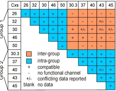
compatibility of different connexins (Figure 1.3). The nomenclature for connexin
subgroups do not effectively account for functional heterotypic compatibility; Cx40 (α
Cx30.3, but not with others (Cx26 and Cx32). These types of discrepancies prompt the
generation of an auxiliary grouping system that takes heterotypic compatibility into
consideration. When looking at the functional compatibility of ten of the most
extensively studied connexins, a certain pattern can be observed (Bai & Wang, 2014).
This pattern allows for the categorization of these ten connexins into two distinct groups
– Group 1 and Group 2. Two initial observations can be made between these two groups.
Firstly, members of the same group (intra-group) can, in most cases, form functional
heterotypic gap junction channels with one another. Secondly, connexins belonging to
different groups (inter-group) cannot usually form functional heterotypic GJs with one
another. The necessity for such a grouping system is further apparent when we observe
that neither Group 1 nor Group 2 consists exclusively of one subgroup of connexins
(Figure 1.3); Group 1 has 3 connexins belonging to β subgroup (Cx26, Cx32 and Cx30)
and 2 belonging to α subgroup (Cx46 and Cx50), while Group 2 has 3 α subgroup (Cx37,
Figure 1.3 Heterotypic compatibility of ten selected connexins.
Ten extensively studied connexins are divided into two groups, Group 1 and Group 2,
depending on their heterotypic compatibility. As a general rule, members of one group
can form functional heterotypic homomeric GJ channels with other intra-group connexins
1.5.1 E2-E2 Interactions
E1 and E2 sequence alignment of the ten Group 1 and Group 2 connexins show
that both extracellular loops are highly conserved, with the E2 being slightly more
variable (Haefliger et al., 1992). The high identity and conservation of the E1 and E2
domain suggests that these regions likely share a similar atomic structure with other
members of the connexin family (Bai & Wang, 2014; Nakagawa et al., 2011). A closer
look at the E2 sequence of Group 1 and Group 2 members showed an interesting pattern.
At a given position in the E2 sequence, most of the residues are either highly variable or
highly conserved, except at the 168 and 176 equivalent positions when aligned using
Cx26 (Figure 1.4). The 168 and 176 positions, along with the 179 and 177 positions, are
important in Cx26 due to their critical role in forming HBs between docking hemichannel
(Maeda et al., 2009). The 179 equivalent residues are highly conserved across both Group
1 and Group 2 except for Cx40, while the 177 equivalent residues are highly variable
throughout. The high variability at the 177 positions across all ten connexins can be
attributed to the fact that the HB was formed on the main peptide chain carbonyl group of
the Cx26 threonine residue, thus having no selective pressure for any specific amino acid.
The patterns of conservation for positions 177 and 179 make it unlikely that they act as
determinants in heterotypic selectivity in docking, leaving positions 168 and 176 as the
likely candidates. For Group 1 connexins, asparagine is completely conserved at the 176
equivalent positions. Group 2 connexins, however, express either a histidine (4/5) or
tyrosine (1/5) at the same equivalent position. While asparagine contains a carboxamide
as a side chain, both histidine and tyrosine contain large aromatics. For the 168
hosting positively charged and long side chains. Group 2 members express a combination
of valine (3/5), alanine (1/5) or threonine (1/5), which are small and non-polar. With
respect to the other HB forming positions, This group-specific phenomenon of
Figure 1.4 Sequence analyses of E1 and E2 domains.
Sequences were aligned for the E1 and E2 of the same ten connexins previously
mentioned (Figure 1.3). Sequence logos were generated for the E2 alignment in order to
investigate conservation at docking HB residues. Generated logos were for Group 1 and
Group 2 human connexins, as well as cumulative Group 1 and Group 2 connexins from
different species. The number of sequences used for the sequence logos were 5, 5, 164,
and 169, respectively. Numbering of residues follow that of Cx26. Arrows indicate
docking HB residues as determined by the Cx26 atomic structure (Maeda et al., 2009).
Bolder arrows point at candidate residues responsible for determining heterotypic
1.5.2 E2 Domain in Docking
In order to test the role and influence of HBs at the E2-E2 interface, a number of
site-specific Cx26 and Cx32 mutants were studied for their functionality. Based on high
sequence identity and putative homology modeling, it was predicted that Cx32 should
behave in a structurally similar manner to Cx26 (Gong et al., 2013; Nakagawa et al.,
2011). The mutants in question changed the residues at Cx26K168 (to K168V and
K168A) and Cx32N175 (to N175H and N175Y). Homology modeling for these mutants
predicted a range of alterations to the total number of E2 HBs in a pair of docked
E2-E2, from the native six HBs all the way down to zero. By recording the transjunctional
conductances (Gj) of these mutants in a range of combinations, the influence of HBs at
the E2 docking interface was elucidated for Cx26/Cx32 channels (Gong et al., 2013). It
was found that a minimum of four HBs between each docked E2-E2 region were
necessary to form functional gap junction channels. This study weighs on the importance
of HBs at the E2-E2 interface, and their role in facilitating the formation of functional
GJs. Group 1 connexins show high sequence identity at their HB forming residues,
suggesting that other Group 1 members may also use the mechanisms observed in Cx26.
However, this prediction has yet to be experimentally proven. Differences at key residues
and the lack of a high-resolution atomic structure for a Group 2 connexins has left us
1.6
Rationale and Hypothesis
Two docking HB forming residues, at positions 168 and 176 of Cx26, are
conserved within but divergent between members of Group 1 and Group 2 connexins. A
connexin from one group cannot usually form functional GJ channels with a connexin
from the apposing group, such as in the case of Cx26 and Cx43. We believe that this is
due to differences in the docking domains between Group 1 and Group 2 connexins. We
hypothesize that the E2 domain of Cx43 and Cx40 share similar structure and use similar
residues as those in Cx26. We further hypothesize that switching the residues at positions
168 and 176 of Cx26 to match the majority residues found in Group 2 connexins (K168V
and N176H) will result in forming functional heterotypic GJs between Cx26 and Group 2
connexins, Cx43 and Cx40.
1.7
Objectives
i. Investigate the docking and functional interaction between human Cx26/Cx43
heterotypic gap junction channels, and explore the morphological and functional
effects of Cx26 single mutants (K168V and N176H) and double mutant
(K168V-N176H) on the formation of functional heterotypic gap junction channels with
Cx43. The mutations at K168 and N176 were introduced individually or in
combination (single and double mutants) to Cx26. Mutant and wildtype constructs
expressing tagged or untagged fluorescent proteins were transfected into mouse
neuroblastoma cells (N2A) for subsequent fluorescent imaging for morphology and
ii. Establish whether the Cx26 variant K168V-N176H can also form functional
heterotypic channels with another Group 2 member, Cx40. Similar to above, tagged
connexins are used to determine morphology and localization, while a combination
of tagged and untagged connexins are utilized in whole-cell patch clamp analysis to
1.8
References
Anumonwo, J. M., Taffet, S. M., Gu, H., Chanson, M., Moreno, A. P., & Delmar, M. (2001). The carboxyl terminal domain regulates the unitary conductance and voltage dependence of connexin40 gap junction channels. Circ Res, 88(7),
666-673.
Bai, D., & Wang, A. H. (2014). Extracellular domains play different roles in gap junction formation and docking compatibility. Biochem J, 458(1), 1-10. doi:
10.1042/bj20131162
Barrio, L. C., Suchyna, T., Bargiello, T., Xu, L. X., Roginski, R. S., Bennett, M. V., et al. (1991). Gap junctions formed by connexins 26 and 32 alone and in combination are differently affected by applied voltage. Proc Natl Acad Sci U S A, 88(19),
8410-8414.
Beyer, E. C., Paul, D. L., & Goodenough, D. A. (1990). Connexin family of gap junction proteins. J Membr Biol, 116(3), 187-194.
Bi, W. L., Parysek, L. M., Warnick, R., & Stambrook, P. J. (1993). In vitro evidence that metabolic cooperation is responsible for the bystander effect observed with HSV tk retroviral gene therapy. Hum Gene Ther, 4(6), 725-731. doi:
10.1089/hum.1993.4.6-725
Bruzzone, R., Veronesi, V., Gomes, D., Bicego, M., Duval, N., Marlin, S., et al. (2003). Loss-of-function and residual channel activity of connexin26 mutations associated with non-syndromic deafness. FEBS Lett, 533(1-3), 79-88.
Bruzzone, R., White, T. W., & Paul, D. L. (1994). Expression of chimeric connexins reveals new properties of the formation and gating behavior of gap junction channels. J Cell Sci, 107(4), 955-967.
Bruzzone, R., White, T. W., & Paul, D. L. (1996). Connections with connexins: the molecular basis of direct intercellular signaling. Eur J Biochem, 238(1), 1-27.
Cronin, M., Anderson, P. N., Cook, J. E., Green, C. R., & Becker, D. L. (2008). Blocking connexin43 expression reduces inflammation and improves functional recovery after spinal cord injury. Mol Cell Neurosci, 39(2), 152-160. doi:
10.1016/j.mcn.2008.06.005
Dahl, G., Nonner, W., & Werner, R. (1994). Attempts to define functional domains of gap junction proteins with synthetic peptides. Biophys J, 67(5), 1816-1822.
Di, W. L., Common, J. E., & Kelsell, D. P. (2001). Connexin 26 expression and mutation analysis in epidermal disease. Cell Commun Adhes, 8(4-6), 415-418.
Gabriel, H. D., Jung, D., Butzler, C., Temme, A., Traub, O., Winterhager, E., et al. (1998). Transplacental uptake of glucose is decreased in embryonic lethal connexin26-deficient mice. J Cell Biol, 140(6), 1453-1461.
Giaume, C., & Theis, M. (2010). Pharmacological and genetic approaches to study connexin-mediated channels in glial cells of the central nervous system. Brain Res Rev, 63(1-2), 160-176. doi: 10.1016/j.brainresrev.2009.11.005
Gorlin, R. J., Miskin, L. H., & St, G. J. (1963). Oculodentodigital dysplasia. J Pediatr, 63, 69-75.
Haefliger, J. A., Bruzzone, R., Jenkins, N. A., Gilbert, D. J., Copeland, N. G., & Paul, D. L. (1992). Four novel members of the connexin family of gap junction proteins. Molecular cloning, expression, and chromosome mapping. J Biol Chem, 267(3),
2057-2064.
Harris, A. L. (2001). Emerging issues of connexin channels: biophysics fills the gap. Q Rev Biophys, 34(3), 325-472.
Iwai, M., Harada, Y., Muramatsu, A., Tanaka, S., Mori, T., Okanoue, T., et al. (2000). Development of gap junctional channels and intercellular communication in rat liver during ontogenesis. J Hepatol, 32(1), 11-18.
Kelsell, D. P., Wilgoss, A. L., Richard, G., Stevens, H. P., Munro, C. S., & Leigh, I. M. (2000). Connexin mutations associated with palmoplantar keratoderma and profound deafness in a single family. Eur J Hum Genet, 8(6), 468. doi:
10.1038/sj.ejhg.5200510
Kibschull, M., Gellhaus, A., & Winterhager, E. (2008). Analogous and unique functions of connexins in mouse and human placental development. Placenta, 29(10),
848-854. doi: 10.1016/j.placenta.2008.07.013
Kidder, G. M., & Mhawi, A. A. (2002). Gap junctions and ovarian folliculogenesis.
Reproduction, 123(5), 613-620.
Kikuchi, T., Kimura, R. S., Paul, D. L., & Adams, J. C. (1995). Gap junctions in the rat cochlea: immunohistochemical and ultrastructural analysis. Anat Embryol (Berl), 191(2), 101-118.
Kumar, N. M., & Gilula, N. B. (1996). The gap junction communication channel. Cell, 84, 381-388.
Lampe, P. D., & Lau, A. F. (2004). The effects of connexin phosphorylation on gap junctional communication. Int J Biochem Cell Biol, 36(7), 1171-1186. doi:
10.1016/s1357-2725(03)00264-4
Lee, J. R., & White, T. W. (2009). Connexin-26 mutations in deafness and skin disease.
Expert Rev Mol Med, 11, e35. doi: 10.1017/s1462399409001276
Loewenstein, W. R. (1981). Junctional intercellular communication: the cell-to-cell membrane channel. Physiol Rev, 61(4), 829-913.
Maeda, S., Nakagawa, S., Suga, M., Yamashita, E., Oshima, A., Fujiyoshi, Y., et al. (2009). Structure of the connexin 26 gap junction channel at 3.5 A resolution.
Nature, 458(7238), 597-602. doi: 10.1038/nature07869
Milks, L. C., Kumar, N. M., Houghten, R., Unwin, N., & Gilula, N. B. (1988). Topology of the 32-kd liver gap junction protein determined by site-directed antibody localizations. Embo j, 7(10), 2967-2975.
Monaghan, P., Clarke, C., Perusinghe, N. P., Moss, D. W., Chen, X. Y., & Evans, W. H. (1996). Gap junction distribution and connexin expression in human breast. Exp Cell Res, 223(1), 29-38. doi: 10.1006/excr.1996.0055
Nakagawa, S., Gong, X. Q., Maeda, S., Dong, Y., Misumi, Y., Tsukihara, T., et al. (2011). Asparagine 175 of Connexin32 Is a Critical Residue for Docking and Forming Functional Heterotypic Gap Junction Channels with Connexin26. J Biol Chem, 286(22), 19672-19681. doi: 10.1074/jbc.M110.204958
Oh, S., Rivkin, S., Tang, Q., Verselis, V. K., & Bargiello, T. A. (2004). Determinants of gating polarity of a connexin 32 hemichannel. Biophys J, 87(2), 912-928. doi:
10.1529/biophysj.103.038448
Oh, S., Rubin, J. B., Bennett, M. V., Verselis, V. K., & Bargiello, T. A. (1999). Molecular determinants of electrical rectification of single channel conductance in gap junctions formed by connexins 26 and 32. J Gen Physiol, 114(3), 339-364.
Oh, S., Verselis, V. K., & Bargiello, T. A. (2008). Charges dispersed over the permeation pathway determine the charge selectivity and conductance of a Cx32 chimeric hemichannel. J Physiol, 586(10), 2445-2461. doi: 10.1113/jphysiol.2008.150805
Orellana, J. A., Avendano, B. C., & Montero, T. D. (2014). Role of connexins and pannexins in ischemic stroke. Curr Med Chem, 21(19), 2165-2182.
Palacios-Prado, N., Chapuis, S., Panjkovich, A., Fregeac, J., Nagy, J. I., & Bukauskas, F. F. (2014). Molecular determinants of magnesium-dependent synaptic plasticity at electrical synapses formed by connexin36. Nat Commun, 5, 4667. doi:
10.1038/ncomms5667
Palacios-Prado, N., Huetteroth, W., & Pereda, A. E. (2014). Hemichannel composition and electrical synaptic transmission: molecular diversity and its implications for electrical rectification. Front Cell Neurosci, 8, 324. doi:
10.3389/fncel.2014.00324
Paznekas, W. A., Karczeski, B., Vermeer, S., Lowry, R. B., Delatycki, M., Laurence, F., et al. (2009). GJA1 mutations, variants, and connexin 43 dysfunction as it relates to the oculodentodigital dysplasia phenotype. Hum Mutat, 30(5), 724-733. doi:
10.1002/humu.20958
Pereda, A. E. (2014). Electrical synapses and their functional interactions with chemical synapses. Nat Rev Neurosci, 15(4), 250-263. doi: 10.1038/nrn3708
Petit, C., Levilliers, J., & Hardelin, J. P. (2001). Molecular genetics of hearing loss. Annu Rev Genet, 35, 589-646. doi: 10.1146/annurev.genet.35.102401.091224
Plum, A., Hallas, G., Magin, T., Dombrowski, F., Hagendorff, A., Schumacher, B., et al. (2000). Unique and shared functions of different connexins in mice. Curr Biol, 10(18), 1083-1091.
Reaume, A. G., de Sousa, P. A., Kulkarni, S., Langille, B. L., Zhu, D., Davies, T. C., et al. (1995). Cardiac malformation in neonatal mice lacking connexin43. Science, 267(5205), 1831-1834.
Roscoe, W. A., Barr, K. J., Mhawi, A. A., Pomerantz, D. K., & Kidder, G. M. (2001). Failure of spermatogenesis in mice lacking connexin43. Biol Reprod, 65(3),
829-838.
Rouan, F., White, T. W., Brown, N., Taylor, A. M., Lucke, T. W., Paul, D. L., et al. (2001). trans-dominant inhibition of connexin-43 by mutant connexin-26: implications for dominant connexin disorders affecting epidermal differentiation.
J Cell Sci, 114(11), 2105-2113.
Saez, J. C., Berthoud, V. M., Branes, M. C., Martinez, A. D., & Beyer, E. C. (2003). Plasma membrane channels formed by connexins: their regulation and functions.
Physiol Rev, 83(4), 1359-1400. doi: 10.1152/physrev.00007.2003
Smith, R. J. (2004). Clinical application of genetic testing for deafness. Am J Med Genet A, 130a(1), 8-12. doi: 10.1002/ajmg.a.30053
Sohl, G., & Willecke, K. (2003) An update on connexin genes and their nomenclature in mouse and man. Cell Commun Adhes, 10(4), 173-180.
Sohl, G., & Willecke, K. (2004). Gap junctions and the connexin protein family.
Cardiovasc Res, 62(2), 228-232. doi: 10.1016/j.cardiores.2003.11.013
Spray, D. C., & Burt, J. M. (1990). Structure-activity relations of the cardiac gap junction channel. Am J Physiol, 258(2), C195-205.
Stewart, M. K., Plante, I., Bechberger, J. F., Naus, C. C., & Laird, D. W. (2014). Mammary gland specific knockdown of the physiological surge in Cx26 during lactation retains normal mammary gland development and function. PLoS One, 9(7), e101546. doi: 10.1371/journal.pone.0101546
Suga, M., Maeda, S., Nakagawa, S., Yamashita, E., & Tsukihara, T. (2009). A description of the structural determination procedures of a gap junction channel at 3.5 A resolution. Acta Crystallogr D Biol Crystallogr, 65(8), 758-766. doi:
10.1107/s0907444909014711
Tranebaerg, L. (2008). Genetics of congenital hearing impairment: a clinical approach.
Int J Audiol, 47(9), 535-545. doi: 10.1080/14992020802249259
Valiunas, V., Polosina, Y. Y., Miller, H., Potapova, I. A., Valiuniene, L., Doronin, S., et al. (2005). Connexin-specific cell-to-cell transfer of short interfering RNA by gap junctions. J Physiol, 568(2), 459-468. doi: 10.1113/jphysiol.2005.090985
Verselis, V. K., Ginter, C. S., & Bargiello, T. A. (1994). Opposite voltage gating polarities of two closely related connexins. Nature, 368(6469), 348-351. doi:
10.1038/368348a0
Wang, N., De Bock, M., Decrock, E., Bol, M., Gadicherla, A., Vinken, M., et al. (2013). Paracrine signaling through plasma membrane hemichannels. Biochim Biophys Acta, 1828(1), 35-50. doi: 10.1016/j.bbamem.2012.07.002
Wang, N., De Vuyst, E., Ponsaerts, R., Boengler, K., Palacios-Prado, N., Wauman, J., et al. (2013). Selective inhibition of Cx43 hemichannels by Gap19 and its impact on myocardial ischemia/reperfusion injury. Basic Res Cardiol, 108(1), 309. doi:
10.1007/s00395-012-0309-x
White, T. W., Bruzzone, R., Wolfram, S., Paul, D. L., & Goodenough, D. A. (1994). Selective interactions among the multiple connexin proteins expressed in the vertebrate lens: the second extracellular domain is a determinant of compatibility between connexins. J Cell Biol, 125(4), 879-892.
White, T. W., & Paul, D. L. (1999). Genetic diseases and gene knockouts reveal diverse connexin functions. Annu Rev Physiol, 61, 283-310. doi:
10.1146/annurev.physiol.61.1.283
Yan, D., & Liu, X. Z. (2008). Cochlear molecules and hereditary deafness. Front Biosci, 13, 4972-4983.
Yeager, M. (1998). Structure of cardiac gap junction intercellular channels. J Struct Biol, 121(2), 231-245. doi: 10.1006/jsbi.1998.3972
Zhao, H. B., Kikuchi, T., Ngezahayo, A., & White, T. W. (2006). Gap junctions and cochlear homeostasis. J Membr Biol, 209(2-3), 177-186. doi:
Chapter 2
– Manuscript
Engineered Cx26 variant established functional heterotypic Cx26/Cx43
and Cx26/Cx40 gap junction channels.
Levent Berk Karademir, Honghong Chen and Donglin Bai
Department of Neuroscience, University of Western Ontario, London, Ontario, Canada
2.1
Abstract
Gap junction (GJ) channel mediates direct intercellular communication and is composed
of two docked hemichannels, which in turn are connexin oligomers. It is well
documented that the docking and formation of functional GJ channels are possible only
between docking compatible hemichannels (or connexins). The underlying mechanisms
of heterotypic docking compatibility are not fully clear. We aligned the protein sequences
of two groups of docking incompatible connexins with that of Cx26, the only connexin
with atomic structure information. We found two putative docking residues on the second
extracellular domain (E2) that are well conserved within docking compatible connexins,
but drastically different between docking incompatible connexins. Switching both of
these residues in Cx26 into the corresponding residues in the docking incompatible
connexins (K168V-N176H) increased morphological and functional heterotypic GJs with
Cx43 (or Cx40), indicating these two residues are important for docking incompatibility
of these and likely other related connexins.
Key words: Gap junction channel, heterotypic docking compatibility, patch clamp,
2.2
Introduction
Communication between cells is crucial in terms of homeostasis and functionality
in multicellular organisms. In humans and animals, gap junctions (GJ) are the only direct
intercellular channels that mediate cell-to-cell communication between adjacent cells
through the exchange of ions, small signaling molecules, and metabolic molecules up to
1kDa in size (Harris, 2001; Kumar & Gilula, 1996; Nicholson, 2003). GJs play an
important role in a number of fundamental biological processes (Levin, 2007; Saez et al.,
2003; Sarieddine et al., 2009), and as such, mutations in these channels can lead to the
development of several inherited diseases, such as hearing loss, cataracts, skin diseases,
peripheral and central neuropathy, cardiac arrhythmias and developmental abnormalities
(Cottrell & Burt, 2005; Kelsell et al., 2000; Laird, 2010).
Gap junction formation requires the head-to-head docking of hemichannels from
neighboring cells, where each hemichannel is composed of a connexin hexamer, also
referred to as a connexon (Kumar & Gilula, 1996). Depending on the connexin
composition, a hemichannel can be heteromeric or homomeric, while the gap junction
may be heterotypic or homotypic. All connexins are believed to share the same topology,
consisting of two extracellular loops (E1 and E2), one intracellular loop, and four
trans-membrane domains, with both the carboxyl and amino terminals in the cytoplasm. Each
tissue expresses a unique set of connexins, with the main physiological function of
mediating metabolic and electrical synchronization between cells (Saez et al., 2003).
The heterotypic docking interactions between several prominent connexins have
Werner et al., 1989; White & Bruzzone, 1996). It has been shown that only compatible
connexins can dock together to form functional gap junction channels with one another.
The extracellular loops, E1 and E2, are believed to play a critical role in determining
docking compatibility, as they are the only domains that exist within the extracellular
matrix capable of interacting with other connexin hemichannels. The E1 loop is believed
to be involved in the formation of the channel pore and parts of the inner channel wall,
whereas the E2 loop is hypothesized to be responsible for determining heterotypic
docking specificity (Harris, 2001; Haubrich et al., 1996). The role of the E2 loop in
docking specificity is based on studies that used chimeric connexins, where the E2
domain of one connexin was replaced with that of another connexin (Bruzzone et al.,
1994; White et al., 1994). In these experiments, chimeric connexins formed functional
gap junction channels based exclusively on the origin of the substituted E2 domain.
Connexins can be separated into two different docking compatible groups, Group 1 and
Group 2, based on their heterotypic docking compatibility (Bai & Wang, 2014). In
general, connexins from the same group (intra-group) are able to dock to form functional
gap junction channels, whereas those from different groups (inter-group) cannot.
Connexins are highly homologous with respect to their amino acid sequences,
especially within the same docking compatible group. The discovery of the
high-resolution structure of Cx26 has given us great insight into the molecular interactions
found at the docking interface of both the E1 and E2 domains (Maeda et al., 2009).
Non-covalent interactions such as hydrogen bonds (HBs) were identified to be prominent
interactions during docking between apposing extracellular loop domains (in both E1 and
formation between Cx26 and Cx32, both of which are Group 1 members (Gong et al.,
2013; Nakagawa et al., 2011). Alterations of docking HB-forming residues have
previously been linked to several diseases, highlighting their importance in connexin
function and subsequent human physiology (Akiyama et al., 2007; Alexandrino et al.,
2009; Richard et al., 2004). High sequence identity and conservation of these docking
HB-forming residues at the E2 domains of Cx26, Cx32 and other docking compatible
connexins in Group 1 (Cx30, Cx46 and Cx50) indicate that these connexins may use a
similar docking mechanism as reported in Cx26 and Cx32. For Group 2 connexins
(mostly docking incompatible to Group 1 connexins), however, the lack of an atomic
structure for any of its members has left us guessing as to the mechanism of their docking
and if they use the corresponding docking residues at their heterotypic docking interface.
The average of the entire sequence identity for ten Group 1 and Group 2
connexins with that of Cx26 is 51%, and 57% for the E2 domain. With such a high level
of sequence identity, it is generally believed that the Cx26 crystal structure is a reliable
3D template for these connexin channels. As such, we hypothesize that Cx43 and Cx40
(members of Group 2 connexins) E2 share similar structure and use similar residues as
those in Cx26 (Group 1). Homology modeling and sequence alignment of Cx43, Cx40,
and other members of Group 2 connexins places docking HB-forming residue equivalents
at positions lining the docking interface, similar to what is observed in Cx26. Sequence
alignment indicates two of these putative docking residues at the 168th and 176th positions
of Cx26 are conserved within docking compatible connexins but divergent between
members of Group 1 and Group 2 (Chapter 1, Figure 1.4). The position and pattern of
channel docking specificity and function. To investigate the validity of this statement, the
two docking HB-forming residues of Cx26 were mutated to match the equivalent residues
found in Group 2 connexins (K168V and N176H). Both single and double mutants of
Cx26 were studied for heterotypic docking compatibility with Cx43 or Cx40, however
mutating both residues (K168V-N176H) are necessary to form morphological and
2.3
Methods
2.3.1 Construction of Cx26 mutants
Human Cx26 cDNA was obtained by polymerase chain reaction (PCR) and
inserted into the pIRES2-EGFP at Xho1 and EcoR1 restriction sites to make
Cx26-IRES-GFP (untagged). Fusion tagged Cx26 was in frame inserted in pTagRFP-N vector as
described earlier (Nakagawa et al., 2011). These untagged and tagged Cx26 constructs
were used as a template for the single point mutations, K168V and N176H, as well as the
double mutant, K168V-N176H. The single mutants were generated using the
QuikChange site-directed mutagenesis kit (Stratagene, La Jolla, CA) with the following
primers:
K168V Forward: 5' GCAGCGGCTGGTGGTCTGCAACGCCTGG 3'
Reverse: 5' CCAGGCGTTGCAGACCACCAGCCGCTGC 3'
N176H Forward: 5' TGGCCTTGTCCCCATACTGTGGACTGC 3'
Reverse: 5' GCAGTCCACAGTATGGGGACAAGGCCA 3'
The double mutant was generated using two sequential mutagenesis processes with these
primers. Human Cx40 and Cx43 cDNA was obtained through PCR and inserted into
pIRES2-EGFP and pTagEGFP-N vectors, as described (Sun et al., 2013).
2.3.2 Cell culture and transient transfections
N2A (mouse neuroblastoma) cells were obtained from ATCC (American Type
Culture Collection, Manassas, VA). Cells were cultured with Dulbecco’s modified
Eagle’s medium (DMEM) containing 10% fetal bovine serum (FBS), acquired from Life
Technologies (Grand Island, New York, USA). Cells were transferred on 35 mm dishes
at 50% confluence to culture overnight. Transfection was performed next
day with 0.7 µg of cDNA and 1.4 µl of X-tremeGENE HP DNA Transfection Reagent
(Roche Applied Sciences, Indianapolis, IN). Constructs containing the connexins of
interest express either tagged or untagged EGFP, RFP or DsRed as reporters. Cells were
incubated with transfection reagents for 4 hours, followed by overnight culture in
DMEM.
2.3.3 Electrophysiological recordings
Gap junctional coupling and Vj - gating properties for paired N2A cells
expressing connexins with fluorescent protein reporters were assessed using dual
whole-cell patch clamp technique as described previously (Bai et al., 2006). For homotypic
channel analysis, transfected cells were replated on to 10 mm glass coverslips and left to
incubate for 30 minutes to 1 hour prior to patch clamp recording. In the case of
heterotypic GJ analysis, RFP or GFP expressing mutant or wildtype cells were detached
separately, mixed and co-cultured on glass cover slips for 1-2 hours prior to patch clamp
recording. Only cell pairs with one red fluorescent cell and one green fluorescent cell
were used for heterotypic channel analysis. Heterotypic designs were incubated longer to
channels. Transjunctional conductance (Gj) was calculated and presented as mean ±
SEM. Offline series resistance compensation was used to improve the accuracy of
measured Gj (Musa et al., 2004).
A cover slip with transfected cells was transferred to a recording chamber on an
upright microscope (BX51WI, Olympus). Cells were then bathed in extracellular fluid
(ECF), which was composed of (in mM): 135 NaCl, 5 KCl, 10 Hepes, 1 MgCl2, 2 CaCl2,
1 BaCl2, 2 CsCl2, 2 Na Pyruvate, 5 D-glucose, pH 7.2-7.4. Paired cells were patched
using two glass micropipettes (pipette resistence 2 – 5 MΩ) filled with intracellular fluid
(ICF) composed of (in mM): 130 CsCl, 10 EGTA, 0.5 CaCl2, 3 MgATP, 2 Na2ATP,
10 Hepes, pH 7.2. Isolated cell pairs of choice were both voltage clamped at 0 mV. To
study transjunctional voltage-dependent gating (Vj-gating), one cell in the pair was
held at 0 mV while the apposing cell was given voltage steps ranging from ± 20 mV to ±
100 mV in 20 mV increments for a duration of 7 seconds.
The macroscopic transjunctional currents (Ij) or unitary channel currents (ij) were
amplified via MultiClamp 700A (Axon Instruments) and then converted to digital signals
via an ADDA converter (Digidata 1322A, Molecular Devices, Sunnyvale, CA) and were
stored in a PC via pClamp9.2 software. The initial amplitude of Ijs was measured at each
tested Vj and was used to generate Ij – Vj plot. Unitary channel currents (ijs) were further
digitally filtered (low-pass Gaussian filter at 200 Hz) for direct measuring current
amplitude. Unitary current ij – Vj plot was also constructed for analysis of rectifying
properties. Linear regressions ij – Vj plot at different range of Vjs were used to estimate
2.3.4 Data analysis
Data are expressed as means ± SEM. One-way ANOVA followed with
Brown-Forsythe’s test was used to compare the coupling conductance (Gj) between different
homotypic and heterotypic pairs (GraphPad, La Jolla, CA). For consistency,
the conductances of all heterotypic Cx26 or its mutant/Cx43 pairs were measured at Vj of
-20 mV (on Cx43 expressing cell). Other comparisons and statistical tests used are
indicated. Statistical probability of p < 0.05 (*), p < 0.01 (**) or p < 0.001 (***) was used
2.4
Results
2.4.1 Designing Cx26 variants to establish docking with Cx43 and Cx40
Fig. 1.3 summarizes ten well-studied connexins for their capacity to from
functional homotypic and heterotypic GJ channels. Generally connexins within the same
docking compatible group, Group 1 or Group 2, are compatible to form functional
heterotypic GJ channels, while connexins between these two groups are rarely able to
form functional heterotypic GJ channels. Cx26, a member belonging to Group 1, is
unable to form heterotypic GJ channels with any member from Group 2. The assignment
of each connexin to its appropriate Group was based on conclusions from previous
functional studies on these ten connexins. The docking HB-forming residues in the E2 of
Cx26 and their equivalent residues showed an interesting pattern between these 2
connexin groups, where 2 out of 4 residues (K168 and N176) were well conserved within
a docking compatible group, but not between these two groups. The other 2 residues
involved in the docking HB-formation (T177 and D179) in Cx26 either showed a lack of
any discernable conservation possibly due to the use of the main chain peptide bond as
the HB forming component (T177), or a nearly total conservation (D179). To test our
hypothesis of Cx43 and Cx40 using similar residues for docking as those in Cx26, we
focused on two putative docking residue differences between the E2 of Cx26 and
members of Group 2 connexins, including Cx43 and Cx40, and generated two point
variants individually (K168V or N176H) or together, a double mutant (K168V-N176H).
K168V was chosen due to the high conservation of lysine (3/5) in the majority of Group
N176H was selected due to complete conservation of asparagine (5/5) in Group 1
connexins, and the nearly complete conservation of histidine (4/5) in Group 2 connexins.
We predict that these single or double Cx26 mutants will increase the likelihood of
2.4.2 Homotypic Cx26 single and double mutants show reduced gap junction
channel function when compared to wildtype Cx26
To test if our designed Cx26 mutants are able to be biosynthesized and reach the
cell-cell interfaces to form morphological GJs, we individually transfected RFP-tagged
wildtype Cx26 and mutants into N2A cells. All single mutants K168V and N176H, and
double mutant K168V-N176H were expressed and able to form GJ plaque-like structures
at the cell-cell interfaces similar to that observed for wildtype Cx26 (Fig. 2.1A). To
determine whether these mutants are capable of forming functional homotypic GJ
channels, dual whole-cell patch clamp was used to measure macroscopic transjunctional
currents (Ijs) in cell pairs expressing one of these mutants. As shown in Fig. 2.1B and C,
majority of cell pairs expressing the single mutants, K168V or N176H, and the double
mutant, K168V-N176H, was coupled similar to that of Cx26. However, the average
coupling conductance (Gj) of the single mutants was significantly reduced (p < 0.001 in
both cases comparing to wildtype Cx26 Gj), while a moderate reduction in Gj was
observed for the double mutant (p < 0.05, Fig. 2.1C). The Gj measurements were
collected through recordings in cell pairs expressing untagged mutants. Under our
experimental conditions, tagged and untagged wildtype Cx26 did not show any detectable
Figure 2.1 Morphological and functional analysis of homotypic Cx26 single and
double mutants.
(A) Fluorescent images (left) and their superimposition on DIC images (right) show
paired/clustered N2A cells expressing RFP-tagged Cx26, K168V, N176H and
K168V-N176H. All homotypic mutant pairs were able to form GJ plaque-like structures (arrows)
at the cell-cell interfaces similar to that of Cx26. (B, C) Dual patch clamp recording was
used to measure transjunctional current (Ij) from N2A cell pairs expressing RFP-tagged
and untagged Cx26, K168V, N176H and K168V-N176H. (B) Representative traces of Ij
at –20 mV transjunctional voltage (Vj) for Cx26, K168V, N176H and K168V-N176H.
(C) Bar graph illustrates the transjunctional coupling conductance (Gj) of homotypic GJ
channels formed in cell pairs expressing untagged Cx26, K168V, N176H,
K168V-N176H, as well as RFP-tagged Cx26. Average Gjs for tagged and untagged Cx26 were
similar. Cell pairs expressing K168V and N176H showed a significant drop in Gj when
compared to Cx26 (p < 0.001). K168V-N176H cell pairs also showed a significant, yet
less prominent, drop in Gj when compared to Cx26 (p < 0.05). Homotypic
K168V-N176H pairs were also significantly different than the negative control (p < 0.05). N2A
cells transfected with the empty vector were used to serve as negative controls for these
2.4.3 Cx26 K168V-N176H, but not L168V or N176H, formed functional
heterotypic gap junction channels with Cx43
After determining the characteristics of the Cx26 mutants, we wanted to see if
these mutants altered heterotypic compatibility when docked with connexins from the
apposing Group. We chose to use Cx43 (Group 2) to heterotypically dock with the Cx26
(Group 1) mutants. In order to establish the morphology and localization of these
heterotypic channels, fluorescent images were taken of tagged, heterotypic pairs
expressing Cx43, with the Cx26 mutants in N2A cells. Wildtype Cx26/Cx43 pairs
showed no GJ plaque formation at the cell-to-cell junction, while the K168V/Cx43,
N176H/Cx43, and K168V-N176H/Cx43 pairs formed distinct GJ plaque-like structures
(Figure 2.2A). To test if these Cx26 variants, K168V, N176H, and K168V-N176H, are
able to form functional heterotypic channels with Cx43, we measured junctional currents
(Ijs) in cell pairs with one expressing Cx26 mutant and the other Cx43. Ijs recorded at a –
20 mV Vj step showed a marginal increase in amplitude for cell pairs with a single
mutant/Cx43, but a substantial increase with the double mutant cell pairing with Cx43,
when compared to wildtype Cx26/Cx43 cell pair (Figure 2.2B). Calculated
transjunctional conductance (Gj) of heterotypic pairs Cx26/Cx43 was very low (0.27 ±
0.09 nS, n = 20), however the probability of observing coupling was evidently higher
than the negative controls (50% vs 0%, n = 27). Statistically single mutants K168V and
N176H failed to show an increase in Gj, while the Gj between cell pairs expressing
double mutant K168V-N176H/Cx43 was significantly increased (p < 0.001 when
comparing to wildtype Cx26/Cx43 Gj) (Figure 2.3C). Due to rectifications being
when the Cx26 mutant-expressing cells were at +Vjs (or the Cx43-expressing cell at –