ABSTRACT
FOGLEMAN, APRIL DANIELLE. The Effect of Storage Time and Temperature on Nutritional Components in Human Breast Milk. (Under the direction of Dr. Jonathan C. Allen).
Breastfeeding is the optimal nutrition for infants and is recommended for the first year of life by the American Academy of Pediatrics. However, mothers who return to work within this time period cannot always breastfeed on demand and often pump their milk and send it with the baby to a day care setting. Infants in the neonatal intensive care unit rely heavily on donor breast milk or their mothers’ own breast milk, all of which is expressed, stored, transported to the hospital, and stored again. There are few guidelines on the length of time that this milk can be kept and the optimum and tolerable storage temperatures for human milk. Guidelines that are sometimes used are based on very few scientific studies of the impact of storage on milk quality and stability. There is a need for science-based standard protocols for milk handling and storage in hospitals and milk banks as well as a need for evidence-based recommendations for safely handling milk at home and in child care facilities.
activity. Samples stored at -20°C only showed a significant increase in NEFA (p < 0.0001). Samples stored at 4°C showed a significant increase in NEFA (p < 0.0001), free amino ends (p < 0.0001), and bacterial growth (p < 0.01). Samples stored at 24°C showed a significant increase in NEFA (p < 0.0001), free amino ends (p < 0.0001), bacterial growth (p < 0.0001), and a decrease in total protein concentration (p < 0.0001). All samples showed a rapid increase in NEFA (p < 0.0001). Total fat, lysozyme, and SIgA remained stable throughout the storage conditions.
The objective of the second study was to observe the difference in bacterial growth in breast milk contaminated by an infant in mother’s own milk and donor milk and determine if it is safe for infants to drink breast milk from a bottle, store it for a period of time, and then drink from it again. Five women volunteered with informed consent and provided parental consent for their infant to participate. Each infant was fed 1 – 2 ounces of breast milk from a bottle prepared with the mother’s pasteurized milk and the mother’s unpasteurized milk. At least one ounce of leftover milk was collected in sterile containers and analyzed for bacterial growth at specific time points during storage. Leftover samples were stored at 4°C for 0 hours, 2, 4, and 6 days, and at 24°C for 0, 3, 6, 9, and 24 hours.
Effect of Storage Time and Temperature on Components in Human Breast Milk
by
April Danielle Fogleman
A thesis submitted to the Graduate Faculty of North Carolina State University
In partial fulfillment of the Requirements for the degree of
Master of Science
Nutrition
Raleigh, North Carolina November 4, 2008
APPROVED BY:
_______________________________ ______________________________ Jonathan C. Allen, PhD George L. Catignani, PhD
DEDICATION
BIOGRAPHY
April was born and raised in Georgia and moved to North Carolina in 1999. While attending college at North Carolina State University with hopes of becoming a medical doctor, she became fascinated with Human Nutrition and the science involved. Her career path changed and she enrolled in the graduate program in Nutrition at NC State, under the direction of Dr. Jonathan C. Allen.
ACKNOWLEDGEMENTS
I give my most sincere thanks to my advisor, Dr. Jonathan C. Allen, for his guidance, support, and expertise during my undergraduate and graduate career. He has provided many opportunities for me to gain experience teaching and experience that will help advance my career in the future. All of his students would agree that he is a fantastic advisor.
I would also like to thank Drs. Lynn Turner and George Catignani for serving on my committee. Each of my committee members spent long hours teaching and helping me to become a better researcher. I am fortunate to have had them on my committee before their retirement.
Special thanks are extended to Dr. Debra A. Clare for her guidance in performing many of the biochemical assays. I would also like to thank Mary Rose Tully and Dr. Christina Stam-Dock.
I am very grateful to Caroline Summers for being such a great person to work with during the development of the Milk and Dairy Products course and through the course of my Masters degree. She is a wonderful friend and a joy to share a lab with. I am also grateful to Katie Patterson, Elizabeth Dixon, Ms. Heather Hickman, and Mrs. Ruth Watkins for always being there to spend time with, answer questions, and to listen during both good and difficult times.
TABLE OF CONTENTS
LIST OF FIGURES ... viii
LIST OF TABLES... ix
INTRODUCTION ...1
CHAPTER 1: Literature Review ...3
1.1. Human Milk Nutrition ...3
1.2. Breastfeeding among Working Mothers...6
1.3. Human Milk Banking ...7
1.4. Current Recommendations for Breast Milk Storage...8
1.5. Effect of Storage Temperature and Time on Compounds in Human Milk...13
1.6. Effect of Storage Temperature and Time on Bacterial Growth in Human Milk… ....17
1.7. Effect of Storage Temperature and Time on IgA Concentration...20
1.8. Protein in Human Breast Milk ...21
1.8.1. Protein Concentration in Human Breast Milk ...21
1.8.2. Casein in Human Breast Milk...22
1.8.3. Whey Proteins in Human Breast Milk...23
1.8.3.1. Lysozyme in Human Breast Milk...25
1.8.4. Immunoglobulins in Human Breast Milk ...26
1.8.4.1. Secretory Immunoglobulin A ...27
1.8.5. Proteolysis...29
1.8.5.1. Proteases Endogenous to Human Breast Milk...33
1.8.5.2. Bacterial Proteases ...34
1.8.6. Proteolysis and Bioavailability ...35
1.9. Lipids in Human Breast Milk ...37
1.9.1. Fatty Acids in Human Breast Milk ...39
1.9.2. Polyunsaturated Fatty Acids in Human Breast Milk ...40
1.9.3. Non-esterified Fatty Acids in Human Breast Milk...44
1.9.3.1. Bioavailability of Non-esterified Fatty Acids in Human Breast Milk...45
1.9.4. Antiviral Activity of Non-esterified Fatty Acids ...45
1.9.5. Non-esterified Fatty Acids and Breast Milk Jaundice ...46
1.9.6. Lipases in Human Breast Milk ...48
1.9.6.1. Bacterial Lipases...50
1.10. Bacteria in Human Breast Milk ...51
1.10.1. Safety and Quality of Human Breast Milk ...52
1.10.2. Milk Safety in the Milk Bank and Hospital Settings ...54
CHAPTER 2: The Effect of Storage Time and Temperature on Total Protein, Proteolysis, Secretory Immunoglobulin A Activity, Lysozyme, Total Lipid, Non-esterified Fatty Acids,
and Bacteria in Freshly Expressed Human Milk ...73
2.1. Abstract ...73
2.2. Introduction...75
2.3. Materials and Methods...78
2.3.1. Collection and Preparation of Human Breast Milk Samples ...78
2.3.2. Storage Environments of Human Breast Milk Samples ...81
2.3.3. Biochemical Assays ...81
2.3.3.1. Total Protein...81
2.3.3.2. Proteolysis...83
2.3.3.3. Preparation of the Escherichia coli Somatic O Antigens ...85
2.3.3.4. SIgA Activity...86
2.3.3.5. Lysozyme ...90
2.3.3.6. Validation of a Breast Milk Method for the SMART Trac Analyzer (CEM) ...91
2.3.3.7. Total Fat Measurement using the SMART Trac Analyzer...92
2.3.3.8. Measurement of Non-esterified Fatty Acids...95
2.3.3.9. Bacterial Analysis ...97
2.4. Statistical Analysis...99
2.5. Results and Discussion ...99
2.5.1. Total Protein...99
2.5.2. Proteolysis...101
2.5.3. SIgA Activity...104
2.5.4. Lysozyme Activity...107
2.5.5. Validation of a Breast Milk Method for the SMART Trac Analyzer...109
2.5.6. Total Fat ...110
2.5.7. Non-esterified Fatty Acids...111
2.5.8. Bacterial Growth ...114
2.6. Conclusion ...116
2.7. Literature Cited ...118
CHAPTER 3: The Effect of Storage Time and Temperature on Bacterial Growth in Human Breast Milk Contaminated by an Infant...132
3.1. Abstract ...132
3.2. Introduction...134
3.3. Materials and Methods...135
3.3.1. Collection and Preparation of Human Breast Milk Samples ...135
3.3.3. Biochemical Assays ...139
3.3.3.1. Bacterial Analysis ...139
3.4. Statistical Analysis...141
3.5. Results and Discussion ...141
3.6. Conclusion ...146
3.7. Literature Cited ...148
CHAPTER 4: Conclusion...154
4.1. Conclusion ...154
4.2. Literature Cited ...159
APPENDICES ...161
Appendix A. Informed Consent Form for Part One of the Study...162
Appendix B. Informative Letter to Participants about the Study...164
LIST OF FIGURES
Figure 2.1 Packages mailed to participants before the study began. Contents include sterile
containers and specific study instructions...79
Figure 2.2 Labeled vials for breast milk storage in appropriate environments ...80
Figure 2.3 Indirect Enzyme-linked Immunosorbent Assay ...88
Figure 2.4 Directions for using the SMART Trac Analyzer ...94
Figure 2.5 SMART Trac Analyzer (CEM corporation)...95
Figure 2.6 Subsequent dilutions...98
Figure 2.7 Change in total protein during breast milk storage ...125
Figure 2.8 Proteolysis during breast milk storage ...126
Figure 2.9 Secretory immunoglobulin A activity during breast milk storage ...127
Figure 2.10 Lysozyme specific activity during breast milk storage ...128
Figure 2.11 Change in total fat during breast milk storage...129
Figure 2.12 Change in non-esterified fatty acids during breast milk storage ...130
Figure 2.13 Change in bacterial growth during breast milk storage...131
Figure 3.1 Storage containers ...138
Figure 3.2 Subsequent dilutions...140
Figure 3.3 Bacterial growth in pasteurized samples stored at 24°C ...150
Figure 3.4 Bacterial growth in unpasteurized samples stored at 24°C ...151
Figure 3.5 Bacterial growth in pasteurized samples stored at 4°C ...152
LIST OF TABLES
Table 1.1 Immune Benefits of Breast Milk at a Glance ...4
Table 1.2 Breastmilk Storage Guidelines ...8
Table 1.3 Amino Acid Composition of Major Human Whey Proteins ...24
Table 1.4 Concentrations of Immunoglobulins in Human Breast Milk...28
Table 1.5 Categories of Bioactivities in Bovine Milk and their General Effects and Major Effectors...29
Table 1.6 Antimicrobial Proteins in Milk...31
Table 1.7 Hydrolysis of Milk Proteins by Gastric Aspirates of Infants...36
Table 1.8 Effects of Lipids in Human Milk...37
Table 1.9 Fatty Acid Composition of Human Breast Milk...40
Table 1.10 Prevalence of Bacterial Isolates of Frozen Human Breast Milk...53
Table 1.11 Summary of HACCP for Handling Expressed Human Milk in the Hospital Setting ...56
Table 2.1 E. coli Serotypes used for E. coli Somatic O Antigen for ELISA...85
Table 2.2 Change in Protein Concentration (g/L) during 3 Weeks of Storage...100
Table 2.3 Change in Free Amino Ends (µM) during 3 Weeks of Storage ...102
Table 2.4 SIgA Activity (µg/µL) during 3 Weeks of Storage ...105
Table 2.5 Lysozyme Activity (units enzyme/mg protein) during 3 Weeks of Storage ...107
Table 2.6 Comparison of Fat Percentages Obtained from Two Methods...109
Table 2.7 Change in Total Fat Percentage during 3 Weeks of Storage ...110
Table 2.8 Change in NEFA Percentage during 3 Weeks of Storage ...112
Table 2.9 Change in Bacterial Growth (CFU/ml) during 3 Weeks of Storage...115
Table 3.1 Bacterial Growth (CFU/ml) in Pasteurized and Unpasteurized Breast Milk Samples during Storage at 4°C and 24°C ...142
INTRODUCTION
Breastfeeding is the optimal nutrition for infants and is recommended for the first year of life by the American Academy of Pediatrics (American Academy of Pediatrics 1997). However, mothers who return to work within this year cannot always breastfeed on demand and often pump their milk and send it with the baby to a day care setting. There are few guidelines on how long this milk can be kept and the optimum and tolerable storage temperatures for human milk. Guidelines that are sometimes used are based on very few scientific studies of the impact of storage on milk quality and safety. There is a need for evidence-based standard protocols for milk handling and storage in hospitals and milk banks as well as a need for evidence-based recommendations for safely handling milk at home and in child care facilities.
Currently, there is a policy within the North Carolina Division of Child Development licensing guidelines that when human breast milk is brought into child care facilities, any remaining milk must be taken home at the end of each day. There are no data comparing the risks for temperature abuse in the short term with the risks of leaving the milk refrigerated at the child care facility for multiple days. This problem presents a need for evidence-based research in order to evaluate this policy and to see if allowing milk to be stored in the child care facility longer than one day is warranted.
sporadic research attention over the past several decades. No research has been done to determine how long breast milk can be saved after a baby has suckled from and contaminated a bottle of pumped breast milk with oral bacteria and salivary enzymes.
The concern for premature and healthy babies presents a need for evidence-based standard protocols for milk handling and storage in hospitals, milk banks, in the home, and in child care facilities. The research will provide current evidence on which to base
recommendations for the proper storage time and temperatures of milk.
The research also investigates common questions as milk storage times and
temperatures. Is it safe for infants to drink breast milk from a bottle, store it for a period of time, and then drink from it again? The findings will be applicable in childcare facilities, milk banks, neonatal intensive care units, as well as in the home.
The effect of storage time and temperature of human breast milk on the total fat, NEFA, total protein, free amino acids, activity of immunological factors such as secretory immunoglobulin A and lysozyme, and bacterial growth was determined. Normally, human milk anti-bacterial factors will reduce the bacterial count after its secretion. With a high bacterial load or after prolonged storage, the antibacterial factors could be used up, bacterial counts could again increase, or the milk could be less effective at attacking bacterial
CHAPTER 1
Literature Review
1.1.Human Milk Nutrition
Human milk supplies infants with the most superior form of nutrition, tailored to their specific needs. It is a remarkable and dynamic life-sustaining fluid that protects the infant from disease and efficiently transfers nutrients from the mother to the infant.
There are three aspects of lactation efficiency (Newburg 2001). The first aspect of lactation efficiency is the way in which nutrients in the maternal diet are brought into the milk. Constant milk composition and synthesis are based on the needs of the infant rather than the nutritional status of the mother. De novo milk synthesis is balanced with the availability of preformed precursors of milk components from the mothers diet as well as body reserves. The second aspect of efficiency is that the rate of milk production matches the rate of milk expression, without limiting nutrition for the infant. The third aspect of
efficiency is that milk components contribute to nutritional support of the infant as well as to immune system development and support (Newburg 2001).
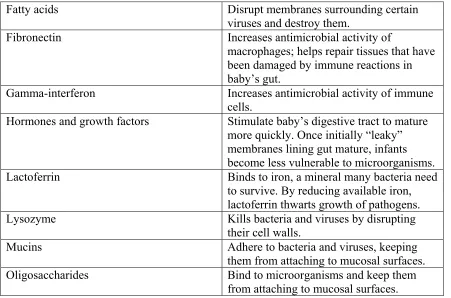
on the mother’s prior exposure to their target pathogens. Components of the innate immune system include constitutive components of human milk such as lysozyme, lactoferrin, and NEFA (Newburg 2001). Table 1.1 illustrates the immune benefits of breast milk (Newman 1995).
Table 1.1 Immune Benefits of Breast Milk at a Glance (Newman 1995)
Component Action
White Blood Cells
B lymphocytes Give rise to antibodies targeted against specific microbes.
Macrophages Kill microbes outright in the baby’s gut, produce lysozyme and activate other components of the immune system. Neutrophils May act as phagocytes, ingesting bacteria
in baby’s digestive system.
T-lymphocytes Kill infected cells directly or send out chemical messages to mobilize other defenses. They proliferate in the presence of organisms that cause serious illness in infants. They also manufacture compounds that can strengthen a child’s own immune response.
Molecules
Antibodies of secretory IgA class Bind to microbes in baby’s digestive tract preventing them from passing through walls of the gut into body’s tissues. B12 binding protein Reduces amount of vitamin B12, which
bacteria need in order to grow.
Table 1.1 Continued
Fatty acids Disrupt membranes surrounding certain
viruses and destroy them.
Fibronectin Increases antimicrobial activity of
macrophages; helps repair tissues that have been damaged by immune reactions in baby’s gut.
Gamma-interferon Increases antimicrobial activity of immune cells.
Hormones and growth factors Stimulate baby’s digestive tract to mature more quickly. Once initially “leaky” membranes lining gut mature, infants become less vulnerable to microorganisms. Lactoferrin Binds to iron, a mineral many bacteria need
to survive. By reducing available iron, lactoferrin thwarts growth of pathogens. Lysozyme Kills bacteria and viruses by disrupting
their cell walls.
Mucins Adhere to bacteria and viruses, keeping
them from attaching to mucosal surfaces. Oligosaccharides Bind to microorganisms and keep them
from attaching to mucosal surfaces.
Many studies have shown a protective effect of breast milk against pathogens, including Escherichia coli (Hayani and others 1992), Vibrio cholerae (Glass and others 1983), rotavirus (Newburg and others 1998), enterotoxigenic E. coli (Cruz and others 1988), Camphylobacter (Ruiz-Palacios and others 1990), and Guardia duodenalis (Walterspiel and others 1994).
The clinical significance of these immune factors has been demonstrated in reports that compared the morbidity and mortality of breast-fed and formula-fed infants (France and others 1980; Jason and others 1984). The availability of these immune factors depends largely on the method of milk storage and processing (Ford and others 1977; Liebhaber and others 1977; Pittard and Bill 1981).
1.2. Breastfeeding among Working Mothers
The American Dietetic Association released their position statement regarding breastfeeding in 2001 (ADA). It is as follows:
“It is the position of the American Dietetic Association (ADA) that broad-based efforts are needed to break the barriers to breastfeeding initiation and duration. Exclusive breastfeeding for 6 months and breastfeeding with complementary foods for at least 12 months is the ideal feeding pattern for infants. Increases in initiation and duration are needed to realize the health, nutritional, immunological, psychological, economical, and environmental benefits of breastfeeding.”
Li and others (2005) used data from the 2002 National Immunization Survey regarding breastfeeding initiation and duration to estimate breastfeeding rates in the United States by characteristics of the child, mother, and family. They found a sharp decline in exclusive breastfeeding between 3 and 5 months, which is often the time in which most mothers return to work or school (Li and others 2005).
Although there are many barriers to continuing breastfeeding, a major barrier is the increasing numbers of mothers around the world have to work soon after delivering their baby. They may still want to continue breast feeding exclusively, but women have a
relatively short maternity leave from work (Fein and Roe 1998), inflexible work hours when returning to work, and the lack of paid breastfeeding or pumping breaks in the workplace (Anonymous 2001). Also, lack of support for lactation in the workplace has been shown to be a major barrier to continuing breastfeeding (Fein and Roe 1998; Lindberg 1996).
1.3. Human Milk Banking
Modern milk banks may be located in large hospitals, while milk donated from the smaller hospitals and voluntary donors often needs to be temporarily stored before being transported to the milk bank. Upon arrival to the milk bank, milk needs to be stored during microbial testing. Therefore, milk donated to milk banks often needs to be stored (Ogundele 2000).
1.4. Current Recommendations for Breast Milk Storage
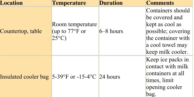
The Centers for Disease Control recommends that breast milk should be stored according to the following guidelines (CDC 2008).
Table 1.2 Breastmilk Storage Guidelines (CDC 2008)
Location Temperature Duration Comments
Countertop, table
Room temperature (up to 77°F or 25°C)
6–8 hours
Containers should be covered and kept as cool as possible; covering the container with a cool towel may keep milk cooler.
Insulated cooler bag 5-39°F or -15-4°C 24 hours
Table 1.2 Continued
Refrigerator 39°F or 4°C 5 days
Store milk in the back of the main body of the refrigerator. Freezer
Freezer
compartment of a
refrigerator 5°F or -15°C 2 weeks Freezer
compartment of refrigerator with separate doors
0°F or -18°C 3–6 months
Chest or upright
deep freezer -4°F or -20°C 6–12 months
Store milk toward the back of the freezer, where temperature is most constant. Milk stored for longer durations in the ranges listed is safe, but some of the lipids in the milk undergo degradation resulting in lower quality.
However, the publication does not cite research-based evidence supporting the fact that breast milk should be stored at these temperatures and times. The book Best Practices
There is limited current research that examines the effects of storage time and temperature on the nutritional components of breast milk such as protein quality and lipid oxidation. No research has been done to answer a question regularly encountered by
clinicians – how long can milk be saved after a baby has drunk from the bottle? Many studies that have been done on human milk storage have limited applicability due to the short time frames of the studies themselves or lack of practical applicability. The time frames of the studies usually run from 0 – 10 days, with 2 days being the longest period of time that refrigerated milk has been stored. Many studies also freeze the milk at -70°C, which is not
applicable to freezers in homes, child care facilities, and hospitals.
Miranda and others (2004) examined changes in glutathione peroxidase (GPx) activity and in the concentration of malondialdehyde (MDA) when milk was kept refrigerated at 4-6°C or frozen at -20°C. Glutathione peroxidase is a key enzyme in an
antioxidant pathway. Malondialdehyde is a final product of lipid peroxidation, so it is used as a marker of the degree of oxidation. The first analysis was done 6 – 8 hours after the
extraction. The second analysis was done on milk stored at refrigeration conditions during 48 hours and the third analysis was done on milk stored under freezing conditions during 10 days. The researchers concluded that storage at -20°C is better than refrigeration to preserve
Monera-Pons and others (1998) evaluated the effect of different storage methods on the stability of the triacylglycerides in human milk. The researchers evaluated the lipolytic activity in human milk during storage at -20°C for four months. Storage at -20°C without
previous heat treatment led to the hydrolysis of triacylglycerides and the appearance of NEFA. Although the researchers stored the milk at -20°C for four months, they only studied
triacylglycerides and they did not study protein quality or degradation (Pons and others 1998).
Other research done on the storage of human breast milk studied the effects of storage time and temperature on proteolysis and lipolysis over a time period of 3 hours to 15 days (Eteng and others 2001; Hamosh and others 1996; Miranda and others 2004; Silvestre and others 2006). The data generally show an increase in proteolysis and lipolysis over time at every storage temperature. However, only milk stored at -20°C was stored for 15 days. The longest amount of time refrigerated breast milk has been stored in these studies is 48 hours.
According to the Dietary Reference Intakes (USDA 2004) people of all ages require 51 mg of lysine per gram of protein. The AI for infants 0-6 months of age is 9.1 g of
protein/day and the RDA for infants 7-12 months of age is 11 g of protein/day (USDA 2004). Based upon these recommendations, infants 0-6 months of age require 464 mg lysine/day and infants 7-12 months of age require 561 mg lysine/day. These recommendations are in
agreement with a review by Tome and Bos (2007). According to Silvestre and others (2006), fresh breast milk contains 161 mg lysine/100 ml milk, the refrigerated breast milk contains 99 mg lysine/100 ml milk, and the frozen breast milk contains 95 mg lysine/100 ml milk. Infants drink between 570-900 ml of breast milk per day, therefore if they drink 570 ml refrigerated breast milk stored for 48 hours, they will still meet the requirements for lysine at 561 mg.
Although studies may find a statistically significant decrease in protein and lipids after cold storage for 48 hours to 10 days, these results may not be clinically significant. In the previous example, although available lysine decreased during cold storage, the remaining lysine was still sufficient to meet the requirements of infants 0 – 12 months of age.
bacteria sequestration activity in the breast milk. Sosa and Barness (1987) also found that storage at refrigeration temperatures for five days decreases the bacterial content in the breast milk (Sosa and Barness 1987). Olowe and others (1987) found that storage of breast milk at refrigeration temperatures for 24 hours resulted in an increase in bacterial growth in 61% of the breast milk samples and inhibition of bacterial growth in 39% of the samples (Olowe and others 1987), most likely due to initial levels of contamination in the breast milk. Other studies examining the effect of storage time and temperature on the bactericidal activity of breast milk either only stored the milk for 24 hours and at ambient temperatures (Eteng and others 2001; Hamosh and others 1996) or they stored the milk at -70 °C, which would not be applicable in child care facilities, hospitals, or in the home. In addition, none of the research addresses the question – how long can breast milk be safely stored after a baby has suckled from a bottle?
1.5. Effect of Storage Temperature and Time on Compounds in Human Milk
A limited amount of research has examined the effect of storage temperature and time on protein, lipids, and bacteria in human milk. The research that has investigated these
factors has looked at the effects of human milk storage for a limited number of days. Although a few studies utilized typical storage temperatures, some studies store samples either at ambient temperatures or at -70°C, which is not available in most homes, hospitals,
A study by Miranda and others (2004) assessed changes in glutathione peroxidase activity and in the concentration of malondialdehyde when milk was kept refrigerated or frozen. Glutathione peroxidase (GPx) is a seleno-enzyme, so it is a source of selenium for the newborn, but it is also an enzyme that protects against free-radical attack. Malondialdehyde (MDA) is a final product of lipid peroxidation, so it is used as a marker of the degree of oxidation. They stored breast milk at 4-6oC for 48 hours and at -20oC for 10 days. The milk was analyzed at 4-6 hours after extraction, at 48 hours, and at 20 days. GPx activity
decreased significantly from the fresh samples in the refrigerated and frozen samples, but activity was greater in the frozen samples than refrigerated samples. Fresh samples show a lower MDA concentration than those kept refrigerated or frozen, but refrigeration storage shows the greatest MDA concentration. The researchers concluded that storage at -20oC is better than refrigeration to preserve the quality of the milk; however, they did not look at refrigeration storage longer than 48 hours (Miranda and others 2004).
Hamosh and others (1996) assessed microbial growth and stability of milk protein and lipid at 15oC, 25oC, and 38oC for up to 24 hours. These temperatures were chosen because they mimic suboptimal storage conditions that may used for human milk storage in developing countries as well as in work situations in industrialized countries. Sixteen healthy women who breastfeed exclusively donated milk. Dissimilar from the present study, the milk was stored at 15oC, 25oC, and 38oC for 1 – 24 hours to determine pH, proteolysis, and
Proteolysis was minimal during milk storage at 15oC or 25oC for 24 hours and was only noticeable after 24 hours of storage at 38oC. However, the differences were not
significant. The greatest rise in proteolysis products was a 40% increase above baseline after 24 hours of storage at 38oC. Lipolysis began during the first hours of storage and increased to 8% at 24 hours of storage. After the milk had been stored for 24 hours at 38oC, the
concentration of NEFA were 440% to 710% higher than in freshly expressed milk. Lipolysis was higher at 24 hours’ storage at 25oC than 15oC or 38oC, possibly due to inactivation of lipoprotein lipase at 38°C. Bacterial growth was mainly limited to nonpathogens. Bacterial
growth was minimal at 15oC within 24 hours, was low at 25oC for the first 4 – 8 hours, and was higher at 38oC at 4 hours (Hamosh and others 1996).
Pons and others (1998) evaluated the effect of various storage methods on the stability of the triacylglyceride fraction of human milk. The purpose was to evaluate the lipolytic activity in human milk during storage at -20oC and to examine the effect of freezing and thawing on samples stored at this same temperature. Thirty milk samples were from six women. The samples were divided into five groups:
1. Storage at -20oC for four months.
2. Rapidly heated at 80oC and held for 1.5 minutes to inactivate bile salt stimulated lipase (BSSL). Samples were then stored at -20oC for 4 months.
3. Storage at -80oC for 4 months. Before analyzing the samples, they were thawed in a 25oC bath and rapidly extracted.
4. Storage at -80oC for 2 months. They were then thawed for an hour in a 25oC bath and stored at -80oC for another 2 months. At the moment of analysis, samples were thawed in the same way and rapidly extracted.
5. Storage at -20oC for 2 months. Then the samples were placed in a refrigerator and thawed slowly by being held for one week at 5oC. Then the samples were stored at -20oC for another month before lipid analysis.
1.6. Effect of Storage Temperature and Time on Bacterial Growth in Human Milk Bacterial growth in human milk actually decreases during storage for five days due to the antibacterial factors contained within the milk (Sosa and Barness 1987). Storage beyond a couple of days will result in an increase in bacterial growth.
A study by Sosa and Barness (1987) was done to assess the potential risks of using unprocessed breast milk to feed premature infants. The researchers studied the bacterial growth in 41 samples of unprocessed human milk for a period of five days under
refrigeration. No bacteria were cultured in eight samples of milk and the bacteria that were cultured in the other 33 samples were similar to bacteria found on the nipple and skin of the breast. However, two samples contained Klebsiella and one sample contained Pseudomonas. The initial concentration of bacteria was low, with a mean of 10,000 colonies. Bacterial colony counts continued to decrease during the five days of refrigeration. It can be assumed that when breast milk is collected under appropriate conditions, the bacteria represent
contamination from normal skin flora. They found that antibacterial factors in breast milk can control bacterial growth when the milk is refrigerated for at least five days (Sosa and Barness 1987).
hours. Cultures were taken at 6 hour intervals. At baseline, 69% were either sterile or contained only normal skin flora and 31% grew potential pathogens. During the 24 hours of storage in the refrigerator, bacteria multiplied in 50 samples and bacterial growth was inhibited in 32 samples. The mean bacterial count at any time during the 24 hours was not significantly different from that at the beginning of the storage in the refrigerator. The researchers concluded that when breast milk is collected under appropriate conditions, the bacteria represent contamination from normal skin flora, and the milk can be safely given to infants within 24 hours of refrigeration (Olowe and others 1987).
and then it sharply declined. The results indicate that loss of bactericidal activity in
refrigerated milk is compensated for by enhanced bacteria sequestration activity, thus stored human milk is safe for infant consumption.
Hernandez and others (1979) evaluated the effects of freezing and pasteurization on the ability of breast milk to suppress the growth of a bacterial inoculum in vitro. Breast milk was obtained from nursing mothers during the second week of postnatal life. Aliquots of the fresh milk were allocated to each of the four study groups.
1. Fresh: untreated and maintained at room temperature.
2. Pasteurized: immersed in boiling water for five minutes, and then cooled to room temperature.
3. Fresh frozen: frozen at -70oC for two minutes, and then thawed to room temperature. 4. Frozen 21 days: frozen at -70oC for three weeks, then thawed, at which time one
aliquot was inoculated directly while another was pasteurized before inoculation. The samples were inoculated within 30 minutes of obtaining the sample. Commercial formula and nutrient broth served as controls. Samples were inoculated 10 to 50 cfu/ml of E. coli or group B streptococcus, and incubated at 37oC. Quantitative growth was measured at eight and 24 hours. Bacterial inhibitory activity was not demonstrated in the control broth, commercial formula, and pasteurized breast milk. Fresh breast milk, fresh frozen breast milk, and breast milk frozen and stored for 21 days demonstrated a significant inhibition of
from the American Academy of Pediatrics that frozen breast milk is a good alternative for feeding premature infants when fresh milk is not available.
1.7. Effect of Storage Temperature and Time on IgA Concentration
1.8. Protein in Human Breast Milk
In addition to providing adequate amounts of essential amino acids, human breast milk contains proteins that provide antimicrobial and immunostimulatory effects, such as SIgA, lactoperoxidase, к-casein, haptocorrin, casein phosphopeptides, and lactadherin. Human breast milk also contains proteins that aid in nutrient absorption in the infant’s gut, such as lactoferrin, bile-salt stimulated lipase, haptocorrin, folate-binding protein, and к -casein (Bernt and Walker 2001).
Milk proteins can be classified into three groups: mucins, casein, and whey proteins. Mucins are milk fat globule membrane proteins that surround the lipid globules in milk (Patton and Huston 1986). Colostrum has a high protein concentration, consisting primarily of whey protein due to the low synthesis of casein during the first few days of lactation (Atkinson and Lonnerdal 1989). The protein concentration of breast milk decreases rapidly during the first month of lactation but it decreases slowly throughout the remainder of lactation. The whey to casein ratio in early lactation is about 80:20 and in late lactation it is about 50:50 (Atkinson and Lonnerdal 1989).
1.8.1. Protein Concentration in Human Breast Milk
The protein concentration of human breast milk is 14 to 16 g/L during early lactation, 8 to 10 g/L at 3 to 4 months of lactation, and 7 to 8 g/L at 6 months and later (Newburg 2001). However, these protein concentrations were determined by measuring nitrogen with the Kjeldahl method, subtracting non-protein nitrogen from total nitrogen, and multiplying by the conventional Kjeldahl factor of 6.25. Non-protein nitrogen is 20-25% of the total nitrogen in human milk; therefore this method of measuring protein in human breast milk may underestimate protein concentration if too large a value is subtracted for non-protein nitrogen. Protein concentrations may also be higher in breast milk of women with higher protein content in their diet.
1.8.2. Casein in Human Breast Milk
The casein micelle in human milk contains β-casein, к-casein, and αs1-casein. The α s1-casein concentration contributes about 0.06% of total protein (Newburg 2001). The main subunit of human milk casein is β-casein, which contains clusters of phosphorylated serine and threonine residues close to the N-terminal end, which are able to complex with calcium ions (Sato and others 1991). Therefore, when β-casein is digested, it may aid in calcium absorption by keeping calcium soluble, leading to the high bioavailability of calcium in human milk (Sato and others 1986).
As opposed to β-casein, which is highly phosphorylated, к-casein is highly
carbohydrate component of к-casein may prevent the adhesion of Helicobacter pylori to the infant’s gastric mucosa (Stromqvist and others 1995).
1.8.3. Whey Proteins in Human Breast Milk
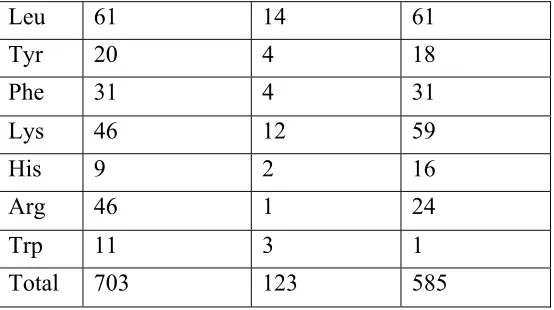
The major whey protein in human milk is α-lactalbumin, which consists of 123 amino acids (Phillips and Jenness 1971). The amino acid composition of α-lactalbumin is listed in table 1.3, along with the amino acid composition of lactoferrin and serum albumin.
It is able to bind calcium in a 1:1 molar ratio and calcium may have a structural role for α -lactalbumin (Lonnerdal and Glazier 1985). The main role of α-lactalbumin is to act as a part of lactose synthase, which is the enzyme responsible for lactose synthesis in the mammary gland. Lactose synthase is composed of α-lactalbumin and galactosyltransferase and together they catalyze the binding of glucose to UDP-galactose to form lactose (Brew and Hill 1975). As a part of this enzyme, α-lactalbumin modifies the catalytic site of galactosyltransferase and promotes the binding of glucose to the enzyme (Atkinson and Lonnerdal 1989).
In addition to aiding in lactose synthesis, α-lactalbumin has a high nutritional value for the infant. Table 1.3 illustrates the amino acid composition of major human whey proteins (Atkinson and Lonnerdal 1989).
The whey protein α-lactalbumin may inhibit growth of potential cancer cells.
Bovine α-lactalbumin also has antiproliferative effects in human colon cancer cell lines (Sternhagen and Allen 2001).
Hakansson and others (2000) reported an antimicrobial activity resulting from the folding of α-lactalbumin into an active complex with oleic acid (C18:1). They found that native α-lactalbumin exerted no bactericidal effects but that it could be converted into an active form by ion exchange chromatography in the presence of human milk casein fractions containing oleic acid. The α-lactalbumin – oleic acid complex is selective for streptococcal strains, including strains of Streptococcus pneumoniae, Staphylococci (gram-positive) and Enterococci (gram-negative) were resistant to this agent (Hakansson and others 2000).
Table 1.3 Amino Acid Composition of Major Human Whey Proteins (Lonnerdal 1989) Amino Acid Residues
Lactoferrin α
-lactalbumin
Serum albumin
Asp 71 17 53
Thr 31 6 28
Ser 50 7 24
Glu 70 15 82
Pro 35 2 24
Gly 56 6 12
Ala 63 6 62
Val 49 2 41
Met 6 2 6
Cys 32 8 35
Table 1.3 Continued
Leu 61 14 61
Tyr 20 4 18
Phe 31 4 31
Lys 46 12 59
His 9 2 16
Arg 46 1 24
Trp 11 3 1
Total 703 123 585
1.8.3.1. Lysozyme in Human Breast Milk
Lysozyme is another major whey protein in human milk, representing 1-4% of whey protein nitrogen (Atkinson and Lonnerdal 1989). Lysozyme is an enzyme found in many secretions, such as human milk, saliva, tears, nasal mucus, and pancreatic juice (Fleming 1922). It is capable of breaking down the outer cell wall of gram-positive bacteria and some gram-negative bacteria by hydrolyzing the β1,4 linkages of N-acetylmuramic acid and 2-acetylamino-2-deoxy-D-glucose residues (Chipman and Sharon 1969).
months of lactation (Bernt and Walker 2001). It is also very resistant to break down by acid in the stomach as well as trypsin, suggesting that adequate amounts of lysozyme reach the intestinal tract (Bernt and Walker 2001).
Lysozyme is active against gram-positive and gram-negative bacteria. Lysozyme has been shown to kill gram-negative bacteria and inhibit the growth of gram-positive bacteria (Shah 2000). Lysozyme may also work together with lactoferrin, which is an iron-binding protein capable of binding two ferric ions (Lonnerdal and Iyer 1995), to kill gram-positive bacteria. Lactoferrin binds lipopolysaccharide (LPS) and removes it from the outer cell membrane of gram-positive bacteria so that lysozyme can degrade the outer membrane and kill the bacteria (Ellison and Giehl 1991).
1.8.4. Immunoglobulins in Human Breast Milk
Paul Ehrlich was the first to show the transfer of maternal antibodies to the neonate by human milk (Bernt and Walker 2001). Immunoglobulins are beneficial because they provide protection to the infant’s immature immune system. Immunoglobulins, or antibodies, that are present in human milk include immunoglobulins A, G, M, D and E, with SIgA being the most abundant (Newman 1995).
disulfide bonds (Goldman and Goldblum 1989). In milk, IgA is in a polymeric form called SIgA, consisting of two four-chained units united by a 15-kD polypeptide called the joining chain and complexed to a 75-kD glycopeptide called the secretory component (Brandtzaeg 1974; Wilde and Koshland 1973).
1.8.4.1. Secretory Immunoglobulin A
SIgA is made from proteins produced by plasma cells and epithelial cells of the mammary gland. The plasma cells that produce dimeric IgA are native to the intestinal and respiratory tract. The dimeric IgA binds to receptor secretory component (SC) molecules on the mammary gland epithelium. The extracellular part of SC attached to an IgA dimer is transported across the cell and secreted as SIgA (Atkinson and Lonnerdal 1989).
Table 1.4 Concentrations of Immunoglobulins (mg/ml; mean ± SD) in Human Breast Milk Duration of Lactation
Immunoglobulin
Concentrations 2-4 days 1 month 6 month 12 month 24 month Total IgA 2.1 ± 2.3 1.0 ± 0.2 0.6 ± 0.1 1.0 ± 0.5 1.1 ± 0.3 SIgA 2.0 ± 2.5 1.0 ± 0.3 0.5 ± 0.1 1.0 ± 0.3 1.1 ± 0.2
IgM 0.12 ± 0.03 0.02 0.02 0.01 b
IgG 0.34 ± 0.01 0.05 ± 0.03 0.03 0.04 b
a Data complied by Goldman and Goldblum from previous studies b Insufficient data
SIgA is able to survive in the hostile environment of the gastrointestinal tract because it is more resistant to degradation from proteases than other immunoglobulins (Atkinson and Lonnerdal 1989). SIgA antibodies against bacterial IgA proteases have been found, including proteases produced by H. influenzae, Streptococcus sanguis, S. pneumoniae, Neisseria gonorrhoeae, Proteus mirabilis, E. coli, and Neisseria meningitidis (Gilbert and others 1983).
SIgA antibodies neutralize bacterial toxins and prevent binding of intestinal bacterial pathogens to epithelial cells by binding to their pili (Newberg 2001). The antibodies
human milk involves blocking the adhesion of microorganisms to the intestinal epithelium and neutralization of microbial toxins (Bernt and Walker 2001).
1.8.5. Proteolysis
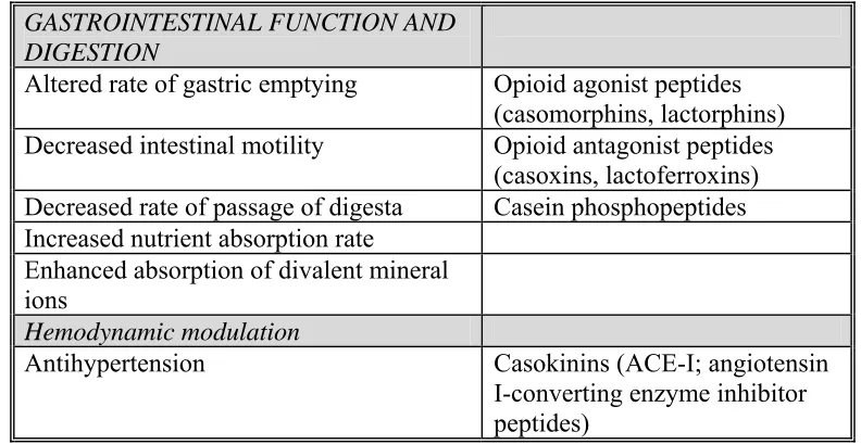
Casein is a source of biologically active peptides (Clare and Swaisgood 2000; Meisel and Bockelmann 1999; Schanbacher and others 1997). Table 1.5 outlines the effects of peptides in human milk (Schanbacher and others 1997). The whole antibacterial effect in milk adds up to be more than the total of the individual contributions of immunoglobulin and nonimmunoglobulin defense proteins due to the fact that the defense proteins work together synergistically (Clare and Swaisgood 2000). Table 1.6 outlines the antimicrobial peptides in bovine milk (Clare and Swaisgood 2000).
Table 1.5 Categories of Bioactivities in Bovine Milk and their General Effects and Major Effecters (Schanbacher and others 1997)
GASTROINTESTINAL FUNCTION AND DIGESTION
Altered rate of gastric emptying Opioid agonist peptides (casomorphins, lactorphins) Decreased intestinal motility Opioid antagonist peptides
(casoxins, lactoferroxins) Decreased rate of passage of digesta Casein phosphopeptides Increased nutrient absorption rate
Enhanced absorption of divalent mineral ions
Hemodynamic modulation
Table 1.5 Continued
Increased blood flow Casoplatenin (K-casein peptides) Lactoferrin and peptides
Probiotic support of intestinal microflora Enhanced growth of Bifidobacteria in GI
tract K-Casein glycomacropeptide Milk oligosaccharides Lactoferrin and lactoferrin peptides
Casomorphins (indirect) Non-immune disease protection
Inhibition of coliform and pathogen growth in gut or mammary gland
Lactoferrin, lysozyme, lactoperoxidase Killing of coliform or pathogenic bacteria
in gut or mammary gland Lactoferrin bactericidal peptides (lactoferricin)
Inhibition of viral infection Glycolipids, sphingolipids Passive immunity
Transfer of immunoglobulins (IgG, IgA; IgM) to blood of neonate
Immunoglobulins IgG, IgA, IgM Continual presence of Ig in lumen of gut Casein immunoregulatory
peptides Enhanced phagocytic activity Cytokines
Growth factors Immunoregulation
Modulation of lymphocyte function Casein immunoregulatory peptides
Modulation of lymphocyte differentiation Lactoferrin and lactoferrin peptides
Modulation of lymphocyte and granulocyte traffic
Cytokines
Enhanced killer cell activity Growth factors Anti-inflammation
Modulation of lymphocyte function Casein immunoregulatory peptides
Reduced release of cytokines by lymphocytes and macrophages
Lactoferrin and lactoferrin peptides
Modulation of polymorphonuclear leukocyte response
Table 1.5 Continued
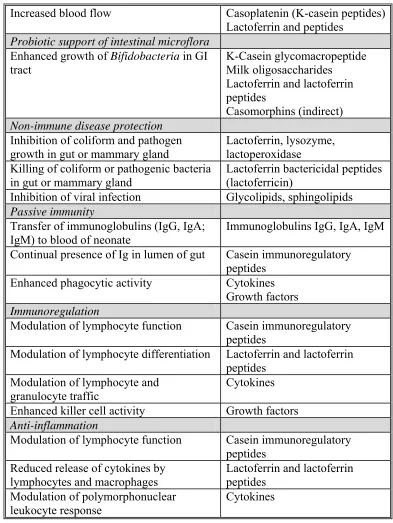
Binding of bacterial lipopolysaccharide (endotoxin)
Growth factors
Growth and development
Enhanced development of intestinal tract Hormones: prolactin, plus many others in milk
Enhanced gastric development and function
Growth factors: IGF, EGF, TGF-a, TGF-P
Enhanced neuroendocrine development Cytokines
Enhanced development of immune system Casein immunoregulatory peptides
Lactoferrin
Table 1.6 Antimicrobial Proteins in Milk (Clare and Swaisgood 2000 Milk Peptide Fragment Release Protease Gram (+) Activity Gram (-) Activity Yeast and Fungi* Casecidin Chymosin and
chymotrypsin Staphylococcus Bacillus subtilis Diplococcus pneumoniae Streptococcus pyogenes Casocidin-I Synthetic peptide Staphylococcus carnosus Escherichia coli
Isracidin Chymosin and
chymotrypsin Staphylococcus aureus Candida albicans Lactoferricin B Lactoferrin
Pepsin Bacillus Listeria Streptococci Staphylococci
Other physiologically active peptides in milk include antihypertensive peptides, antithrombotic peptides, caseinophosphopeptides, immunomodulatory peptides, opoid milk peptides, casomorphins, caseinomacropeptide, and atriopeptin (Clare and Swaisgood 2000). As mentioned previously, peptides from к-casein exert antimicrobial (Stromqvist and others 1995) and antithrombotic effects, in particular the к-caseinglycopeptide (hCGP) (Chabance and others 1995).
The bioactive peptides in human milk proteins are dormant until released from the parent protein and activated by enzymatic proteolysis, such as during digestion or food processing (Meisel and Bockelmann 1999). Ferranti and others (2004) found that most peptides identified in human milk originate from β-casein and not the most abundant whey protein, α-lactalbumin. They identified these proteins using mass spectroscopy and outlined the proteolytic steps that β-casein follows (Ferranti and others 2004). They outlined three proteolytic steps in β-casein breakdown. The first step involves the action of a plasmin-like enzyme at Lys18, Lys23, and Lys160. The next step involves the action of endogenous
endopeptidases at Ala73, Ala180, and Glu186. Finally, these peptides are substrates for other exopeptidases, including aminopeptidases, carboxypeptidases, and peptidases cleaving after proline residues, which all require short peptides as substrates (Ferranti and others 2004).
stranded DNA occurs, autolytic enzymes, such as amidases that degrade the peptidoglycan layer, are stimulated, bactericidal molecules, such as hydrogen peroxide, are produced, and/or binding of cationic peptides to cellular nucleic acids results in antimicrobial effects (Clare and others 2003).
1.8.5.1. Proteases Endogenous to Human Breast Milk
The existence of proteolytic enzymes in human milk provides the infant with
significant digestive assistance during the period of adjustment from prenatal nutrition by the placenta to postnatal oral feeding (Storrs and Hull 1956). In human milk there are proteolytic enzymes, a fibrinolytic system, plasmin, and a plasminogen activator (Atkinson and
Lonnerdal 1989).
Lys107-Glu108 to give three β-casein fragments γ1-, γ2-, and γ3-caseins as well as proteose-peptones (Chen and others 2003). Plasmin does not hydrolyze α-lactalbumin.
There are also several protease inhibitors in human milk, including α
1-antichymotrypsin and α1-antitrypsin. Protease inhibiting activity was found in two thirds of milk samples collected during the first 3 days of lactation and in all the samples collected from later stages of lactation, including inhibitors of trypsin, chymotrypsin, and elastase (Lindberg 1979). The same samples had low proteolytic activity and the milk protease inhibitors formed complexes with added trypsin and chymotrypsin, but not pepsin (Lindberg 1979). These results suggest that the proteases were inactive due to the complexes with the inhibitors (Lindberg and others 1982).
1.8.5.2. Bacterial Proteases
Gram-negative psychrotrophic bacteria include Pseudomonas, Aeromonas, Serratia, Acinetobacter, Alcaligenes, Achromobacter, Enterobacter, and Flavobacterium. Gram-positive psychrotrophic bacteria include Bacillus, Clostridium, Corynebacterium, Microbacterium, Micrococcus Streptococcus, Staphylococcus, and Lactobacillus. The organisms most commonly isolated from breast milk, including gram-positive, coagulase-negative staphylococci and gram-coagulase-negative Acinetobacter are also known skin colonizers (Botsford and others 1986) (el-Mohandes and others 1993).
Similar to plasmin, proteases from psychrotrophic bacteria preferentially hydrolyze casein over whey, particularly к-casein, resulting in the cleavage of the Phe105 – Met106 peptide bond, leading to the release of the C-terminal caseinomacropeptide (CMP) (Chen and others 2003) (Dupont and others 2007).
1.8.6. Proteolysis and Bioavailability
The pre-term infant may be more dependent upon the immunoprotective actions of lysozyme, lactoferrin, and SIgA than the term infant, due to its immature immune function, but there is no doubt that the term infant is dependent upon these components as well. Proteolysis of proteins during storage of human milk may be of concern because these proteins need to be intact in the GI tract in order to exert their protective functions. As previously mentioned, SIgA is able to endure in the hostile environment of the
of human milk are stable when stored at room temperature for 8 hours, when stored at 0 - 4°C for three days, or when frozen at -20°C for 12 months (Lawrence 2001).
Studies have indicated minimal protein digestion in the infant’s stomach due to a low pepsin output (Agunod and others 1969; Weisselberg and others 1992; Yahav and others 1987) and high postprandial gastric pH (Hamosh and others 1978; Mason 1962). Because infants have a higher pH in their gastrointestinal tract than do adults, proteins are not as readily denatured. Therefore, proteolysis of milk proteins prior to ingestion may increase bioavailability of protein to the infant because they are already broken-down before ingestion. Table 1.7 summarizes current knowledge on gastric proteolysis in infants (Henderson and others 2001).
Table 1.7 Hydrolysis of Milk Proteins by Gastric Aspirates of Infants (Henderson and others 2001)
• Gastric pH is 5.0 to 6.0 during the 1st hour postprandial
• Pepsin activity and output are significantly lower in infants (especially preterm) than in children and adults
• Peptic activity tested in vitro with specific human or bovine milk proteins is very low to nil at pH > 4.5
• Hydrolysis of human or bovine milk protein by infant duodenal juice in vitro is also low • Hydrolysis of milk whey proteins, under optimal in vitro conditions, by infant gastric or
duodenal juice is lower than that of casein
1.9. Lipids in Human Breast Milk
Triacylglycerols comprise 98% of total lipids on human milk, phospholipids 0.8%, and cholesterol 0.5% (Jensen 1999). The majority of the fatty acids are medium and long chain fatty acids (C:10 to C:22) (Manson and Weaver 1997). Lipids are present in milk between 3% and 5%, and they exist as an emulsion in the aqueous phase (87%) of human milk (Jensen 1999). The fat globules are surrounded by a membrane called the milk fat globule membrane (MFGM), which is composed of phospholipid proteins,
mucopolysaccharides, cholesterol, and enzymes (Jensen 1999). Table 1.8 summarizes the effects of lipids in human milk (Jensen 1999).
Table 1.8 Effects of Lipids in Human Milk (Jensen 1999)
1. Source of 50–60% of the calories, about 70 kcal/dL, in human milk. Not usually responsive to diet.
2. Fatty acids combined into triacylglycerols to maintain a bulk melting point below 38°C. 3. Provide about 15 mg of cholesterol/dL. May predispose the infant to efficiently metabolize dietary cholesterol as an adult. Precursor of steroid hormones and other derivatives in
humans. Not affected by changes in maternal diet.
4. Contain the essential polyunsaturated fatty acids 18:2n-6 and 18:3n-3 and their products, 20:4n-6, 20:5n-3, and 22:6n-3. Required in maternal diet.
5. The 20:4n-6, 20:5n-3, and 22:6n-3 in proper balance may be required for maturation and optimal function of the visual process and brain and nervous system.
6. Contain 8:0–14:0, which if absorbed in the stomach, are transported to the liver and oxidized in decreasing amounts as the molecular weight increases. Quantities in milk dependent on amount of carbohydrate in diet.
7. If present, the conjugated fatty acid, c9, t11-18:2, or rumenic acid, may exert
Table 1.8 Continued
8. Trans unsaturates believed by some to adversely affect infants growth. Some positional isomers of c-18:1 may ameliorate atherosclerosis.
9. When produced by lipolysis in the stomach and small intestine, 12:0 and 18:2 and their monoacylglycerols have potent cidal effects against some microorganisms. Soluble and dispersible salts of fatty acids may act against microorganisms.
10. Milk gangliosides inactivate cholera and other enterotoxins.
11. Contain eicosanoids and their precursors, which act as first and second messengers. Precursors respond to diet.
12. Lipolysis of triacylglycerols by gastric lipase, regioselective for sn-3/sn-1,3/1; produces sn-1,2-diacylglycerols, which can be second messengers in stomach and small intestine. 13. Milk lipid globule membrane binds, protects, and releases bioactive compounds as needed. Graded doses of lipid provided. Membrane stabilizes globules in an oil/water emulsion.
14. Increases in lipid content and numbers of globules during a nursing may help develop appetite and its control in the infant by approaching satiety and by the tactile effect of the globules.
15. Lipid content increases as lactation progresses to help provide for growth and development of infant.
16. Carrier of fat-soluble vitamins.
17. Can transport undesirable compounds such as dioxins.
18. When milk is stored in the frozen state, it can be the source of soapy flavor due to free 12:0 and to oxidized flavors (cardboardy) due to polyunsaturated fatty acids.
19. Carotenoids, tocopherols, and conjugated 18:2 may be antioxidants.
The fat content in human milk increases from 2.0g/dL in colostrum to 4.9g/dL in mature milk, which is a reflection of the rising energy requirement of the growing infant (Manson and Weaver 1997). Fat content also varies during feedings and throughout the day. Fat content is about 3.0 g/dL in midday foremilk and 4.0 g/dL in midday hindmilk. Fat content is about 3.0 g/dL in early morning milk and 4.5 g/dL in evening milk (Jensen and others 1978).
1.9.1. Fatty Acids in Human Breast Milk
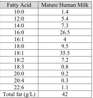
Table 1.9 Fatty Acid Composition of Human Milk (Manson and Weaver 1997) Fatty Acid Mature Human Milk
10:0 1.4 12:0 5.4 14:0 7.3 16:0 26.5 16:1 4 18:0 9.5 18:1 35.5 18:2 7.2 18:3 0.8 20:0 0.2 20:4 0.3 22:6 1.1 Total fat (g/L) 42
1.9.2. Polyunsaturated Fatty Acids in Human Breast Milk
The lipids in human milk and formula are very important for the growth and
development of infants. Lipids are the major energy source for infants and are tolerated better than proteins and carbohydrates. Human milk provides linoleic acid (18:2 n-6, LA), α
-linolenic acid (18:3 n-3, ALA), docosahexaenoic acid (22:6 n-3, DHA), arachidonic acid (20:4 n-6, AA), and other long chain polyunsaturated fatty acids (LCPUFA).
accumulate more AA than DHA, even when there is sufficient ALA for DHA synthesis (Fleith and Clandinin 2005). The reason AA may be synthesized more so than DHA is because AA synthesis is subjected to less regulatory steps than is the synthesis of DHA. DHA synthesis requires a second Δ6-desaturase step and β-oxidation in the peroxisomes.
Humans can desaturate and elongate ALA into EPA and DHA only when the ratio of LA (n-6) to ALA (n-3) is low. Excess n-6 fatty acids in the diet saturate the enzymes and prevent conversion of ALA into EPA and DHA (Mahan and Escott-Stump 2004). Therefore, the amount of DHA that an infant can synthesize is limited by the ratio of LA (n-6) to ALA (n-3) in dietary lipids. Human infants can synthesize small amounts of LCPUFA from LA and ALA, but the rate of synthesis is not adequate for LCPUFA accumulation in body tissues (Fleith and Clandinin 2005).
During the third trimester, the neonate accumulates about 40 to 60 mg n-3 PUFA per kg body weight per day (Koletzko and others 2008). LCPUFA intake is important for
have found that visual attention, problem-solving, and global development is improved in infants fed LCPUFA (Jensen and others 1978).
Makrides and others (2000) compared fatty acid status, visual evoked potential acuity, and growth of term infants who were fed formula containing LA:ALA of 10:1 or 5:1 to those of exclusively breast-fed infants (Makrides and others 2000). The formula-fed infants were randomly assigned in a double blind manner to formula containing LA:ALA of 10:1 or 5:1. ALA content was increased by replacing soy oil with low-erucic acid canola oil. Growth and fatty acid status in all groups were assessed at 6, 16, and 34 weeks of age, and visual evoked potential acuity was assessed at 16 and 34 weeks. They found that infants fed the 5:1 LA:ALA formula had significantly greater DHA plasma and erythrocyte
phospholipid concentrations than did infants fed the 10:1 formula, but concentrations in both formula groups were still significantly lower than those in the breast-fed infants. However, there were no differences seen in visual evoked potential acuity, weight, length, or head circumference within the groups. These results lead to the conclusion that lowering the LA:ALA ratio in infant formula by using low-erucic acid canola oil results in a significant increase in plasma DHA, but the concentrations still do not match those of the breast fed infants (Makrides and others 2000).
approximately 16% of total fatty acids as LA and 0.4%, 1.0%, 1.7%, or 3.2% as ALA, resulting in LA:ALA ratios of 44, 18.2, 9.7, and 4.8. The fatty acid profile of plasma phospholipids was determined at 21, 60, and 120 days of age in the formula fed groups. Anthropometric data to determine growth were assessed at 21, 60, 120, and 240 days of age in the formula fed groups. Transient visual evoked responses (VERs) to determine visual function were measured at 120 and 240 days of age in all groups. The results showed that VER latency and amplitude did not differ within any groups. Infants who received formula with an LA:ALA ratio of 4.8 had higher plasma DHA concentrations, but lower AA acid concentrations at 21, 60, and 120 days of age. The average weight of this group at 120 days was 760 grams (1.67 pounds) less than the average weight of the group fed the formula with the LA:ALA ratio of 44. However, this association was not found at any other time points. The authors did correct for birth weight, but they did not report any influence of gender on weight. Although plasma DHA increased as the LA:ALA ratio decreased, weight gain significantly decreased.
A review of the literature by Fleith and Clandinin (2005) concluded that LCPUFA should be supplied to preterm infants in the range provided by feeding human milk in Western countries. The range can be achieved by feeding AA and DHA at a ratio of 1.5 AA to DHA, with DHA at 0.4%. Addition of LCPUFA in infant formulas, in reasonable
LCPUFA accumulation due to disruption of the placental supply. Another review by Koletzko and others (2001) reports conclusions of a workshop on the role of LCPUFA in maternal and child health. For healthy infants, the authors support breastfeeding as the preferred method of feeding.
1.9.3. Non-esterified Fatty Acids in Human Breast Milk
It has been demonstrated that NEFA increase in stored breast milk due to the lipolysis of triacylglycerols. Hamosh and others (1996) found that when breast milk is stored at
ambient temperatures for 24 hours, there is a 440% - 710% increase in free fatty acid
concentration above that in freshly expressed milk (Hamosh and others 1996). The enzymes thought to be responsible for lipolysis of triacylglycerols are the bile-salt stimulated lipase (BSSL), lipoprotein lipase, or both (Atkinson and Lonnerdal 1989; Hamosh and others 1996). However, BSSL, as the name implies, is inactive without added bile salts, which should not be present in human milk in the concentration needed for BSSL activation (Atkinson and Lonnerdal 1989). Therefore, it is more likely that lipoprotein lipase is responsible for the release of non-esterified fatty acids from the triacylglycerols.
An increase in NEFA in stored breast milk causes an increase in titratable acidity, which has been of concern because of the belief that it may be caused by bacterial
1.9.3.1. Bioavailability of Non-esterified Fatty Acids in Human Breast Milk
The consequences of increased NEFA on human milk quality and digestibility are unknown. Hernell and Blackberg (1982) suggested that NEFA are absorbed better than esterified fatty acids when intraduodenal bile salts are low in the infant (Hernell and Blackberg 1982). Therefore, it is a possibility that an increase in NEFA could increase bioavailability to the infant. On the other hand, Patton and Carey (1979) suggested that NEFA may bind to calcium or other components of the milk and make them unavailable for absorption (Patton and Carey 1979).
Ionized fatty acids (fatty acid soap) may form during storage and bind with other milk components and make them unavailable for absorption. Lavine and Clark (1987) measured ionized fatty acids in breast milk and found trace amounts (<1.0mg/dL) in freshly expressed milk (Lavine and Clark 1987). As storage time and temperature progressed, there was increased evidence of ionized fatty acids but in small quantities. The greatest amount of ionized fatty acids found was in milk stored for 8 weeks at -11°C, containing 2.2 mg/dL,
which was only 1.4% of the total fatty acids in the milk (Lavine and Clark 1987).
1.9.4. Antiviral Activity of Non-esterified Fatty Acids
NEFA have also caused membrane disruption of enveloped viruses in cultured cells. Thormar and others (1987) found that lipids in fresh human milk do not inactivate viruses but they become antiviral after storage at 4°C and 23°C, probably due to the release of NEFA
from milk triglycerides. They compared the effect of fatty acids and monoglycerides on enveloped viruses. They found that short chain fatty acids, such as butyric, caproic, and caprylic, as well as long-chain saturated fatty aids, such as palmitic and stearic, had no or very small antiviral effects at the highest concentration tested. However, the medium-chain saturated and long-chain unsaturated fatty acids were all antiviral but at different
concentrations. The polyunsaturated fatty acids had the most antiviral activity. The level of lipoprotein lipase in the breast milk was correlated with the antiviral activity of the milk, indicating that it is caused by the release of NEFA or other products of lipid hydrolysis (Thormar and others 1987). They also found that the antiviral effect is caused by
disintegration of viral envelopes by fatty acids, and these results were confirmed by Sarkar and others (1973), who treated mouse mammary virus with the cream fraction of human milk and found that the viral envelope disintegrated (Sarkar and others 1973).
1.9.5. Non-esterified Fatty Acids and Breast Milk Jaundice
Lavine and Clark (1987) determined total lipid and free fatty acid concentrations in breast milk samples obtained from eight mothers and stored at 25°C, 4°C, or -11°C for 6, 12,
of NEFA changed as storage progressed; with free 16:0 decreasing and 18:2 increasing, and long-chain polyunsaturated fatty acids increasing (Lavine and Clark 1987). Hargreaves (1973) demonstrated that unsaturated fatty acids inhibit bilirubin conjugation in vitro, with the inhibition increasing with the degree of unsaturation (Hargreaves 1973). Therefore, it is a possibility that high amounts of polyunsaturated fatty acids in breast milk could inhibit bilirubin conjugation, leading to jaundice in the infant.
It has been proposed that mothers with jaundiced infants have increased lipolytic activity, which results in large amounts of NEFA in the milk after storage (Poland and others 1980). One hypothesized cause of jaundice is that NEFA inhibit bilirubin conjugation, causing neonatal unconjugated hyperbilirubinemia (Lavine and Clark 1987). Another hypothesized cause of jaundice is altered intestinal bilirubin absorption (Gartner and others 1983). Gartner and others (1983) observed in rats that normal milk decreased bilirubin absorption in the intestine. However, milk from mothers with jaundiced infants enhanced bilirubin absorption and increased the bilirubin in the liver (Gartner and others 1983).
(Constantopoulos and others 1980). Jalili and others (1985) also could not confirm the
observation that increased levels of NEFA are associated with breast milk jaundice (Jalili and others 1985). They analyzed breast milk samples and serum fatty acids in mothers with jaundiced infants and mothers without jaundiced infants. The concentrations of FFA
increased after storage of the milk from both groups, but no difference in the composition of milk FFA or serum fatty acids were found (Jalili and others 1985).
More recent studies have focused on the enzyme β-glucuronidase as a contributory factor in neonatal jaundice. This enzyme may lead to production of unconjugated bilirubin by cleaving the ester bond of bilirubin glucuronide in the neonatal intestine (Poland and Odell 1971). Ince and others (1995) analyzed breast milk samples from breast milk of mothers with jaundiced infants and breast milk of mothers with non-jaundiced infants for β-glucuronidase activity (Ince and others 1995). Enzyme activity was slightly higher in the control group, but the difference was not statistically significant. Based on these results, β-glucuronidase may be one of the factors contributing to breast milk jaundice, but it is not the only factor.
1.9.6. Lipases in Human Breast Milk
Triacylglycerols can not be absorbed into the intestinal wall and must first be