Georgia State University Georgia State University
ScholarWorks @ Georgia State University
ScholarWorks @ Georgia State University
Nutrition Theses Department of Nutrition
Fall 1-6-2012
Factors Associated with Bone Mineral Density in Elite Female
Factors Associated with Bone Mineral Density in Elite Female
Gymnasts
Gymnasts
Erin C. Millson
Georgia State University
Follow this and additional works at: https://scholarworks.gsu.edu/nutrition_theses Part of the Nutrition Commons
Recommended Citation Recommended Citation
Millson, Erin C., "Factors Associated with Bone Mineral Density in Elite Female Gymnasts." Thesis, Georgia State University, 2012.
https://scholarworks.gsu.edu/nutrition_theses/31
This Thesis is brought to you for free and open access by the Department of Nutrition at ScholarWorks @ Georgia State University. It has been accepted for inclusion in Nutrition Theses by an authorized administrator of
i FACTORS ASSOCIATED WITH BONE MINERAL DENSITY
IN ELITE FEMALE GYMNASTS
By
ERIN MILLSON
A Thesis Submitted to the Graduate Faculty of Georgia State University in Partial Fulfillment of the
Requirements for the Degree
MASTER OF SCIENCE
HEALTH SCIENCES: SPECIALIZATION IN NUTRITION
ATLANTA, GEORGIA
ii ACKNOWEDGEMENTS
Several people have been instrumental in helping me through the process of writing this thesis, and I would like to take the time to thank them. First and foremost, I would like to thank Dr. Dan Benardot for his enthusiasm, unending patience, and
expertise while guiding me through this process. Dr. Benardot sparked my interest in the field of nutrition during an undergraduate elective course that I took years ago, and for that, I‟ll always be grateful. I would like to thank Barb Hopkins for being a committee member, and also for being a wonderfully enthusiastic and knowledgeable teacher during my time in the nutrition program at Georgia State. I always looked forward to attending any class that she taught, and I specifically remember becoming excited about the profession of dietetics during the summer that I took my first semester of Medical Nutrition Therapy. I would also like to thank Dr. Walt Thompson for being a committee member and taking the time to read my thesis. Finally, I would like to thank my
iii AUTHOR’S STATEMENT
In presenting this thesis as a partial fulfillment of the requirements for an
advanced degree from Georgia State University, I agree that the Library of the University shall make it available for the inspection and circulation in accordance with its
regulations governing materials of this type. I agree that permission to quote from, to copy from, or to publish this thesis may be granted by the author or, in his/her absence, by the Associate Dean, College of Health and Human Sciences. Such quoting, copying, or publishing must be solely for scholarly purposes and will not involve potential
financial gain. It is understood that any copying from or publication of this thesis which involves potential financial gain will not be allowed without written permission of the author.
iv NOTICE TO BORROWERS
All theses deposited in the Georgia State University Library must be used in accordance with the stipulations prescribed by the author in the preceding statement.
The author of this thesis is:
Erin Millson
168 Garden Lane, Apt. A Decatur, GA 30030
The Chair of the committee for this thesis is:
Dan Benardot, PhD, RD, LD, DHC, FACSM Professor, Division of Nutrition
Georgia State University Atlanta, Georgia 30303
Users of this thesis who are not regularly enrolled as students at Georgia State University are required to attest acceptance of the preceding stipulation by signing below. Libraries borrowing this thesis for use of their patrons are required to see that each user records here the information requested.
NAME OF USER ADDRESS DATE TYPE OF USE
v VITA
2000-2004 Boston University Boston, Massachusetts Bachelor of Arts in History
2008-2011 Georgia State University Atlanta, Georgia
Master of Science in Health Sciences: Specialization in Nutrition (GPA: 3.93)
Graduate Research Assistantship: Critical Thinking through Writing Consultant
2010-2011 Dietetic Internship
Southern Regional Medical Center Riverdale, Georgia
2011-present Bionutrition Research Manager
Atlanta Clinical and Translational Science Institute Emory University Hospital
vi ABSTRACT
Introduction: Gymnastics participation involves movements that place repeated strain on bone and muscle, which positively affects bone mineralization. Although increased bone mineralization is protective against the development of osteopenia and osteoporosis later in life, the elite female gymnastics population is at risk for the development of the Female Athlete Triad, which can negatively affect bone mineral density (BMD). This study looks at factors that can influence BMD in this population.
Purpose: To investigate the influence of muscle mass, menstrual status, calcium intake, sunlight exposure, and other related factors on BMD in elite female gymnasts.
Methods: This study represents a secondary analysis of existing data that were obtained between 1994 and 1996. Using dual-energy X-ray absorptiometry, a group of US
National Team female gymnasts (n=43) in this study was screened at the Laboratory for Elite Athlete Performance at Georgia State University. BMD values (g/cm2) were collected on total body, arms, legs, trunk, ribs, pelvis, and spine. These measurements were compared to other variables and assessed for statistical significance.
Results: Age of gymnasts was positively associated with BMD at all measured sites (p <0.001; r=0.62-0.68). Weight was positively associated with BMD at all measured sites (p <0.001; r=0.82-0.90). Lean body mass was positively associated with BMD at all measured sites (p <0.001; r=0.74-0.87). Body fat percentage was positively associated with BMD at all measured sites (p <0.001-p=0.01; r=0.39-0.54). However, calcium intake was not significantly associated with any of the BMD sites. Sunlight exposure and indirect estimates of vitamin D were not significantly associated with any of the BMD sites; all r-values indicated a weak positive association with BMD. Of the gymnasts who had experienced menses (n=15), those with regular menstrual periods (n=8) had
significantly higher BMD values at the arm, leg, trunk, rib, and spine, and total body than those who did not have regular menstrual periods (n=7). There was no significant
difference in BMD for gymnasts who had regular periods at the pelvis. A regression analysis was performed. The predictors total BMD values from the regression equation were the following: regular menses, height, weight, percent kilocalorie requirement consumed from predicted kilocalorie needs, calcium intake with supplements, lean body mass, hourly deficits >300 kilocalories from predicted kilocalorie needs, and hourly surpluses >300 kilocalories from predicted kilocalorie needs.
vii TABLE OF CONTENTS
Page
ACKNOWLEDGMENTS...……… ii
LIST OF TABLES……… viii
LIST OF ABBREVIATIONS...……….. ix
CHAPTER I. INTRODUCTION………... 1
Purpose……….. 1
Hypotheses………. 2
II. REVIEW OF LITERATURE………... 4
III.METHODS………... 24
IV.RESULTS………... 26
Descriptive Statistics………... 26
Statistical Analysis……….. 32
V. DISCUSSION AND CONCLUSIONS……… 37
viii LIST OF TABLES
Page
TABLE 1 Demographics of Assessed USA National Team Female Gymnasts……26
TABLE 2 Ethnicity of Assessed USA National Team Female Gymnasts………….27
TABLE 3 Bone Mineral Density of Assessed USA National Team Female Gymnasts………...28
TABLE 4 Energy Consumption, Requirements, & Balance of Assessed USA
National Team Female Gymnasts………..29
TABLE 5 Time Spent Outside in Calorie Deficit and Surplus of Assessed USA National Team Female Gymnasts………...30
TABLE 6 Calcium Intake/Time Spent Outside of Assessed USA National Team Female Gymnasts………...30
TABLE 7 Menstrual Status of Assessed USA National Team Female Gymnasts….31
TABLE 8 Age at First Period of Assessed USA National Team Female Gymnasts..31
TABLE 9 Menstrual Regularity of Assessed USA National Team Female
Gymnasts………32 TABLE 10 Bone Mineral Density and Related Factors of USA National Team
Female Gymnasts………...34 TABLE 11 Regular Menstrual Status and Bone Mineral Density of Assessed USA
National Team Female Gymnasts………..35
ix LIST OF ABBREVIATIONS
ADA American Dietetic Association AI Adequate intake
ANOVA Analysis of variance BMD Bone Mineral Density cm Centimeter
DEXA Dual X-Ray absorptiometry DRI Dietary Reference Intake F.A.T. Female Athlete Triad FFM Fat-free mass
g Gram
g/cm2 Grams per square centimeter hr Hour
IRB Institutional Review Board IU International Units
kcal Kilocalorie kg Kilogram
LH Luteinizing hormone mg Milligram
NOF National Osteoporosis Foundation PTH Parathyroid hormone
1 CHAPTER 1: INTRODUCTION
Title
Factors associated with bone mineral density in elite female gymnasts
Purpose
The purpose of this study was to determine factors associated with bone mineral
density (BMD) in elite female gymnasts. Gymnastics places repeated load-bearing force
on bone and muscle, which has been shown to positively influence BMD (Egan et al.,
2006). Maximal accrual of BMD during the adolescent years may be protective of bone
and may help prevent or delay the development of bone-related diseases such as
osteopenia and osteoporosis later in life (Kudlac et al., 2004). Generally, adolescence is
marked with the onset of puberty; in females, the hormones that are associated with
regular menstrual cycles help increase BMD. However, gymnasts performing at an elite
level, such as those assessed for this study, often have delayed development of secondary
sexual characteristics that is associated with a later onset of menstruation; those who
begin menstruating often have irregular menstrual cycles (Hobart & Smucker, 2000;
Manore, 2002). As a result, gymnasts may not experience optimal BMD values during
this crucial period of adolescence, which could potentially affect osteopenia or
2
This study aims to assess factors associated with dietary intake, sunlight exposure,
activity, and menstrual status that are the most significantly related to higher BMD values
in this sample of elite female gymnasts. To achieve this goal, a secondary analysis of
longitudinal data collected by Dr. Dan Benardot from 1994-1996, as part of an
IRB-approved protocol, was assessed. After an extensive literature review to determine the
primary factors that consistently influence BMD in female athletes and the general
population, these data were analyzed and compared to findings in the literature. So as to
structure the data analysis and review of the literature, four different hypotheses and null
hypotheses were formulated.
Hypotheses
Hypothesis 1: Higher muscle mass is positively associated with BMD in elite female
gymnasts.
Null Hypothesis 1: Higher muscle mass is not positively associated with BMD in elite
female gymnasts.
Hypothesis 2: Regular menstruation is positively associated with BMD in elite female
gymnasts.
Null Hypothesis 2: Regular menstruation is not positively associated with BMD in elite
female gymnasts.
Hypothesis 3: Sunlight exposure (hours/week) is positively associated with BMD in elite
female gymnasts.
Null Hypothesis 3: Sunlight exposure (hours/week) is not positively associated with
3
Hypothesis 4: Dietary calcium intake (mg/day) is positively associated with BMD in
elite female gymnasts.
Null Hypothesis 4: Dietary calcium intake (mg/day) is not positively associated with
4 CHAPTER 2: REVIEW OF LITERATURE
INTRODUCTION
Osteoporosis and osteopenia affect nearly fifty-five percent of Americans over the
age of fifty. The National Osteoporosis Foundation (NOF) estimates that around eighty
percent of this population is female (National Osteoporosis Foundation, 2010). There are
many risk factors that predispose a person to developing bone-related problems later in
life, but there are also factors that help prevent the development of osteoporosis,
including regular exercise and weight resistance training (Daly et al., 2004). The
development of peak bone mass during childhood and adolescence, when the skeleton is
developing quickly, is thought to be a major determinant in the prevention of later
osteopenia and osteoporosis. Most elite female gymnasts begin training at a young age,
and participate in intensive, load-bearing exercise that can potentially have protective
effects on bone mineralization and overall bone health later in life. However, these
athletes may also be at risk for factors that negatively impact bone mineralization and
overall bone health, leading to questions about how protective this intensive exercise and
5
This literature review explores the factors that are associated with BMD in both the
general population and in elite female gymnasts.
BONE MINERAL DENSITY
During childhood and adolescence the skeleton undergoes rapid change, which
should result in large gains in bone mass (Bass et al., 2000). Factors that influence
accrual of BMD may vary with gender and genotype. Studies have shown that regular
load bearing exercise is strongly correlated with increased BMD in children and
adolescents (Daly et al., 2004; Egan et al., 2006; Bass, 2000). Large gains in BMD in
adolescence and puberty seem to carry over to adulthood; these gains during adolescence
may influence risk factors for bone-related diseases such as osteopenia and osteoporosis
later in life (Kudlac et al., 2004; Bass, 2000).
Bone growth does not occur consistently throughout the life cycle. Bones form
rapidly in infancy and then again during the onset of the adolescent growth spurt. After
this growth spurt is complete, the majority of bone mass is formed (Bertelloni et al.,
2006). Thus, accrual of maximal bone mass is a necessity during this crucial period of
adolescent growth. In females, accrual of BMD may occur at a slower rate until the early
to mid-twenties, but the exact age at which bone growth ceases is still unclear (Gropper et
al., 2009; Parfitt et al., 2000). After peak BMD has been reached, bone is then
maintained by a process of remodeling. Bone remodeling refers to a process that
involves both bone formation and bone resorption. This process occurs constantly in the
body and helps maintain calcium homeostasis in extracellular fluids (Seeman, 2008).
6
maintain these levels through a process called resorption. Osteoclasts resorb minerals,
including calcium, and organic materials from the bone. During this process, osteoclasts
remove microscopic portions of minerals and organic material from the surface of the
bone, thereby leaving small deficits in the bone (Prentice, 2004). In contrast, osteoblasts
replace, or repair, the deficits left in bone during resorption (Confavreux, 2011). Ideally,
the process of osteoblastic and osteoclastic activity would occur in equilibrium.
However, if dietary calcium intake and vitamin D status are inadequate, osteoclastic
activity may occur at a faster rate than osteoblastic activity, leading to decreased BMD
(Parfitt et al., 2000). Furthermore, as one ages, osteoblastic activity slows even more,
sometimes taking up to a year or more to fill the deficits left by osteoclastic activity
(Mahan & Escott-Stump, 2008). Thus, accrual of maximal BMD during adolescence and
maintenance of adequate vitamin and mineral status during the lifespan may delay, or
even prevent, the development of osteoporosis later in life.
In gymnasts, BMD is directly and indirectly correlated to a number of different
factors. Females who participate in athletics during childhood can benefit from
increased BMD, especially if the sport involves load-bearing exercise (Daly et al., 2004).
Load-bearing exercise refers to any type of exercise that places strain on muscle and
bone, which can lead to increased muscle mass and increased BMD. Artistic gymnastics
is an example of this type of sport. It is well documented that females who practice
artistic gymnastics at an elite level and from a young age generally have
higher-than-normal BMD for their age (Dowthwaite et al., 2006; Mudd et al., 2007; Bass et al., 2000).
7
strain on muscle and bone. These movements increase muscle mass and also enhance
BMD (Taaffe & Marcus, 2004).
Types of Bone
There are two main types of bone that make up the framework of the skeleton:
cortical and trabecular bone, with the majority of bone (around 80%) consisting of
cortical bone. Cortical bone forms the hard outer layer of long bones, while trabecular
(cancellous) bone is present in the lining of bone marrow, the ends of long bones, and in
ribs, scapulae, vertebrae, and pelvic bones (Mahan & Escott-Stump, 2008). Cortical bone
is rigid and is mostly calcified, so it has a high BMD when compared to trabecular bone.
Trabecular bone, on the other hand, is less dense than cortical bone, has a larger surface
area, and has a spongy appearance. Large gains in BMD during periods of growth are
seen in trabecular bone due to this increased surface area (Mahan & Escott-Stump, 2008).
Measurement of BMD
According to the World Health Organization (WHO), the most accurate current
means of measuring BMD in human subjects is by a dual energy x-ray absorptiometry
(DEXA) scan (Egan et al., 2006). This type of scan uses dual low frequency x-ray waves
to differentiate between tissues (muscle, bone, adipose) and provides an accurate
measurement of BMD in whole-body and regional bone density values (Egan et al.,
2006). DEXA results are given in bone mineral content (total grams) and BMD (g/cm2).
8
scans should be implemented. Most of the current literature involving BMD and athletes
involves this type of scan (Sherman & Thompson, 2004).
FAT-FREE MASS
Fat-free mass (FFM) refers to body mass that is free of adipose tissue (e.g.,
muscle, water, and bone). The relationship between muscle and bone is dynamic and has
significant implications regarding athletic performance. This is largely because changes
in muscle in response to higher loading forces and more intense exercise is one of the
predominant determining factors of changes in bone size, mass, and density (Daly et al.,
2004). In a controlled animal study, it was shown that jumping and bounding exercises
significantly increased muscle strength, stimulated bone formation, and improved the
overall structure of bone by stimulating osteoblastic activity and slowing osteoclastic
activity (Notomi et al., 2000). There are important implications from these findings for
the human model. Jumping and bounding exercises are integral to gymnastic
performance, and this type of mechanical loading puts an enormous amount of force on
both muscle and bone. Gymnastic exercises such as vaulting, tumbling, and beam work
can “generate vertical ground reaction forces of approximately 3.5 to 10 times body
weight,” in upper and lower extremities (Dowthwaite & Scerpella, 2009). To be able to
withstand this type of force, BMD must increase concomitantly with muscle mass. If
deficits are present in bone in this population, the likelihood of developing stress
fractures as a result of repeated jumping and bounding activities also increases (Loud et
9
Deficits in BMD in relation to muscle mass in elite female gymnasts often result
from the requirements for success that are inherent to the sport. To perform at the elite
level, a certain body type is necessary for success. This body type is “characterized by
short stature, light body mass, narrow hips with relatively broad shoulders…and a low
percentage body fat with a high fat-free mass” (Claessens et al., 1999). The literature
suggests that certain genetic factors predispose a gymnast to success later in life; elite
gymnasts tend to be shorter in stature long before they begin training, which may make it
easier to achieve success and compete at an elite level (Bass et al., 2000). Although
genetic factors may play a role in athletic predisposition, the maintenance of the
abovementioned characteristics in elite female gymnasts can result in bone deficits.
Gymnasts with low body weight are generally able to achieve higher performance
scores than those who have higher body weights or higher percentages of overall body fat
(Classesns et al., 1999). Thus, maintaining a low percentage of body fat and a high
percentage of muscle mass may determine success in this sport. Often, gymnasts achieve
this type of physique by restricting energy intake so that adiposity is not increased
(Lindholm et al., 1995). However, long-term energy restriction and chronic low body fat
percentage can lead to delayed puberty (Manore, 2002). This can prevent the accrual of
maximal BMD due to the lack of bone-building hormones that are present in adolescent
females who achieve puberty during adolescence. A study published in the Journal of
Pediatrics found a correlation between delayed skeletal maturation and restriction of
energy intake in gymnasts; the authors also stated that onset of puberty may impair
gymnastic performance (Bass et al., 2000). Taken together, these facts suggest that elite
10
not be able to maximize BMD, which could lead to stress fractures, increased risk for
injury, and impaired performance.
FEMALE ATHLETE TRIAD (F.A.T.)
Female athletes, including those who participate in aesthetic sports at an elite
level, are at increased risk for developing a set of symptoms that characterize a disorder
referred to as the Female Athlete Triad (F.A.T.). The interrelated symptoms that
characterize the F.A.T. are disordered eating, amenorrhea, and osteoporosis (Sherman &
Thompson, 2004). According to an article published in the Journal of Sports Sciences,
females who participate in sports that emphasize low body weight, such as artistic
gymnastics, may be at greater risk for developing this disorder because these females are
more likely to restrict energy intake (Manore et al., 2007). This restriction of energy
intake happens as these athletes improve or progress in a sport.
Adolescent females who begin to compete at elite levels may become preoccupied
with achieving an „ideal‟ physique for competition, which involves loss of body weight
as a result of energy restriction (Steen, 1996). These behaviors are often encouraged by
coaches or parents, but also can be brought about by the athlete herself (Steen, 1996).
Due to this energy restriction, which may be associated with disordered eating, the other
symptoms of the F.A.T. may concomitantly occur. Inadequate nutrient consumption is
associated with restricted energy intake. In fact, the primary minerals that have been
shown to be lacking in the diets of athletes are “calcium, vitamin D, iron, zinc, and
magnesium, as well as some antioxidants such as vitamins C and E, beta carotene, and
11
in female athletes, can decrease the mineralization of bone and increase the risk of low
BMD, which may influence stress fracture risk and the development of osteoporosis later
in life (American Dietetic Association, 2009). Development of the F.A.T. in adolescence
can have detrimental consequences for these athletes. It is important for these athletes,
their coaches, and their parents to understand the long- and short-term consequences and
implications of developing the symptoms associated with the F.A.T.
Disordered Eating
One symptom of the F.A.T. is disordered eating. The term „disordered eating‟
should not be confused with the term „eating disorder.‟ The former refers to a broad
range of habits, tendencies, and dispositions associated with food consumption and
exercise. In contrast, the latter refers to clusters of symptoms linked by specific
physiological and psychological etiologies defined by the American Psychiatric
Association (e.g., anorexia nervosa and bulimia nervosa). Disordered eating is of
concern among elite female gymnasts due to the sport‟s aesthetic nature, which includes
an emphasis on low body weight and subjective performance scoring by judges (West,
1998). Much of the time, disordered eating includes a restriction of caloric intake in an
attempt to achieve a desirable lower body weight. In addition, some athletes may engage
in behaviors other than simply restricting caloric intake. These behaviors may include
purging behaviors such as vomiting, and the use of diet pills, diuretics, or laxatives
(West, 1998). Restriction of caloric intake and the aforementioned purging behaviors
may result in electrolyte and fluid imbalances in the body (Hobart & Smucker, 2000).
12
decreased strength, endurance, and speed, as well as an inability to concentrate (West,
1998). Furthermore, inadequate macronutrient and micronutrient intake, which may
result from purging and/or restriction of caloric intake, can affect optimal growth and
development of an athlete (Jonnalagadda et al., 2000).
It is important to note that disordered eating can be directly related to the other
facets of the F.A.T.: abnormal menstrual function and decreased BMD. Inadequate
energy intake and low body weight may cause dysmennorhea (menstrual dysfunction) in
athletes. This disrupted menstrual function may lead to decreased BMD (Manore et al.,
2007). Female athletes with disordered eating have increased risk of developing the other
features of the F.A.T. To avoid the long-term health consequences associated with all
components of the F.A.T., coaches, parents, and the athletes should be aware of the
dangers associated with disordered eating and make sure that caloric intake is on par with
the amount of calories expended during training and daily exercise.
Abnormal Menstrual Function (Amenorrhea)
Menstrual status in adolescence and adulthood can be described using several
different terms. Amenorrhea refers to the absence of a menstrual period in a female who
has reached reproductive age. This term can be further classified into two categories:
primary amenorrhea and secondary amenorrhea. A female with primary amenorrhea is
defined as someone who has never had a menstrual period by the age of sixteen, or has
not reached sexual development by the age of fourteen (West, 1998). A female with
secondary amenorrhea has previously had a menstrual period, but has had an absence of
13
irregular menstrual cycle, with menstrual cycles occurring more than thirty-five days
apart (Manore et al., 2007). These abnormalities in menstrual function are common
among young elite athletes, and may even be “viewed as normal by athletes and sports
personnel” (Sherman & Thompson, 2004). The causes of amenorrhea and
oligomenorrhea vary, and may be comorbid with the other symptoms associated with the
F.A.T.
Dysmenorrhea in female adolescent athletes can be caused by a number of
different factors. As previously mentioned, many young females who participate in
aesthetic sports restrict energy intake to achieve a desired physique, which may
temporarily and positively influence athletic performance. Elite female gymnasts with
lower percentages of body fat tend to have higher performance scores when compared to
gymnasts who have a higher percentage of body fat or are more endomorphic (Claessens
et al., 1999). When athletes consume too little energy in comparison with their daily
energy expenditure, menstrual dysfunction can occur (Sherman & Thompson, 2004).
Amenorrhea caused by low energy intake is “classified as functional hypothalamic
amenorrhea” (Manore et al., 2007). In this type of amenorrhea, a reproductive hormone
called luteinizing hormone is expressed at a lower rate than that of eumenorrheic females,
causing decreased ovulation and ovarian function (Manore et al., 2007). Another
hormonal consequence associated with amenorrhea in adolescent females is a lower
concentration of estrogen in circulation. Low levels of estrogen can have significant
effects on bone health. This is due to decreased calcium homeostasis in the kidneys and
14
There is an increased need for dietary calcium, and if calcium in the diet is deficient, the
risk for osteoporosis later in life increases (West, 1998).
Problems with BMD
Another problem associated with development of the F.A.T. is increased risk of
developing premature bone loss, namely, osteopenia and osteoporosis. Osteopenia refers
to lower-than-normal BMD, but does not classify as osteoporosis, while osteoporosis is
“a disease characterized by low bone mass and structural deterioration of bone tissue,
leading to bone fragility and an increased susceptibility to fractures” (National
Osteoporosis Foundation, 2010). The importance of bone development in childhood
and adolescence is crucial; osteoporosis has been called “a pediatric disease with geriatric
consequences” (Golden, 2000). Thus, problems associated with the F.A.T. in childhood
and adolescence can lead to long-term skeletal problems associated with osteoporosis.
Chronic hypoestrogenemia in adolescence, when bone formation and metabolism
is at a critical stage, may significantly increase the chances of a female developing
osteoporosis as an adult (Cupisti et al., 2000). Amenorrhea and later onset of menarche
in female athletes can also decrease peak bone mass. Aside from increasing the risk for
osteoporosis, decreased peak bone mass in female athletes may increase susceptibility for
stress fractures (Yingling, 2009). Stress fractures are problematic for elite female
gymnasts and may affect performance and training schedule for long periods of time.
The symptoms associated with the F.A.T are intertwined and often result from a
15
Smucker, 2000; Manore et al., 2007; West, 1998). However, the restriction of calories
and other behaviors associated with disordered eating can lead to amenorrhea, which can
lead to decreased bone mineralization during adolescence. All of these behaviors can be
detrimental to the female athlete and may cause irreversible long-term consequences.
Therefore, elite female gymnasts, among other athletes, should be aware of the dangers
associated with the F.A.T., and should turn to other methods of improving performance
than those associated with the F.A.T.
CALCIUM INTAKE
Calcium functions as one of the major macrominerals in the body. The majority
of the calcium present in the body is stored in bones, and a small amount (around one
percent) is present in plasma. Adequate calcium consumption throughout the lifecycle
may help in the prevention of chronic diseases such as osteoporosis, hypertension, and
certain types of cancer later in life (Gropper et al., 2009). However, average calcium
intake in adolescents is significantly lower than the recommended dietary allowance
(RDA) that is suggested for calcium (Steen, 1996). Furthermore, athletes who participate
in aesthetic sports like gymnastics often have low intakes of calcium, which may
correlate with inadequate energy intake and dietary restriction of calcium-rich foods, such
as dairy products (Manore, 2002). As a result, adolescent elite female gymnasts are
particularly susceptible to long- and short-term effects of suboptimal calcium intake.
Calcium is present in a variety of different foods. However, some foods are better
sources of calcium than others. The best sources of dietary calcium are dairy products
16
include broccoli, leafy greens, dried fruits, and legumes (Gropper et al., 2009). There is
an RDA for calcium, which varies among gender and age group. For adults between the
ages of 19 and 50 years, the RDA for calcium is 1000 mg per day. However, for
adolescents and teenagers between the ages of 9 and 18 years, the RDA for calcium is
1300 mg per day (Office of Dietary Supplements – National Institutes of Health). This is
equivalent to approximately four glasses of milk per day.
Calcium Intake in Adolescence
An adequate intake of dietary calcium supports bone formation, especially during
times of growth (Quintas et al., 2003). Because elite female gymnasts train while they
are still maturing, it is important for them to maintain adequate calcium intake throughout
their careers. Moreover, some studies have shown that athletes may need more calcium
than non-athletes (Kudlac et al., 2004). This potential increased calcium requirement
may present problems in gymnasts who are still growing, yet restrict energy intake.
According to the American Dietetic Association (ADA), calcium is one of the
primary minerals that is often inadequately consumed by athletes. This may be
attributed, at least in part, to both energy restriction and the restriction of dairy products
in their diets (American Dietetic Association, 2009). In addition, recent research has
shown that mean daily intake of calcium tends to decrease during the transition from
middle adolescence to young adulthood. This has been correlated with lower
consumption of dairy products during these years (Larson et al., 2009). Because dairy
consumption tends to decrease during this transitional period, adolescents and teenagers
17
risk for developing osteoporosis or other bone-related issues later in life (Larson et al.,
2009). This evidence has serious implications for elite female gymnasts because most
competitive gymnasts train throughout this period. This information, combined with an
increased propensity towards restricting energy intake and decreased estrogen levels due
to amenorrhea, may have detrimental effects on BMD and the risk for osteoporosis later
in life.
Calcium and BMD
The vast majority of calcium in the body is found in bones and teeth. Thus, one
main function of calcium in the body is bone mineralization. Bones are comprised of
both inorganic and organic substances. Much of the inorganic material in bone is made
up of minerals, such as calcium, phosphorous, and magnesium, while the organic material
is made up of different proteins that function together to form the bone matrix (Gropper
et al., 2009). Calcium helps the proteins that are present in bone to interact with each
other and form a stable matrix. As previously mentioned, osteoblasts are specialized
cells that help form bone. Although the exact processes of bone mineralization at the
cellular level are not clearly outlined at this point in time, it is thought that osteoblasts
release substances on the surface of the bone that help to bind calcium and increase
mineral deposition in bone (Mahan & Escott-Stump, 2008). Osteoclasts, on the other
hand, are specialized bone cells that promote the break-down of bone. When plasma
calcium concentrations are low, osteoclastic activity in the bone will increase. This, in
turn, causes the release of calcium into the blood. Although plasma calcium
18
consumption of calcium can negatively affect bone mineralization, which may increase
the likelihood of developing osteopenia or osteoporosis later in life.
Regulators of calcium in the body
The hormones that regulate calcium homeostasis in the body are interrelated, and
work in conjunction with one another. If one hormone or regulator is deficient or
under-produced, calcium homeostasis must be maintained at the expense of bone
demineralization. Thus, maintaining adequate vitamin and mineral status and having
adequate function of organs that maintain calcium homeostasis are imperative to
maintaining BMD and bone health.
Parathyroid Hormone
One vital hormone that helps regulate calcium in the body is parathyroid hormone
(PTH), which is secreted by the parathyroid gland when plasma calcium concentration is
low. There are specialized sensors on the parathyroid gland that monitor plasma calcium
levels. The presence of PTH in the blood causes resorption of calcium from bones and
stimulates production of calcitriol (the active form of vitamin D), which stimulates the
reabsorption of calcium from the kidneys. Reabsorbed calcium from the kidneys enters
the bloodstream, maintaining plasma calcium concentrations (Perez et al., 2008). In
addition, PTH is also thought to bind with osteoblastic receptors, signaling cessation of
osteoblastic activity. Thus, osteoclastic activity is enhanced and calcium is also released
19
Vitamin D
Adequate vitamin D status in the body helps with the absorption of calcium. Vitamin
D, stored in the kidneys as calcidiol, must be converted to the active form of vitamin D
(calcitriol) to promote reabsorption of calcium from the kidneys (Mahan & Escott-Stump,
2008). This conversion occurs via an enzyme called renal-hydroxylase to convert
calcidiol to calcitriol (1,25 dihydroxycholecalciferol) (Gropper et al., 2009). Once
calcitriol is released from the kidneys and into the blood, calcium absorption in the
intestine increases, which leads to slightly increased plasma calcium levels. More
importantly, though, this release of PTH and calcitriol into the blood stimulates
resorption of calcium from the bone via osteoclastic activity (Gropper et al., 2009). If
vitamin D is not present in the body in adequate amounts, the dynamic process involving
PTH and calcitriol may be compromised. Long-term deficiency of vitamin D in the body
leads to lower calcium absorption in the intestine, and can result in low calcium levels in
the blood (Perez et al., 2008). Consistently low plasma calcium levels result in
consistently high plasma PTH levels; this disruption of calcium homeostasis can cause
problems with bone mineralization and leads to softening of the bone, or osteomalacia
(Bhan et al, 2010). Vitamin D production and its relation to elite female athletes and
BMD will be discussed in further detail below.
Calcitonin
Calcitonin essentially has the opposite effect of PTH on calcium status in the body.
When calcitonin is released by the thyroid gland, plasma calcium levels are lowered,
20
calcitonin stimulates the construction of the bone and helps to minimize bone resorption
(Mahan & Escott-Stump, 2008). In addition, the conversion of calcidiol to calcitriol in
the kidneys is decreased when plasma calcitonin levels are increased. This promotes the
excretion of calcium from the kidneys into urine instead of promoting renal calcium
reabsorption (Gropper et al., 2009). PTH, vitamin D, and calcitonin work in conjunction
to carefully maintain calcium homeostasis. However, in females, other hormones, such
as estrogen, affect bone mineralization and disrupt equilibrium of the remodeling process.
Estrogen
Estrogen is a female sex hormone that plays an important role in maintenance of
BMD. In females, estrogen production in the ovaries is stimulated by luteinizing
hormone (LH) during each menstrual cycle. Estrogen production increases when an
adolescent female begins her menstrual cycle; this increase in estrogen leads to
hormone-induced gains in bone mineralization and generally coincides with the cessation of the
adolescent growth spurt. Females who are amenorrheic experience a significant decrease
in the production of reproductive hormones – particularly estrogen (Manore, 2002). Low
circulating levels of estrogen in females with primary or secondary ameorrhea may cause
problems with BMD and bone metabolism; this is concerning especially when combined
with low calcium intake (Cupisti et al., 2000). Several theories have been proposed about
why menstrual dysfunction and decreased estrogen production commonly occur in female
athletes. Recent evidence has suggested that decreased energy take and decreased energy
availability to muscle and other body tissues may be the source of menstrual dysfunction
21
a decreased production of estrogen (Loucks, 2003). Adequate energy balance and energy
intake in gymnasts, therefore, may be protective against menstrual dysfunction and later
development of bone-related diseases.
SUNLIGHT EXPOSURE/VITAMIN D
Vitamin D is a fat-soluble vitamin that is present in many commonly consumed
foods. Good sources of vitamin D include flesh of fatty fish, fish liver oils, and fortified
products such as milk, cheese, and margarines (Mahan & Escott-Stump, 2008). In 2010,
the Institute of Medicine stated that the dietary reference intake (DRI) for vitamin D in
healthy adults is 600 international units (IU) per day (Institute of Medicine, 2010). This
vitamin has a steroid structure, and is considered to be a “seco-steroid” due to incomplete
formation of one of its four steroid rings (Gropper et al., 2009). Since vitamin D status in
the body has implications for the maintenance of bone health and BMD throughout the
life cycle, it is important that people at risk for decreased BMD consume enough dietary
vitamin D, or that they synthesize an adequate amount of vitamin D endogenously.
Vitamin D is present in the body in several different forms, but the ones most
important to bone health are calcidiol and calcitriol. There are two main sources of
dietary vitamin D that serve as the precursors for calcidiol and calcitriol in the body.
These precursors are ergocalciferol, or vitamin D2, and cholecalciferol, or vitamin D3.
Cholecalciferol is also produced via thermal isomerization in the body after exposure to
sunlight. Thus, vitamin D can be consumed in the diet or produced endogenously via
sunlight exposure (Gropper et al., 2009). Once cholecalciferol is absorbed from the
22
via an enzyme called 25-hydroxylase and stored in the kidney as calcidiol. This is the
form of vitamin D that needs to be present in the body for production of calcitriol to
occur (Gropper et al., 2009).
As indicated previously, vitamin D is converted from calcidiol to calcitriol via renal
hydroxylase and is regulated by PTH secretion by the parathyroid gland in response to
low plasma calcium levels. Elite female gymnasts may be at risk for under-production of
calcitriol and therefore may be at a higher risk for developing bone-related consequences
later in life (Quintas et al., 2003). This is due to a couple of different factors. These
athletes may restrict energy intake as a means to keep body weight low, and may not
consume enough dietary sources of vitamin D, particularly if dairy products are not
consumed on a regular basis (Gabel, 2006). Furthermore, elite female gymnasts train
indoors and may not receive enough sunlight exposure during training, resulting in
inadequate production of vitamin D via thermal isomerization (American Dietetic
Association, 2009). If these athletes do not consume enough dietary vitamin D and do
not produce enough via sunlight, it is likely that calcitriol production will decrease. This
can lead to decreased calcium absorption from the intestine and low plasma calcium
levels. Osteomalacia can be a long-term consequence of inadequate vitamin D status in
the body (Bhan et al., 2010). Osteomalacia is another bone-related disease that may
affect elite female gymnasts later in life if sunlight exposure is limited and dietary
consumption of vitamin D is inadequate. However, it has been shown that vitamin D
supplementation in children and adolescents with low serum vitamin D may help prevent
bone-related problems by correcting the insufficiency and clinically improve overall bone
23 CONCLUSION
BMD in both males and females is influenced by many different factors, some of
which are controllable (e.g., adequate vitamin and mineral intake, consistent exercise
throughout the life cycle, etc.) and some of which are determined before birth (e.g.,
genotype). During adolescence, female gymnasts typically experience large gains in
muscle mass, which can positively influence mineralization of bone and possibly protect
from the risk of developing osteopenia or osteoporosis later in life, even after intensive
gymnastic training has ceased (Kudlac et al., 2004). However, elite female gymnasts are
also at risk for developing the F.A.T., which includes menstrual abnormalities, disordered
eating, and problems with BMD. Taken together, it is unclear whether or not intense
gains in muscle mass and higher-than-normal BMD during adolescence can “make up”
for hormonal disruptions and vitamin and mineral deficiencies that are common among
this population. Currently, there are no long-term longitudinal studies available that
examine BMD in adolescent female gymnasts during intensive training in comparison to
BMD in the same female gymnasts after their gymnastic career is over (i.e., when they
have aged, are post-menopausal, and are more susceptible to developing osteoporosis or
other bone-related problems). Therefore, at this point, one can only speculate what the
correlations may look like for this population in relation to bone health over the life span.
Further studies are needed before definitive statements can be made about which factors
have the strongest influence on BMD in elite female gymnasts over the course of the life
24 CHAPTER 3: METHODS
The data used in this study were collected by Dr. Dan Benardot over a two-year
period from 1994-1996 at Georgia State University through an IRB-approved protocol.
This thesis represents a secondary analysis of those data. USA National Team female
gymnasts were included in the data acquisition and analysis. A second IRB approval for
the current analysis of data was received on July 23, 2009. Anthropometric data,
including height and weight, were collected for each of the gymnasts. The gymnasts were
also asked to complete a 24-hour dietary recall to obtain information about nutritional
status. Energy needs were predicted using the obtained activity, anthropometric, and
dietary intake data. Additional questions were asked about menstrual status, time (hours)
spent outside weekly, vitamin/mineral supplementation (including calcium
supplementation), and history of weight loss and dieting. Wingate tests were performed
to assess anaerobic power and anaerobic endurance. DEXA scans were performed on
each volunteer participant to measure the following BMD sites: arm, leg, trunk, rib,
pelvis, spine, and total BMD. The DEXA scans also provided an assessment of body
composition that included fat mass, fat-free mass, and body fat percent.
For the secondary analysis of the questionnaires and nutrition status, two-tailed
25
between BMD and other collected variables. A one-way ANOVA with a Bonferroni
post-hoc test was performed to assess the variance explained in BMD by age category
(12-13 years; 14-15 years; > 16 years). Regression analyses were also performed to
determine if BMD could be predicted from selected variables including the following:
height, weight, energy balance, total calcium intake, lean body mass, time spent in energy
deficits (> 300 kcals), and time spent in energy surpluses (> 300 kcals). All data were
assessed for significance using a probability level of p < 0.05 and the results were
26 CHAPTER 4: RESULTS
DESCRIPTIVE STATISTICS
Demographics
A total of forty-three USA National Team female gymnasts were recruited for this study.
The average age for the participants was approximately 15 years (+ 1.9 years). The
average height was 151.4 cm (+ 7.1 cm), and the average weight was 46.8 kg (+ 8.2 kg).
Body fat percentage was predicted using two methods: skinfold measurement using
calipers and DEXA measurement. The average body fat percentage using skinfold
measurements and DEXA were 11.7% (+ 1.99%) and 15.4% (+ 3.64%), respectively.
[image:36.612.120.429.514.683.2](See Table 1)
Table 1: Demographics of Assessed USA National Team Female Gymnasts (N=43)
Mean Std. Deviation
Age(yr) 15.07 1.882
Height (cm) 151.3558 7.10393
Weight (kg) 46.8463 8.22603
Skinfold Body Fat % 11.7028 1.98682
27
Of the forty-three gymnasts included in this study, the majority (n=38) were Caucasian.
[image:37.612.105.452.239.380.2]Four gymnasts were African American, and one gymnast was Asian. (See Table 2)
Table 2: Ethnicity of Assessed USA National Team Female Gymnasts
Frequency Valid Percent
Cumulative Percent
Valid Caucasian 38 88.4 88.4
African American 4 9.3 97.7
Asian 1 2.3 100.0
Total 43 100.0
Bone Mineral Density
BMD measurements were obtained using full-body DEXA scans which provided BMD values for
seven anatomical sites on all forty-three participants. All BMD values are provided as cross
sectional area (g/cm2). The average total BMD was 1.15 g/cm2 (+ 0.11 g/cm2). The following are
average BMD values for the other sites measured: arm (0.91 g/cm2, + 0.11 g/cm2), leg (1.25
g/cm2, + 0.13 g/cm2), trunk (0.97 g/cm2, + 0.11 g/cm2), rib (0.74 g/cm2, + 0.08 g/cm2), pelvis
28 Table 3: Bone Mineral Density of Assessed USA National Team
Female Gymnasts (N=43)
Mean Std. Deviation
BMD arm (g/cm2) 0.91077 0.110718
BMD leg (g/cm2) 1.25400 0.133435
BMD trunk (g/cm2) 0.96907 0.107326
BMD rib (g/cm2) 0.73907 0.075010
BMD pelvis (g/cm2) 1.20353 0.145648
BMD spine (g/cm2) 1.17586 0.181664
BMD total (g/cm2) 1.15260 0.112812
Valid N (listwise)
Energy Balance Dynamics
Energy requirements were predicted in forty-two of the forty-three participants; resting
energy expenditure (REE) was measured using indirect calorimetry. The difference
between these values was then calculated. All of these values were measured in hourly
energy requirements. The average predicted REE was 38.9 kcals/hour (+ 7.0 kcals/hour),
or 933 kcals/day. The measured REE using indirect calorimetry was 52.5 kcals/hour (+
11.3 kcals/hour), or 1260 kcals/day. The difference between the predicted and measured
REE values was 13.5 kcals/hour (+ 9.0 kcals/hour), or 324 kcals/day. Energy
consumption was measured for all forty-three participants using the 24-hour dietary recall
filled out by each participant. Energy expenditure was predicted using the
29
kcals/day), while the average energy expenditure was 2150 kcals/day (+ 151 kcals/day).
The average percentage of kcals consumed when compared to energy expended was
72.3% (+ 27.7%). Hourly energy deficits and surpluses were calculated using the
24-hour dietary recall. The largest average energy deficit over any one-24-hour period during
24 hours was 398 kcals (+ 341 kcals), while the largest energy surplus over a one-hour
[image:39.612.108.535.307.544.2]period was 357 kcals (+ 397 kcals). (See Table 4)
Table 4: Energy Consumption, Requirements, & Balance of Assessed USA National Team Female Gymnasts
N Mean Std. Deviation
Predicted REE (kcals/hr) 42 38.931 6.9993
Actual REE (kcals/hr) 42 52.455 11.2698
REE Difference (kcals/hr) 42 13.5264 9.00120
Energy Consumed (kcal) 43 1536.93 528.356
Energy Expended (kcal) 43 2149.58 150.831
Percent Kcal Requirement 43 72.26 27.725
Largest Energy Deficit (kcal) 43 397.63 341.307
Largest Energy Surplus (kcal) 42 357.19 397.954
Average time (hours) spent in energy deficits and surpluses was calculated using the
completed 24-hour dietary recall for forty-one participants. The average amount of time
spent in a deficit of > 300 kcals/day was 2.7 hours (+ 2.7 hours) and the average amount
30 Table 5: Time Spent in Kcal Deficit and Surplus of Assessed
USA National Team Female Gymnasts (N=41)
Mean Std. Deviation
Time Line Deficits > 300 (hr) 2.7073 2.70411
Time Line Surpluses > 300 (hr) 2.7805 3.83740
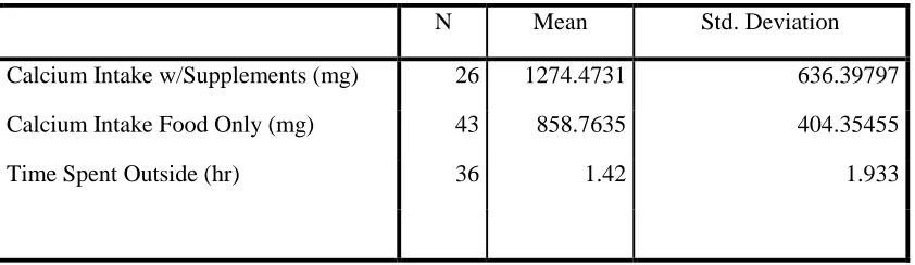
Factors Affecting BMD: Calcium and Sunlight Exposure (Vitamin D)
Calcium intake for all forty-three participants was calculated using the completed
24-hour dietary recall. The average calcium intake from food was 859 mg/day (+ 404
mg/day). Of the participants taking supplements (n=26), the average calcium intake with
food and supplements was 1274 mg/day (+ 636 mg/day). All participants were asked if
they spent time outside, and were asked to indicate how many hours they spent outside
per week. Of the participants who spent time outside every week (n=36), the average
amount of time spent outside was 1.4 hours/week (+ 1.9 hours/week). (See Table 6)
Table 6: Calcium Intake and Time Spent Outside of Assessed USA National Team Female Gymnasts
N Mean Std. Deviation
Calcium Intake w/Supplements (mg) 26 1274.4731 636.39797
Calcium Intake Food Only (mg) 43 858.7635 404.35455
[image:40.612.109.530.519.641.2]31 Menstrual Status
All participants (n=43) were asked about their menstrual status. Of the participants who
had begun menstruating (n=15), the average age of menstrual onset was approximately
[image:41.612.112.388.231.331.2]15.1 years (+ 1.4 years). (See Table 7 and Table 8)
Table 7: Menstrual Status of Assessed USA National Team Female Gymnasts
Have period? Frequency Percent Valid Percent
Valid Yes 15 34.9 34.9
No 28 65.1 65.1
Total 43 100.0 100.0
Table 8: Age at First Period of Assessed USA National Team Female Gymnasts
N Mean Std. Deviation
What Age 1st Period? 15 15.13 1.356
Valid N (listwise) 15
The participants who had begun menstruating (n=15) were also asked if they had regular
menstrual cycles. Of these participants, eight indicated that they had regular menstrual
[image:41.612.108.489.384.458.2]32
Table 9: Menstrual Regularity of Assessed USA National Team Female Gymnasts
Regular Period? Frequency Percent Valid Percent
Valid N/A 27 62.8 64.3
Yes 8 18.6 19.0
No 7 16.3 16.7
Total 42 97.7 100.0
Missing System 1 2.3
Total 43 100.0
STATISTICAL ANALYSIS
To determine statistical relationships between BMD and related factors among subjects,
Pearson Correlation statistics were assessed using two-tailed significance. This
assessment includes correlations (r-values) and probabilities (p-values) to assess
significance levels.
Demographic Data Analysis
Age of the gymnasts was positively and significantly associated with BMD in all
categories (p <0.001 for arm, leg, trunk, rib, pelvis, spine, and total BMD). All of the
r-values for BMD indicated a strong positive association with BMD (range r=0.62 to
r=0.68), except for BMD in the pelvis, which had a moderate positive association
(r=0.55). Statistical analysis was performed using a t-test to assess differences in BMD
between Caucasian (n=38) and non-Caucasian (n=5) subjects. There was no significant
33 Anthropometric Data Analysis
Weight was significantly associated with BMD in all categories (p <0.001 for arm, leg,
trunk, rib, pelvis, spine, and total BMD). All of the r-values indicated a very strong
positive association with BMD, ranging from r=0.82 to r= 0.90 for all sites. Lean body
mass, assessed by DEXA scans, was significantly associated with BMD in all categories
(p<0.001 for arm, leg, trunk, rib, pelvis, spine and total BMD). All r-values for lean body
mass and BMD indicated a strong positive association, ranging from r=0.74 to r=0.87 for
all sites. Body fat percentage, assessed by DEXA scans, was also significantly associated
with BMD at all sites, with p-values ranging from p<0.001 to p=0.01. The r-values for
BMD and percent body fat showed moderate positive associations, ranging from r=0.39
to r=0.54.
Vitamin Status Data Analysis
Calcium intake was not significantly associated with any of the BMD sites, and all of the
r-values indicated a weak negative association with BMD. Similarly, sunlight exposure
and indirect measures of vitamin D were not significantly associated with any of the
BMD sites, and all of the r-values indicated a weak positive association with BMD. (See
34
Table 10: BMD and Related Factors of Assessed USA National Team Female Gymnasts
BMD (g/cm2)
Age r (p) Weight r(p) Calcium r(p) Sunlight exposure r(p) Lean Body Mass r(p) Percent Body Fat DEXA r(p)
Arm 0.68
(<0.001) 0.83 (<0.001) -0.17 (0.28) 0.082 (0.63) 0.82 (<0.001) 0.44 (0.003)
Leg 0.64
(<0.001) 0.90 (<0.001) -0.14 (0.37) 0.13 (0.44) 0.84 (<0.001) 0.53 (<0.001)
Trunk 0.63
(<0.001) 0.89 (<0.001) -0.17 (0.28) 0.20 (0.24) 0.87 (<0.001) 0.47 (0.002)
Rib 0.64
(<0.001) 0.82 (<0.001) -0.20 (0.20) 0.13 (0.45) 0.74 (<0.001) 0.52 (<0.001) Pelvis 0.55
(<0.001) 0.87 (<0.001) -0.21 (0.18) 0.23 (0.19) 0.86 (<0.001) 0.39 (0.01)
Spine 0.62
(<0.001) 0.87 (<0.001) -0.16 (0.30) 0.15 (0.40) 0.82 (<0.001) 0.47 (0.002)
Total 0.68
(<0.001) 0.87 (<0.001) -0.20 (0.20) 0.12 (0.48) 0.81 (<0.001) 0.54 (<0.001)
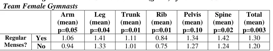
Menstrual Status Data Analysis
Regular menstrual status was assessed among the fifteen gymnasts who had begun
menstruating. The average BMD values were assessed for each site for participants who
had regular periods and those who did not have regular periods. The significance
between these values was then assessed. The average arm BMD for gymnasts with
regular periods was 1.06 g/cm2, while the average arm BMD for gymnasts without
regular periods was 0.94 g/cm2 (p=0.05). The average leg BMD for gymnasts with
regular periods was 1.41 g/cm2, while the average leg BMD for gymnasts without a
regular period was 1.33 g/cm2 (p=0.04). The average trunk BMD for gymnasts with
regular periods was 1.11 g/cm2, while the average trunk BMD for gymnasts without
regular periods was 1.01 g/cm2 (p=0.01). The average rib BMD for gymnasts with
35
periods was 0.75 g/cm2 (p=0.01). The average pelvic BMD for gymnasts without regular
periods was 1.34 g/cm2, while the average pelvic BMD for gymnast with regular periods
was 1.27 g/cm2 (p=0.10). The average spine BMD for gymnasts with regular periods was
1.42 g/cm2, while the average spine BMD for gymnasts without regular periods was 1.24
g/cm2 (p=0.02). The average total BMD for gymnasts with regular periods was 1.30
g/cm2, while the average total BMD for gymnasts without regular periods was 1.20 g/cm2
[image:45.612.103.541.304.387.2](p=0.003). (See Table 11)
Table 11: Regular Menstrual Status and BMD (g/cm2) of Assessed USA National
Team Female Gymnasts
Arm (mean) p=0.05 Leg (mean) p=0.04 Trunk (mean) p=0.01 Rib (mean) p=0.01 Pelvis (mean) p=0.10 Spine (mean) p=0.02 Total (mean) p=0.003 Regular Menses?
Yes 1.06 1.41 1.11 0.84 1.34 1.42 1.30 No 0.94 1.33 1.01 0.75 1.27 1.24 1.20
Predictors of Total BMD
A regression analysis was performed to determine which factors were strong predictors of
total BMD values in the assessed gymnasts. This dependent variable in this analysis was
total BMD (g/cm2) and the independent variables were regular menses, height, weight,
percent of kcals consumed from predicted kcal requirement (from indirect calorimetry),
calcium intake with supplements, lean body mass (from DEXA), time line deficits >300
kcals, and time line surpluses >300 kcals. The R2 value for this equation was 0.999, the
adjusted R2 value was 0.997, and the standard error of the estimate (SEE) was 0.005244
36
Table 12: Regression Equation to Predict Total BMD (g/cm2) in Assessed USA National Team Female Gymnasts
BMD total (g/cm2) = regular menses(-0.21) + height(0.014) + weight (0.001) + percent kcal requirement (0) + calcium intake w/supplements(-0.00006) + lean body mass (-0.003) + time line deficits >300(-0.01) + time line surpluses >300(-0.01) + 0.005244
37 CHAPTER 5: DISCUSSION AND CONCLUSIONS
DISCUSSION
The purpose of this study was to assess factors that are associated with BMD in
elite female gymnasts. Gymnastics is a high-impact sport that can positively affect
BMD, due to increased muscle mass and repeated load-bearing movements (Dowthwaite
& Scerpella, 2009). As osteoporosis and osteopenia affect nearly fifty-five million
Americans, it is important to maximize accrual of BMD during the formative years of
adolescence (National Osteoporosis Foundation, 2010). In this study, there were several
strong predictors of increased BMD in the assessed USA National Team female
gymnasts. Increased age was positively associated with increased BMD, which indicates
that the gymnasts involved in the study experienced age-related gains in BMD that are
similar to those seen in non-gymnast adolescents. Increased weight and increased
fat-free mass had strong positive associations with increased BMD in this population. It is
well-documented in the literature that increased muscle mass has positive effects on
BMD (Bertelloni et al., 2006; Daly et al., 2004; Egan et al., 2006; Taaffe & Marcus,
2004). Since muscle adds weight to the body, these factors are intertwined (i.e.,
increased muscle mass = increased body weight). Increased muscle mass can also lead to
increased power and better performance overall (Claessens et al., 1999). Although
38
only moderately associated with increased BMD. Since the average body fat percentage
for the assessed gymnasts was quite low in comparison to average body fat percentage in
non-gymnast adolescents, it may be difficult to predict BMD with regards to body fat
percentage in this population.
One unexpected result of the data assessment done in this study was that there
was no statistically significant association with either sunlight exposure or calcium intake
with BMD. Calcium and vitamin D play important roles in the maintenance and building
of bones. It has been shown in the literature that vitamin D status and calcium intake are
positively associated with increased BMD in the general population, but that was not the
case with this population (Bhan et al., 2010; Larson et al., 2009; Prentice, 2004). Since
the dietary information given by the participants was in the form of a 24-hour dietary
recall, there could have been discrepancies in normal calcium intake among the
participants. The calcium status may have been under- or over-estimated among the
gymnasts. In addition, there was a large standard deviation in calcium intake (mean
intake without supplements = 859 mg/day + 404 mg/day). This large standard deviation
may indicate a wide distribution of data, and may have affected the significance of the
data. Sunlight exposure was the sole measure of vitamin D status in these gymnasts, but
vitamin D status is also influenced by dietary intake and supplementation. Therefore,
sunlight exposure alone may not be a strong enough predictor of increased BMD values.
Regular menstrual status was significantly associated with BMD at all sites. Of
the fifteen gymnasts who had begun menstruating, the gymnasts with regular periods had
significantly higher BMD at all measured sites than those who did not have regular
39
menstruation among female adolescents is associated with higher BMD during puberty
and into adulthood (Loud et al., 2005; Yingling, 2009). It is also indicative of a larger
problem among the assessed gymnasts in this study. Of forty-three gymnasts, only eight
had regular periods (approximately 19% of the study population). The gymnasts who had
regular periods had significantly higher BMD than the gymnasts who did not have
regular periods, which indicates that regular menstrual status provides protective effects
to BMD and can positively influence maximal accrual of BMD. Since the majority of the
assessed gymnasts had not begun menstruating, and the average age of menstruation was
15 years, it may be assumed that these gymnasts are not benefiting from maximal BMD
accrual during their adolescent years. This could have detrimental effects later in life,
and they could be a higher at risk for developing osteopenia and osteoporosis than those
who had regular periods.
The regression analysis to predict total BMD had a very strong positive predictive
R2 value of 0.999 (adjusted R2 value=0.997, SEE=0.005244). The regression equation
developed from the regression analysis indicates that 99.9% of the variance in the
dependent variable (total BMD) can be predicted by the using the following independent
variables: regular menstruation, height, weight, percent kcal intake from predicted kcal
requirement (from indirect calorimetry), calcium intake (with supplements), lean body
mass (from DEXA), time line deficits >300 kcals in any one-hour period over 24 hours,
and time line surpluses >300 kcals in any one-hour period over 24 hours. This regression
equation should be able to accurately predict total BMD only when used with
age-matched elite female gymnasts. It is important to note that this is a limited population, as