S T U D Y P R O T O C O L
Open Access
Cellular versus acellular matrix devices in
treatment of diabetic foot ulcers: study protocol
for a comparative efficacy randomized controlled
trial
Hadar Lev-Tov
1,2, Chin-Shang Li
3, Sara Dahle
1,2*and Roslyn Rivkah Isseroff
1,2*Abstract
Background:Diabetic foot ulcers (DFUs) represent a significant source of morbidity and an enormous financial burden. Standard care for DFUs involves systemic glucose control, ensuring adequate perfusion, debridement of nonviable tissue, off-loading, control of infection, local wound care and patient education, all administered by a multidisciplinary team. Unfortunately, even with the best standard of care (SOC) available, only 24% or 30% of DFUs will heal at weeks 12 or 20, respectively.
The extracellular matrix (ECM) in DFUs is abnormal and its impairment has been proposed as a key target for new therapeutic devices. These devices intend to replace the aberrant ECM by implanting a matrix, either devoid of cells or enhanced with fibroblasts, keratinocytes or both as well as various growth factors. These new bioengineered skin substitutes are proposed to encourage angiogenesis and in-growth of new tissue, and to utilize living cells to generate cytokines needed for wound repair.
To date, the efficacy of bioengineered ECM containing live cellular elements for improving healing above that of a SOC control group has not been compared with the efficacy of an ECM devoid of cells relative to the same SOC. Our hypothesis is that there is no difference in the improved healing effected by either of these two product types relative to SOC.
Methods/Design:To test this hypothesis we propose a randomized, single-blind, clinical trial with three arms: SOC, SOC plus DermagraftW(bioengineered ECM containing living fibroblasts) and SOC plus OasisW(ECM devoid of living cells) in patients with nonhealing DFUs. The primary outcome is the percentage of subjects that achieved complete wound closure by week 12.
Discussion:If our hypothesis is correct, then immense cost savings could be realized by using the
orders-of-magnitude less expensive acellular ECM device without compromising patient health outcomes. The article describes the protocol proposed to test our hypothesis.
Trial registration:ClinicalTrials.gov: NCT01450943. Registered: 7 October 2011
Keywords:Diabetic foot ulcer, Chronic wounds, Nonhealing wounds, Oasis, Dermagraft, Wound matrix
* Correspondence:[email protected];[email protected] 1
Veterans Affairs Medical Center, Northern California Healthcare System, 10535 Hospital Way, Mather, CA 95655, USA
2
Department of Dermatology, University of California Davis, 3301 C Street, Sacramento, CA 95816, USA
Full list of author information is available at the end of the article
Background
Diabetes affects nearly one-third of the adult population in the United States [1]. For individuals with diabetes, the lifetime probability of developing a diabetic foot ulcer (DFU) is estimated at 10 to 25% [2]. DFUs repre-sent a significant source of morbidity and an enormous financial burden [3,4]. Diabetes is the leading cause of nontraumatic lower-extremity amputations in the United States [1] and a DFU is often the initial insult leading to these amputations [5]. The pathophysiology of DFUs is thought to result from the combined comorbidities of neuropathy, vascular deficits, impaired immunity, infec-tion and trauma, all occurring in no particular order and overlapping to produce a vicious cycle [6]. The standard of care (SOC) for DFUs involves systemic glucose con-trol, ensuring adequate extremity perfusion, debridement of nonviable tissue, off-loading, control of infection, local wound care and patient education, all administered by a multidisciplinary team [7-10]. Unfortunately, even with the best SOC available, only 24% or 30% of DFUs will heal at weeks 12 or 20, respectively [11].
Classic wound repair has been described as a rela-tively linear process (albeit with significant complexity and overlap), progressing through three general phases: inflammatory, including platelet aggregation, release of proinflammatory cytokines and recruitment of neutro-phils and macrophages; proliferative, including fibro-blast proliferation, angiogenesis and formation of granulation tissue and keratinocyte proliferation and re-epithelialization; and remodeling, the longest phase, including myofibroblast transformation and slow re-structuring of the healed wound to increase its strength [6,12].
In nonhealing DFUs the normal healing process is stalled. The exact mechanisms involved in this impair-ment are not fully understood but an increasing number of pathways are suggested with varying levels of evidence and are summarized herein as reviewed in a number of recent excellent articles [13-18]. Hyperglycemia is thought to adversely affect healing by increased levels of advanced glycation end products that inhibit normal extracellular matrix (ECM) deposition and upregulate activity of matrix metalloproteinases (MMPs). Prolonged hyperglycemia may also offset the delicate balance be-tween reactive oxygen species and various antioxidants by depletion of nicotinamide adenine dinucleotide phosphate on one hand and increasing reactive oxygen species production on the other. DFUs have increased activity of MMPs coupled with decreased activity of MMP inhibitors (tissue inhibitors of metalloprotei-nases). Fibroblasts in DFUs are often senescent, ex-hibit decreased proliferation and are less responsive to growth factors when compared with fibroblasts from age-matched diabetic controls without ulceration.
Keratinocytes in diabetic wounds exhibit impaired mi-gration, which may be mediated by c-Myc andβ-catenin. A drastic decrease in available inorganic phosphate leads to decreased levels of ATP in DFUs–a devastating blow to all healing-related processes. Immune response is impaired, with reduced inflammatory cell recruitment at the initial phase and later a skewed immune response towards macrophage and B-cell infiltrates. That phase is also characterized by increased TNFα and IL-1β expres-sion, which are known MMP stimulators. Leukocyte function and intracellular killing are impaired in DFUs, rendering these ulcers exceptionally susceptible to super-ficial infections and resistant biofilms. Neuropathy leads to decreased levels of neuropeptides that nor-mally contribute to healing. In addition, neuropathy reduces capillary blood flow. Impaired gap junction function has recently immerged as an additional pathological mechanism leading to impaired wound healing in DFUs.
As described above, the ECM is abnormal in DFUs and its impairment has been proposed as a key target for new therapeutic devices [19]. These devices intend to replace the aberrant ECM by implanting a matrix, either devoid of cells or enhanced with fibroblasts, keratino-cytes or both as well as various growth factors. These new bioengineered skin substitutes are proposed to en-courage angiogenesis and in-growth of new tissue, and to utilize living cells to generate cytokines needed for wound repair. DermagraftW (Shire Regenerative Medi-cine, Inc. La Jolla, California, United States ) is one ex-ample of bioengineered matrix supplemented with fibroblasts (cellular matrix (CM)). Other “ new-gener-ation”ECM replacement tissues, devoid of cellular com-ponents, have been created for wound repair, and the OasisW (Healthpoint, Ltd Fort Worth, Texas, United States ) acellular matrix (ACM) is an example of this cat-egory. Interestingly, in industry-supported randomized controlled trials, the reported rates of wound closure at week 12 are approximately 50% for both devices [20,21]. A success rate of 50% represents about 20% added bene-fit to the use of such devices over the SOC. This min-imal benefit may be disproportionate to the expenditure these devices accumulate (for example, up to $1,800 per application, and up to eight applications as the recom-mended regimen).
expensive acellular ECM device without compromising patient health outcomes. To test this hypothesis we pro-posed a randomized, single-blind, clinical trial with three arms: SOC, SOC plus DermagraftW(bioengineered ECM containing living fibroblasts) and SOC plus OasisW(ECM devoid of living cells) in patients with nonhealing DFUs.
Methods/Design Participants
The study protocol is approved by both the Veterans Affairs’ Institutional Research and Development Com-mittee and their Institutional Review Board (IRB). Study participants are veterans eligible for Veterans Affairs’ medical benefits. Patients are recruited from all clinics at the Veterans Affairs’ Northern California Healthcare System via provider referral and IRB-approved flyers. The study is supported by a Veterans Affairs’ MERIT award (project ID SURG-369-10S). During the “run-in” phase (see Table 1) all participants are screened for eligi-bility based on the inclusion and exclusion criteria. All study-related procedures (except diagnostic tests such as radiography or blood testing) are performed by study
staff at participating Veterans Affairs Northern Califor-nia Healthcare System wound centers.
Inclusion criteria
Institutional Review Board (IRB) Informed Consent Form is signed and dated prior to any study-related activities.
Area of the study ulcer after debridement is between 1 and 25 cm2at week 0/visit 3.
Subject is between 18 and 85 years of age.
Subject's highest Ankle Brachial Pressure Index (ABPI)/Ankle Arm Index (AAI)≥0.80 and < 1.4 (Highest ABPI/AAI value from three
measurements within last 6 months shall apply) or toe–arm index≥0.6.
[image:3.595.55.540.370.716.2]Patient has one or more diabetic ulcers on the target foot with only one ulcer selected as the study (target) ulcer. The target ulcer must be at least 4 cm from a nontarget ulcer and, in the investigator’s opinion, be unlikely to coalesce with another ulcer within 12 weeks of randomization.
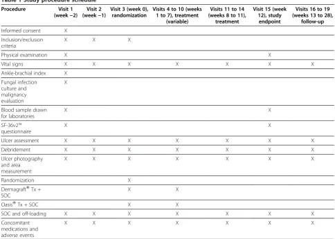
Table 1 Study procedure schedule
Procedure Visit 1 (week−2)
Visit 2 (week−1)
Visit 3 (week 0), randomization
Visits 4 to 10 (weeks 1 to 7), treatment
(variable)
Visits 11 to 14 (weeks 8 to 11),
treatment
Visit 15 (week 12), study
endpoint
Visits 16 to 19 (weeks 13 to 28),
follow-up
Informed consent X
Inclusion/exclusion criteria
X X X
Physical examination X X
Vital signs X X X X X X X
Ankle-brachial index X
Fungal infection culture and malignancy evaluation
X
Blood sample drawn for laboratories
X X
SF-36v2™ questionnaire
X X
Ulcer assessment X X X X X X X
Debridement X X X X X X X
Ulcer photography and area measurement
X X X X X X X
Randomization X
DermagraftWTx + SOC
X X
OasisWTx + SOC X X
SOC and off-loading X X X X X X X
Concomitant medications and adverse events
X X X X X X X
Subject's study ulcer is full thickness and does not extend to bone, muscle, or tendon.
Subject's study ulcer has been present at least 4 weeks prior to the initial screening (visit 1) or 6 weeks at randomization (visit 3).
Subject has been diagnosed with type 1 or type 2 diabetes and hemoglobin A1c < 10%.
Study ulcer has no clinical feature of infection (two signs of inflammation and elevated bacterial load of the wound).
Female subjects of childbearing age potential have a negative pregnancy test and are lactating for the duration of the study.
Subject understands the requirements of this study and is willing to comply with all the study requirements.
Exclusion criteria
Subject is diagnosed with cancer and is undergoing treatment with immunosuppressive or
chemotherapeutic agents, radiotherapy or systemic corticosteroids < 30 days before enrollment.
Subject is diagnosed with HIV/AIDS.
Subject is diagnosed with any bleeding disorders.
Subject is diagnosed with any connective tissue diseases.
For female subjects, the subject is pregnant or lactating.
Subject has a history of illicit drug use within 1 year of enrollment.
In the past year, the subject experienced episodes of drinking more than five alcoholic beverages in <2 hours and/or drinking alcohol became a problem in their interpersonal relationships, work, driving and/ or their behavior in general.
Subject has any active infected wounds or
osteomyelitis (confirmed by bone biopsy, magnetic resonance imaging or bone scan).
Subject had Doppler examination within the last 365 days, and it demonstrated reflux > 0.5 seconds.
Subject is diagnosed with active Charcot as described by Saunder’s classification system.
Subject manifests signs of poor nutritional status and/or albumin level < 2.9 g/dl.
Subject received DermagraftWand/or OasisWin the last 60 days.
Study ulcer size < 1.0 cm2or >25 cm2.
Subject has any porcine allergy or cow product allergy.
Subject’s recent (last 30 days) chemistry test’s serum creatinine is two times above the upper limit of normal and/or liver enzymes (AST, ALT) are three times above the upper limit of normal.
Between week−2/visit 1 and week 0/visit 3
(randomization) the study ulcer decreased in size by > 40%, or increased in size by > 50%.
DermagraftW (Advanced BioHealing, Inc. La Jolla, California, United States), OasisW (Healthpoint, Ltd Fort Worth, Texas, United States).
Participant consent
Detailed informed consent is obtained from all partici-pants and a HIPAA Health Insurance Portability and Ac-countability Act release form is signed. Prior to any study-related intervention, study staff provide each pro-spective subject with a detailed explanation of the nature and purpose of the trial. Once study staff are assured that the prospective subject understands all study proce-dures and potential implications, an IRB-approved informed consent form is signed. The signed copy becomes part of the subject’s record. An additional copy of the informed consent is provided to the subject for future reference.
Criteria for withdrawal
Subjects are informed that they may withdraw from the trial for any reason at any time and that the investigator may decide to withdraw them from the trial for safety-related issues. Study staff explain that failure to comply with the study’s procedure may result in withdrawal from the trial. In the case of such an event, the reason for withdrawal is documented in the subject’s file and the subject is referred to the wound clinic for further follow-up.
Participant compliance
Overall participant compliance with study-related proce-dures is assessed at each study visit by the staff. Specific-ally, detailed questioning regarding compliance with off-loading and any tampering with study-related dressings is performed at each study visit and any deviations are noted in the subject’s file for later analysis.
Summarized details of products compared (interventions)
The CM of this study, DermagraftW, is a cryopreserved human fibroblast-derived dermal substitute composed of viable newborn foreskin fibroblasts, seeded onto a bioab-sorbable polyglactin mesh. The product is supplied fro-zen (−75°C) in a clear bag containing a 2 inch×3 inch matrix [22]. The product is stored according to the man-ufacturer’s instructions and records of quality control, including constant temperature monitoring, are kept by study staff. DermagraftW is applied according to the manufacturer’s instructions [22].
The SOC dressing is comprised of IodosorbWgel (Smith & Nephew Largo, Florida, United States), AdapticW (Johnson & Johnson Gargrave, Skipton, United Kingdom ) and gauze. This dressing also serves as the secondary dressing for the two intervention arms. If a subject has an allergy to iodine products, then Bacitracin antibiotic oint-ment is used as a replaceoint-ment for Iodosorb.
We will provide all subjects with a removable walking boot, such as a camwalker or diabetic conformer Bledsoe boot. The clinician will make further accommodation/ adjustment to offload plantar ulcers with excavation or aperature using plastazote, foam or felt padding. Those patients that use exclusively manual or motorized wheel-chairs will be identified to differentiate patients that are fully ambulatory from those with limited ambulatory capacity. While total contact casting is considered the gold standard for offloading plantar ulcers by many aca-demicians, in our experience total contact casting is not in standard use owing to time constraints with cast ap-plication and accessibility to materials. Indeed, a recent study found that fewer than 2% of diabetic foot specia-lists utilize this form of offloading [24]. Our study there-fore models the standard care for all arms after realistic clinical practice.
All products used in this trial are approved by the Food and Drug Administration for use in treatment of DFUs.
Hypothesis (objectives)
There is no significant difference in healing at weeks 12 and week 20 or in the rate of healing when treating DFUs with SOC as compared with CM or ACM.
Alternative hypothesis
There is a significant difference in healing at weeks 12 and week 20 or in the rate of healing when treating DFUs with SOC as compared with CM or ACM.
Primary outcome
The primary outcome is the percentage of subjects that achieved complete wound closure by week 12. Complete healing is defined as full re-epithelialization of the ulcer with no drainage, or callus formation with underlying ulcer on two consecutive visits 1 week apart. Callus (hyperkeratosis) formation will be debrided at the wound site to determine whether complete closure has been obtained. The wound is deemed healed if there is no underlying open lesion after debridement (complete epithelialization).
Secondary outcomes
The first secondary outcome is the percentage of subjects that achieved complete wound closure by week 20. Complete healing will be defined as full re-epithelialization
with no drainage, or callus formation with underlying ulcer on two consecutive visits 1 week apart.
Another secondary outcome is the rate of wound heal-ing to achieve complete closure based on weekly wound area measurements.
Third is the incidence of ulcer recurrence at week 20. Recurrence will be defined as an ulcer occurring at the same location as the healed study ulcer.
Fourth is the association of wound healing (overall and given a particular treatment) with the following wound characteristics: peri-ulcer erythema, induration, tender-ness, pain, local warmth, size, depth, undermining, ulcer location, ulcer age at presentation, ulcer precipitating event and total wound assessment score at presentation.
Another outcome is the association of wound healing (overall and given a particular treatment) with the fol-lowing patient characteristics on presentation: age, sex, body mass index, smoking history, family history of dia-betes, complete blood count results, chemistry panel results, erythrocyte sedimentation rate and C-reactive protein serum concentration and hemoglobin A1C serum concentration.
Sixth is the association of a particular treatment with the incidence of the following during the study period: cellulitis, osteomyelitis, acute Charcot disease and over-all adverse events rate.
Another outcome is the association of a particular treatment with a change in quality of life assessments (quality-adjusted life-years).
The final secondary outcome is cost-effectiveness of a particular treatment compared with SOC.
Sample size
Based on previous trials [20,21] it is estimated that ap-proximately 50% of the subjects in the intervention arms will achieve complete wound closure at week 12 (pri-mary outcome) and approximately 25% of the SOC arm will achieve complete wound closure by week 12. The sample size was estimated based on 80% power and a 0.05 significance level to detect a 25% difference between the intervention and control arms (Stplan 4.5 statistical software, Department of Biomathematics, University of Texas MD Anderson Cancer Center, Houston, TX, USA, 2010). Based on the method of normal approximation to the arcsin transformation of the binomial distribution and, to be conservative, a two-sided test, enrollment of 57 subjects in each arm is required.
Randomization Sequence generation
60 through 69 years old and 70 through 85 years old) and race (black and nonblack). We used a computer-generated algorithm to randomly assign treatments to subjects within a block and to randomly determine the block sequence. Subjects presenting to the study will be entered into their appropriate block in a strict chrono-logical order.
Allocation concealment and implementation
The randomization table was generated prior to study initiation by the study’s biostatistician. Allocation is con-cealed by the Northern California Veterans Affairs Inves-tigational Drug Service, an independent third party, until the moment of randomization.
Blinding
Owing to the nature of the interventions studied, complete blinding is not realistic (for example, Derma-graftWrequires storage at −75 ± 10°C and a specific ma-nipulation immediately prior to application). An independent observer was therefore assigned to assess the primary and secondary outcomes. Clinical images will be captured at each visit before and after debride-ment and will be de-identified and uploaded to an exter-nal server (Silhouette CentralW Aranz Medical, Christchurch, New Zealand). The observer will log on to the server independently and determine whether the wound has closed or not. In addition, the observer will determine whether the margins used for area measure-ment are accurate.
Statistical methods
All data will be analyzed based on intention to treat. The primary outcome, wound closure by week 12, will be analyzed using the chi-square test or Fisher’s exact test when appropriate to compare the percentages of subjects with complete closure in each group. We will also perform exploratory analysis with logistic regres-sion, using complete wound closure at 12 weeks as the dependent variable and the independent variables would include the characteristics described as the fourth to sixth items in Secondary outcomes above. We will run preliminary analysis on selected covariates to determine whether there are significant differences.
Cost-effectiveness analysis will be performed to com-pare the treatment groups DermagraftW and OasisWand the standard care group using the analysis of varianceF test or Kruskal–Wallis test according to the data distri-bution pattern. If significant, we will further analyze with pair-wise comparisons using Tukey’s test or Dunn’s test. A multiple-comparison post-hoc test for Kruskal–Wallis analysis will be performed with the Statistical Analysis System (Cary, NC, USA) macro developed by Elliott and Hynan [27].
We will evaluate the cost-effectiveness of OasisW com-pared with DermagraftW and the SOC by measuring quality-adjusted life-years as the measure of effective-ness, based on results obtained from Short Form-36 (SF-36v2™) questionnaires. Multiple regression will be used to evaluate continuous dependent variable outcomes, in-cluding change in SF-36v2™physical and mental compo-nent summaries scores between baseline and the end of the study.
Rates of healing among the groups will be analyzed using a log-rank test to compare the time of healing within 20 weeks.
Secondary outcomes such as complete healing at 20 weeks and rate of ulcer recurrence at 20 weeks will be analyzed using chi-square tests or Fisher’s exact tests when appropriate. In addition, demographics, smoking history, and other characteristics mentioned in Second-ary outcomes will be summarized in a table. For com-parison of nominal categorical secondary outcome variables, we will use the chi-square test or Fisher’s exact test when appropriate. For comparison of ordinal cat-egorical variables, we will use the Wilcoxon rank-sum test (for two independent group comparisons) and the Kruskal–Wallis test (for three independent group comparisons).
Data management
Data will be managed by the study’s biostatistician (C-SL) at the University of California Davis Clinical and Transla-tional Science Center. Data will be automatically imported from digital subject records into pre-designed spread sheets using ExcelWand analyzed using Statistical Analysis System, version 9.3 [28].
Plan and trial design
This is a randomized, controlled, single-blind, trial com-paring the efficacy of ACM with that of CM in treatment of DFUs. A total of 171 subjects will be enrolled and randomly assigned to one of three treatment groups: ACM, CM and SOC. Subjects are followed for a total of 30 weeks in three major phases: run-in phase, treatment phase and follow-up phase. The study’s procedures are detailed below.
Run-in phase
comorbidities and establish a baseline: vital signs, body mass index, pregnancy test, Ankle-Brachial Index, quan-titative bacterial cultures, fungal cultures, tissue path-ology, complete blood count, comprehensive metabolic panels (including liver enzymes and albumin levels), erythrocyte sedimentation rate, C-reactive protein level, hemoglobin A1C level and lower-extremity X-rays. Once eligibility is established, subjects are randomized (as described above) to one of three treatment arms. SF-36 questionnaires are filled out by the subjects.
Treatment phase
This phase is divided into a variable treatment period (weeks 0 through 7) and a SOC treatment period (weeks 8 through 11). Subjects will be evaluated on a weekly basis.
During the variable treatment period, subjects in each arm will receive its corresponding primary dressing treatment (that is, ACM, CM or SOC). During each visit, vital signs will be measured, a comprehensive lower-extremity assessment will be performed (including pho-tography and ulcer area measurement), health status and medication changes will be recorded and adverse events will be assessed. Compliance with the off-loading device will also be assessed.
During the SOC treatment phase the primary dressing applied to all subjects will be identical and will comprise the SOC dressing. The rest of the visit will be identical to the variable treatment period as described above.
On week 12, a study endpoint visit will be conducted. During this visit, vital signs will be measured, a compre-hensive lower-extremity assessment will be performed (including photography and ulcer area measurement), health status and medication changes will be recorded and adverse events will be assessed. Compliance with the off-loading device will also be assessed. In addition, the SF-36 questionnaire is filled out for the second time by the subjects and repeat complete blood count and comprehensive metabolic panels are ordered. SOC treat-ment will be provided to all subjects.
Follow-up phase
This phase is comprised of four monthly visits (weeks 13 through 28). During each visit, vital signs are measured, a comprehensive lower-extremity assessment is per-formed (including photography and ulcer area measure-ment), health status and medication changes are recorded and adverse events are assessed. Compliance with the off-loading device is also assessed. During the follow-up phase the primary dressing applied to all sub-jects is identical and will comprise the SOC dressing. SOC treatment is provided to all subjects.
In the event of early wound closure (before week 12), the subject will return for follow-up visits every 4 weeks
from the confirmatory visit and in week 12 or in week 12 only, whichever comes first, and will return to the schedule above thereafter. In the event of failure to heal after week 12, subjects will be followed at the wound clinic based on clinical need but will return for all follow-up phase mandated visits.
Ethical considerations
The DOLCE trial is approved by the Veterans Affairs Northern California Healthcare System IRB and the Re-search and Development Committee. The primary inves-tigators will ensure that the study is conducted in full compliance with the protocol and IRB regulations as well as international standards on human subjects’ re-search. The primary investigators will ensure compliance with institutional regulations as well as local and na-tional law. Compliance will be administered by a dedi-cated, certified Clinical Research Coordinator. A data monitoring committee has been set up to review safety data. All adverse events will be reported to the IRB as stipulated by the IRB in its protocol-approval letter.
Discussion
Trial status
Actively recruiting.
Abbreviations
ACM: acellular matrix; CM: cellular matrix; DFU: diabetic foot ulcer; ECM: extracellular matrix; IL: interleukin; IRB: Institutional Review Board; MMP: matrix metalloproteinase; SF-36: Short Form-36; SOC: standard of care; TNF: tumor necrosis factor.
Competing interests
The authors declare that they have no competing interests.
Authors’contributions
HL-T contributed to the protocol design, wrote IRB amendments, and wrote the first draft of the manuscript. C-SL contributed to the statistical design and edited the manuscript. SD contributed to the overall study design, wrote the first draft of the protocol, edited the manuscript, and wrote the MERIT grant. RI conceived the study idea, supervised the study design and protocol development, edited the manuscript, edited IRB documents, and wrote the MERIT grant. All authors read and approved the final manuscript.
Acknowledgements
The authors acknowledge the seminal contributions of Dr Huong L Le in her role developing this project with her overarching goal of providing the best care to her patients. Sadly, her untimely death prevented her from completing this task. This trial is supported by a Veteran Affairs’MERIT award (Project ID SURG-369-10S). Statistical services are supported by the National Center for Advancing Translational Sciences, National Institutes of Health, through grant #UL1 TR000002.
Author details
1
Veterans Affairs Medical Center, Northern California Healthcare System, 10535 Hospital Way, Mather, CA 95655, USA.2Department of Dermatology, University of California Davis, 3301 C Street, Sacramento, CA 95816, USA. 3Department of Public Health Sciences, Division of Biostatistics, University of
California Davis, MS1C Room 145, Davis, CA 95616, USA.
Received: 22 July 2012 Accepted: 18 December 2012 Published: 9 January 2013
References
1. Center for Disease Control and Prevention:National Diabetes Fact Sheet: National Estimates and General Information on Diabetes and Prediabetes in the United States, 2011. Atlanta, GA: Department of Health and Human Services; 2011.
2. Margolis DJ, Hoffstad O, Nafash J, Leonard CE, Freeman CP, Hennessy S, Wiebe DJ:Location, location, location: geographic clustering of lower-extremity amputation among Medicare beneficiaries with diabetes.
Diabetes Care2011,34:2363–2367.
3. Boulton AJ, Vileikyte L, Ragnarson-Tennvall G, Apelqvist J:The global burden of diabetic foot disease.Lancet2005,366:1719–1724. 4. Sen CK, Gordillo GM, Roy S, Kirsner R, Lambert L, Hunt TK, Gottrup F,
Gurtner GC, Longaker MT:Human skin wounds: a major and snowballing threat to public health and the economy.Wound Repair Regen2009,
17:763–771.
5. Apelqvist J, Bakker K, van Houtum WH, Schaper NC:Practical guidelines on the management and prevention of the diabetic foot: based upon the International Consensus on the Diabetic Foot (2007) Prepared by the International Working Group on the Diabetic Foot.Diabetes Metab Res Rev
2008,24(Suppl 1):S181–S187.
6. Falanga V:Wound healing and its impairment in the diabetic foot.Lancet
2005,366:1736–1743.
7. Brem H, Sheehan P, Boulton AJ:Protocol for treatment of diabetic foot ulcers.Am J Surg2004,187:1S–10S.
8. Game FL, Hinchliffe RJ, Apelqvist J, Armstrong DG, Bakker K, Hartemann A, Löndahl M, Price PE, Jeffcoate WJ, International Working Group on Diabetic Foot:Specific guidelines on wound and wound-bed management 2011.
Diabetes Metab Res Rev2012,28(Suppl 1):232–233. 9. Bakker K, Apelqvist J, Schaper NC:Practical guidelines on the
management and prevention of the diabetic foot 2011.Diabetes Metab Res Rev2012,28(Suppl 1):225–231.
10. Lepantalo M, Apelqvist J, Setacci C, Ricco JB, de Donato G, Becker F, Robert-Ebadi H, Cao P, Eckstein HH, De Rango P, Diehm N, Schmidli J, Teraa M, Moll FL, Dick F, Davies AH:Chapter V: Diabetic foot.Eur J Vasc Endovasc Surg2011,42(Suppl 2):S60–S74.
11. Margolis DJ, Kantor J, Berlin JA:Healing of diabetic neuropathic foot ulcers receiving standard treatment. A meta-analysis.Diabetes Care1999,
22:692–695.
12. Singer AJ, Clark RA:Cutaneous wound healing.N Engl J Med1999,
341:738–746.
13. Gary Sibbald R, Woo KY:The biology of chronic foot ulcers in persons with diabetes.Diabetes Metab Res Rev2008,24(Suppl 1):S25–S30. 14. Loots MA, Lamme EN, Zeegelaar J, Mekkes JR, Bos JD, Middelkoop E:
Differences in cellular infiltrate and extracellular matrix of chronic diabetic and venous ulcers versus acute wounds.J Invest Dermatol1998,
111:850–857.
15. Medina A, Scott PG, Ghahary A, Tredget EE:Pathophysiology of chronic nonhealing wounds.J Burn Care Rehabil2005,26:306–319.
16. Mendoza-Naranjo A, Cormie P, Serrano AE, Wang CM, Thrasivoulou C, Sutcliffe JE, Gilmartin DJ, Tsui J, Serena TE, Phillips AR, Becker DL:
Overexpression of the gap junction protein Cx43 as found in diabetic foot ulcers can retard fibroblast migration.Cell Biol Int2012,36:661–667. 17. Neut D, Tijdens-Creusen EJ, Bulstra SK, van der Mei HC, Busscher HJ:
Biofilms in chronic diabetic foot ulcers–a study of 2 cases.Acta Orthop
2011,82:383–385.
18. Bajpai S, Shukla VK, Tripathi K, Srikrishna S, Singh RK:Targeting connexin 43 in diabetic wound healing: future perspectives.J Postgrad Med2009,
55:143–149.
19. Panuncialman J, Falanga V:The science of wound bed preparation.Surg Clin North Am2009,89:611–626.
20. Gentzkow GD, Iwasaki SD, Hershon KS, Mengel M, Prendergast JJ, Ricotta JJ, Steed DP, Lipkin S:Use of dermagraft, a cultured human dermis, to treat diabetic foot ulcers.Diabetes Care1996,19:350–354.
21. Niezgoda JA, Van Gils CC, Frykberg RG, Hodde JP:Randomized clinical trial comparing OASIS Wound Matrix to Regranex Gel for diabetic ulcers.Adv Skin Wound Care2005,18(5 Pt 1):258–266.
22. For Non-healing Diabetic Foot Ulcers.[http://www.dermagraft.com/about/ overview]
23. Healthpoint Offers Two OasisWMatrix Products.[http://www. oasiswoundmatrix.com/]
24. Wu SC, Jensen JL, Weber AK, Robinson DE, Armstrong DG:Use of pressure offloading devices in diabetic foot ulcers: do we practice what we preach?Diabetes Care2008,31:2118–2119.
25. Ranjita Misra LL, David V, Ashley M, Khanna SR, Sen CK:Predictors of Diabetic Wound Healing by Racial/Ethnic Categories [poster]. Columbus OH: The Ohio State University Comprehensive Wound Center; 2012 [http://misra.tamu. edu/chronic_healing.html]
26. Goodson WH, Goodson WH, Hunt TK:Wound healing and aging.J Invest Dermatol1979,73:88–91.
27. Elliott AC, Hynan LS:A SASWmacro implementation of a multiple comparison post hoc test for a Kruskal–Wallis analysis.Comput Methods Programs Biomed2011,102:75–80.
28. Statistical Analysis System.[www.sas.com]
29. Lundh A, Krogsboll LT, Gotzsche PC:Sponsors' participation in conduct and reporting of industry trials: a descriptive study.Trials2012,13:146. doi:10.1186/1745-6215-14-8