http://dx.doi.org/10.4236/abb.2015.66040
How to cite this paper: Flores-Rozas, H., Jaafar, L. and Xia, L. (2015) The Role of DNA Mismatch Repair and Recombination in the Processing of DNA Alkylating Damage in Living Yeast Cells. Advances in Bioscience and Biotechnology, 6, 408-418. http://dx.doi.org/10.4236/abb.2015.66040
The Role of DNA Mismatch Repair and
Recombination in the Processing of
DNA Alkylating Damage in Living
Yeast Cells
Hernan Flores-Rozas
1,2*#, Lahcen Jaafar
2#, Ling Xia
2#1College of Pharmacy and Pharmaceutical Sciences, Florida A&M University, Tallahassee, USA 2Institute of Molecular Medicine and Genetics, Georgia Regents University, Augusta, USA
Email: *[email protected]
Received 25 March 2015; accepted 8 June 2015; published 11 June 2015
Copyright © 2015 by authors and Scientific Research Publishing Inc.
This work is licensed under the Creative Commons Attribution International License (CC BY). http://creativecommons.org/licenses/by/4.0/
Abstract
It is proposed that mismatch repair (MMR) mediates the cytotoxic effects of DNA damaging agents by exerting a futile repair pathway which leads to double strand breaks (DSBs). Previous reports indicate that the sensitivity of cells defective in homologous recombination (HR) to DNA alkylation is reduced by defects in MMR genes. We have assessed the contribution of different MMR genes to the processing of alkylation damage in vivo. We have directly visualized recombination complexes formed upon DNA damage using fluorescent protein (FP) fusions. We find that msh6 mutants are more resistant than wild type cells to MNNG, and that an msh6 mutation rescues the sensitivity of
rad52 strains more efficiently than an msh3 mutation. Analysis of RAD52-GFP tagged strains
indi-cate that MNNG increases repair foci formation, and that the inactivation of the MHS2 and MSH6 genes but not the MSH3 gene result in a reduction of the number of foci formed. In addition, in the absence of HR, NHEJ could process the MNNG-induced DSBs as indicated by the formation of NHEJ- GFP tagged foci. These data suggest that processing of the alkylation damage by MMR, mainly by MSH2-MSH6, is required for recruitment of recombination proteins to the damage site for repair.
Keywords
DNA Mismatch Repair, Recombination, DNA Damage, Non-Homologous End Joining
*Corresponding author. #
1. Introduction
Mismatch repair (MMR) plays a crucial role in the maintenance of the genome by repairing nucleotide misin-corporations that occur during DNA replication and by preventing recombination between diverged sequences [1] [2], for review see [3]. Defects in MMR genes lead to the accumulation of mutations and microsattelites instability
[4] which is the underlying defect in Hereditary Non Polyposis Colon Cancer (HNPCC). To date, six MMR genes have been identified in eukaryotes: MSH2, MSH3 and MSH6, which are homologs of E. coli MutS, and MLH1, MLH3 and PMS2 (PMS1 in yeast) which are homologous to MutL [2]. The recognition step, which is the rate-li- miting step of the reaction, is carried out by two heterodimers that possess differential specificity for mismatches. The MSH2-MSH6 complex repairs base-base mismatches and small loops, while the MSH2-MSH3 complex binds and repairs large loops [5]. Inactivation of MSH6 or MSH3 results in modest phenotype due to their re-dundancy, whereas inactivation of MSH2 completely impairs the recognition step. Besides their important role in correcting replication errors, MMR proteins have been recently reported to participate in cellular responses to some forms of DNA damage induced by certain anticancer drugs [5]. Among these are the SN1-type methylat-ing agents like N-methyl-N’-nitro-N-nitrosoguanidine (MNNG). The O6-methylguanine (O6-meG) is a portion of the DNA damage caused by SN1 methylating drugs. The O6MeG DNA methyl-transferase (MTase) effi-ciently removes the methyl group [6] and puts back normal base pairing, preventing cells from mutagenesis and death [7]. However, in MTase-deficient bacterial, yeast and mammalian cells, the O6-meG persists and forms during replication, base pairs with both C and T generating O6-MeG:T or O6-MeG:C mismatches [8]. Cells defi-cient in the MMR proteins cannot recognize O6-MeG: T and are therefore highly resistant to toxic effects of methylating agents [9]-[11].
The mechanism by which MMR proteins mediate cytotoxic responses to MNNG has not yet been elucidated. More than one model exists to describe this effect. The most extensively studied model proposes that MMR me-diates the cytotoxic effects of DNA damaging agents by exerting a futile repair pathway in which O6-meG con-tinually produces a template for MMR system [12] and leads to repeated cycles of repair resulting in the forma-tion of a single-strand gaps [13] opposite to O6-meG which finally results, during the following cycle of DNA replication, in double strand breaks (DSBs) which lead cell death if not repaired [14] [15]. The DSBs, caused by the collapse of replication forks, are in the most part repaired either by homologous recombination (HR) or nonhomologous end joining (NHEJ) [16]. However, the contribution of both repair pathways depends mostly on the cell-cycle stage, with NHEJ being used in G1 in which the two DSBsends are directly rejoined. On the other hand, HR is mainly active during S/G2 phases where sister chromatids are present [17]. The choice of the path-way differs also among species. In yeast, HR is the most dominant whereas, NHEJ is most used in higher euka-ryotes [18]. In S. cerevisiae, HR is controlled by the RAD52 epistasis group which includes RAD50, RAD51, RAD52, RAD54, RAD55, RAD57, RAD59, RDH54/TID1, MRE11 and XRS2 genes [19] [20]. Among these, the RAD52 gene seems to be involved in all HR events including single-strand annealing and gene conversion [21]. When RAD52 is deleted, severe phenotype defects in recombination are manifested in these strains suggesting the fundamental role of this gene compared to other mutants of the same group [22]. S. cerevisiae also uses NHEJ to repair DSBs. The components of yeast NHEJ pathway are divided into three protein complexes [23]
that include the exonuclease complex MRX (Mre11/Rad50/Xrs2) [24], the DNA end recognition protein com-plex yKu (YKU70/YKU80) which is initially recruited to the DNA damage site [25] and the DNA ligase (Dnl4/ Lif1) [25] [26].
The goal of our study is to determine the role of the different MMR genes in the processing of DNA alkylat-ing damage to DSBs and to visualize the recruitment of the recombination pathways duralkylat-ing the repair process. Our data suggest that the MSH2-MSH6 and not the MSH2-MSH3 complex mediates the processing of alkylat-ing DNAdamage to DSBs and that both NHEJ and HR recombination pathways are recruited to the DNA dam-age site for repair.
2. Materials and Methods
2.1. General Genetic Methods and Strains
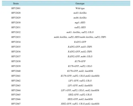
Table 1. Yeast strains used in this study.
Strain Genotype
HFY2001 Wild type
HFY2028 msh3::hisGhis
HFY2029 msh6::hisGhis
HFY2030 mgt1::HIS3
HFY2031 rad52::HIS3
HFY2032 msh3:: hisGhis, rad52::URA3
HFY2033 msh6::hisGhis, rad52::HIS3/msh6::hisGhis, rad52::TRP1
HFY2034 RAD52-GFP
HFY2035 RAD52-GFP, msh3::TRP1
HFY2036 RAD52-GFP, msh2::TRP1
HFY2037 RAD52-GFP, msh6::URA3
HFY2038 KU70-GFP
HFY2039 KU70-GFP, rad52::URA3
HFY2040 KU70-GFP, msh2::kanMX4
HFY2041 KU70-GFP, rad52::URA3,msh2::kanMX4
HFY2042 LIF1-GFP, rad52::URA3
HFY2043 LIF1-GFP, msh2::kanMX4
HFY2044 LIF1-GFP, rad52::URA3, msh2::kanMX4
HFY2045 XRS2-GFP, rad52::URA3
HFY2046 XRS2-GFP, msh2::kanMX4
HFY2047 XRS2-GFP, rad52::URA3,msh2::kanMX4
All strains are derivatives of HFY2001 (MATa his3Δ200 ura3-52 leu2Δ1trp1Δ63 ade2Δ1 ade8 hom3-10lys2ΔBgl), except for the NHEJ-GFP strains, which are derivatives of BY4741 (MATa his3-1 leu2 met15ura3).
2.2. Strains
Gene-disrupted strains were constructed using a 3-step PCR method. We created DNA cassettes containing ex-ogenic regions of the gene of interest (i.e. RAD52), flanking a selective marker (i.e. HIS3, TRP1, URA3). These disruption cassettes were integrated into the genomes by homologous recombination. Double knockout mutants were obtained by sequential gene disruption using different selective markers. All strains were confirmed by PCR. Strains expressing GFP-tagged proteins were constructed using a 3-step PCR method we created DNA fragments that contain the 5’ end of the gene of interest (i.e. MSH2, RAD52) fused, in frame, to genes encoding fluorescent proteins (i.e. GFP, CFP, YFP). These PCR products were integrated into the genomes of either wild-type (RKY3023) or MMR-deficient cells (i.e. msh2, msh6 mutant) by homologous recombination, and the strains containing the proper fusion were selected and confirmed by PCR and sequencing. Strains containing two differently tagged genes were obtained by sequential transformation into the same strain.
2.3. Survival Assay
- 4 hrs, when the cells were again in an exponential growth phase. The cells were then mock-treated and treated with several concentrations of freshly diluted MNNG for 1hr. They were harvested, washed, and spotted (~10 µl drops) at serial dilutions (1 × 10⁻¹ - 10−5) on YPD plates. The plates were evaluated after 3 days of cultivation at 30˚C. The results shown are based on 3 - 5 independent experiments.
2.4. Live Cell Imaging and Fluorescent Microscopy
The cells were prepared as described in spot assay. For visualizing DNA in living cells, 10 mg/ml DAPI was added to the culture 30 min before imaging. Next, 1 ml aliquots were washed, pelleted and resuspended in 200µl of fresh synthetic minimal medium (SD). Then 5 ml volume of cells were immobilized on a glass slide pre- coated with 0.1% poly-L-lysine solutions (Sigma), to prevent evaporation, the cover glass was sealed with Cy-tosealTM XYL mounting medium (Richard-Allan Scientific). Cells were viewed under a Zeiss Axioplan 2 imag-ing microscope (Carl Zeiss, Thornwood, NY) with a water-immersion Achroplan 63/0.9W/DIC III objective. The illumination source was a 100-W mercury arc lamp. Cell images were taken using an AxioCamHRm digital camera operated via AxioVision 4.5 software. Confocal images were captured with a LSM 510 META system operated via META 3.2 software. The wavelengths of the filters used to visualize the RAD52-GFP (excitation 488 nm; emission 509 nm) and 4’,6-diamidino-2-phenylindole (DAPI; excitation 358 nm, emission 463 nm). Image acquisition times for RAD52-GFP was 1100 ms. At least 300 nuclei were counted for total of 6 - 10 field of cells, the observations of RAD52-GFP foci formation are from 1.5 to 6 hr after MNNG treatment, the results shown are based on 3 independent experiments. Unless otherwise noted, all experiments were performed at room temperature.
2.5. Immunofluorescence Staining
Yeast Cells were grown and treated with MNNG exactly as in the spot tests. Immunofluorescence staining was carried out as previously described [29] with some modifications: Paraformaldehyde was added to 2 - 5 ml cul-tures to a final concentration of 4% and incubated at room temperature for 1 hr with gentle agitation. Cells were washed five times with a solution containing 5 mM MgCl2, 40 mM KH2PO4 and resuspended in 0.5 ml of diges-tion buffer containing 5 mM MgCl2, 40 mM KH2PO4, and 1.2 M sorbitol. Zymolase (1 mg/ml final concentra-tion) was added and cells were incubated at 30˚ for 45 - 60 min. After a wash with digestion buffer, cells were spotted on slides precoated with poly-L-lysine (Sigma-Aldrich) and incubated 15 - 30 min. Slides were then in-cubated in blocking buffer (1.5% BSA in PBS, 0.5% Tween 20, and 0.1% Triton X-100) for 30 min. Incubation with the primary antibody directed against H2A phosphoserine 129 (1:1000 dilution, gift from Dr. Bonner, NIH) was performed overnight at room temperature. The slides were then washed two to three times with PBS, once with blocking buffer and then incubated for 1 hr at room temperature with a secondary antibody conjugated with the green fluorescent dye Alexa 594 (Molecular probes, 1:500 dilution in blocking buffer). After three washes with PBS, slides were then stained with (10 µg/ml) of 4,6-diamino-2-phenylindole (DAPI), for 5 min at room temperature. The samples were then visualized under microscope.
2.6. Statistical Analysis
Data analysis and graphing was performed using the GraphPad Prism 4 software package. Specific analysis for each experiment is indicated in the figure legend. In most cases the mean of at least three experiments is plotted together with the standard deviation.
3. Results
3.1. Inactivation of MSH6 but Not MSH3 Rescues the Sensitivity of HR-Deficient Strains
to MNNG
(a)
[image:5.595.167.430.86.545.2](b)
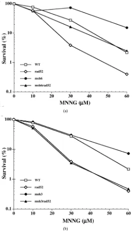
Figure 1. MNNG sensitivity of the MMR-deficient (msh3 or msh6) and/ or HR-deficient (rad52) strains. Mid-log phase cells were treated with the indicated concentrations of MNNG, harvested, washed and spotted onto YPD plates at proper serial dilutions as described under Materials and Methods. Each data point represents the mean of 3 independent experiments. (a) Sensitivity of msh6 and msh6-derived strains. The msh6 mutations increases the survival of cells to MNNG exposure and rescues rad52 mutants; (b) Sensitivity of msh3 and msh3-derived mutants to MNNG. Inactivation of MSH3 does not increase the survival of cells to MNNG, and does not rescue the sensitivity of the rad52 strain.
3.2. Exposure of Cells to MNNG Results in Formation of Repair Foci
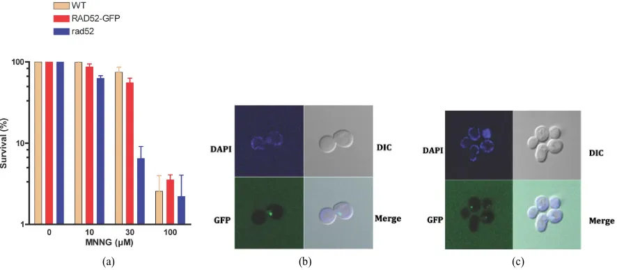
To study the involvement of MMR genes in the processing of alkylation damage to DSBs, we constructed a se-ries of strains that contain as a reporter RAD52 fused to a GFP at the C-terminal end. The RAD52-GFP strain behaves similarly to the wild type strain when exposed to DNA damaging agents that cause DSBs (Figure 2(a)) displaying a survival that is similar to that of wild type cells and significantly higher than that of the rad52 mu-tant, indicating that the GFP tag does not affect the activity of RAD52 and that the RAD52-GFP strain is com-petent in HR. The strain was tested for its ability to display repair foci after DNA damage. Treatment of the cells with ionizing radiation (γ-rays) resulted in a single defined foci, restricted to the nucleus, which appears within 30 min after exposure (Figure 2(b)) consistent with previous reports [30]. Similarly, exposure of the RAD52- GFP strain to MNNG also induced RAD52 foci formation (Figure 2(c)), indicating that MNNG generates a damage that serves as a substrate for the HR machinery. In both treatment procedures the majority of the cells (>98%) that display RAD52 foci present a single foci and very few present two or more foci (~2%) (data not shown).
3.3. Formation of RAD52 Foci upon Treatment with MNNG Requires the Activity
MSH6 but Not MSH3
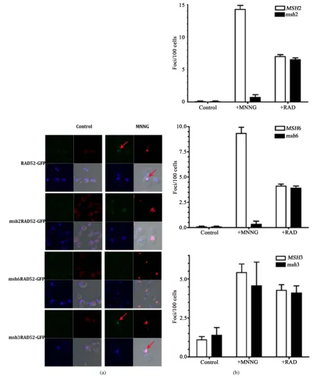
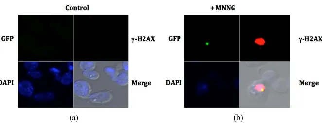
To determine if the recruitment of homologous recombination is altered in a MMR defective background, we inactivated MMR genes in the strain containing the RAD52-GFP fusion. Spontaneous foci form at very low frequency in the absence of exposure to the DNA alkylating agent (less than 1 per 100 cells, Figure 3(a)top left panel) and is increased at least 15-fold after treatment (Figure 3(a) top right panel and Figure 3(a)). Inactiva-tion of MSH2 results in a considerable reducInactiva-tion of the RAD52 foci formed after exposure to MNNG (Figure 3(a)) to levels similar to those of unexposed cells (14-fold reduction, Figure 3(b)). Similarly, the inactivation of the MSH6 gene also reduces the accumulation of RAD52 foci (Figure 3(a)) by approximately 9-fold (Figure 3(b)). As expected, the inactivation of the MSH3 gene did not significantly reduced the formation of RAD52 fo-ci upon treatment with MNNG. As a control, exposure of the cells to ionizing radiation (+RAD) did not require a proficient MMR system since MMR mutants strains accumulate similar number of foci compared to the wild type strain (Figure 3(b)). In all cases that a RAD52 foci was observed, it was restricted to a specific damage area as indicated by histone γ-H2AX activation. Activation of γ-H2AX occurs predominantly after exposure to MNNG (Figure 3(a)). Interestingly, histone γ-H2AX still becomes activated in strains defective in MMR, which do not process the alkylation damage to DSB. This is consistent with observation that γ-H2AX activation is not restricted to DSBs but is a signal for DNA damage in general [31]. Consistent with the survival data (Figure 1(a)
[image:6.595.91.534.482.676.2]
(a) (b) (c)
[image:7.595.80.535.85.632.2]
(a) (b)
Figure 3. The MSH2-MSH6 complex is required for foci formation upon DNA damage. (a) Visualization of DNA dam-age sites as indicated by histone H2AX phosphorylation (red regions). MNNG treated or untreated (control) cells were fixed immediately after treatment with MNNG and processed for direct immunofluorescence microscopy using yeast γ- H2AX antibody as described in the Materials and Methods. Foci of Rad52-GFP (green spots) are indicated by an arrow. Nuclei were demarked by DAPI-stained (blue); (b) The numbers of RAD52-GFP foci formed was determined from 3 in-dependent experiments visualizing at least 400 live yeast cells per experiment. Error bars indicate standard deviation. Strains used were wild type (RAD52-GFP), msh2 mutants (msh2 RAD52-GFP), msh3 mutants (msh3 RAD52-GFP) and
and Figure 1(b)), MSH2 and MSH6 genes, but not MSH3 gene, are required to process alkylation damage to an injury that requires HR for repair, most likely a DSB, as indicated by the formation of repair centers that contain RAD52.
3.4. Nonhomologous End Joining Can Be Recruited to DNA Damage Sites in the Absence
of Homologous Recombination in a MMR Dependent Manner
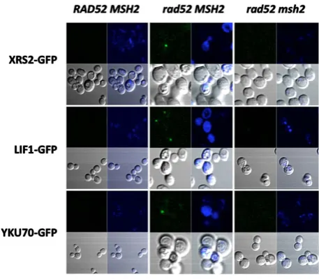
To determine if the repair of DNA damage resulting from the processing of DNA alkylation by MMR can also be repaired by NHEJ we constructed a series of strains containing GFP-tagged NHEJ proteins. Initial attempts to visualize DNA damage-induced foci of NHEJ proteins were unsuccessful. We reasoned that the presence of HR in the NHEJ-GFP tagged strains competes for DNA ends preventing the loading of NHEJ. We proceeded to in-activate HR in these strains and determine if foci form in a mismatch repair-dependent manner after exposure to MNNG. The strains defective in HR, which also contain fusions of a GFP at the C-terminus of YKu70, XRS2 and LIF1, appeared to be functional in DNA end joining as determined by their ability to repair a restricted plasmid compared to the NHEJ knock-out (data not shown). As shown in Figure 4, no NHEJ foci form in the presence of HR in the XRS2-GFP, LIF1-GFP and YKU70-GFP strains. However, upon inactivation of HR we observe NHEJ foci formation, although the frequency is much lower than what we observed with RAD52-GFP strains (only 3 - 5 fold over untreated cells). In addition, NHEJ foci formation after treatment with MNNG is al-so dependent on a proficient MMR pathway, since inactivation of MSH2 leads to a significant reduction of the observed foci. As in the RAD52 foci, the NHEJ foci is also restricted to sites of DNA damage as indicated by the colocalization of LIF1-GFP and activated histone γ-H2AX (Figure 5). These data suggests that NHEJ can also repair DNA damage that results from the processing of alkylated DNA by MMR.
4. Discussion
[image:8.595.184.413.433.633.2]Defects in mismatch repair have been associated to increased tolerance to DNA damage. In particular, alkylation damage that results in methylation of O6-G can be processed to DSBs in a process that requires misincorporation by the DNA polymerase to form a T-methyl-O6-G that can be recognized by the MMR system. Attempts to re-pair this mismatch results in a futile cycle where MMR replaces the misincorporated T and DNA polymerase
(a) (b)
Figure 5. NHEJ repair foci assemble at damage sites in HR-defective strain. Upon exposure to MNNG, the rad52 LIF1-GFP strain displays repair foci (green spot) that localizes within DNA damage sites, as determined by activation of histone H2AX (red region). Cell nucleus was stained with DAPI (blue).
reintroduces it as long as the methyl-O6-G persists. It is speculated that this continuous cycle of repair eventually leads to DSBs which are toxic to the cell. We have investigated which component of MMR involved in the rec-ognition step are required to process methylation damage to a DSB. We find that inactivation of MSH6, leads to an increased tolerance to MNNG, and that inactivation of MSH6 can rescue the sensitivity of the HR mutant rad52 to the alkylating agent. In contrast, inactivation of MSH3 does not alter the sensitivity of cells to DNA methylation and does not rescue the rad52 strain. This results are consistent with the fact that polymerase mi-sincorporation opposite then methyl-O6-G leads to a base-base mispair, which are recognized by MSH2-MSH6 complex rather than the MSH2-MSH3 complex. A recent study that screened for mutants that display differen-tial sensitivity to alkylation damage concluded that MMR is the only pathway that sensitizes cells to alkylation damage and that MSH3 has no involvement in this process [9], consistent with our results.
We have also shown that upon MNNG treatment, RAD52 foci form consistent with the involvement of HR in the repair of these lesions, and that the assembly of the repair center requires that MMR processes the DNA al-kylation to a DSB. In fact, inactivation of MSH2 or MSH6 reduces the accumulation of RAD52 foci by 14- and 9-fold, respectively. On the contrary, inactivation of MSH3, did not affect the appearance of foci after treatment with MNNG. These results are consistent with our data on the sensitivity of the strains to exposure to the drug and suggest that the inability of recombination complexes to assemble and repair the damage results in cell death. We also determine if the alternative recombination pathway of non-homologous end joining could also be re-cruited to DNA damage ends. Strains that harbor GFP fusions of NHEJ components failed to accumulate foci upon treatment with MNNG unless HR has been inactivated. Although NHEJ foci were clearly visible, the fre-quency of foci formed was significantly lower than that observed for HR. This is consistent with the observation that in S. cerevisiae the main pathway for recombination is HR and that NHEJ plays a minor role in repair of DSB that may be restricted to G1 phase of the cell cycle. In fact the frequency of spontaneous NHEJ foci was at least 10 times lower (1 in 1000 cells) to that of RAD52 (1 in 100 cells) and upon DNA damage it only increased 3 - 5 fold. This suggests that even when inactivating HR, NHEJ does not become the default pathway, and that there may be additional mechanisms that control (limit) the recruitment of NHEJ complexes to DNA ends in ad-dition to a potential competition by HR. Both repair foci formed (either HR or NHEJ) were limited to regions of DNA damage as indicated by activation of histone γ-H2AX. In eukaryotes, H2AX has been used extensively as s marker for DSBs. It is one of the earliest proteins to move to the DNA break where in yeast is phosphorylated in Ser-129 of the carboxy-terminal [32]. Interestingly, we observed that even in the absence of MSH2 or MSH6, H2AX becomes activated. However, no recruitment of RAD52 was visible, suggesting that a DSB has not been generated at that site. Although we cannot exclude other types of DNA damage (i.e. single DNA breaks, nicks, etc.) have been generated. It appears that activation of H2AX is not limited to generation of DSB, an observation that has been realized by others [31].
Acknowledgements
References
[1] Chen, W. and Jinks-Robertson, S. (1998) Mismatch Repair Proteins Regulate Heteroduplex Formation during Mitotic Recombination in Yeast. Molecular and Cellular Biology, 18, 6525-6537.
[2] Kolodner, R.D. and Marsischky, G.T. (1999) Eukaryotic DNA Mismatch Repair. Current Opinion in Genetics & Deve -lopment, 9, 89-96. http://dx.doi.org/10.1016/S0959-437X(99)80013-6
[3] Jiricny, J. (2006) The Multifaceted Mismatch-Repair System. Nature Reviews Molecular Cell Biology, 7, 335-346. http://dx.doi.org/10.1038/nrm1907
[4] Eshleman, J.R., Lang, E.Z., Bowerfind, G.K., Parsons, R., Vogelstein, B., Willson, J.K., Veigl, M.L., Sedwick, W.D. and Markowitz, S.D. (1995) Increased Mutation Rate at the Hprt Locus Accompanies Microsatellite Instability in Co-lon Cancer. Oncogene, 10, 33-37.
[5] Iyer, R.R., Pluciennik, A., Burdett, V. and Modrich, P.L. (2006) DNA Mismatch Repair: Functions and Mechanisms. Chemical Reviews, 106, 302-323. http://dx.doi.org/10.1021/cr0404794
[6] Pegg, A.E. (2000) Repair of O6-Alkylguanine by Alkyltransferases. Mutation Research/Reviews in Mutation Research, 462, 83-100. http://dx.doi.org/10.1016/S1383-5742(00)00017-X
[7] Xiao, W., Derfler, B., Chen, J. and Samson, L. (1991) Primary Sequence and Biological Functions of a Saccharomyces cerevisiae O6-Methylguanine/O4-Methylthymine DNA Repair Methyltransferase Gene. EMBO Journal, 10, 2179- 2186.
[8] Swann, P.F. (1990) Why Do O6-Alkylguanine and O4-Alkylthymine Miscode? The Relationship between the Structure of DNA Containing O6-Alkylguanine and O4-Alkylthymine and the Mutagenic Properties of These Bases. Mutation Research, 233, 81-94. http://dx.doi.org/10.1016/0027-5107(90)90153-U
[9] Cejka, P. and Jiricny, J. (2008) Interplay of DNA Repair Pathways Controls Methylation Damage Toxicity in Saccha-romyces cerevisiae. Genetics, 179, 1835-1844. http://dx.doi.org/10.1534/genetics.108.089979
[10] Cejka, P., Stojic, L., Mojas, N., Russell, A.M., Heinimann, K., Cannavo, E., di Pietro, M., Marra, G. and Jiricny, J. (2003) Methylation-Induced G(2)/M Arrest Requires a Full Complement of the Mismatch Repair Protein hMLH1. Embo Journal, 22, 2245-2254. http://dx.doi.org/10.1093/emboj/cdg216
[11] Pegg, A.E., Dolan, M.E. and Moschel, R.C. (1995) Structure, Function, and Inhibition of O6-Alkylguanine-DNA Al-kyltransferase. Progress in Nucleic Acid Research and Molecular Biology, 51, 167-223.
http://dx.doi.org/10.1016/S0079-6603(08)60879-X
[12] Haracska, L., Prakash, S. and Prakash, L. (2000) Replication past O(6)-Methylguanine by Yeast and Human DNA Po-lymerase Eta. Molecular and Cellular Biology, 20, 8001-8007. http://dx.doi.org/10.1128/MCB.20.21.8001-8007.2000
[13] York, S.J. and Modrich, P. (2006) Mismatch Repair-Dependent Iterative Excision at Irreparable O6-Methylguanine Le-sions in Human Nuclear Extracts. Journal of Biological Chemistry, 281, 22674-22683.
http://dx.doi.org/10.1074/jbc.M603667200
[14] Karran, P. (2001) Mechanisms of Tolerance to DNA Damaging Therapeutic Drugs. Carcinogenesis, 22, 1931-1937. http://dx.doi.org/10.1093/carcin/22.12.1931
[15] Karran, P. and Hampson, R. (1996) Genomic Instability and Tolerance to Alkylating Agents. Journal of Cancer Survi -vorship, 28, 69-85.
[16] van Gent, D.C., Hoeijmakers, J.H. and Kanaar, R. (2001) Chromosomal Stability and the DNA Double-Stranded Break Connection. Nature Reviews Genetics, 2, 196-206. http://dx.doi.org/10.1038/85243
[17] Sonoda, E., Hochegger, H., Saberi, A., Taniguchi, Y. and Takeda, S. (2006) Differential Usage of Non-Homologous End-Joining and Homologous Recombination in Double Strand Break Repair. DNA Repair, 5, 1021-1029.
http://dx.doi.org/10.1016/j.dnarep.2006.05.022
[18] Jeggo, P.A. (1998) Identification of Genes Involved in Repair of DNA Double-Strand Breaks in Mammalian Cells. Ra -diation Research, 150, S80-S91. http://dx.doi.org/10.2307/3579810
[19] Sung, P., Trujillo, K.M. and Van Komen, S. (2000) Recombination Factors of Saccharomyces cerevisiae. Mutation Re -search, 451, 257-275. http://dx.doi.org/10.1016/S0027-5107(00)00054-3
[20] van den Bosch, M., Lohman, P.H. and Pastink, A. (2002) DNA Double-Strand Break Repair by Homologous Recombi- nation. Journal of Biological Chemistry, 383, 873-892.
[21] Haber, J.E. (1995) In Vivo Biochemistry: Physical Monitoring of Recombination Induced by Site-Specific Endonuc-leases. Bioessays, 17, 609-620. http://dx.doi.org/10.1002/bies.950170707
[22] Symington, L.S. (2002) Role of RAD52 Epistasis Group Genes in Homologous Recombination and Double-Strand
[23] Daley, J.M., Palmbos, P.L., Wu, D. and Wilson, T.E. (2005) Nonhomologous End Joining in Yeast. Annual Review of Genetics, 39, 431-451. http://dx.doi.org/10.1146/annurev.genet.39.073003.113340
[24] Dudasova, Z., Dudas, A. and Chovanec, M. (2004) Non-Homologous End-Joining Factors of Saccharomyces cerevi-siae. FEMS Microbiology Reviews, 28, 581-601. http://dx.doi.org/10.1016/j.femsre.2004.06.001
[25] Martin, S.G., Laroche, T., Suka, N., Grunstein, M. and Gasser, S.M. (1999) Relocalization of Telomeric Ku and SIR Proteins in Response to DNA Strand Breaks in Yeast. Cell, 97, 621-633.
http://dx.doi.org/10.1016/S0092-8674(00)80773-4
[26] Chen, L., Trujillo, K., Ramos, W., Sung, P. and Tomkinson, A.E. (2001) Promotion of Dnl4-Catalyzed DNA End- Joining by the Rad50/Mre11/Xrs2 and Hdf1/Hdf2 Complexes. Molecular Cell, 8, 1105-1115.
http://dx.doi.org/10.1016/S1097-2765(01)00388-4
[27] Alani, E.R., Reenan, A. and Kolodner, R.D. (1994) Interaction between Mismatch Repair and Genetic Recombination in Saccharomyces cerevisiae. Genetics, 137, 19-39.
[28] Tishkoff, D.X., Filosi, N., Gaida, G.M. and Kolodner, R.D. (1997) A Novel Mutation Avoidance Mechanism Depen-dent on Saccharomyces cerevisiae RAD27 Is Distinct from DNA Mismatch Repair. Cell, 88, 253-263.
http://dx.doi.org/10.1016/S0092-8674(00)81846-2
[29] Redon, C., Pilch, D.R. and Bonner, W.M. (2006) Genetic Analysis of Saccharomyces cerevisiae H2A Serine 129 Mu-tant Suggests a Functional Relationship between H2A and the Sister-Chromatid Cohesion Partners Csm3-Tof1 for the Repair of Topoisomerase I-Induced DNA Damage. Genetics, 172, 67-76.
http://dx.doi.org/10.1534/genetics.105.046128
[30] Lisby, M., Rothstein, R. and Mortensen, U.H. (2001) Rad52 Forms DNA Repair and Recombination Centers during S Phase. Proceedings of the National Academy of Sciences of the United States of America, 98, 8276-8282.
http://dx.doi.org/10.1073/pnas.121006298
[31] Takahashi, A. and Ohnishi, T. (2005) Does γH2AX Foci Formation Depend on the Presence of DNA Double Strand Breaks? Cancer Letters, 229, 171-179. http://dx.doi.org/10.1016/j.canlet.2005.07.016