Cardiac muscarinic receptors decrease with
age. In vitro and in vivo studies.
O E Brodde, … , J Radke, H R Zerkowski
J Clin Invest.
1998;
101(2)
:471-478.
https://doi.org/10.1172/JCI1113
.
The M1 muscarinic receptor antagonist pirenzepine in low doses decreases resting heart
rate; this effect declines with age (Poller, U., G. Nedelka, J. Radke, K. Pönicke, and O.-E.
Brodde. 1997. J. Am. Coll. Cardiol. 29:187-193). To study possible mechanisms underlying
this effect, we assessed (a) in six young (26 yr old) and six older volunteers (61 yr old),
pirenzepine effects (0.32 and 0.64 mg intravenous [i.v.] bolus) on isoprenaline-induced
heart rate increases; (b) in five heart transplant recipients, pirenzepine effects (0.05-10 mg
i.v. bolus) on resting heart rate in the recipient's native and transplanted sinus nodes; and
(c) in right atria from 39 patients of different ages (5 d-76 yr) undergoing open heart surgery,
M2 muscarinic receptor density (by [3H]N-methyl-scopolamine binding) and adenylyl
cyclase activity. (a) Pirenzepine at both doses decreased heart rate in young volunteers
significantly more than in older volunteers; (b) pirenzepine (< 1 mg) decreased resting heart
rate in the recipient's native but not transplanted sinus node; and (c) M2 receptor density
and carbachol-induced inhibition of forskolin-stimulated adenylyl cyclase activity decreased
significantly with the age of the patients. We conclude that pirenzepine decreases heart rate
via inhibition of presynaptic M1 autoreceptors, thereby releasing endogenous acetylcholine,
and that the heart rate-decreasing effect of acetylcholine declines with age because right
atrial M2 receptor density and function decrease.
Research Article
Find the latest version:
J. Clin. Invest.
© The American Society for Clinical Investigation, Inc. 0021-9738/98/01/0471/08 $2.00
Volume 101, Number 2, January 1998, 471–478 http://www.jci.org
Cardiac Muscarinic Receptors Decrease with Age
In Vitro and In Vivo Studies
Otto-Erich Brodde,* Ulrich Konschak,* Karin Becker,* Florian Rüter,‡ Ulrike Poller,* Jens Jakubetz,§ Joachim Radke,§ and Hans-Reinhard Zerkowski‡
*Institute of Pharmacology and Toxicology, ‡Clinic for Cardio-Thoracic Surgery, and §Department of Anesthesiology, Martin Luther
University Halle-Wittenberg, D-06097 Halle/Saale, Germany
Abstract
The M
1muscarinic receptor antagonist pirenzepine in low
doses decreases resting heart rate; this effect declines with
age (Poller, U., G. Nedelka, J. Radke, K. Pönicke, and O.-E.
Brodde. 1997.
J. Am. Coll. Cardiol.
29:187–193). To study
possible mechanisms underlying this effect, we assessed
(
a
)
in six young (26 yr old) and six older volunteers (61 yr old),
pirenzepine effects (0.32 and 0.64 mg intravenous [i.v.]
bo-lus) on isoprenaline-induced heart rate increases; (
b
) in five
heart transplant recipients, pirenzepine effects (0.05–10 mg
i.v. bolus) on resting heart rate in the recipient’s native and
transplanted sinus nodes; and (
c
) in right atria from 39
pa-tients of different ages (5 d–76 yr) undergoing open heart
surgery, M
2muscarinic receptor density (by [
3H]
N-
methyl-scopolamine binding) and adenylyl cyclase activity. (
a
)
Pirenzepine at both doses decreased heart rate in young
vol-unteers significantly more than in older volvol-unteers; (
b
)
pirenzepine (
,
1 mg) decreased resting heart rate in the
re-cipient’s native but not transplanted sinus node; and (
c
) M
2receptor density and carbachol-induced inhibition of
for-skolin-stimulated adenylyl cyclase activity decreased
signif-icantly with the age of the patients. We conclude that
piren-zepine decreases heart rate via inhibition of presynaptic M
1autoreceptors, thereby releasing endogenous acetylcholine,
and that the heart rate–decreasing effect of acetylcholine
declines with age because right atrial M
2receptor density
and function decrease. (
J. Clin. Invest.
1998. 101:471-478.)
Key words: human cardiac muscarinic receptors
•heart
rate
•aging
•adenylyl cyclase
•heart transplantation
Introduction
Aging is associated with a decline in the function of many hor-mone and neurotransmitter receptors (for a review, see refer-ence 1). While such age-dependent changes in human adrener-gic receptors have been studied extensively (for reviews, see references 2–4), relatively little is known about possible age-dependent alterations in human cholinergic receptors. In the
human heart, there are muscarinic receptors that are predomi-nantly of the M2 subtype and that couple to the inhibitory G protein Gi (5, 6). Stimulation of these receptors causes inhibi-tion of adenylyl cyclase activity and a decrease in heart rate as well as in b-adrenoceptor–mediated increases in ventricular contractility (6–9).
We have shown recently that in healthy volunteers, the M1 muscarinic receptor antagonist pirenzepine in low doses caused parasympathomimetic effects in an age-dependent manner: it decreased resting heart rate in young volunteers to a significantly greater extent than in older volunteers (10). The aim of this study was to investigate possible mechanisms un-derlying this age-dependent decrease in muscarinic receptor function in the human heart. For this purpose, we assessed (a) in vivo in six young (age 26 yr) and six older volunteers (age 61 yr) whether pirenzepine might also reduce isoprenaline-induced heart rate increases in an age-dependent manner (b) in vivo in five heart transplant recipients whether an intact parasympa-thetic innervation is essential for the heart rate–decreasing ef-fect of pirenzepine; and (c) in vitro in right atria from 39 pa-tients of different ages (range 5 d–76 yr) whether there are age-dependent changes in the density (assessed by [3H]N -methyl-scopolamine ([3H]NMS)1 binding) and function (as-sessed by carbachol-induced inhibition of forskolin-stimulated adenylyl cyclase activity) of muscarinic receptors.
Methods
In vivo studies
pirenzepine effects in healthy volunteers
Subjects.The study included six young healthy male volunteers (23– 29 yr old, mean age 26.260.6 yr) and six older healthy volunteers (53– 68 yr old, mean age 60.861.4 yr). At baseline, systolic/diastolic blood pressure was 11062/7661 vs. 12363/7862 mm Hg; heart rate was 61.962 vs. 60.562 beats per min (bpm); and body weight was 72.5 vs. 66.5 kg in young and older volunteers, respectively. Normal health status was previously established by medical history, physical exami-nation, biochemical and hematologic screening, and electrocardio-gram (ECG). All volunteers had undergone this examination to exclude asthma, chronic pulmonary disease, diabetes mellitus, hyper-tension, cardiac disease, and other symptoms pertaining to the car-diovascular system. They were of average physical fitness, and had not taken any medication during the last 6 wk before entry into the study. All had fasted from 11 p.m. on the evening before the study. Smoking was prohibited on the morning of the study.
The experimental procedure and its purpose were explained thor-oughly to all subjects, and written consent was obtained. The study protocol was approved by the Ethical Committee of the University of Halle-Wittenberg.
Address correspondence to Otto-Erich Brodde, Ph.D., Institute of Pharmacology and Toxicology, University of Halle-Wittenberg, Magdeburger Str. 4, D-06097 Halle/Saale, Germany. Phone: 49-345-557-1773; FAX: 49-345-557-1835.
Received for publication 7 July 1997 and accepted in revised form 10 November 1997.
Study protocol. All experiments were carried out in a quiet air-conditioned room with the volunteers in supine position. Subjects were studied on three occasions, with at least 1 wk between each treatment regimen. After arrival at the clinical laboratory at 7 a.m. and after placement of the instruments, indwelling polythene cathe-ters were positioned in an antecubital vein of each arm. Blood sam-ples were drawn from the left arm, and drugs were administered into the right arm. After at least 30 min of rest in supine position, the ex-periments were begun.
Examination of muscarinic receptor function. For the isoprena-line response, continuous systemic intravenous (i.v.) infusion of isoprenaline was administered with a perfusion pump (B. Braun Melsungen AG, Melsungen, Germany). Incremental doses of isopren-aline (sequential doses of 3.5, 7, 17, and 35 ng/kg/min for 10 min each) were infused. These experiments were repeated with the volunteers pretreated with atropine (15 mg/kg/body wt as bolus, followed by i.v. infusion of 0.15 mg/kg/min until the end of the isoprenaline infusion). 30 min after the start of the atropine administration, i.v. isoprenaline dose–response curves were assessed. Blood pressure and systolic time intervals (STIs) were determined immediately before atropine ad-ministration, immediately before the start of the isoprenaline infu-sion, and at the end of each dose step of the isoprenaline infusion as detailed elsewhere (11, 12).
For the pirenzepine experiments, volunteers were infused i.v. with 17 ng/kg/min isoprenaline for 60 min. 20 min after the beginning of the isoprenaline infusion, pirenzepine was injected over 5 min in doses of 0.32 and 0.64 mg, with each dose step requiring 20 min as de-scribed recently (10). Blood pressure and STIs were determined at 5, 10, 15, and 20 min of each dose step. All measurements were per-formed with the subjects in supine position.
Measurements. Systolic and diastolic (phase V) blood pressure were measured with an automatic device (Dinamap; Critikon, Johnson & Johnson Medical, Inc., Norderstedt, Germany). Measuments of STIs were obtained noninvasively from simultaneous re-cordings of an ECG lead, a phonocardiogram, and a carotid pulse tracing at high paper speed (100 mm/s) using a multichannel recorder (Bioset 8000; Hörmann Medizintechnik, Zwönitz, Germany). The following parameters were measured: 20 RR intervals (ms) of the ECG from which heart rate (bpm) was calculated; duration of the electro-mechanical systole (ms); duration of the left ventricular ejection time (ms); and duration of preejection period (ms) derived mathematically by subtracting left ventricular ejection time from electromechanical systole (for details, see references 10–12).
pirenzepine effects in heart transplant (hxt) recipients
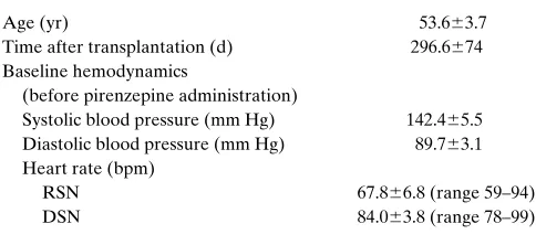
Five orthotopic cardiac transplant (HTX) patients were studied during routine surveillance follow-up on the occasion of routine endo-myocardial biopsy. The study protocol was approved by the Ethical Committee of the University of Halle-Wittenberg, and written in-formed consent was obtained from all patients. Patients were included in the study if they showed no evidence of acute infection or clinical signs of acute rejection. Patients were in a stable maintenance immuno-suppressed state (the standard regimen at our institution consists of cy-closporine A and azathioprine, exclusively), and showed no evidence of graft atherosclerosis proven by coronary angiography within the pre-vious 6 mo, or evidence of rejection at the time of investigation proven by histology of the biopsies obtained 30 min after the end of the infu-sion study. All patients showed histology grades of A0–A2 IUC.
The average cyclosporine A trough level was 229.2639.0 ng/ml. The clinical data for the patients at the time of study are given in Table I. In addition to cyclosporine A and azathioprine, patients were treated with angiotensin-converting enzyme inhibitors (n5 2), di-uretics (n5 3), nitrates (n5 5), acetyl salicylic acid (n5 5), and pred-nisolone (n5 1), alone or in combination; none of the patients had received catecholamines and/or cholinergic drugs for at least 6 wk be-fore the study.
Study protocol. After physical examination, a conventional
bi-opsy sheath was placed into the right internal jugular vein. The pa-tients were prepared for ECG and blood pressure monitoring, and rested for 30 min in supine position. Thereafter, an intraatrial multi-ple bipolar probe was placed such that recipient sinus node (RSN) and donor sinus node (DSN) rates could be recorded simultaneously. Pirenzepine was injected over 5 min in increasing doses of 0.05, 0.10, 0.25, 0.50, 1.0, and 10 mg bolus, with every dose step requiring 20 min. Systolic and diastolic blood pressure (phase V) were mea-sured with the Dinamap automatic device. Heart rate and surface ECG as well as peripheral capillary oxygen saturation were recorded continuously. The RSN (remaining sinus node of the recipient) and DSN (actual transplanted heart) were measured via the intracardiac electrode catheter (Bard Electrophysiology, Bellerica, MA) and re-corded by a 12-channel recorder at a paper speed of 50 mm/s in the last min of each dose step. Endomyocardial biopsies were taken 30 min after the end of the infusion using a commercial right ventricular bioptome (9 French, 2.8-mm head diameter; Scholten Surgical Instru-ments, Redwood City, CA).
In vitro studies
Right atrial appendages were obtained from 14 children (8 female, 6 male) with acyanotic congenital heart disease who underwent open heart surgery because of ventricular septal defect (n5 5), atrioven-tricular septal defect (n5 3), atrial septal defect (n5 3), or aortic stenosis (n5 3). Their parents had given informed written consent. None of the children suffered from acute heart failure or had been treated with sympathomimetic (i.e., catecholamines) or cholinergic drugs for at least 3 wk before surgery.
Anesthesiological premedication usually consisted of 1 mg/kg i.v. diazepam given immediately before surgery in infants with a body weight , 10 kg, 0.3 mg/kg rectally in children with a body weight of 10–25 kg, and 0.4 mg/kg p.o. in children with a body weight . 25 kg. The operation was carried out under balanced anesthesia with mida-zolam and isoflurane (up to 1.5 vol percent to effects). N2O was avoided in all cases. Fentanyl was added for analgesia. Controlled ventilation was performed with an inspired oxygen fraction of 50– 100%. Right atrial appendages were removed immediately after in-stallation of the cardiopulmonary bypass.
[image:3.612.314.556.73.184.2]Right atrial appendages were also obtained from 25 adult patients (18 male, 7 female) undergoing elective coronary artery bypass graft without apparent heart failure (New York Heart Association [NYHA] class I–II, n5 14) or undergoing open heart surgery be-cause of aortic valve disease (n5 9, NYHA class I–II except one pa-tient class II–III) or mitral valve insufficiency (n5 2, NYHA class III), having given informed written consent. None of these patients had been treated with b-adrenoceptor agonists or cholinergic drugs for at least 6 wk before the operation. However, patients had re-ceived nitrates (n5 17), b-adrenoceptor antagonists (n5 9), calcium antagonists (n5 6), angiotensin-converting enzyme inhibitors (n5 10), diuretics (n5 7), digitalis glycosides (n5 4), heparin (n5 13), 3-hydroxy-3-methylglutaryl CoA reductase inhibitors (n5 8), acetyl salicylic acid (n5 5), and antibiotics (n5 4), alone or in combination.
Table I. Clinical Characteristics of the Five HTX Recipients
Age (yr) 53.663.7
Time after transplantation (d) 296.6674 Baseline hemodynamics
(before pirenzepine administration)
Systolic blood pressure (mm Hg) 142.465.5 Diastolic blood pressure (mm Hg) 89.763.1 Heart rate (bpm)
RSN 67.866.8 (range 59–94)
DSN 84.063.8 (range 78–99)
Premedication usually consisted of 0.5 mg lorazepam given orally the evening before and 0.5–1.0 mg given orally the morning of sur-gery. The operation was carried out under balanced anesthesia con-sisting of midazolam, fentanyl, and pancuronium bromide as muscle relaxant, as well as isoflurane (0.6–1.0% vol/vol) for narcosis; N2O was avoided in all cases. Controlled ventilation was performed with an inspired oxygen fraction of 50–100%. In all patients, right atrial appendages were removed during installation of the cardiopulmo-nary bypass. Immediately after excision, all specimens were quickly frozen in liquid nitrogen.
Patients were arbitrarily divided into three age-groups: children (group A, , 20 yr), adults (group B, 20–50 yr), and old subjects (group C, . 50 yr). The mean ages of the three groups were as fol-lows: group A, 7.361.5 yr (range 5 d–15 yr), n 5 14; group B, 32.662.5 yr (range 22–43 yr), n5 9; and group C, 66.861.8 yr (range 55–76 yr), n5 16.
Radioligand binding studies
For radioligand binding studies, tissues were minced with scissors and homogenized in 10 vol of ice-cold 1 mM KHCO3 with an Ultra Tur-rax (Janke & Kunkel, Staufen, Germany) twice for 20 s at full speed in 1-min intervals. The homogenate was diluted with 1 mM KHCO3 to 50 ml, and centrifuged at 700 g for 15 min; the supernatant was passed through four layers of cheesecloth and centrifuged at 21,000 g for 45 min; the pellets were washed once by resuspension and recen-trifugation. The final pellets were resuspended in incubation buffer (10 mM Na2HPO4, 10 mM NaH2PO4, pH 7.4) to yield a protein con-centration of 0.6–0.7 mg/ml. Protein content was determined by the method of Bradford (13) using bovine IgG as the standard.
[3H]NMS and all drugs used in these experiments were prepared in 10 mM Na2HPO4, 10 mM NaH2PO4 buffer, pH 7.4. If not stated otherwise, an aliquot of the membrane suspension (150 ml) was incu-bated with [3H]NMS in a final volume of 250 ml. Incubations were carried out for 60 min at 258C and terminated by adding 10 ml of washing buffer to the entire incubation mixture, followed by rapid fil-tration over Whatman GF/C glass fiber filters that had been soaked previously in 1 mM unlabeled NMS.
Each filter was washed with an additional 10 ml of washing buffer. The filters were then dried and transferred to vials containing 5 ml of Lumasafe plus scintillator (Lumac-LSC B.V., Groningen, The Netherlands), and radioactivity was determined in a scintillation counter (LS 6000; Beckman Instruments, Inc., Fullerton, CA) at an efficiency of 42%. Nonspecific binding of [3H]NMS was defined as ra-dioactivity bound to membranes that was not displaced by a high con-centration (1 mM) of atropine. Specific binding of [3H]NMS was de-fined as total binding minus nonspecific binding, and was 80–90% (at 0.5–3 nM) and 70% (at 10 nM) of [3H]NMS.
For determination of the density of muscarinic receptors in mem-branes from right atrial tissues, the amount of specifically bound [3H]NMS was determined at six different concentrations ranging from 0.1 to 10 nM.
Adenylyl cyclase determination
Adenylyl cyclase activity was assessed as described by Salomon et al. (14) with minor modifications as detailed elsewhere (15). Membranes (35–45 mg of protein) were incubated for 10 min at 308C in a final vol-ume of 100 ml containing 40 mM Hepes buffer, pH 7.4, 5 mM MgCl2, 1 mM EDTA, 10 mM GTP, 500 mM ATP, z 1,000,000 cpm [a-32 P]-ATP, 100 mM cAMP, and an ATP regenerating system (5 mM phos-phocreatine and 50 U/ml creatine phosphokinase) in the presence or absence of isoprenaline (10 mM), forskolin (10 mM), and various con-centrations of carbachol (10 nM–100 mM). The reaction was stopped by addition of 100 ml buffer containing 50 mM Tris, 40 mM ATP, 1.4 mM cAMP, 2% SDS, and [3H]cAMP (z 10,000 cpm) at pH 7.5; 800
ml water was then added.
The mixture was poured into Dowex AG 50W-X4 anion-exchange columns (200–400 mesh, hydrogen form), and ATP was eluted twice with 1 ml water. The Dowex columns were then placed over neutral
alumina columns, and cAMP was eluted from the Dowex columns with 4 ml water. The alumina columns were placed over scintillation vials, and the cAMP was eluted from the alumina columns with 5 ml 0.1 M imidazole (pH 7.3). Lumasafe plus scintillator (15 ml) was added to the eluate and counted at 42% efficiency. The determined amount of [3H]cAMP in each vial was used to calculate the recovery of cAMP for each column, and the amount of [32P]cAMP collected from each column was corrected for the recovery rate (usually 70– 80%).
Statistical evaluations
Data given are mean6SEM of n experiments. The equilibrium disso-ciation constant (KD) and maximal number of binding sites (Bmax) for [3H]NMS were calculated from plots according to Scatchard (16). For calculation of EC50 values of carbachol-induced adenylyl cyclase inhi-bition, data were fitted to sigmoid curves; in these calculations, the bottom of the curve was fixed at 0% inhibition, and inhibition of ade-nylyl cyclase by 100 mM carbachol was considered maximal inhibi-tion; the Hill slopes were kept variable. From these curves, EC50 val-ues were obtained that were not considerably different (maximal difference a factor of 2) from those calculated with a Hill slope fixed at 1.0. Experimental data were analyzed by computer-supported iter-ative nonlinear regression analysis using the InPlot program (Graph-PAD Software for Science, San Diego, CA).
Linear regression analysis of the data was performed by the least squares method (model:[3H]NMS binding sites 5 a 3 age 1 b). Sta-tistical significance of differences was analyzed by one-way ANOVA followed by Bonferroni testing for multiple comparisons and by re-peated measures analysis (17), or, if appropriate, by nonpaired, two-tailed Student’s t test (for specification, see citations to the figures and Table II). A P value , 0.05 was considered significant. Statistical calculations were performed with the Instat program (GraphPAD Software for Science) or with the SPSS Advanced Statistics program, version 7.5 (SPSS Inc., Chicago, IL).
Drugs used
[image:4.612.316.557.93.200.2]For infusion, atropine (Atropin-sulfat solution) was purchased from Fresenius AG (Bad Homburg, Germany), isoprenaline (Aleudrina) from Boehringer Ingelheim (Ingelheim, Germany), and pirenzepine (Gastrozepin) from Thomae (Biberach an der Riss, Germany). Atro-pine sulfate, (2)-isoprenaline bitartrate, carbachol chloride, and for-skolin were obtained from Sigma-Aldrich Chemie GmbH (Deisen-hofen, Germany), N-methyl-scopolamine nitrate from Merck KGaA (Darmstadt, Germany), [3H]NMS (specific activity 85 Ci/mmole), [a-32P]ATP (specific activity 30 Ci/mmole), and [3H]cAMP (specific
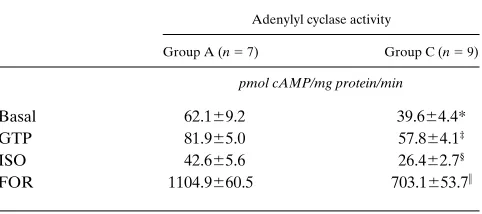
Table II. Adenylyl Cyclase Activity in Right Atrial Membranes from Two Groups of Patients without Apparent Heart Failure Undergoing Open Heart Surgery
Adenylyl cyclase activity
Group A (n 5 7) Group C (n 5 9)
pmol cAMP/mg protein/min
Basal 62.169.2 39.664.4*
GTP 81.965.0 57.864.1‡
ISO 42.665.6 26.462.7§
FOR 1104.9660.5 703.1653.7i
*P 5 0.0324, ‡P 5 0.0021, §P 5 0.0148, and iP , 0.0003 vs. the
activity 44.5 Ci/mmole) from New England Nuclear (Dreieich, Ger-many). All other chemicals were of the purest commercially available grade.
Results
In vitro studies
Right atrial muscarinic receptor density. The mean right atrial
muscarinic receptor density decreased across the three groups,
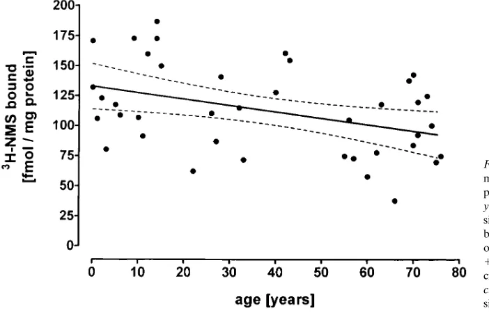
from group A (patients , 20 yr old) via group B (patients 20–50 yr old) to group C (patients . 50 yr old; Fig. 1); the dif-ference between groups A and C was highly significant. More-over, for all 39 patients there was a significant negative corre-lation between right atrial muscarinic receptor density and the age of the patients (Fig. 2). KD values for [3H]NMS were not significantly different in the three groups (see citation to Fig. 1).
Right atrial adenylyl cyclase activity. Because of limited
amount of tissue, we could assess right atrial adenylyl cyclase activity in only seven patients from group A (mean age 10.461.8 yr) and nine patients from group C (mean age 67.362.7 yr). In agreement with our recently published data (15), basal, 10 mM GTP-, 10 mM isoprenaline-, and 10 mM for-skolin-stimulated adenylyl cyclase activity was significantly higher in group A than in group C (Table II).
The muscarinic receptor agonist carbachol (10 nM–100
mM) inhibited 10 mM forskolin-stimulated adenylyl cyclase ac-tivity in a concentration-dependent manner (Fig. 3). This in-hibitory effect was antagonized by the muscarinic receptor an-tagonist atropine with a pKi value of 8.42 (data not shown), indicating that it is mediated by muscarinic receptor stimulation. However, inhibition of 10 mM forskolin-stimulated ade-nylyl cyclase activity was significantly more pronounced at each concentration of carbachol in group A than in group C (Fig. 3). In addition, the EC50 value for carbachol was signifi-cantly lower in group A (0.7660.22 mM) than in group C (7.262.1 mM, P , 0.05).
In vivo studies
Pirenzepine effects in young and older volunteers. As
men-tioned in the Introduction, we have shown recently that piren-zepine in doses of 0.04–1.25 mg decreases basal heart rate in healthy volunteers; maximum effects were obtained at doses of 0.32 and 0.64 mg pirenzepine (10). Therefore, we used these two doses to study whether pirenzepine might also decrease isoprenaline-induced increases in heart rate, and whether this effect is also diminished in older volunteers.
To assess the dose of isoprenaline that increases heart rate by z 20 bpm, we first performed dose–response curves for
[image:5.612.58.261.58.267.2]iso-Figure 1. Muscarinic receptor density in right atria from three groups of patients undergoing open heart surgery: group A (, 20 yr old, white bars), group B (20–50 yr old, striated bars), and group C (. 50 yr old, black bars) (see Methods). Right atrial muscarinic receptor density is expressed in femtomoles of [3H]NMS specifically bound per milligram of protein (Mean6SEM). KD values for [3H]NMS were 1.4360.26 nM (n 5 14) for group A, 1.3560.27 nM (n 5 9) for group B, and 1.4360.34 nM (n 5 16) for group C. **P , 0.01 vs. group A (one-way ANOVA followed by the Bonferroni test for multiple com-parisons).
Figure 2. Correlation between right atrial muscarinic receptor density and age of 39 patients undergoing open heart surgery. y axis, right atrial muscarinic receptor den-sity in femtomoles of [3H]NMS specifically bound per milligram of protein; x axis, age of the patients in years. y 520.531x
[image:5.612.57.408.516.741.2]prenaline-induced increases in heart rate in six young and six older volunteers. As shown in Fig. 4, the dose–response curves for isoprenaline-induced increases in heart rate were very sim-ilar in both groups; however, when these experiments were re-peated in the presence of atropine, thereby inhibiting vagal tone, isoprenaline-induced increases in heart rate were en-hanced significantly in the young volunteers, but were affected only marginally and not significantly in the older volunteers. Moreover, atropine increased resting heart rate to a signifi-cantly greater extent in the young volunteers (1 36.964.3 bpm) than in the older volunteers (1 18.864.0 bpm, P , 0.02). Based on these experiments, we choose a dose of 17 ng/kg/min isoprenaline for the pirenzepine experiments.
At this dose, isoprenaline increased heart rate by 20.761.4 bpm in the young and 24.064.1 bpm in the older volunteers. Heart rate remained elevated throughout the 1-h infusion. When pirenzepine was added 20 min after the start of isopren-aline infusion, heart rate declined rapidly; however, at both doses of pirenzepine, the decrease in heart rate was markedly more pronounced in the young than in the older volunteers (Fig. 5). Thus, at the 0.64 mg dose of pirenzepine, heart rate in young volunteers returned almost to baseline levels (despite the continuous infusion of isoprenaline), whereas heart rate in the older volunteers was still increased by 13–14 bpm (Fig. 5). On the other hand, pirenzepine in neither dose significantly af-fected isoprenaline-induced changes in blood pressure and STI (data not shown).
Studies in heart transplant recipients. For this study,
[image:6.612.56.295.61.381.2]piren-zepine was given in doses of 0.05–10 mg. Application of the in-tracavitary multipolar probe allowed assessment of sinus node
[image:6.612.57.402.499.733.2]Figure 4. Effects of atropine (15 mg/kg/ body as bolus, followed by i.v. infusion of 0.15 mg/kg/min until the end of the isopren-aline infusion) on isoprenisopren-aline infusion– induced heart rate increases in six older (left, filled symbols) and six young volun-teers (right, open symbols) (see Methods). y axis, heart rate increases (D Beats/min); x axis, dose of isoprenaline in nanograms per kilogram per minute for 10 min each. Mean6SEM of six experiments each. Baseline heart rate in young volunteers was 63.763.4 bpm before and 97.364.3 bpm after 30 min of atropine infusion; in older volunteers, baseline heart rate was 61.562.9 bpm before and 82.468.7 bpm af-ter atropine infusion. **P , 0.01, *P , 0.05 vs. the corresponding values in the absence of atropine (one-way ANOVA followed by the Bonferroni test for multiple compari-sons). When the data were analyzed by re-peated measures analysis (17), isoprenaline dose–response curve in the presence of at-ropine (squares) was significantly different from control (circles) in young (P 5 0.002) but not in older volunteers (P 5 0.312). Figure 3. Inhibition of 10 mM forskolin-stimulated adenylyl cyclase
activity by carbachol in right atria from seven patients of group A (, 20 yr old, white bars) and nine patients of group C (. 50 yr old, black bars) (see Results). y axis, net decrease in 10 mM forskolin-stimulated right atrial adenylyl cyclase activity upon carbachol stimu-lation in picomoles of cAMP formed per milligram of protein per minute (mean6SEM); x axis, molar concentration of carbachol. For
rates in the native atrial cuffs as well as in the transplanted do-nor hearts. Pirenzepine in doses of 0.05–0.5 mg caused dose-dependent decreases in sinus node rate in the native atrial cuffs but not in the transplanted donor hearts (Fig. 6); the max-imal decrease at 0.5 mg was 4.961.1 bpm. At the 10-mg dose, pirenzepine increased heart rate significantly in the RSN, by 10.562.4 bpm, and again, did not affect heart rate significantly in the DSN (Fig. 6).
Discussion
In healthy volunteers, atropine and the M1-selective musca-rinic receptor antagonist pirenzepine cause biphasic effects on resting heart rate: in low (M1-selective) doses, they decrease heart rate, whereas in higher doses, they lead to the well-known increase in heart rate (18, 19). We have shown recently that in healthy volunteers, the heart rate–decreasing effects of low doses of atropine and pirenzepine were significantly greater in young than in older volunteers (10). The results of this study confirm and extend these findings: they show that pirenzepine in low doses can also inhibit isoprenaline-induced heart rate increases; again, this effect was markedly more pro-nounced in young than in older volunteers.
The mechanism of the heart rate–decreasing effects of low doses of pirenzepine is not completely understood. According to its affinity profile for M1, M2, M3, and M4 muscarinic recep-tors at the doses used in these experiments, pirenzepine should act selectively at M1 muscarinic receptors (20). Therefore, it has been suggested that the heart rate–decreasing effect of pirenzepine is due to inhibition of presynaptic M1 muscarinic autoreceptors; this leads to an increased release of endogenous acetylcholine (ACh) that decreases heart rate via stimulation
of postsynaptic M2 muscarinic receptors (10, 21). The existence of such presynaptic ACh autoreceptors modulating ACh re-lease has been demonstrated in chicken, rat, rabbit, and guinea pig atria but not yet in human atria: they are species-depen-dent either of the M1 (chicken [22, 23]) or the M2 subtype (guinea pig [22], rat [24], rabbit [25]).
[image:7.612.59.270.55.218.2]Studies in HTX patients support the idea of an involve-ment of presynaptic receptors modulating ACh release in the negative chronotropic effects of atropine and pirenzepine. Ep-stein et al. (26) have shown that in HTX patients, atropine de-creases heart rate only in the (innervated) native RSN and not in the (denervated) DSN, and almost identical results were ob-tained in our study in HTX patients: pirenzepine in low doses decreased heart rate only in the native RSN but not in the DSN. These results strongly indicate that pirenzepine (and at-ropine) can exert their negative chronotropic effects only when innervation is intact; therefore, it is quite likely that the negative chronotropic effects of low doses of pirenzepine are due to inhibition of presynaptic M1 muscarinic receptors, thereby releasing endogenous ACh, although the final experi-mental proof of the existence of such presynaptic M1 musca-rinic receptors in the human right atrium is still lacking. It should be noted that in HTX patients, sympathetic reinnerva-tion can occur long-term after transplantareinnerva-tion (27, 28), and
Figure 5. Effects of pirenzepine (0.32 and 0.64 mg i.v. bolus) on iso-prenaline infusion (17 ng/kg/min throughout)–induced increases in heart rate in six young (s) and six older volunteers (d). After 20 min of isoprenaline infusion, pirenzepine was added, and heart rate was assessed (see Methods). y axis, heart rate changes (D Beats/min); x axis, time in minutes. Mean6SEM of six experiments each. **P , 0.01, *P , 0.05 vs. the corresponding value after 20 min of isoprena-line infusion (one-way ANOVA followed by the Bonferroni test for multiple comparisons); a), P , 0.05 vs. the corresponding values for older volunteers (one-way ANOVA followed by the Bonferroni test for multiple comparisons). When the effects of pirenzepine on iso-prenaline-induced heart rate increases were analyzed by repeated measures analysis (17), the difference between young and older vol-unteers was just under statistical significance (P 5 0.065).
[image:7.612.316.515.353.610.2]very recently, preliminary evidence for vagal reinnervation has also been obtained (29). However, in this study, HTX patients were investigated who had undergone transplantation less than a year before (see Table I); therefore, it is very unlikely that any sympathetic or parasympathetic reinnervation oc-curred in these patients.
The negative chronotropic effect of pirenzepine on resting heart rate (10) as well as on isoprenaline-stimulated heart rate (this study) was markedly reduced in the elderly. Assuming that pirenzepine induces this negative chronotropic effect by inhibition of presynaptic M1 muscarinic receptors with subse-quent release of endogenous ACh, the age-dependent reduc-tion in this effect could be due either to a diminished release of ACh in the elderly or to a diminished response of the postsyn-aptic M2 muscarinic receptors to the released ACh. To address this question, in this study, we have measured the density of muscarinic receptors in right atria from patients of ages rang-ing from 5 d to 76 yr usrang-ing [3H]NMS binding studies. By this method, only M2 muscarinic receptors are identified in the hu-man heart (5, 6). There was a significant decline in receptor number from group A (patients , 20 yr old) via group B (pa-tients 20–50 yr old) to group C (pa(pa-tients . 50 yr old); in addi-tion, right atrial muscarinic receptor density was significantly negatively correlated with patient age. In this study, musca-rinic receptor density has been assessed in crude membrane preparations from right atria. It has been shown that the aging heart is associated with a loss of myocytes and with fibrosis, which might contribute considerably to the age-dependent de-crease in muscarinic receptors observed in this study. We can-not exclude this possibility, but it seems very unlikely, since several groups have demonstrated that in severely failing hu-man hearts that exhibit a greater loss of cardiomyocytes and fi-brosis than the aging human heart, muscarinic receptor density is unchanged compared with nonfailing human hearts (7, 30, 31). The age-dependent decrease in muscarinic receptor den-sity was accompanied by an age-dependent decline in car-bachol-induced inhibition of forskolin-stimulated adenylyl cyclase mediated via M2 muscarinic receptor stimulation; moreover, affinity of carbachol was reduced significantly in ag-ing atrial tissues (Fig. 3). Taken together, these results indicate that an age-dependent decrease in right atrial M2 muscarinic receptor density and function might contribute considerably to the age-dependence of the negative chronotropic effect of pirenzepine. Whether the decrease in receptor density or the decrease in agonist affinity is more important cannot be deter-mined from this study; however, the fact that correlation be-tween age and receptor density was weak and that many points were outside the 95% confidence limit might favor the idea that decreased agonist affinity is more important for the age-dependent decrease in M2 receptor function. In addition, since we cannot measure ACh in the intact human heart, we cannot exclude completely the possibility that a diminished release of ACh in the elderly might also contribute to this effect.
It is interesting to note that in this study, the decrease in isoprenaline-increased heart rate induced by pirenzepine was greater in both young and older volunteers than we had re-cently observed for resting heart rate (Fig. 5, and reference 10). This is obviously due to the very well-known phenome-non, “accentuated antagonism,” where the inhibitory effects of muscarinic receptor stimulation are enhanced when sympa-thetic activity is increased (32). In contrast, in this study, iso-prenaline-induced shortening of STIs (as a measure of positive
inotropism) was not affected significantly by low dose piren-zepine, although it has been shown in vitro and in vivo that
b-adrenoceptor agonist–induced increases in left ventricular con-tractile force can be inhibited in a concentration-dependent manner by carbachol via M2 muscarinic receptor stimulation (6–9, 33, 34). On the other hand, in vivo and in vitro studies have shown that muscarinic stimulation does not affect basal contractile force of the left ventricular myocardium (6, 8, 9, 33), and muscarinic receptor blockade by atropine augmented only slightly b-adrenoceptor agonist–induced increases in left ventricular contractility (9, 34), if at all (8, 10). These weak parasympathomimetic effects are very likely due to the fact that human ventricular myocardium is only sparsely parasym-pathetically innervated (35, 36). Such a sparse parasympathetic innervation might also explain the findings of this study that low dose pirenzepine failed to antagonize isoprenaline-induced increases in ventricular contractility: it might well be that the concentration of ACh released under these conditions is too low to lead to a considerable stimulation of the M2 muscarinic receptor. Another possibility is that in ventricular myocar-dium, in contrast to the atria, presynaptic M1 muscarinic recep-tors modulating ACh release may not exist. Finally, the fact that as in rat, rabbit (37), and chick hearts (38), in human ven-tricular myocardium the number of M2 muscarinic receptors is less than in atrial tissue (6), might also contribute to the lack of the negative inotropic effect of low dose pirenzepine.
It is generally accepted that in the human heart, b -adreno-ceptor function declines with aging (for reviews, see references 2, 3, and 39). The mechanism underlying this effect is not com-pletely understood, but it might be due to an age-dependent decrease in the catalytic unit of adenylyl cyclase (right atrium [15]) or decrease in b-adrenoceptor number (left ventricle [40]). However, in this study, isoprenaline infusion induced the same increase in heart rate in young as it did in older volun-teers. It has been shown that during isoprenaline infusion, va-gal tone increases, thus damping isoprenaline effects (41). As discussed above, human right atrial M2 muscarinic receptor function declines with age. Thus, the depressing effects of in-creases in vagal tone on isoprenaline infusion–induced heart rate increases are lower in older than in young volunteers; this obviously can mask the age-dependent decrease in cardiac
b-adrenoceptor–mediated effects. In fact, after blockade of va-gal activity by atropine, isoprenaline showed significantly greater increases in heart rate in the young than in the older volunteers (Fig. 4), thus supporting our recent findings that right atrial b-adrenoceptor function is decreased in the elderly (15). These results are in good agreement with recently pub-lished data by White and Leenen (42), who also found a de-creased chronotropic response to isoprenaline in older vol-unteers only after ganglionic blockade by trimethaphan; moreover, these authors demonstrated that an age-dependent decrease in positive inotropic responses to isoprenaline could also be obtained only after trimethaphan treatment. And fi-nally, we have found recently that only in the presence of atro-pine could differences in the heart rate response to noradrena-line and tyramine infusion be demonstrated between young and older volunteers (12).
decreases heart rate via activation of postsynaptic M2 musca-rinic receptors. The heart rate–decreasing effect of endoge-nous ACh is diminished with age, because M2 muscarinic re-ceptor density and function decline. Such an age-dependent decrease in M2 muscarinic receptor density may well lead to a decrease in cardiac parasympathetic activity, as suggested pre-viously (43), and might contribute significantly to the well-known decrease in baroreflex activity with aging (2–4).
Acknowledgments
The skillful technical assistance of Mrs. Pia Matthes is gratefully ac-knowledged. The authors are thankful to Dr. Johannes Haerting, In-stitute of Medical Epidemiology, Biometry, and Medical Informatics (University of Halle-Wittenberg), for his help with statistical analysis of the data.
This work was supported by grants from the Deutsche Forsch-ungsgemeinschaft (DFG Br 526/3-3 to O.-E. Brodde, and DFG Ze 218/3-3 to H.-R. Zerkowski).
References
1. Roth, G.S. 1979. Hormone receptor changes during adulthood and senes-cence: significance for aging research. Fed. Proc. 38:1910–1914.
2. Docherty, J.R. 1990. Cardiovascular responses in aging: a review.
Phar-macol. Rev. 42:103–125.
3. Lakatta, E.G. 1993. Cardiovascular regulatory mechanisms in advanced age. Physiol. Rev. 73:413–467.
4. Folkow, B., and A. Svanborg. 1993. Physiology of cardiovascular aging.
Physiol. Rev. 73:725–764.
5. Giraldo, E., F. Martos, A. Gomez, A. Garcia, M.A. Vigano, H. Ladinsky, and F. Sanchez de la Cuesta. 1988. Characterization of muscarinic receptor sub-types in human tissues. Life Sci. 43:1507–1515.
6. Deighton, N.M., S. Motomura, D. Borquez, H.-R. Zerkowski, N. Doetsch, and O.-E. Brodde. 1990. Muscarinic cholinoceptors in the human heart: demon-stration, subclassification, and distribution. Naunyn-Schmiedeberg’s Arch.
Phar-macol. 341:414–421.
7. Böhm, M., P. Gierschik, K.H. Jakobs, B. Piesk, P. Schnabel, M. Ungerer, and E. Erdmann. 1990. Increase of Gi in human hearts with dilated but not
ischemic cardiomyopathy. Circulation. 82:1249–1265.
8. Von Scheidt, W., M. Böhm, A. Stäblein, G. Autenrieth, and E. Erdmann. 1992. Antiadrenergic effect of M-cholinoceptor stimulation on human ventricu-lar contractility in vivo. Am. J. Physiol. 263:H1927–H1931.
9. Landzberg, J.S., J.D. Parker, D.F. Gauthier, and W.S. Colucci. 1994. Ef-fect of intracoronary acetylcholine and atropine on basal and dobutamine-stim-ulated left ventricular contractility. Circulation. 89:164–168.
10. Poller, U., G. Nedelka, J. Radke, K. Pönicke, and O.-E. Brodde. 1997. Age-dependent changes in cardiac muscarinic receptor function in healthy vol-unteers. J. Am. Coll. Cardiol. 29:187–193.
11. Schäfers, R.F., S. Adler, A. Daul, G. Zeitler, M. Vogelsang, H.-R. Zerkowski, and O.-E. Brodde. 1994. Positive inotropic effects of the beta2
-adrenoceptor agonist terbutaline in the human heart: effects of long-term beta1
-adrenoceptor antagonist treatment. J. Am. Coll. Cardiol. 23:1224–1233. 12. Poller, U., R.F. Schäfers, S. Schmuck, J. Jakubetz, J. Radke, A.E. Daul, K. Pönicke, and O.-E. Brodde. 1997. Influence of atropine on the cardiovascu-lar effects of noradrenaline and tyramine in elder volunteers.
Naunyn-Schmiedeberg’s Arch. Pharmacol. 356:100–106.
13. Bradford, M.M. 1976. A rapid and sensitive method for the quantitation of microgram quantities of protein utilizing the principle of protein-dye bind-ing. Anal. Biochem. 72:248–254.
14. Salomon, Y., C. Londos, and M. Rodbell. 1974. A highly sensitive ade-nylate cyclase assay. Anal. Biochem. 58:541–548.
15. Brodde, O.-E., H.-R. Zerkowski, D. Schranz, A. Broede-Sitz, M. Michel-Reher, E. Schäfer-Beisenbusch, J.A. Piotrowski, and H. Oelert. 1995. Age-dependent changes of the b-adrenoceptor-G-protein(s)-adenylyl cyclase system in human right atrium. J. Cardiovasc. Pharmacol. 26:20–26.
16. Scatchard, G. 1949. The attraction of proteins for small molecules and ions. Ann. NY Acad. Sci. 51:660–672.
17. Winer, B.J., D.R. Brown, and K.M. Michels. 1991. Statistical Principles in Experimental Design. McGraw-Hill Inc., New York.907 pp.
18. Wellstein, A., and H.F. Pitschner. 1988. Complex dose-response curves of atropine in man explained by different functions of M1- and M2
-cholinocep-tors. Naunyn-Schmiedeberg’s Arch. Pharmacol. 338:19–27.
19. Pitschner, H.F., and A. Wellstein. 1988. Dose-response curves of piren-zepine in man in relation to M1- and M2-cholinoceptor occupancy.
Naunyn-Schmiedeberg’s Arch. Pharmacol. 338:207–210.
20. Eglen, R.M., and N. Watson. 1996. Selective muscarinic receptor ago-nists and antagoago-nists. Pharmacol. Toxicol. 78:59–68.
21. Pitschner, H.F., M. Schlepper, B. Schulte, C. Volz, D. Palm, and A. Wellstein. 1989. Selective antagonists reveal different functions of M choli-noceptor subtypes in humans. Trends Pharmacol. Sci. 10(Suppl.):92–96.
22. Jeck, D., R. Lindmar, K. Löffelholz, and M. Wanke. 1988. Subtypes of muscarinic receptor on cholinergic nerves and atrial cells of chicken and guinea-pig hearts. Br. J. Pharmacol. 93:357–366.
23. Brehm, G., R. Lindmar, and K. Löffelholz. 1992. Inhibitory and excita-tory muscarinic receptors modulating the release of acetylcholine from the postganglionic parasympathetic neuron of the chicken heart.
Naunyn-Schmiedeberg’s Arch. Pharmacol. 346:375–382.
24. Bognar, I.T., B. Beinhauer, P. Kann, and H. Fuder. 1990. Different mus-carinic receptors mediate autoinhibition of acetylcholine release and vagally-induced vasoconstriction in the rat isolated perfused heart.
Naunyn-Schmiede-berg’s Arch. Pharmacol. 341:279–287.
25. Habermeier-Muth, A., U. Altes, K.M. Forsyth, and E. Muscholl. 1990. A presynaptic excitatory M1 muscarine receptor at postganglionic cardiac
nor-adrenergic nerve fibres that is activated by endogenous acetylcholine.
Naunyn-Schmiedeberg’s Arch. Pharmacol. 342:483–489.
26. Epstein, A.E., B.I. Hirschowitz, J.K. Kirklin, K.A. Kirk, G.N. Kay, and V.J. Plumb. 1990. Evidence for a central site of action to explain the negative chronotropic effect of atropine: studies on the human transplanted heart. J.
Am. Coll. Cardiol. 15:1610–1617.
27. Wilson, R.F., B.V. Christensen, and A. Simon. 1991. Evidence for struc-tural sympathetic reinnervation after human orthotopic cardiac transplantation.
Circulation. 83:1210–1220.
28. Koglin, J., T. Gross, P. Überfuhr, and W. Von Scheidt. 1997. Time-dependent decrease of presynaptic inotropic supersensitivity: physiological evi-dence of sympathetic reinnervation after heart transplantation. J. Heart Lung
Transplant. 16:621–628.
29. Frey, A.W., M. Dambacher, H. Achakri, M. Schwaiger, P. Überfuhr, G. Spadacini, L. Bernardi, and H. Roskamm. 1997. First evidence of parallel vagal and sympathetic reinnervation long term after heart transplantation. J. Heart
Lung Transplant. 16:88. (Abstr.)
30. Brodde, O.-E., S. Hillemann, K. Kunde, M. Vogelsang, and H.-R. Zerkowski. 1992. Receptor systems affecting force of contraction in the human heart and their alterations in chronic heart failure. J. Heart Lung Transplant. 11: S164–S174.
31. Bristow, M.R. 1993. Changes in myocardial and vascular receptors in heart failure. J. Am. Coll. Cardiol. 22(Suppl. 4):61A–71A.
32. Levy, M.N. 1971. Sympathetic-parasympathetic interactions in the heart. Circ. Res. 29:437–445.
33. Koglin, J., M. Böhm, W. Von Scheidt, A. Stäblein, and E. Erdmann. 1994. Antiadrenergic effect of carbachol but not of adenosine on contractility in the intact human ventricle in vivo. J. Am. Coll. Cardiol. 23:678–683.
34. Newton, G.E., A.B. Parker, J.S. Landzberg, W.S. Colucci, and J.D. Parker. 1996. Muscarinic receptor modulation of basal and b-adrenergic stimu-lated function of the failing human left ventricle. J. Clin. Invest. 98:2756–2763.
35. Kent, K.M., S.E. Epstein, T. Cooper, and D.M. Jacobowitz. 1974. Cho-linergic innervation of the canine and human ventricular conducting system. Anatomic and electrophysiologic correlations. Circulation. 50:948–955.
36. Löffelholz, K., and A.J. Pappano. 1985. The parasympathetic neuroef-fector junction of the heart. Pharmacol. Rev. 37:1–24.
37. Fields, J.Z., W.R. Roeske, E. Morkin, and H.I. Yamamura. 1978. Car-diac muscarinic cholinergic receptors, biochemical identification and character-ization. J. Biol. Chem. 253:3251–3258.
38. Sorota, S., L.P. Adam, and A.J. Pappano. 1986. Comparison of muscar-inic receptor properties in hatched chick heart atrium and ventricle. J.
Pharma-col. Exp. Ther. 236:602–609.
39. Feldman, R.D. 1986. Physiological and molecular correlates of age-related changes in the human b-adrenergic receptor system. Fed. Proc. 45:48– 50.
40. White, M., R. Roden, W. Minobe, M.F. Khan, P. Larrabee, M. Wollmer-ing, D. Port, F. Anderson, D. Campbell, A.M. Feldman, and M.R. Bristow. 1994. Age-related changes in b-adrenergic neuroeffector systems in the human heart. Circulation.. 90:1225–1238.
41. Arnold, J.M.O., and D.G. McDevitt. 1984. Vagal activity is increased during intravenous isoprenaline infusion in man. Br. J. Clin. Pharmacol. 18: 311–316.
42. White, M., and F.H.H. Leenen. 1994. Aging and cardiovascular respon-siveness to b-agonist in humans: role of changes in b-receptor responses versus baroreflex activity. Clin. Pharmacol. Ther. 56:543–553.