RESEARCH ARTICLE
The microRNA
bantam
regulates a developmental transition in
epithelial cells that restricts sensory dendrite growth
Nan Jiang1, Peter Soba2, Edward Parker3, Charles C. Kim4and Jay Z. Parrish1,*ABSTRACT
As animals grow, many early born structures grow by cell expansion rather than cell addition; thus growth of distinct structures must be coordinated to maintain proportionality. This phenomenon is particularly widespread in the nervous system, with dendrite arbors of many neurons expanding in concert with their substrate to sustain connectivity and maintain receptive field coverage as animals grow. After rapidly growing to establish body wall coverage, dendrites of Drosophila class IV dendrite arborization (C4da) neurons grow synchronously with their substrate, the body wall epithelium, providing a system to study how proportionality is maintained during animal growth. Here, we show that the microRNA bantam (ban) ensures coordinated growth of C4da dendrites and the epithelium through regulation of epithelial endoreplication, a modified cell cycle that entails genome amplification without cell division. InDrosophila larvae, epithelial endoreplication leads to progressive changes in dendrite-extracellular matrix (ECM) and dendrite-epithelium contacts, coupling dendrite/substrate expansion and restricting dendrite growth beyond established boundaries. Moreover, changes in epithelial expression of cell adhesion molecules, including the beta-integrin myospheroid(mys), accompany this developmental transition. Finally, endoreplication and the accompanying changes in epithelial mys expression are required to constrain late-stage dendrite growth and structural plasticity. Hence, modulating epithelium-ECM attachment probably influences substrate permissivity for dendrite growth and contributes to the dendrite-substrate coupling that ensures proportional expansion of the two cell types.
KEY WORDS: Dendrite, Endoreplication, Extracellular matrix, Growth control, Plasticity,Drosophila
INTRODUCTION
A central question in growth control of multicellular organisms is how growing organisms maintain proportionality. This problem is particularly complex when different types of interacting cells must grow in a coordinated fashion, a scenario that is widespread in the nervous system. For example, as animals grow, dendrite arbors of many neurons expand proportionally to sustain proper connectivity and maintain receptive field coverage (Bentley and Toroian-Raymond, 1981; Bloomfield and Hitchcock, 1991; Hitchcock, 1987; Parrish et al., 2009; Truman and Reiss, 1988). This scalar expansion of dendrite arbors to accommodate growth is widely
documented in sensory systems, including many types of invertebrate sensory neurons and vertebrate retinal ganglion cells (RGCs). In many cases, dendrites outpace substrate growth to establish appropriate coverage, and subsequently scale with substrate expansion to maintain coverage. Thus, neurons and their substrates differentially respond to common growth cues, receive distinct growth cues, or some combination of both.
Several observations suggest that neuron non-autonomous growth-inhibitory signals contribute to the fidelity of dendrite arbor expansion by restricting dendrite arbors to target fields. Following ablation of RGCs or starburst amacrine cells, the surviving cells developed regularly spaced dendrite arbors that exhibited a limited ability to expand into unoccupied territory (Farajian et al., 2004; Lin et al., 2004). Therefore, interactions between neighboring dendrites are largely dispensable for maintenance of coverage in these neurons and unknown constraints limit their growth potential. Supporting the argument in favor of an extrinsic component, these dendrite arbors expand in sync with retinal growth, whereas exuberant growth is limited.
Drosophila peripheral nervous system (PNS) class IV dendrite arborization (C4da) neurons completely and non-redundantly cover (‘tile’) the larval epidermis early in development and maintain this tiling by growing in precise synchrony with their substrate, the body wall epithelium (Emoto et al., 2006; Grueber et al., 2002; Parrish et al., 2007, 2009). Before establishment of tiling, ablating C4da neurons leads to dendrite growth into vacated territory by adjacent neurons (Grueber et al., 2003; Parrish et al., 2009; Sugimura et al., 2003). However, after tiling is established the invasive growth potential is lost; growth occurs only to maintain proportional receptive field coverage, showing that, as with RGCs, signals constrain late-stage growth of these dendrites. Notably, this signaling does not involve the homotypic repulsion required to establish tiling. Instead, epithelium-derived signals restrict exuberant arbor expansion; the miRNAbanacts in epithelial cells to regulate substrate-derived growth-inhibitory signals that constrain dendrite growth (Parrish et al., 2009).
Here, we report our characterization of theban-regulated epithelial signaling that regulates dendrite growth. We found thatbanregulates epithelial endoreplication, which is required for postembryonic body wall epithelial growth, and that manipulating epithelial endoreplication usingban-independent approaches recapitulatesban-mediated effects on dendrite growth. Endoreplication influences epithelium-dendrite and epithelium-extracellular matrix (ECM) interactions, providing the cellular basis for the reduced dendrite growth potential that accompanies larval development. At a molecular level, endoreplication alters epithelial expression of cell adhesion molecules, including the integrin Mys, which is required for proper coupling of dendrite and substrate expansion and to restrict dendritic structural plasticity. Thus, a developmental transition in epithelial growth constrains dendrite growth to ensure synchronous expansion of dendrites and their substrate.
Received 26 December 2013; Accepted 6 May 2014
1
Department of Biology, University of Washington, Seattle, WA 98195, USA.2Center for Molecular Neurobiology, Hamburg D-20251, Germany.3Department of Ophthalmology, University of Washington, School of Medicine, Seattle, WA 98195, USA.4Division of Experimental Medicine, Department of Medicine, University of California San Francisco, San Francisco, CA 94110, USA.
*Author for correspondence ( [email protected])
DEVEL
O
RESULTS
Identification ofban-regulated pathways
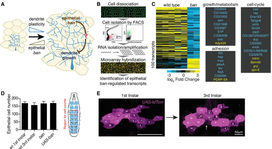
The microRNAbanfunctions as a regulatory switch for substrate-derived signaling that restricts PNS dendrite growth/plasticity and ensures proportional expansion of dendrite and substrate (Fig. 1A) (Parrish et al., 2009). To identify the substrate-derived factors required for proportional dendrite/substrate growth, we conducted microarray-based expression profiling of epithelial cells from wild-type andbanmutant larvae (Fig. 1B). We identified∼100 transcripts that were significantly deregulated inban mutant epithelial cells (Fig. 1C; supplementary material Table S1). Lexical analysis (Kim and Falkow, 2003) indicated that genes associated with the cell cycle were enriched in this dataset, suggesting thatbanregulates the cell cycle in epithelial cells; we also identified a large number of transcripts associated with cell growth and adhesion.
Drosophila increase their mass ∼200-fold during larval development, and this growth is accomplished by cell expansion rather than cell addition (Britton and Edgar, 1998; Church and Robertson, 1966). Indeed, we found that body wall epithelial cell number is constant from late embryogenesis to late larval stages, and that epithelial cells infrequently turn over, as epithelial cell clones persist through larval development >98% of the time (n=200; Fig. 1D,E). Body wall epithelial cell number is unresponsive toban activity (Fig. 1D); thus ban regulation of epithelial growth must involve growth of existing cells rather than proliferation. Although ban is known to regulate proliferation, and hence growth, in mitotically active tissues (Brennecke et al., 2003; Hipfner et al., 2002), howbanregulates growth of differentiated cells is unknown. To facilitate growth, many larval cell types undergo endoreplication, a modified cell cycle that entails DNA replication without cell division (Britton and Edgar, 1998; Smith and Orr-Weaver, 1991). Consistent with a change in endoreplication, ban
mutant epithelial cells exhibited dysregulation of cell-cycle-associated genes, including reduced expression of two regulators of endoreplication, double parked (dup) and retina aberrant in pattern (rap) (Park and Asano, 2008; Pimentel and Venkatesh, 2005; Sigrist and Lehner, 1997; Zielke et al., 2008), which encode orthologs of the DNA replication factor CDT1 and the APC/C activator CDH1/FZR1, respectively (Fig. 1C). We therefore hypothesized that ban regulates growth of mitotic (e.g. imaginal discs) and postmitotic body wall epithelial cells by regulating different forms of the cell cycle.
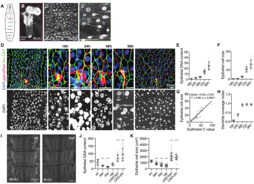
To monitor larval endoreplication during the period of ban activity required for dendrite growth, we fed first instar larvae BrdU for 1 day and monitored BrdU incorporation in third instar larvae. As a positive control we monitored BrdU incorporation in the ventral ganglion, which contains mitotically active neuroblasts and endoreplicating glia (Truman and Bate, 1988; Unhavaithaya and Orr-Weaver, 2012), and observed extensive labeling (Fig. 2A,B). Likewise, we observed extensive labeling of epithelia and muscle, but no labeling of sensory neurons, even when BrdU was constantly administered (Fig. 2C). We therefore conclude that larval body wall epithelia and muscle, but not sensory neurons, endoreplicate.
[image:2.612.82.531.416.662.2]To monitor the timing and extent of endoreplication in the larval epidermis we measured DNA content in epithelial cells over development. During embryogenesis, epithelial cells and PNS neurons had comparable levels of DAPI staining, and hence DNA content (Fig. 2D,E). Similar to other larval tissues (Britton and Edgar, 1998), body wall epithelia exhibited low levels of endoreplication in first instar larvae; DNA content in epithelial cells was 2.8-fold higher than in PNS neurons. Epithelial ploidy increased throughout larval development, and the rate of endoreplication increased dramatically at 48 h after egg laying (AEL), leading to a∼25-fold increase in genome content by 96 h AEL. Epithelial cell size and ploidy increased at
Fig. 1. Identification of substrate-derived regulators of dendrite growth.(A) Schematic depicting the role thatbanplays in regulating dendrite structural plasticity. (B) Workflow for microarray expression profiling. (C) Heat map depictingban-regulated epithelial transcripts. Transcripts involved in cell growth/ metabolism, adhesion, and the cell cycle are listed. (D) Body wall epithelial cell number counts in a region bounded on the anterior and posterior ends by segment borders and on the left and right by C4da neuron cell bodies (hatched rectangle) using DAPI to label nuclei and anti-Mys staining to demarcate cell boundaries. Counts were restricted to segments A2-A4;n≥10 segments for each genotype. (E) Time-lapse imaging of epithelial cell clones in first instar (left) and third instar larvae (right). In this example, all of the cells labeled in first instar (numbered) are present in the same relative position in third instar larvae. Occasionally,
additional cells are weakly labeled in third instar larvae (arrow); these cells were present in first instar but tdTomato was undetectable.
DEVEL
O
comparable rates, with the pace of epithelial growth dramatically increasing after 48 h AEL (Fig. 2F). Indeed, we observed a strong linear relationship between epithelial DNA content and cell size, suggesting that the two are tightly coupled (Fig. 2G). By contrast, C4da dendrites expanded most rapidly before 48 h AEL, when dendrites establish coverage of a fixed portion of the body wall (Fig. 2H) (Parrish et al., 2009).
banis required in epithelial cells to dampen dendrite growth beginning at ∼48 h AEL (Parrish et al., 2009), after dendrites tile the body wall and coincident with the rapid increase in epithelial endoreplication. We therefore assayed forbanfunction in endoreplication by monitoring BrdU incorporation in ban mutant larvae and found that BrdU incorporation was reduced by an average of 56% in ban mutant body wall epithelial cells compared with controls (Fig. 2I). Likewise, epithelial ploidy was significantly reduced inbanmutants, as indupmutants and larvae
with epithelium-specific expression of dup(RNAi) (Fig. 2J; supplementary material Figs S2 and S3); rap mutants exhibited reduced ploidy, but the change was not significant, perhaps owing to protein perdurance. By contrast, epithelial ploidy was significantly increased by overexpression of ban or diminutive (dm; encodes Drosophila Myc), which promotes endoreplication in a variety of Drosophilacell types (Pierce et al., 2004). Thus,banis necessary and sufficient to promote endoreplication in larval body wall epithelial cells, and the major wave of endoreplication is initiated in second instar larvae, corresponding to the time when ban functions in epithelial cells to coordinate dendrite/epithelial growth (Parrish et al., 2009).
[image:3.612.50.563.55.426.2]We next investigated effects ofbanactivity and endoreplication on epithelial growth. Previously, we found thatbanmutant body wall lysates had decreased levels of phosphorylated Akt ( p-Akt) (Parrish et al., 2009), which is often associated with cell growth. Likewise,
Fig. 2.banregulates a developmental growth transition in epithelial cells.(A-C) BrdU incorporation inDrosophilalarvae (A), the larval brain (B), and a single dorsal abdominal hemisegment of the larval body wall (C). (C0) Region containing dorsal PNS neurons; these neurons do not incorporate BrdU. (D) Embryos (18 h) or larvae carryingppk-tdTomato(C4da dendrites) andNrx-IV::GFP(epithelia) were injected with DAPI and imaged at the indicated time to allow simultaneous measurement of DNA content, cell size and dendrite coverage. Arrows, epithelial nuclei; carats, neuronal nuclei. (E) Scatter plot, epithelial cell size normalized to the mean epithelial size at 18 h AEL. (F) Scatter plot, epithelial DNA content over development.n≥50 cells for each time point in E and F. (G) Regression analysis of the relationship between epithelial cell size and DNA content. Ten measurements each from 18 h, 24 h, 36 h, 48 h, 72 h, and 96 h were used for the analysis. The pattern of residuals supports a linear model. (H) Scatter plot depicting dendrite coverage index, the proportion of the body wall covered by C4da dendrites (n≥8 dendrites at each time point). (I) BrdU incorporation in wild-type (wt) andbanmutant larvae. Wild-type andbanlarvae were processed in the same tube and imaged under identical conditions. (J,K) Scatter plots depict effects of endoreplication on (J) DNA content and (K) cell size in 120 h AEL larvae of the indicated genotype. For J,K, larva were filleted, fixed, and stained with DAPI and Mys antibodies;n≥20 cells for each genotype. Error bars depict the mean and 95% confidence interval; each data point represents a single cell. *P<0.05, **P<0.01, ***P<0.001, ns, not significant, compared to wild type; one-way ANOVA with a post-hoc Dunnett’s test. Scale bars: 25μm for B-D, 50μm for H. wt, wild type.
DEVEL
O
epithelial p-Akt levels were significantly decreased inbanmutant and dupmutant larvae, whereas p-Akt levels were significantly increased in epithelial cells overexpressingbanordm(supplementary material Fig. S1), consistent withbanand endoreplication promoting epithelial growth. Indeed,banmutant larvae exhibited significant decreases in epithelial cell size, as didduporrapmutants, whereasbanordm overexpression significantly increased cell size (Fig. 2K). Thus,ban regulates larval endoreplication, which promotes growth of larval epithelial cells.
Epithelial endoreplication influences dendrite growth C4da dendrite growth outpaces substrate growth to establish tiling, but growth is altered coincident with onset of epithelial endoreplication, such that dendrite arbors expand synchronously with the epidermis to maintain tiling of the growing body wall (Parrish et al., 2009). C4da dendrites inbanmutants tile the body wall properly, but dendrite and substrate growth are not synchronized
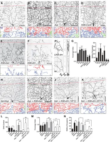
at the first/second instar transition, hence dendrite growth outpaces substrate growth, causing dendrites to occupy larger territories and more densely populate the body wall. As shown in Fig. 3, C4da dendrites in ban mutant larvae exhibit a ∼30% increase in the territory they cover (coverage index) and a∼140% increase in dorsal midline occupancy as a result of unchecked late-stage growth.
[image:4.612.50.418.259.737.2]We hypothesized that local signals, independent of systemic cues that promote larval growth and dendrite expansion, coordinate dendrite/substrate growth and that epithelial endoreplication may be a crucial component of this local substrate-derived control of dendrite growth. We therefore assayed effects of epithelial endoreplication on dendrite growth. First, we monitored effects of reducing endoreplication on dendrite growth. In larvae homozygous for mutations indupor rap, which attenuate epithelial endoreplication (Fig. 2J), we observed exuberant late-stage dendrite growth similar to banmutants (Fig. 3C,G). Expression ofdup(RNAi) with an epithelium-specific driver (supplementary material Fig. S2) similarly reduced
Fig. 3. Epithelial endoreplication influences dendrite growth.
(A-D) Effects of reduced epithelial endoreplication on C4da dendrite coverage. Compared to wild type (A), blocking endoreplication with mutations in ban(B),dup(C), or epithelialCyclin E overexpression (D) increased dendrite growth, including growth beyond normal boundaries (e.g. dorsal midline, hatched line: red in images, black in traces). (E,F) Effects of excess epithelial endoreplication on C4da dendrite growth. Epithelial overexpression ofban(E) ordm (F) led to reduction in dendrite growth, dendrite wrapping of epithelial cells (arrows) and increased 3D dendrite occupancy. (F0) 3D orientation of dendrites.yzcross-section and 3D rendering of region of interest (boxed in F) is shown. A corresponding region is shown for a wild-type C4da dendrite. (G) Quantification of dendrite coverage index and midline occupancy (μm dendrite length/1000μm2). (H-N) Epistasis analysis ofbanand endoreplication machinery. Representative dendrite images ofban/duptrans-heterozygotes (H),dupmutant larvae with epithelial banoverexpression (I), andbanmutants overexpressingdmin epithelial cells reared at 18°C for moderatedm expression (J) or 25°C for highdm expression (K). Traces depict midline dendrites.n≥8 neurons for each measurement in G-J. (L-N) Quantification of DNA content, dendrite coverage, and dendrite occupancy from
ban-endoreplication epistasis analyses. Error bars represent standard deviation. *P<0.05, **P<0.01, **P<0.001; ns, not significant; one-way ANOVA with a post-hoc Dunnett’s test.
DEVEL
O
epithelial endoreplication and caused exuberant dendrite growth, whereas resupplying UAS-dup to epithelial cells rescued the dup mutant dendrite growth defects (supplementary material Fig. S3), demonstrating that dup is required in epithelial cells to regulate endoreplication and dendrite growth. Finally, sustained epithelial expression ofCyclin E, which inhibits progress through endoreplication cycles (Weiss et al., 1998), caused dendrite defects similar toban mutants (Fig. 3D,G), demonstrating that epithelial endoreplication is necessary for modulation of C4da dendrite growth. By contrast,banor dm overexpression, which increases epithelial endoreplication, dampened late-stage dendrite growth, leading to decreased dendrite coverage and midline occupancy (Fig. 3E-G), consistent with a role for epithelial endoreplication in constraining dendrite growth. As with epithelial ban overexpression (Parrish et al., 2009), epithelial dm overexpression caused dendritic‘wrapping’of epithelial cells, possibly reflecting tighter dendrite-epithelium coupling (Fig. 3F,G). Whereas wild-type C4da dendrites were confined to a thin cross-sectional area along the basal surface of epithelial cells (Fig. 3F0) (Han et al., 2012; Kim et al., 2012), epithelialdmorbanoverexpression caused dendrites to occupy a larger three-dimensional space, particularly in regions exhibiting the‘wrapping’behavior.
To test whetherbancontrol of dendrite growth involves epithelial endoreplication, we examined the epistatic relationship between banand endoreplication regulators (Fig. 3H-N). First, we assayed for genetic interactions between ban and dup. On its own, heterozygosity for mutations in either gene had no significant effect on dendrite coverage or epithelial ploidy, but larvae doubly heterozygous for ban and dup mutations exhibited modest but significant increases in dendrite coverage and reductions in epithelial ploidy (Fig. 3H,L-N), suggesting that ban and the endoreplication effector dup function in a genetic pathway to regulate epithelial endoreplication and hence dendrite growth. Second, we overexpressedbanindupmutant larvae and found that reducing dup function blocked the effects of epithelial ban overexpression on dendrite growth and epithelial ploidy (Fig. 3I), demonstrating the requirement for endoreplication inban-mediated dendrite growth control. Third, we overexpresseddmin epithelial cells ofban mutant larvae, taking advantage of the temperature-sensitive nature of the Gal4-UAS system to drivedmexpression at different levels. Consistent withdmfunctioning downstream ofban to promote endoreplication and constrain dendrite growth, dm expression suppressed the dendrite overgrowth and epithelial endoreplication defects of ban mutants in a dose-dependent fashion (Fig. 3J,K). Altogether, these results demonstrate thatban regulates epithelial endoreplication to modulate dendrite growth.
Developmental control of dendrite-epithelium interactions Increased epithelial endoreplication alters the relative position of dendrites and epithelial cells (Fig. 3F). We hypothesized that epithelial endoreplication affects dendrite-substrate interactions by promoting epithelium-dendrite adhesion, modulating ECM permissivity to dendrite growth, or some combination of the two. Using a genetically encoded proximity sensor, high-resolution confocal imaging of dendrite/ECM markers, and transmission electron microscopy (TEM) of the dendrite/epithelium interface we examined whether dendrite/epithelium interactions change over developmental time in response tobanand endoreplication.
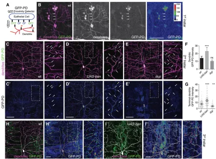
We used GFP reconstitution as a proximity detector (GFP-PD) to monitor dendrite-epithelium apposition with the underlying hypothesis that increased dendrite-epithelium adhesion would be manifest as increased dendrite-epithelium proximity. Based on physical dimensions of the components (Becker et al., 1989; Morell
et al., 2008), GFP reconstitution indicates dendrite-substrate proximity of <30 nm, a distance spanned by known adhesion molecules (Fig. 4A). GFP reassembly occurs slowly (Pédelacq et al., 2006); thus dendrite-epithelium apposition must be stable to generate GFP-PD signal. Expressing either half of the proximity sensor produced no GFP-PD signalin vivo, whereas neuronal co-expression of both split-GFP fragments resulted in split-GFP-PD signal throughout the dendritic arbor of first instar larvae (supplementary material Fig. S4), demonstrating that GFP reconstitution occurs on a timescale amenable to analysis of larval dendrite-epithelium proximity.
To monitor dendrite-epithelium juxtaposition we expressed one half of the proximity sensor in C4da neurons and the other half in epithelial cells. In first instar larvae, before the surge in epithelial endoreplication, GFP-PD signal was almost undetectable (Fig. 4B). Epithelial ploidy rapidly increases in second instar larvae, and we likewise observed dendritic accumulation of GFP-PD in second instar larvae (Fig. 4C). Epithelial overexpression of ban ordm led to a significant increase in GFP-PD in second instar larvae (Fig. 4D,F,G), suggesting that endoreplication promotes dendrite-epithelium juxtaposition. Conversely,dupmutants exhibited reduced GFP-PD signal that was most pronounced in terminal dendrites (Fig. 4E-G). We conclude that epithelial endoreplication is necessary and sufficient to trigger developmental changes in dendrite-epithelium juxtaposition.
GFP-PD signal was apparent throughout the majority of the dendritic arbor in third instar larvae (Fig. 4H), suggesting that dendrite-epithelium apposition, and probably adhesion, progressively increases throughout larval development. However, GFP-PD signal was unevenly distributed and markedly reduced/absent from many terminal dendrites, suggesting that dendrite-epithelium apposition varies across the dendritic arbor, with the most dynamic portions (terminal dendrites) coupled to epithelial cells to a lesser degree (Fig. 4H). Epithelialbanordmoverexpression, by contrast, led to increased levels of GFP-PD signal throughout the dendrite arbor, including terminal dendrites (Fig. 4I), consistent with a role for epithelial endoreplication in promoting dendrite-epithelium adhesion. C4da dendrites grow along the basal surface of epithelial cells, attached to the ECM by virtue of dendritic integrins, with a small proportion of dendrites embedded in epithelial cells (Han et al., 2012; Kim et al., 2012). Based on our observation that dendritic and epithelial membranes become more closely juxtaposed as larval development progresses, we hypothesized that dendrite-ECM interactions might be developmentally regulated as well. Using high-resolution confocal imaging (Han et al., 2012), we monitored colocalization of dendrites and ECM components labeled by GFP exon traps, including Collagen IV (vkg-gfp), and Perlecan (trol-gfp) (Fig. 5; supplementary material Figs S5 and S6) (Morin et al., 2001). In first instar larvae, >98% of dendrites co-localize with ECM markers (Fig. 5A,G), but 12% of dendrites in third instar larvae were detached from the ECM and apically shifted (Fig. 5B,G; supplementary material Fig. S5); these detached dendrites are likely embedded in epithelial cells (Han et al., 2012). Significantly fewer dendrites were detached from the ECM inbanordupmutant third instar larvae, whereas epithelial ban or dm overexpression significantly increased apical ECM detachment of dendrites (>25% of dendrites; Fig. 5C-G; supplementary material Fig. S6). Taken together, these results demonstrate that dendrite-epithelium proximity and dendrite-epithelium interactions (embedding) are developmentally regulated by epithelial endoreplication. Thus, after dendritic coverage of the body wall is established, dendrites become increasingly coupled to epithelial cells.
To corroborate our finding that dendrite-epithelium interactions are developmentally regulated by endoreplication, we examined
DEVEL
O
dendrite-epithelium interactions using TEM. In thin sections of abdominal segments cut along the apicobasal body wall axis, we monitored distribution of dendrites (identified as processes near the basal epithelial surface containing arrays of parallel microtubules) and the frequency of plasma membrane invaginations, as internalized dendrites are frequently found in membrane invaginations (Han et al., 2012; Kim et al., 2012). In first instar larvae, most dendrites were positioned at the surface of epithelial cells in direct contact with the ECM; only 3/50 dendrites were enclosed in epithelial cells (Fig. 5H; supplementary material Fig. S7). We observed a substantial increase in epithelium-embedded dendrites in third instar larvae (27/91 dendrites; Fig. 5H). Thus, dendrite-epithelium interactions change substantially from early larval development when dendrites establish body wall coverage to late larval development when dendrites expand proportionally with their substrate. The frequency of membrane invaginations significantly increased in third instar larvae (supplementary material Fig. S7), and this may facilitate dendrite enclosure by epithelial cells. Mutations that inhibited endoreplication blocked the developmental increase in epithelial plasma membrane invagination and dendrite enclosure, whereas treatments that increased epithelial endoreplication had the opposite effects (Fig. 5H;
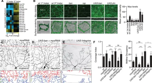
[image:6.612.84.526.55.382.2]supplementary material Fig. S7). These findings confirm our results using a GFP-based proximity sensor and our in vivo imaging of dendrite-ECM colocalization; altogether, these studies indicate that dendrite-epithelium interactions are developmentally regulated and that endoreplication in epithelial cells is a crucial component of this control. Next, we set out to identify molecular mediators of this developmentally regulated change in dendrite-epithelium interaction. Mostban-responsive adhesion-related transcripts were differentially expressed in first and third instar epithelial cells, consistent withban playing a role in developmental control of epithelial adhesion (Fig. 1C and Fig. 6A). We therefore examined whether expression of these adhesion-related genes was responsive to endoreplication. Notable among these transcripts weremys, which encodes the lone somatically expressed Drosophila β-integrin, and hep, which encodes a Drosophila JNK kinase; integrins are key mediators of cell-ECM interactions including epidermis-basement membrane attachment (DiPersio et al., 1997), and JNK regulates adhesive properties of Drosophilaepithelial cells (Jacinto et al., 2000; Jasper et al., 2001; Martin-Blanco et al., 2000). Epithelial Mys levels, in particular on the basal surface of epithelial cells outside of the basolateral junctional domain (Fig. 6B; arrows, adherens junctions), significantly increased
Fig. 4. Dendrite-epithelium proximity is developmentally regulated.(A) GFP-PD schematic. Expressing one membrane-tethered portion of GFP (GF; GFP beta sheet strands 1-10) in epithelial cells and the other membrane-tethered portion in neurons (P; GFP beta sheet strand 11) facilitates high resolution in vivoanalysis of dendrite-substrate proximity. (B) Dendrite-epithelium GFP-PD signal in first instar larvae.ppk-spGFP11-CD4-tdTomatolabels dendrites independent of GFP-PD signal. GFP-PD signal is only slightly above background in dendrites (arrows) at this stage; GFP-PD intensity is color-coded according to a lookup table (right). (C-G) Dendrite-epithelia proximity in second instar larvae.Top, merged image showing distribution of GFP-PD in the dendrite arbor of wild type (C), epithelialbanoverexpressing (D) ordupmutant (E) larvae.Bottom, GFP-PD intensity. Arrows mark tips of a subset of terminal dendrites. (F) Quantification of dendrite-epithelium GFP-PD signal from second instar larvae. Intensity values were measured along the entire dendrite arbor; mean values (following background subtraction) for ten neurons of each genotype are plotted. Error bars, standard deviation. (G) Scatter plot showing the mean and 95% confidence interval for terminal dendrite GFP-PD intensity (n≥200 terminal dendrites for each genotype). *P<0.05, **P<0.01, ***P<0.001, ns, not significant, compared to wild type; one-way ANOVA with a post-hoc Dunnett’s test. (H-I) Dendrite-epithelia GFP-PD signal in third instar larvae. Epithelialbanoverexpression leads to increased GFP-PD signal intensity, especially in terminal dendrites, suggesting thatbanpromotes dendrite-substrate proximity. Scale bars: 50μm.
DEVEL
O
during larval development, concomitant with the rapid increase in larval endoreplication, suggesting that Mys expression is coupled to endoreplication. Indeed, Mys expression was dampened by mutations that reduced endoreplication, and epithelialbanordmoverexpression, which increase endoreplication, further enhanced Mys levels (Fig. 6B). Likewise, phosphorylated JNK (P-Jnk) levels significantly increased during larval development and this increase was dependent onbanas well as endoreplication (supplementary material Fig. S8). Thus, developmental changes in epithelial expression of adhesion-related genes are triggered by endoreplication. We therefore hypothesized that endoreplication affects dendrite growth via changes in epithelial adhesion.
Epithelium-ECM interactions influence dendrite growth and plasticity
To examine whether epithelial integrins influence dendrite growth, we expressedUAS-mys(RNAi) in epithelial cells, which attenuated epithelial Mys protein levels (supplementary material Fig. S9), and monitored effects on dendrite patterning in third instar larvae. Epithelialmys(RNAi) caused exuberant late-stage dendrite growth similar tobanmutants or other endoreplication-defective mutants, albeit to a lesser extent (Fig. 6C,F). Thus, increased expression of Mys, which mediates ECM attachment, contributes to coordination of dendrite/epithelial growth by constraining late-stage dendrite growth. One model to account for this finding is that increased epithelium-ECM attachment makes the ECM less permissive to dendrite growth. Alternatively, increased epithelium-ECM attachment may potentiate epithelium-dendrite contacts that constrain dendrite growth.
To ascertain whether developmental control of Mys expression is a functionally relevant output of ban in regulating dendrite growth, we examined the epistatic relationship between epithelial ban and mysin control of C4da dendrite development. First, we simultaneously overexpressed ban and knocked down mys in epithelial cells and found thatmys(RNAi) attenuated the dendrite
growth defect of ban overexpression, resulting in dendrite overextension beyond normal boundaries (Fig. 6D,F). Second, we assayed effects of epithelial overexpression of integrins (UAS-mys +UAS-inflated) on dendrite growth and found that integrin overexpression caused a dendrite undergrowth phenotype similar to that of ban or dm overexpression (Fig. 6E,F). Third, we overexpressed integrins in ban mutant larvae and found that epithelial integrin overexpression only partially suppressed the dendrite overgrowth ofban mutants (Fig. 6F). We conclude that increased epithelial Mys expression is necessary but not sufficient forban-mediated control of dendrite growth. Taken together with our observation that modulating integrin expression causes less severe dendrite defects than modulation ofbanor endoreplication, these results suggest that additional epithelium-derived factors contribute toban-mediated, and hence endoreplication-dependent, epithelial control of dendrite growth.
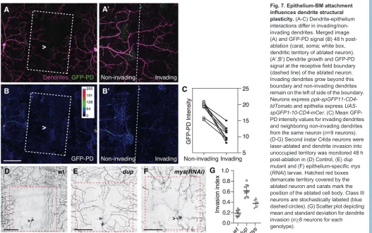
Ablating C4da neurons before establishment of tiling leads to exuberant dendrite growth into unoccupied territory by spared neurons and this invasive growth potential is lost concomitant with the onset of epithelial endoreplication (Grueber et al., 2003; Parrish et al., 2009; Sugimura et al., 2003). We therefore hypothesized that developmental restriction in C4da dendrite plasticity is the result of alterations in dendrite-substrate and dendrite-epithelium interactions triggered by epithelial endoreplication, including increased proximity dendrite-epithelium proximity. Indeed, following ablation of a C4da neuron, invading dendrites of spared neurons were less closely associated with epithelial cells than neighboring non-invading dendrites, as assessed by relative levels of GFP-PD intensity (Fig. 7A-C).
[image:7.612.106.505.59.260.2]ECM modification plays important roles in regulating dendrite structural plasticity in several contexts (Mataga et al., 2002; Oray et al., 2004; Yasunaga et al., 2010), thus we reasoned thatban-regulated changes in epithelium-ECM attachment might alter substrate permissivity for dendrite growth and hence C4da dendrite plasticity. To test this possibility, we ablated second instar Cd4a neurons and monitored dendrite invasion of the unoccupied territory by spared
Fig. 5. Epithelial endoreplication influences dendrite-ECM attachment.Live imaging of dendrites and ECM using a neuronal membrane marker (ppk-CD4-tdTomato) and acollagen IVexon trap (vkg-GFP) to monitor dendrite-ECM colocalization in first instar wild-type (A), third instar wild-type (B),ban mutant (C), epithelialbanoverexpressing (D),dupmutant (E) and epithelialdmoverexpressing larvae (F). Representative maximum projections of 3D stacks are shown (A,B). Following deconvolution, colocalization was measured between dendrites and Vkg-GFP (see supplementary material Fig. S5); in traces, green depicts dendrites that colocalize with Vkg-GFP and magenta depicts detached (apically shifted) dendrites. Similar results were obtained using an additional ECM marker (supplementary material Fig. S6). (G) Quantification of dendrite detachment from the ECM (n≥10 neurons for each genotype). (H) TEM analysis. The fraction of dendrites enclosed inside epithelial cells is shown (n≥50 dendrites for all genotypes). Other than wild-type first instar samples, all samples were from third instar larvae. *P<0.05, **P<0.01, ***P<0.001 compared to wild-type third instar; one-way ANOVA with a post-hoc Dunnett’s test.
DEVEL
O
neurons. In this paradigm, wild-type dendrites exhibit little invasive activity, covering∼18% of vacated territory (Fig. 7D,G). By contrast, when we ablated C4da neurons in dup mutant larvae, which are defective in endoreplication and the associated developmental changes in dendrite-epithelium interactions, we observed robust invasive activity comparable tobanmutants (Fig. 7E,G). Likewise, when we reduced epithelial Mys levels via epithelia-specific mys(RNAi), we observed a significant potentiation of dendrite invasion (Fig. 7F,G). Notably, mutations inban ordup potentiate dendrite invasion to a greater degree thanmys(RNAi) (Fig. 7G; Parrish et al., 2009), suggesting that factors other than mys function downstream of endoreplication to control dendrite expansion. We conclude that the developmentally programmed growth transition to endoreplication in epithelial cells regulates substrate permissivity for dendrite growth, in part by regulating epithelium-ECM interactions.
DISCUSSION
Local and systemic control of dendrite growth
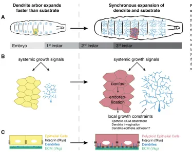
During embryonic and early larval development, C4da dendrites expand faster than their substrate to achieve complete body wall coverage. C4da neurons respond to nociceptive stimuli (Tracey et al., 2003), and the rapid early dendrite growth ensures that the larval body wall is completely covered by these sensory dendrites shortly after hatching, when larvae must interact with their surroundings. Subsequently, C4da dendrites expand synchronously with the body wall epithelium as larvae grow while maintaining proportionality (Fig. 8A). Thus, multiple growth signals are likely to be at work in this system. In response to systemic growth cues, dendrites and
epithelial cells continuously expand during larval development. Blocking these growth cues, for example by ablating dILP neurons or compromising insulin signaling, similarly affects both neuron and substrate, resulting in growth-arrested larvae with properly scaled dendrite arbors (Parrish et al., 2009). Beginning in second instar larvae, sensory neurons and epithelial cells respond differently to growth cues: epithelial growth relies on endoreplication; sensory neuron growth does not (Fig. 8B). During this latter period of larval growth, epithelium-derived signals constrain dendrite expansion to ensure synchronous dendrite/substrate growth. Larval activation of banin epithelial cells is an essential component of this signaling cascade (Parrish et al., 2009), and here we demonstrate that ban functions in epithelial cells to regulate endoreplication. By altering adhesive properties of epithelial cells, hence epithelium-ECM and epithelium-dendrite interactions, endoreplication curtails C4da dendrite growth and plasticity (Fig. 8C). Many types of neuron expand their arbors synchronously with their substrate to maintain proportional coverage of their receptive field; developmental control of substrate adhesion may be similarly regulated in other contexts to couple dendrite/substrate growth, and to regulate dendrite structural plasticity.
Endoreplication in nervous system development
[image:8.612.55.555.60.332.2]Growth control is particularly complex in the nervous system, where different cell types are continuously incorporated and cells must grow while maintaining connections. In some contexts, programmed polyploidy facilitates neuronal growth. For example, in the terrestrial slug Limax valentianus endoreplication occurs
Fig. 6. Developmental changes in epithelial adhesion influence dendrite growth.(A) Heat map depicting developmental expression ofban-regulated adhesion molecules in epithelial cells. (B) Developmental regulation of epithelial Mys expression. Top, larval fillets of the indicated genotype stained with Mys antibody. Images are maximum projections ofz-stacks; dashed lines, position of orthogonal slices. Bottom, 3D rendering of Mys distribution. Right, Quantification of Mys immunoreactivity in the indicated genotypes.n≥50 cells for each genotype. (C-F) Epithelial integrin influences dendrite growth. Representative C4da dendrites and midline dendrite traces for epithelialmys(RNAi) (C), epithelial co-expression ofUAS-mys(RNAi) andUAS-ban(D), and epithelial expression of UAS-integrins(E). (F) Quantification of dendrite coverage index and midline occupancy (μm dendrite length/1000μm2).n≥8 neurons for each measurement. Error bars represent standard deviation. *P<0.05, **P<0.01, ***P<0.001, ns, not significant; one-way ANOVA with a post-hoc Dunnett’s test. Scale bars: 25μm in B, top; 10μm in B, bottom; 50μm in C-E.
DEVEL
O
throughout the nervous system in proportion to animal growth, presumably to facilitate neuron expansion (Yamagishi et al., 2011). Neuronal polyploidy has been documented in vertebrates as well; tetraploid neurons exist in the retina and cortex (López-Sánchez and Frade, 2013; Morillo et al., 2010), and Purkinje neurons may be polyploid (Lapham, 1968; Mann et al., 1978).
In addition to supporting neuronal growth, developmentally regulated polyploidy (endoreplication) is suited for tissue growth in circumstances where division might disrupt patterning or connectivity. For example, endoreplication allows glial cells that wrap the Drosophila ventral ganglion to maintain blood-brain barrier integrity even as glia grow to accommodate brain expansion (Unhavaithaya and Orr-Weaver, 2012). Likewise, we found that epithelial endoreplication allows for substrate growth without cell divisions that might disrupt body wall innervation. In addition, epithelial endoreplication drives a differentiation program that regulates epithelium-dendrite interactions to influence sensory dendrite growth and patterning. Notable among these interactions is an increase in epithelial dendrite embedding, which may serve a number of purposes. First, as suggested in prior studies, embedding may have functional consequences (Han et al., 2012; Kim et al., 2012). Second, embedding may provide points of contact that constrain dendrite expansion. Third, embedding may facilitate generation of tensile forces as the body wall expands; such forces could contribute to dendrite growth, analogous to axon elongation in response to mechanical tension (Bray, 1984). How genome amplification potentiates certain signaling pathways is unknown, but Drosophila endoreplication entails under-replication of specific genomic regions in a cell-type-specific manner (Sher et al., 2013), thus endoreplication may facilitate expression of an
epithelium-differentiation program. It remains to be seen whether endoreplication regulates substrate or target-derived signals that influence neuron growth and patterning in other systems.
Substrate control of dendrite plasticity
Here, we demonstrate that dendrite plasticity is tied to growth control. We found that alterations in substrate adhesive properties constrain dendrite plasticity in C4da neurons concomitant with proportional expansion of dendrites and substrate. First, decreased plasticity is accompanied by increased dendrite-epithelium proximity during development; manipulations that increase plasticity decrease dendrite-epithelium proximity, and vice versa. Second, epithelial dendrite embedding, which may serve to tether dendrites to epithelial cells, increases as the capacity for structural plasticity decreases, and manipulations that increase plasticity decrease the prevalence of epithelium-embedded dendrites. Third, epithelial adhesion changes as dendritic plasticity is restricted and epithelial integrin expression contributes to restriction of plasticity. Large-scale plasticity would seem to be incompatible with synchronous growth of neurons and their substrate, so it will be intriguing to see whether dendritic plasticity is broadly constrained during periods of growth that maintain proportionality.
MATERIALS AND METHODS
Fly stocks
See supplementary material Table S2 for alleles used in this study.
Live imaging
[image:9.612.48.575.55.386.2]Embryos were collected on yeasted grape juice agar plates, aged at 25°C in a moist chamber, mounted in 90% glycerol under coverslips sealed with
Fig. 7. Epithelium-BM attachment influences dendrite structural plasticity.(A-C) Dendrite-epithelium interactions differ in invading/non-invading dendrites. Merged image (A) and GFP-PD signal (B) 48 h post-ablation (carat, soma; white box, dendritic territory of ablated neuron). (A0,B0) Dendrite growth and GFP-PD signal at the receptive field boundary (dashed line) of the ablated neuron. Invading dendrites grow beyond this boundary and non-invading dendrites remain on the left of side of the boundary. Neurons express ppk-spGFP11-CD4-tdTomatoand epithelia express UAS-spGFP1-10-CD4-mCer.(C) Mean GFP-PD intensity values for invading dendrites and neighboring non-invading dendrites from the same neuron (n=9 neurons). (D-G) Second instar C4da neurons were laser-ablated and dendrite invasion into unoccupied territory was monitored 48 h post-ablation in (D) Control, (E)dup mutant and (F) epithelium-specificmys (RNAi) larvae. Hatched red boxes demarcate territory covered by the ablated neuron and carats mark the position of the ablated cell body. Class III neurons are stochastically labeled (blue dashed circles). (G) Scatter plot depicting mean and standard deviation for dendrite invasion (n≥8 neurons for each genotype).
DEVEL
O
grease and imaged using a Leica SP5 microscope with a 40×, 1.25NA lens. To analyze dendrite phenotypes, image stacks of dendrites in segments A2-A4 were captured from 8-10 larvae. For high-resolution imaging of the dendrite-ECM interface, larvae were anesthetized with ether before mounting and stacks with a 0.2 µmz-step were acquired.
GFP reconstitution
GFP proximity detection (GFP-PD) is based on GRASP constructs (Feinberg et al., 2008) using extracellular GFP fragments tethered to the transmembrane carrier CD4. Briefly, CD4-spGFP1-10 was PCR amplified and cloned into pUAST containing a C-terminal mCerulean tag. Transgenic lines were obtained from BestGene (Chino Hills, CA).
UAS-spGFP1-10-CD4-mCerwas recombined with
ppk-spGFP11-CD4-tdTomato(Han et al., 2012) and epithelium-dendrite GFP reconstitution
was monitored using the epithelium-specificA58-Gal4driver to express
UAS-spGFP1-10-CD4-mCer with the C4da neuron-specific
ppk-spGFP11-CD4-tdTomato. GFP-PD signal was imaged under identical
conditions for all samples of a given time point, taking care to avoid pixel saturation. Dendrite arbors were used to generate a mask, and mean GFP-PD pixel intensity within the mask was measured using ImageJ (NIH).
Laser ablation
A single larva was mounted, as for live imaging, and the nucleus of a C4da neuron was targeted under a 100×-1.4NA objective using a 337 nm pulsed nitrogen laser (Andor Micropoint; 12 Hz, 15 s) mounted on a Leica DM550 microscope. Following ablation, animals were recovered to cornmeal agar and imaged 48 h later.
Immunohistochemistry
Larval immunohistochemistry was as described (Grueber et al., 2002) using the following: HRP-Cy2 or Cy3 (1:200; Jackson ImmunoResearch), mCD8 (1:100; Life Technologies), phospho-D-Akt Ser505 (1:500; Cell Signaling), Myospheroid-CF.6G11 (1:20; Developmental Studies Hybridoma Bank), BrdU (1:250; Abcam), phospho-JNK (1:500; Cell Signaling), DAPI (50 ng/ml; Life Technologies) and secondary antibodies from Jackson ImmunoResearch. For BrdU labeling, larvae were fed
cornmeal-molasses food containing BrdU (10μg/ml; Sigma), dissected in PBS, fixed in 4% formaldehyde/PBS, permeabilized in PBS-Tx, acid treated with 2.5 N HCl for 30 min, neutralized with 100 mM sodium tetraborate, and processed for immunostaining.
TEM
Larvae were perforated with insect pins, fixed in 2.5% glutaraldehyde/0.1 M sodium cacodylate buffer with centrifugation [15,000 rpm (21,330g), 1 h], washed 4×5 min in PBS and post-fixed at 4°C in buffered 2% osmium tetroxide overnight. Samples were spun [12,500 rpm (14,674 g), 1 h], washed with distilled water in 20 ml scintillation vials (3×20 min), and dehydrated in a graded series of ethanol, followed by two changes of propylene oxide. This was followed by infiltration in a 1:1 mixture of propylene oxide:epon-araldite overnight, two changes in epon-araldite (2 h each), and overnight polymerization (60°C). 70 nm sections were stained with Reynolds’lead citrate and viewed on a JEOL-1230 microscope with an AMT XR80 camera.
Microarray analysis
Fluorescence-activated cell sorting (FACS) cell purification and microarray analysis were conducted as described (Parrish et al., 2014). Details are available in in the supplementary materials and methods.
Measurements
Dendrite metrics: 2D projections of z-stacks were used for computer-assisted dendrite tracing (Neurolucida); arbor features were measured using the traces. Details are provided in the supplementary materials and methods.
DNA content: DAPI quantitation was as described (Unhavaithaya and Orr-Weaver, 2012). Briefly, we measured pixel intensity of DAPI stain in each optical section ofz-stacks (500 nm step) and normalized epithelial DAPI intensity to mean DAPI intensity of ten diploid PNS neurons from the same fillet imaged using identical settings. PNS neuron DAPI intensity varied less than 10%.
[image:10.612.47.421.52.349.2]Cell size: We traced the outline of Nrx-IV-GFP or anti-Mys immunoreactivity for≥50 epithelial cells of each genotype using ImageJ (NIH). We obtained similar results with volume measurements of epithelial cells.
Fig. 8. Biphasic control of larval dendrite expansion.(A) Dendrites outgrow their substrate to establish complete coverage of the epidermis. After the first/second instar transition, dendrites expand in proportion to their substrate. (B) Larval dendrites and epithelial cells constantly grow in response to systemic cues. Epithelial endoreplication alters adhesive properties of epithelial cells, locally constraining dendrite growth. (C) Epithelial expression ofβ-Integrin (black dashed lines) and epithelial invagination of dendrites (blue) are developmentally regulated.
DEVEL
O
Statistical analysis
Differences between group means were analyzed via ANOVA with a post-hoc Dunnett’s test. Significance of microarray expression was calculated using Significance analysis of microarrays (SAM) (Tusher et al., 2001) using a 5% false discovery rate and fold-change threshold of 1.5-fold. Lexical analysis was as described (Kim and Falkow, 2003).
Accession number
Microarray data are available in the NCBI Gene Expression Omnibus (GSE50540).
Acknowledgements
We thank the Bloomington Stock Center, Chun Han, Yuh Nung Jan and Paul Lasko for fly stocks; the DSHB for antibodies; the CHDD for assistance with TEM; Hui Li and Jon Ahn for assistance with stereology; and Michael Kim for critical reading of the manuscript.
Competing interests
The authors declare no competing financial interests.
Author contributions
N.J. and J.P. conceived and designed the experiments. N.J. and J.P. conducted the experiments with assistance from P.S. on GFP reconstitution assays, C.K. on microarray analysis, and E.P. on TEM. N.J., C.K. and J.P. analyzed the data. J.P. wrote the manuscript.
Funding
This work was supported by the National Institutes of Health (NIH) [NIMH-R00MH084277 and NINDS-R01NS076614], by a Basil O’Connor Starter Scholar Award, by a Klingenstein Fellowship in Neuroscience (J.Z.P.) and by a Benjamin Hall Graduate Fellowship (N.J.). Deposited in PMC for release after 12 months.
Supplementary material
Supplementary material available online at
http://dev.biologists.org/lookup/suppl/doi:10.1242/dev.107573/-/DC1
References
Becker, J. W., Erickson, H. P., Hoffman, S., Cunningham, B. A. and Edelman, G. M.
(1989). Topology of cell adhesion molecules.Proc. Natl. Acad. Sci. U.S.A.86, 1088-1092.
Bentley, D. and Toroian-Raymond, A.(1981). Embryonic and postembryonic morphogenesis of a grasshopper interneuron.J. Comp. Neurol.201, 507-518.
Bloomfield, S. A. and Hitchcock, P. F.(1991). Dendritic arbors of large-field ganglion cells show scaled growth during expansion of the goldfish retina: a study of morphometric and electrotonic properties. J. Neurosci. 11, 910-917.
Bray, D.(1984). Axonal growth in response to experimentally applied mechanical tension.Dev. Biol.102, 379-389.
Brennecke, J., Hipfner, D. R., Stark, A., Russell, R. B. and Cohen, S. M.(2003). bantam encodes a developmentally regulated microRNA that controls cell proliferation and regulates the proapoptotic gene hid in Drosophila.Cell113, 25-36.
Britton, J. S. and Edgar, B. A.(1998). Environmental control of the cell cycle in Drosophila: nutrition activates mitotic and endoreplicative cells by distinct mechanisms.Development125, 2149-2158.
Church, R. B. and Robertson, F. W.(1966). Biochemical analysis of genetic differences in the growth of Drosophila.Genet. Res.7, 383-407.
DiPersio, C. M., Hodivala-Dilke, K. M., Jaenisch, R., Kreidberg, J. A. and Hynes, R. O.(1997).α3β1 Integrin is required for normal development of the epidermal basement membrane.J. Cell Biol.137, 729-742.
Emoto, K., Parrish, J. Z., Jan, L. Y. and Jan, Y.-N.(2006). The tumour suppressor Hippo acts with the NDR kinases in dendritic tiling and maintenance.Nature443, 210-213.
Farajian, R., Raven, M. A., Cusato, K. and Reese, B. E. (2004). Cellular positioning and dendritic field size of cholinergic amacrine cells are impervious to early ablation of neighboring cells in the mouse retina.Vis. Neurosci.21, 13-22.
Feinberg, E. H., Vanhoven, M. K., Bendesky, A., Wang, G., Fetter, R. D., Shen, K. and Bargmann, C. I. (2008). GFP Reconstitution Across Synaptic Partners (GRASP) defines cell contacts and synapses in living nervous systems.Neuron 57, 353-363.
Grueber, W. B., Jan, L. Y. and Jan, Y. N.(2002). Tiling of the Drosophila epidermis by multidendritic sensory neurons.Development129, 2867-2878.
Grueber, W. B., Ye, B., Moore, A. W., Jan, L. Y. and Jan, Y. N.(2003). Dendrites of distinct classes of Drosophila sensory neurons show different capacities for homotypic repulsion.Curr. Biol.13, 618-626.
Han, C., Wang, D., Soba, P., Zhu, S., Lin, X., Jan, L. Y. and Jan, Y.-N.(2012). Integrins regulate repulsion-mediated dendritic patterning of drosophila sensory neurons by restricting dendrites in a 2D space.Neuron73, 64-78.
Hipfner, D. R., Weigmann, K. and Cohen, S. M. (2002). The bantam gene regulates Drosophila growth.Genetics161, 1527-1537.
Hitchcock, P. F.(1987). Constant dendritic coverage by ganglion cells with growth of the goldfish’s retina.Vis. Res.27, 17-22.
Jacinto, A., Wood, W., Balayo, T., Turmaine, M., Martinez-Arias, A. and Martin, P.
(2000). Dynamic actin-based epithelial adhesion and cell matching during Drosophila dorsal closure.Curr. Biol.10, 1420-1426.
Jasper, H., Benes, V., Schwager, C., Sauer, S., Clauder-Münster, S., Ansorge, W. and Bohmann, D. (2001). The genomic response of the Drosophila embryo to JNK signaling.Dev. Cell1, 579-586.
Kim, C. C. and Falkow, S.(2003). Significance analysis of lexical bias in microarray data.BMC Bioinformatics4, 12.
Kim, M. E., Shrestha, B. R., Blazeski, R., Mason, C. A. and Grueber, W. B.
(2012). Integrins establish dendrite-substrate relationships that promote dendritic self-avoidance and patterning in drosophila sensory neurons.
Neuron73, 79-91.
Lapham, L. W.(1968). Tetraploid DNA content of Purkinje neurons of human cerebellar cortex.Science159, 310-312.
Lin, B., Wang, S. W. and Masland, R. H.(2004). Retinal ganglion cell type, size, and spacing can be specified independent of homotypic dendritic contacts.
Neuron43, 475-485.
López-Sánchez, N. and Frade, J. M.(2013). Genetic evidence for p75NTR-dependent tetraploidy in cortical projection neurons from adult mice.J. Neurosci. 33, 7488-7500.
Mann, D. M. A., Yates, P. O. and Barton, C. M.(1978). The DNA content of Purkinje cells in mammals.J. Comp. Neurol.180, 345-347.
Martin-Blanco, E., Pastor-Pareja, J. C. and Garcia-Bellido, A.(2000). JNK and decapentaplegic signaling control adhesiveness and cytoskeleton dynamics during thorax closure in Drosophila.Proc. Natl. Acad. Sci. U.S.A.97, 7888-7893.
Mataga, N., Nagai, N. and Hensch, T. K.(2002). Permissive proteolytic activity for visual cortical plasticity.Proc. Natl. Acad. Sci. U.S.A.99, 7717-7721.
Morell, M., Czihal, P., Hoffmann, R., Otvos, L., Avilés, F. X. and Ventura, S.(2008). Monitoring the interference of protein-protein interactions in vivo by bimolecular fluorescence complementation: the DnaK case.Proteomics8, 3433-3442.
Morillo, S. M., Escoll, P., de la Hera, A. and Frade, J. M.(2010). Somatic tetraploidy in specific chick retinal ganglion cells induced by nerve growth factor.
Proc. Natl. Acad. Sci. U.S.A.107, 109-114.
Morin, X., Daneman, R., Zavortink, M. and Chia, W.(2001). A protein trap strategy to detect GFP-tagged proteins expressed from their endogenous loci in Drosophila.Proc. Natl. Acad. Sci. U.S.A.98, 15050-15055.
Oray, S., Majewska, A. and Sur, M.(2004). Dendritic spine dynamics are regulated by monocular deprivation and extracellular matrix degradation. Neuron 44, 1021-1030.
Park, S. Y. and Asano, M.(2008). The origin recognition complex is dispensable for endoreplication in Drosophila. Proc. Natl. Acad. Sci. U.S.A. 105, 12343-12348.
Parrish, J. Z., Emoto, K., Jan, L. Y. and Jan, Y. N.(2007). Polycomb genes interact with the tumor suppressor genes hippo and warts in the maintenance of Drosophila sensory neuron dendrites.Genes Dev.21, 956-972.
Parrish, J. Z., Xu, P., Kim, C. C., Jan, L. Y. and Jan, Y. N.(2009). The microRNA bantam functions in epithelial cells to regulate scaling growth of dendrite arbors in drosophila sensory neurons.Neuron63, 788-802.
Parrish, J. Z., Kim, C. C., Tang, L., Bergquist, S., Wang, T., DeRisi, J. L., Jan, Y. N., Jan, L. Y. and Davis, G. W.(2014). Krüppel mediates the selective rebalancing of ion channel expression.Neuron82, 537-544.
Pédelacq, J.-D., Cabantous, S., Tran, T., Terwilliger, T. C. and Waldo, G. S.
(2006). Engineering and characterization of a superfolder green fluorescent protein.Nat. Biotechnol.24, 79-88.
Pierce, S. B., Yost, C., Britton, J. S., Loo, L. W. M., Flynn, E. M., Edgar, B. A. and Eisenman, R. N.(2004). dMyc is required for larval growth and endoreplication in Drosophila.Development131, 2317-2327.
Pimentel, A. C. and Venkatesh, T. R.(2005). rap gene encodes Fizzy-related protein (Fzr) and regulates cell proliferation and pattern formation in the developing Drosophila eye-antennal disc.Dev. Biol.285, 436-446.
Sher, N., Von Stetina, J. R., Bell, G. W., Matsuura, S., Ravid, K. and Orr-Weaver, T. L.
(2013). Fundamental differences in endoreplication in mammals and Drosophila revealed by analysis of endocycling and endomitotic cells.Proc. Natl. Acad. Sci. U.S.A.110, 9368-9373.
Sigrist, S. J. and Lehner, C. F.(1997). Drosophila fizzy-related down-regulates mitotic cyclins and is required for cell proliferation arrest and entry into endocycles.
Cell90, 671-681.
Smith, A. V. and Orr-Weaver, T. L.(1991). The regulation of the cell cycle during Drosophila embryogenesis: the transition to polyteny. Development 112,
997-1008.
DEVEL
O
Sugimura, K., Yamamoto, M., Niwa, R., Satoh, D., Goto, S., Taniguchi, M., Hayashi, S. and Uemura, T.(2003). Distinct developmental modes and lesion-induced reactions of dendrites of two classes of Drosophila sensory neurons.
J. Neurosci.23, 3752-3760.
Tracey, W. D., Jr, Wilson, R. I., Laurent, G. and Benzer, S.(2003). painless, a Drosophila gene essential for nociception.Cell113, 261-273.
Truman, J. W. and Bate, M.(1988). Spatial and temporal patterns of neurogenesis in the central nervous system of Drosophila melanogaster. Dev. Biol. 125, 145-157.
Truman, J. W. and Reiss, S. E.(1988). Hormonal regulation of the shape of identified motoneurons in the moth Manduca sexta.J. Neurosci.8, 765-775.
Tusher, V. G., Tibshirani, R. and Chu, G.(2001). Significance analysis of microarrays applied to the ionizing radiation response. Proc. Natl. Acad. Sci. U.S.A. 98, 5116-5121.
Unhavaithaya, Y. and Orr-Weaver, T. L.(2012). Polyploidization of glia in neural development links tissue growth to blood-brain barrier integrity.Genes Dev.26, 31-36.
Weiss, A., Herzig, A., Jacobs, H. and Lehner, C. F.(1998). Continuous Cyclin E expression inhibits progression through endoreduplication cycles in Drosophila.
Curr. Biol.8, 239-242.
Yamagishi, M., Ito, E. and Matsuo, R.(2011). DNA endoreplication in the brain neurons during body growth of an adult slug.J. Neurosci.31, 5596-5604.
Yasunaga, K.-i., Kanamori, T., Morikawa, R., Suzuki, E. and Emoto, K.(2010). Dendrite reshaping of adult Drosophila sensory neurons requires matrix metalloproteinase-mediated modification of the basement membranes. Dev. Cell18, 621-632.
Zielke, N., Querings, S., Rottig, C., Lehner, C. and Sprenger, F.(2008). The anaphase-promoting complex/cyclosome (APC/C) is required for rereplication control in endoreplication cycles.Genes Dev.22, 1690-1703.