RESEARCH ARTICLE
Specification of neuronal subtypes by different levels of
Hunchback
Marta Moris-Sanz, Alicia Estacio-Gómez, Javier Álvarez-Rivero and Fernando J. Dıaz-Benjumea*́
ABSTRACT
During the development of the central nervous system, neural progenitors generate an enormous number of distinct types of neuron and glial cells by asymmetric division. Intrinsic genetic programs define the combinations of transcription factors that determine the fate of each cell, but the precise mechanisms by which all these factors are integrated at the level of individual cells are poorly understood. Here, we analyzed the specification of the neurons in the ventral nerve cord of Drosophila that express Crustacean cardioactive peptide (CCAP).There are two types of CCAP neurons: interneurons and efferent neurons. We found that both are specified during the Hunchback temporal window of neuroblast 3-5, but are not sibling cells. Further, this temporal window generates two ganglion mother cells that give rise to four neurons, which can be identified by the expression ofempty spiracles. We show that the expression of Hunchback in the neuroblast increases over time and provide evidence that the absolute levels of Hunchback expression specify the two different CCAP neuronal fates.
KEY WORDS:Drosophila, Central nervous system, CCAP, Bursicon, Cell fate specification, Temporal identity factors, Hunchback
INTRODUCTION
During embryonic development, neuronal stem cells actively proliferate and generate the enormous variety of cell types found in the central nervous system (CNS). It is generally assumed that combinations of transcription factors in the progenitor cell, which are spatially and temporally regulated in very sophisticated ways, determine the sets of characteristics that define the different cell types. How the corresponding states are set at the cellular level is far from clear (Gaspard and Vanderhaeghen, 2010). Given the complexity of the process, the choice of an appropriate model system for its study is crucial. The Drosophilaventral nerve cord (VNC), which is the equivalent of the vertebrate spinal cord, has provided an important model system for studying the molecular mechanisms underlying neuronal cell fate specification (Technau et al., 2006).
A combination of lineage studies and molecular genetics approaches has revealed some general rules by which the embryonic neuroectoderm is patterned along the anterior-posterior and dorsal-ventral axes of the Drosophila embryo. The process generates an invariant array of 60 neuroblasts (NBs) per segment, bilaterally located in mirror-image hemisegments. Each one of the 30 pairs of NBs generates, by several rounds of asymmetric
division, an invariant and unique lineage. In each division the NB self-renews and buds off a daughter cell, called a ganglion mother cell (GMC), which divides once to generate two sibling cells that differentiate as neurons or glial cells (Doe, 2008; Knoblich, 2008). NBs sequentially express a series of genes, with periods of expression that define temporal windows, in the sequence: hunchback (hb)→Kruppel (Kr)→pdm1 (nubbin – FlyBase)/pdm2 (henceforth pdm)→castor (cas)→grainy head (grh) (Brody and Odenwald, 2000; Cleary and Doe, 2006; Grosskortenhaus et al., 2005, 2006; Isshiki et al., 2001; Kambadur et al., 1998; Mettler et al., 2006; Novotny et al., 2002; Pearson and Doe, 2003; Tran and Doe, 2008). The expression of these temporal identity factors confers the competence to specify particular cell fates; thus, early-born neurons are specified by the expression of Hb, and, if expression of Hb is artificially extended, early neuronal fates continue to be specified (Isshiki et al., 2001; Pearson and Doe, 2003).
Many lineages contain broad temporal windows within which more than one neural fate is generated. This raises the question of how such temporal windows are subdivided. This issue is poorly understood and has only been addressed for the Cas temporal window of the well-characterized lineage of NB5-6. In this case, two sequential feed-forward loops triggered by Cas specify four distinct neuronal fates (Baumgardt et al., 2009).
Because of their restricted patterns of expression, neuropeptides are often used as terminal differentiation markers for specific subsets of neurons (Nässel, 2002). In this report we have chosen the set of neurons that express Crustacean cardioactive peptide (CCAP) and Bursicon (Burs) to study the patterning of the CNS. The CCAP neuropeptide is widespread in invertebrates. In addition to its cardioacceleratory action, it is also involved in the control of ecdysis (Dulcis et al., 2005; Ewer, 2005; Mesce and Fahrbach, 2002; Park et al., 2003). The Burs neuropeptide, which is also found in other insects, is a tanning factor involved in the control of ecdysis. The active form of Burs is a heterodimer composed of Bursαand Bursβ (Pburs– FlyBase). CCAP and Burs are co-expressed in a set of neurons of the ventral ganglion, and genetic evidence confirms that these neurons play a key role in head eversion and leg and wing expansion at pupal ecdysis (Dewey et al., 2004; Peabody et al., 2008). CCAP/Burs-expressing neurons in the most anterior abdominal segments also express myoinhibitory peptides (Mip, also known as AstB), which are also involved in regulating ecdysis (reviewed by Nässel, 2002; Nässel and Winther, 2010). In summary, the complex set of neuropeptides expressed by these neurons confers their crucial roles in the networks that control the different phases of ecdysis (Kim et al., 2006).
In the VNC of first instar larvae there are two types of CCAP-expressing neurons: interneurons and efferent neurons. Our goals in this work were to identify the progenitor NB(s) of these neurons and the mechanisms by which their different identities are established. We found all CCAP neurons belong to the lineage of NB3-5 that can be identified by the expression ofempty spiracles(ems). Furthermore,
Received 27 May 2014; Accepted 3 September 2014
Centro de Biologıa Molecular-Severo Ochoa (CSIC-UAM), c./Nicolá ́s Cabrera 1, Universidad Autónoma, Madrid 28049, Spain.
*Author for correspondence ([email protected])
DEVEL
O
these neurons are not sibling cells but are generated in the same temporal window, namely that of Hb. In addition, we provide evidence that different levels of Hb expression in the NB subdivide its temporal window and determine the two different fates of the postmitotic neurons, and these in turn express different levels of Hb, which is crucial for the specification of different neuronal subtypes.
RESULTS
CCAP neurons as a model system to study neural fate specification
The onset of CCAP expression in the VNC starts in the 18-h-old embryo and is observed in a group of 13 interneurons (INs) per hemiganglion (the right or left half of the ventral ganglion): one in each hemisegment from the first subesophagic segment (SE1) to the seventh abdominal segment (A7) (Fig. 1A,B). Distinct CCAP-expressing neurons appear in the T3-A4 segments in the first instar larva; these are efferent neurons (ENs) that exit the ganglion via the lateral segmental nerve and can be identified by the expression of
Dachshund (Dac) (Fig. 1C) (Park et al., 2003; Santos et al., 2007; Veverytsa and Allan, 2011; Vomel and Wegener, 2007). Although the ENs of segment T3 always express CCAP in the first instar larva, in A1-4 segments only one or two ENs per hemiganglion usually expresses CCAP at this stage, with expression in the others appearing sequentially during larval development (Fig. 1D). Later, in pupal development, one EN in each of hemisegments A5-7 expresses CCAP. It is important to note that, in first instar larvae, CCAP expression is stronger in the more anterior segments and gradually decays in the more posterior segments.
Bursαexpression was observed in the first instar larva in the same cells that express CCAP and, in addition, in the ENs of segments A1-4, which will express CCAP in the third instar larva (Kim et al., 2006). Thus, CCAP and Bursα are co-expressed in the VNC of mature larvae. Unlike CCAP, Bursαexpression was stronger in the more posterior segments. Hereafter, we will refer to those cells that express CCAP/Bursα as CCAP neurons, although in several experiments, for convenience, we examined the expression of Bursα. CCAP expression in segments 8-9 of first instar larva ganglion was very weak and difficult to score when it appeared, and Bursαexpression showed a very low penetrance; thus, neurons of these segments were not quantified.
All CCAP-expressing neurons are generated from NB3-5 We first aimed to identify the progenitor NB of the CCAP neurons. We used a set of previously described molecular markers that allow the identification of NBs at different embryonic stages (Doe, 1992). The expression of many of these markers is maintained, at least temporarily, in the progeny that they generate. CCAP neurons do not expressmirror(mirr)-lacZ,gooseberry(gsb)-lacZor Engrailed (En) (Fig. 2A-C), which indicates that they derive either from a NB of row 4, or from NB3-1, NB3-3 or NB3-5.
Although CCAP neurons did not express Ems, some expression of β-galactosidase (ems-Gal4>UAS-lacZ) was observed, which suggests that they expressed it during embryogenesis (Fig. 2D; supplementary material Fig. S1D), and, moreover, CCAP neurons are mostly lost inems1mutants (Fig. 2E; supplementary material Table S1).
Only three NBs express Ems in the ventral ganglion (NB3-3, NB3-5 and NB4-4; supplementary material Fig. S1A-C) (Birkholz et al., 2013). We excluded NB3-3, as CCAP neurons do not express eagle(eg)-Gal4>UAS-GFPand it has been shown thategexpression is maintained by the NB3-3 lineage at least until late embryogenesis (Fig. 2F; supplementary material Fig. S1E,E′) (Tsuji et al., 2008). NB4-4 expresseshuckebein(hkb)-lacZ, and although CCAP neurons do not express it (Fig. 2G), we cannot definitively exclude NB4-4 as hkbexpression is not maintained. Together, these results, in addition to their lateral position, suggest that CCAP neurons derive from either NB3-5 or NB4-4. The same results were obtained from scoring either CCAP-INs or CCAP-ENs in the different segments (supplementary material Fig. S1D-E′; data not shown).
[image:2.612.86.262.273.630.2]NB4-4 delaminates at early stage 11 and NB3-5 delaminates at stage 8. To distinguish between these two NBs, we labeled the neurons generated at or before stage 10 (withelav-Gal4 UAS-flp tub-Gal80[ts]Act5C>stop>lacZ; raised at 29°C and shifted to 17°C at stage 10; see Materials and Methods for details). If CCAP neurons are labeled this would indicate that they come from NB3-5, and this is indeed what we observed (Fig. 2H). As a control we labeled the abdominal leucokinergic (ABLK) neuron, which derives from NB5-5 and also delaminates at stage 11 (Benito-Sipos et al., 2010), and we never observed labeled ABLK neurons (data not shown). We confirmed this result usingems-Gal4under the same experimental
Fig. 1. Pattern of expression of CCAP and Bursαin the VNC.
(A) Scheme showing the pattern of expression of neuropeptides CCAP and Bursαin the ventral ganglion of first (left) and third (right) instarDrosophila
larvae. Efferent neurons, identified by the expression of Dac, are indicated. (B-B″) Expression of CCAP (red) and Bursα(green) in first instar ventral ganglia. Merged and separate channels are shown. (C-C″) Magnified view of a T3 segment showing the expression of CCAP (red), Bursα(green) and Dac (blue); the midline is on the left. (D-D″) ExpressionCCAP-Gal4 UAS-GFP(red) and Bursα(green) in third instar ventral ganglia. White bars indicate boundaries between subesophagic (SE), thoracic (Th) and abdominal (Ab)
segments. Here and in subsequent figures, anterior is up in all ganglia.
DEVEL
O
conditions; this cassette will only be activated in theems-expressing early delaminating NBs, and since NB3-3 and NB4-4 delaminate at stage 11, neither of them, nor their progeny, should be labeled. Again, the CCAP neurons were labeled (Fig. 2I), which strongly suggests that NB3-5 is their sole progenitor (Fig. 2J). Thus, we conclude that all CCAP neurons from the ventral ganglion derive from NB3-5.
All CCAP-expressing neurons are generated in the Hb temporal window
Embryonic NBs progress through a cascade of temporal windows defined by the sequential expression of a series of transcription factors (Brody and Odenwald, 2000; Isshiki et al., 2001; Kambadur et al., 1998). These temporal factors are not mere molecular markers, since their expression in NBs contributes to the identity of the cells in which they are expressed.
To identify the temporal window in which CCAP neurons are generated we tested the expression of CCAP in mutants for all temporal genes (supplementary material Table S1), and found a clearcut loss of CCAP expression only in hb mutants (Fig. 3A). Individual mutants forKr,pdm,casorgrh, or embryos in which these genes were overexpressed, showed no significant changes in the pattern of CCAP expression (supplementary material Fig. S2). To confirm this result we overexpressedhbwith a pan-NB driver ( insc-Gal4 UAS-hb) and observed one or two additional CCAP neurons per hemisegment (Fig. 3B,D). We also tested CCAP expression in a seven up(svp) mutant;svpencodes a COUP-TF nuclear receptor and is required to close the Hb temporal window (Grosskortenhaus et al., 2005; Kanai et al., 2005; Mettler et al., 2006). Insvpmutants we expect to find extra CCAP neurons, as whenhbis overexpressed, and this is indeed what we observed (Fig. 3C,D). We observed the same outcome in all the subesophagic, thoracic and abdominal segments and thus conclude that all CCAP neurons from the ventral ganglion are generated during the Hb temporal window.
It is important to note that, in both the Hb overexpression experiment and thesvpmutant, the extra CCAP neurons (red circles
in Fig. 3D) are INs, as shown by the absence of Dac expression. This led us to suggest that the CCAP-EN is generated first, followed by the CCAP-IN (see below).
Analysis of the early lineage of NB3-5
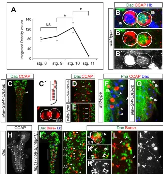
We next aimed to establish in more detail the early lineage of NB3-5. We took advantage of the fact that the only NB that expresses Ems before stage 11 is NB3-5. We stained for Ems, Hb, Kr, Pdm and, to distinguish GMCs from neurons, Deadpan (Dpn), a marker for NBs and GMCs (Fig. 4A-E; supplementary material Fig. S1A-C and Fig. S3A-D′) (Bier et al., 1992).
[image:3.612.49.365.55.320.2]NB3-5 initially expresses Hb, Kr and Pdm and generates a first GMC that divides to produce two cells that express all of these markers. At stage 10 the expression of Pdm is lost, but the expression of Hb and Kr is maintained and the NB divides to generate the second GMC that generates two more cells. At early stage 11 the NB only expresses Kr and generates the third GMC that produces two cells. At late stage 11 it expresses Pdm and generates the fourth GMC that again produces two cells. From this stage onwards it was difficult to follow the lineage, as NB3-3 and NB4-4, which also express Ems, have delaminated by this time, and cells from the three lineages become intermingled, which does not permit an unambiguous identification of the NB3-5 progeny. We conclude that, in the Hb temporal window, NB3-5 divides twice to generate four cells, here consisting of CCAP-EN and CCAP-IN, and two other cells with unknown fates (supplementary material Fig. S3E). As in segments T3-A4, two cells express CCAP, namely one IN and one EN (Fig. 1A). We aimed to establish whether they are sibling cells and, if not, what their order of appearance is in the lineage. Unfortunately, we lack molecular markers that would allow us to identify CCAP neurons at these early stages, since CCAP expression begins at stage 17 of embryogenesis. To circumvent this problem we analyzed GFP expression in CCAP neurons from embryos of the genotypeelav-Gal4 UAS-GFP tub-Gal80tsraised at 30°C and shifted to 17°C at early stage 9, since the data presented above indicate that CCAP neurons are born between stage 9 and
Fig. 2. NB3-5 is the progenitor of all CCAP neurons in the VNC.(A-G) Labeling for CCAP andβ-galactosidase (β-gal) inmirr-lacZ(A),gsb-lacZ(B),ems-Gal4 UAS-lacZ
(D) andhkb-lacZ(G), for CCAP and En in wild type (C), for CCAP inems1(E) and for CCAP and GFP ineg-Gal4 UAS-GFP(F). CCAP, red (except white in E);β-gal (A,B,D,G), En (C) or GFP (F), green. The segments shown in each figure are indicated bottom right. (H,I) Labeling for CCAP (red),β-gal (green) and Dac (blue) inelav-Gal4 UAS-flp tub-Gal80tsAct5C>stop>lacZ (H) orems-Gal4 UAS-flp tub-Gal80tsAct5C>stop>lacZ(I) grown at 29°C and shifted to 17°C at stage 10 of embryonic development. CCAP-expressing neurons were labeled in both experiments, indicating that they had been generated before stage 10. All images correspond to first instar larval ganglia. (J) Summary of the results presented in this figure, showing the pattern of NBs in a right hemisegment of a stage 11 embryo. NB3-5 is indicated. The pattern of expression of the different markers is illustrated. Anterior is up and the midline is on the left.
DEVEL
O
early stage 11. If we found cases in which only one of the CCAP neurons was labeled, this would mean that they were not siblings, and the labeled cell would be that which was generated first. In fact, we found several cases in which the CCAP-EN was the only CCAP neuron labeled, and the CCAP-IN was not labeled at all. This indicates that the two CCAP neurons are not siblings and that the CCAP-EN is generated first (Fig. 4F; supplementary material Fig. S4).
To corroborate these conclusions we conducted a cell lineage assay, labeling the progeny of NBs by injecting BrdU into stage 9 embryos (see Materials and Methods for details). BrdU is incorporated during the S phase of proliferating cells and labels all their progeny from then on; therefore, if CCAP neurons were not siblings, we would expect to find cases in which only one of them was labeled, as the NB would have incorporated BrdU in the S phase between the two mitoses that generated the two neurons (Novotny et al., 2002; Prokop and Technau, 1991); in this situation, the
labeled neuron would be that which was generated second. We observed several cases in which only the CCAP-IN was labeled, and no instances in which only the CCAP-EN was labeled (Fig. 4G-H″).
Thus, these results all agree in showing that the two CCAP neurons are not sibling cells and that the CCAP-EN is generated first.
Role of Notch in CCAP specification
[image:4.612.332.543.54.430.2]NBs divide asymmetrically to self-renew and generate a GMC that divides once to generate two sibling cells that usually acquire different
Fig. 3. All CCAP neurons in the VNC are specified in the Hb temporal window.(A-A″) Leucokinin (Lk; green) and CCAP (red) expression inhbFB hbP1. Lk expression is unaffected, whereas CCAP expression is lost. Lk is shown as a control, as Lk-expressing cells are generated in the Cas temporal window (Benito-Sipos et al., 2010). (B-C′) Dac (green) and CCAP (red) expression ininsc-Gal4 UAS-hb(B) andsvp1(C). Magnified views of hemisegments A4 (B,B′) and A1 (C,C′) (arrowheads) are shown at the bottom of each figure. (D) Schematic representation of right hemiganglia summarizing the phenotypes observed. Circles indicate wild-type (black) and new (red) cells expressing CCAP. Gray circles indicate CCAP expression that appears randomly in A1-4 segment wild-type ganglia of first instar larvae (see Fig. 1A). Full and empty circles represent EN and IN, respectively. Horizontal bars indicate the boundaries between subesophagus, thorax and abdomen.
Fig. 4. The early lineage of NB3-5.(A-E) Staining for Dpn (green), Hb (red) and Ems (blue) in embryos of stages 9/10 (A,B), 10 (C,D) and 11 (E). White circles indicate NB3-5 and its progeny, identified by expression of Ems. Dpn allows the identification of the NB and GMCs. C and D are two focal planes of the same ganglion showing the NB (C) and GMC2 (D). The green and red channels of A,B,D,E are shown in A′,B′,D′,E′. To the right are graphic representations in which circles represent cells expressing the indicated markers; NB3-5 and GMCs are indicated; midline is to the right, anterior is up. (F) Staining for GFP (green), CCAP (red) and Dac (blue) inelav-Gal4 UAS-GFP tub-Gal80tsfirst instar larvae grown at 30°C and shifted to 17°C at stage 9. Only the CCAP-EN is labeled with GFP. To the right is a graphic representation of the experimental scheme and of the patterns of expression. Separate channels of this figure are shown in supplementary material Fig. S4. (G-H″) Staining for BrdU (green), Hb (red) and Bursα(blue) in first instar larvae of embryos injected with BrdU at stage 9. The high level of Hb expression permits identification of the IN. (G) Only the IN is labeled with BrdU, suggesting that BrdU incorporation took place after generation of the EN and before generation of the IN. (H) Both EN and IN are labeled with BrdU. Green/red (G′,H′) and blue (G″,H″; here in white) channels are shown.
DEVEL
O
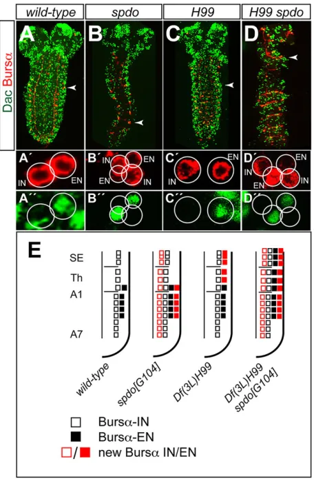
[image:4.612.76.274.56.407.2]fates. The Notch signaling pathway plays a role in distinguishing the fates of these two postmitotic cells (fates A and B) (Udolph et al., 2009). To address the role of Notch in the specification of CCAP neurons we examined its expression in mutants ofsanpodo(spdo), a gene required for asymmetric cell division (Babaoglan et al., 2009). Althoughspdomutants barely survive until late embryogenesis, we observed that both CCAP neurons, i.e. IN and EN, were duplicated in thoracic and abdominal segments (Fig. 5A-B″,E; in these experiments we labeled the expression of Bursα instead of CCAP, as Bursα expression is stronger in the abdominal segments at this stage and thus easier to identify). This confirms that CCAP-EN and CCAP-IN are not sibling cells and suggests that Notch signaling is OFF (fate B) in CCAP neurons and ON (fate A) in their respective sibling cells. Thus, compromising Notch signaling is sufficient to transform fate A into fate B and generate two CCAP-ENs and two CCAP-INs.
Programmed cell death in the NB3-5 lineage
A number of cells generated during embryonic neurogenesis undergo programmed cell death (PCD). A comparative analysis of the number of cells generated during embryonic neurogenesis in
wild-type and in PCD-deficient [Df(3L)H99] embryos indicated that∼37% of the cells died by apoptosis (Rogulja-Ortmann et al., 2007). Specifically, in the NB3-5 lineage, there were seven more cells inDf(3L)H99than in wild-type embryos [19-24 cells in wild type and 26-31 cells in Df(3L)H99]. We did not observe any increase in the number of Ems/Hb-expressing cells inDf(3L)H99 embryos at early stage 11 (data not shown). We examined whether the number of CCAP neurons was altered inDf(3L)H99embryos and found that an extra CCAP cell appeared in segments SE1-T2, whereas in T3 and the abdominal segments the number was not altered (Fig. 5C-C″,E). Since these extra CCAP cells expressed Dac, these results suggest that the CCAP-EN, which in the wild-type ganglion is only found in segments T3-A4, dies by apoptosis in segments SE1-T2. To confirm this we examined Df(3L)H99 spdoG104ganglia and observed an additive phenotype. This was especially informative in the SE segments where, on the one hand, the apoptosis of the EN was rescued, and, on the other hand, both the EN and the IN were duplicated (Fig. 5D-D″,E).
Different levels of Hb expression determine distinct neuronal fates
The above results indicate that both CCAP neurons are generated in the Hb temporal window. We then considered how these two cell fates were specified. One possibility is that different levels of Hb expression determine the different identities. During early embryo development Hb activates or represses different target genes in a concentration-dependent manner (Hülskamp et al., 1990; Schulz and Tautz, 1994; Struhl et al., 1992).
To assess whether different levels of Hb expression determine distinct neuronal fates, we first analyzed Hb expression in NB3-5 and observed that it increased over time from late stage 8 to stage 10, falling off quickly at stage 11 (Fig. 6A; supplementary material Table S2). We then stained Hb in CCAP neurons and observed that CCAP-IN (Dac–) expressed a high level of Hb and CCAP-EN (Dac+) a very low level of Hb (Fig. 6B-B″). This result suggests that the CCAP-IN fate is specified in the NB that expresses the higher level of Hb, and that specification also requires the maintenance of a high level of Hb expression in the postmitotic cell. To test this idea, we overexpressed Hb in neurons and monitored EN fate by assessing the expression of Dac (elav-Gal4 UAS-hb). In first instar larvae we found, in each segment from SE1 to A7, two CCAP cells, neither of which expressed Dac (Fig. 6C,C′). It is important to note that in wild-type ganglia we only observe two CCAP cells (one IN and one EN) in T3-A4, since in the more anterior segments (SE1-T2) CCAP-EN is not present and in the more posterior segments (A5-7) CCAP expression in EN starts later, although these cells are themselves generated during embryonic neurogenesis (Veverytsa and Allan, 2012). Thus, the lack of expression of Dac and the early onset of CCAP expression indicate that the sustained high level of Hb expression in postmitotic cells transforms CCAP-ENs into CCAP-INs. We also observed that the number of cells expressing Dac was strongly reduced compared with wild-type ganglia (Fig. 6D,E). These results suggest thatdaccan be a direct target of Hb (see Discussion).
[image:5.612.61.289.299.648.2]To strengthen this conclusion we monitored the axon projections of the CCAP-ENs in both wild-type andelav-Gal4 UAS-hbganglia of first instar larvae. In wild type we observed an axon emerging from the EN, whereas inelav-Gal4 UAS-hbwe never observed these efferent axonal projections, indicating that they no longer exit the ganglion as EN but instead seem to behave as CCAP-IN (Fig. 6F,G). It is also important to note that in adac mutant we observed no change in the pattern or onset of CCAP expression or in the number
Fig. 5. The role of Notch and PCD in the specification of CCAP neurons.
(A-D) Expression of Dac (green) and Bursα(red) in wild-type (A),spdoG104(B),
Df(3L)H99(C) andDf(3L)H99 spdoG104(D) first instar larvae. The red (A′-D′) and green (A″-D″) channels of a magnified hemisegment (arrowheads) are shown separately beneath. (E) Summary of the phenotypes in this figure. Squares represent cells expressing Bursαin the ventral ganglion of first instar larvae (see Fig. 1A); other symbols as in Fig. 3. Note that Bursα/Dac labeling allows the detection of ENs in T3-A4 in first instar larvae; the onset of CCAP
expression in these neurons occurs later on development (Fig. 1A).
DEVEL
O
of CCAP neurons, which clearly indicates that overexpression of Hb not only removes Dac expression from CCAP-ENs but also transforms them into CCAP-INs (Fig. 6H).
The model that we propose is that the absolute level of Hb expression in the NB determines the fate of the CCAP neuron: high in CCAP-IN and low in CCAP-EN. If so, reducing the level of Hb expression should convert the CCAP-INs to CCAP-ENs. To test this prediction we labeled Bursα expression in an hb hypomorphic condition (hb1/hbFB hbP1) and observed that INs in SE1-T2 segments were lost. In segments T3-A4 we observed one or two CCAP neurons per hemisegment, all of which expressed Dac (Fig. 6I-K′) and produced axons that exited the ganglion (Fig. 6L,L′). These findings indicated that, in the hb hypomorphic condition, CCAP-INs are either lost or transformed into ENs.
Together, these results support the view that the absolute level of Hb expression in NB3-5 determines CCAP neuronal subtype.
DISCUSSION
In this study we analyzed how CCAP-expressing neurons are specified. We obtained evidence that both the CCAP-ENs and CCAP-INs of all embryonic segments are generated by NB3-5. Our results also indicate that CCAP neurons are generated in the Hb temporal window, are not sibling cells and that the CCAP-ENs are generated first followed by the CCAP-INs (Fig. 7). Although the Hb temporal window in NB3-5 generates two GMCs that can be distinguished by the expression of Pdm in GMC1, Pdm does not seem to play any role in the specification of these neurons, as we did not observe any phenotype inpdmmutants.
[image:6.612.141.469.54.404.2]These findings raised the question of how these two neuronal fates are generated, and the results that we present here suggest that different levels of Hb expression specify them. The evidence for this is as follows. First, Hb expression in NB3-5 increases over time from stage 9 to early stage 11, then its expression quickly fades,
Fig. 6. CCAP-IN and CCAP-EN express different levels of Hb.(A) Levels of Hb expression (integrated density values in thousands of pixels;n=6 at each stage; for details see Materials and Methods and supplementary material Table S2) in the NB3-5 of stages 8-11. *P<0.01, two-tailed Student’st-test; NS, not significant. (B-B″) Expression of Dac (green), CCAP (red) and Hb (blue) in the CCAP neurons of a wild-type first instar larva. Green/red (B′) and blue (B″) channels are shown separately. Hb expression is very strong in the CCAP-IN and very weak in the CCAP-EN (B″), as identified by the expression of Dac in the EN (B′). (C) Expression of Dac (green) and CCAP (red) inelav-Gal4 UAS-hb. A magnified view of a hemisegment (arrowhead) is shown to the right. There are two CCAP neurons in each hemisegment and neither expresses Dac. (C′) Schematic of the phenotype in C. (D,E) Expression of Dac (green) and CCAP (red) inelav-Gal4 UAS-hb(D) and wild type (E). Three abdominal segments of first instar larva ganglia are shown (white bars). Note the reduction in the number of cells inelav-Gal4 UAS-hbexpressing Dac. (F,G) Phalloidin labeling (green) and labeling for CCAP (red) and Dac (blue), showing the projection of efferent axons (arrowheads) in wild-type (F) andelav-Gal4 UAS-hb(G) first instar larvae. (H) CCAP expression indac4is unaffected. (I,I′) Dac (green), Bursα(red) and Lk (blue) expression inhb1/hbFBhbP1first instar larva. The expression of Lk is unaffected inhbmutants, which permits the identification of the abdominal segments (Benito-Sipos et al., 2010). The white bar indicates the area magnified in I′(in which only green and red channels are shown). There are one or two ENs per hemisegment (arrowheads in I′) and INs are not found, suggesting they are lost or converted into ENs. (J) Magnified view showing anhb1/hbFBhbP1abdominal segment with two ENs. (K,K′) Magnified view showing duplications of ENs on both sides of two segments (arrowheads). (L,L′) Magnified view of CCAP-ENs showing the efferent contralateral axons (arrowheads). The red channel (Bursα) is shown separately in white (K′,L′).
DEVEL
O
coinciding with the reported expression of Svp, which is known to close the Hb temporal window (Broadus et al., 1995; Kanai et al., 2005; Mettler et al., 2006). During this time window, NB3-5 divides twice and generates four neurons. Second, overexpression of high levels of Hb using a pan-NB driver extends the IN fate. Third, in an hb hypomorphic condition CCAP-INs are lost or converted into ENs, as monitored by the expression of Dac and the presence of axons that exit the ganglion.
This mechanism for generating distinct neuronal fates is different from that proposed for subdividing the Cas temporal window in NB5-6, which involves two sequential feed-forward loops and several genes to define the fates of four cells (Ap1-4) that are sequentially generated and form the Apterous (Ap) cluster of neurons. However, the mechanism that we propose is very similar to the role that the grh gene plays in the Ap cluster, since Grh expression increases gradually over time from Ap1 to Ap4, and overexpression of Grh converts all four Ap neurons into Ap4 (Baumgardt et al., 2009).
In addition to the different levels of Hb expression observed in NB3-5, we found that CCAP-ENs and CCAP-INs express low and high levels of Hb, respectively, and overexpression of Hb in postmitotic cells convert the ENs into INs. These observations raise the question of how a high level of Hb expression in the NB leads to a high level of expression in the neuron. A recent analysis of thehb regulatory region revealed a specific postmitotic enhancer (Hirono et al., 2012), so it would be tempting to propose that this enhancer is only activated in neurons that are generated by a NB expressing a high level of Hb. However, we failed to detect expression of this enhancer in any of the CCAP neurons, and overexpression of Hb in the NB did not lead to activation of the enhancer in neurons (data not shown). Therefore, further work is needed to identify the mechanism by which only a subset of the neurons generated in the Hb temporal window expresses a high level of Hb and how this is translated into different neuronal fates.
We have observed that CCAP-INs express a high level of Hb and do not express Dac, and that upon Hb overexpression the expression
of Dac is lost in many, although not all, cells. This could placedacas a direct target of Hb. Analysis ofdaccis-regulatory domains indicates the presence of a 5.8 kb domain in the first intron that, when placed in a Gal4 vector, was sufficient to drive GFP expressionin vivoin many neurons of late embryos (Pfeiffer et al., 2008). A preliminary analysis of the sequence of this domain suggests the presence of conserved regions and putative Hb binding sites (supplementary material Fig. S5A,B). Further analysis will be required to confirm the presence and elucidate the function of such sequences.
Ikaros(orIkzf1), a mouse ortholog of Hb, is expressed in all early retinal progenitor cells (RPCs) of the developing retina. Its expression in RPCs is necessary and sufficient to confer the competence to generate early-born neurons (Elliott et al., 2008). These and other observations suggest that, as in the Drosophila CNS, cell-intrinsic mechanisms act in the RPC to control temporal competence (Cayouette et al., 2006). Ikaros is expressed in the early RPCs that give rise to several cell types, namely horizontal, amacrine and gangion cells; however, it is unclear whether distinct levels of Ikaros expression are responsible for the production of these different cell types (Elliott et al., 2008).
In the early embryo, different concentrations of Hb seem to elicit different cellular responses. At low concentrations, Hb monomers function as activators, whereas at high concentrations they form dimers that either repress transcription or block activation (Hülskamp et al., 1990; Papatsenko and Levine, 2008; Schulz and Tautz, 1994). Analysis of the Hb protein has led to the identification of two conserved domains: a DNA-binding domain and a dimerization domain (McCarty et al., 2003). More recently, it has been shown that, in CNS development, Hb repressor function is required to maintain early NB competence and to specify early-born neuronal identity (Tran et al., 2010). These results are compatible with the evidence presented here that it is the absolute level of Hb in a NB that determines whether it is expressed in the postmitotic progeny and so specifies the different neuronal subtypes.
MATERIALS AND METHODS Fly strains
The fly stocks used were: hbFB hbP1 and Kr1 KrCD (which prevent,
respectively,hbandKrCNS expression but rescue segmentation expression)
(Isshiki et al., 2001), hb7, hb1 (hypomorphic allele), Df(2L)ED773
(which deletespdm1andpdm2),casΔ1,casΔ3,grhIM,spdoG104,svp1,dac4,
ems1,Df(3L)H99,y w UAS-flp122; Act5C>y+>lacZandCanton-Sas
wild-type stock.
Gal4/Gal80 lines were:elavC155-Gal4,insc-Gal4,wor-Gal4,ems-Gal4,
eg-Gal4,CCAP-Gal4,tub-Gal80ts.
UAS lines were: UAS-GFP, UAS-lacZ, UAS-hb, UAS-Kr, UAS-pdm,
UAS-cas,UAS-grh.
lacZlines were:ems-lacZ,hkb-lacZ,mirr-lacZ,gsb01155-lacZ(FlyBase).
Immunohistochemistry and confocal imaging
Primary antibodies used were: mouse anti-GFP (1:200; Roche #11814460), rabbit anti-GFP (1:200; Invitrogen #A6455); mouse anti-BrdU (1:10; Roche #11296736001); rabbit anti-Lk (1:100; provided by D. Nässel, Stockholm
University, Sweden); rat anti-CCAP (1:100; this work); rabbit anti-Bursα
(1:250; provided by B. White, National Institute of Mental Health, Bethesda, USA); rabbit anti-Dpn (1:40; provided by D. Strutt, University of Sheffield, UK); guinea pig anti-Hb (1:200; provided by I. Miguel-Aliaga, Imperial College, London, UK); rabbit anti-Kr (1:500; provided by P. Carrera, Max Planck Institute, Göttingen, Germany); rabbit anti-Pdm1 (1:500; Terriente et al., 2008); guinea pig anti-Cas (1:500; provided by T. Isshiki, National Institute of Genetics, Mishima, Japan); rat anti-Ems (1:10; provided by U. Walldorf,
University of Saarland, Homburg/Saar, Germany); mouse anti-β-galactosidase
[image:7.612.80.268.55.224.2](1:50; DSHB #40-1a); mouse anti-En (1:50; DSHB #4D9); and mouse anti-Dac (1:50; DSHB #mAbdac2-3). Phalloidin-TRITC was from Sigma (#P1951). Fig. 7. Summary of the genetic network involved in CCAP neuron
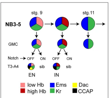
specification.Summary of the Hb temporal window in NB3-5. From stage 9 to stage 11, NB3-5 divides twice in the Hb/Kr temporal window; at stage 11 Hb expression is switched off. The first GMC generates the CCAP-EN, which is identified by the expression of Ems, CCAP, Bursαand Dac in T3-A4. In first instar larva this neuron expresses low levels of Hb. The second GMC generates the CCAP-IN in segments SE1-A7; in first instar larva this neuron expresses high levels of Hb and Ems, CCAP and Bursα. The third GMC expresses Ems and Kr. CCAP neurons are NotchOFF; the identity of their sibling cells is unknown.
DEVEL
O
The level of Hb expression in the early lineage of NB3-5 was evaluated by staining embryos of stages 8-11, first with guinea pig anti-Hb primary antibody and then with Alexa Fluor 555 goat anti-guinea pig secondary antibody (1:500; Molecular Probes #21435). The fluorescence of NB images was measured by calculating mean gray values (average gray values within selected areas) using ImageJ software (NIH). Integrated density (area × mean gray value) was used to compare fluorescence intensities.
Immunostaining was performed according to Benito-Sipos et al. (2010) and confocal image stacks were collected using a Zeiss LSM710 or LSM510 confocal microscope.
Antibody production
To generate anti-CCAP antibody, two rats were immunized with the peptide KRPFCNAFTGCGRKC, which includes the sequence of the mature peptide. The terminal Cys residue was added to couple the peptide to keyhole limpet hemocyanin carrier protein. After five immunizations, the rats were bled and the resulting sera were tested for CCAP-specific staining of the larval CNS.
BrdU labeling
Embryos at stage 9 were collected, dechorionated and injected with 10 mM
5-bromo-2′-deoxyuridine solution (BrdU detection kit; Roche #11296736001).
First instar larvae were fixed by standard procedures, treated with DNase (RQ1, Promega #M199A) and stained with mouse anti-BrdU and guinea piganti-Hb and anti-Dac.
Acknowledgements
We thank P. Carrera, C. Doe, J. Ewer, T. Isshiki, D. Nässel, I. Miguel-Aliaga, D. Strutt, S. Thor, U. Walldorf, B. White and the Bloomington Drosophila Stock Center (BDSC) for providing fly stocks and/or reagents; E. Caminero, B. Fraile, A. Montes and the confocal microscopy service of the CBM-SO for technical help. The mouse anti-Dac monoclonal antibody developed by G. Rubin was obtained from the Developmental Studies Hybridoma Bank, which was created by the NICHD of the NIH and is maintained at The University of Iowa, Department of Biology and guinea pig Iowa City, IA 52242, USA.
Competing interests
The authors declare no competing financial interests.
Author contributions
M.M.-S. and F.J.D.-B. conceived and designed the experiments and analyzed the data. M.M.-S., A.E.-G. and J.Á.-R. performed the experiments. F.J.D.-B. wrote the paper.
Funding
This work was supported by a pre-doctoral fellowship from the C.S.I.C. to M.M.-S. [JAEPre-08-01279] and from the Ministerio de Educación to A.E.-G. [AP2008-00397], by grants from the Ministerio de Ciencia e Innovación [CSD2007-00008 and BFU2011-24315] to F.J.D.-B. and by an institutional grant from the Fundación Ramón Areces to the CBM-SO.
Supplementary material
Supplementary material available online at
http://dev.biologists.org/lookup/suppl/doi:10.1242/dev.113381/-/DC1
References
Babaoglan, A. B., O’Connor-Giles, K. M., Mistry, H., Schickedanz, A., Wilson, B. A. and Skeath, J. B.(2009). Sanpodo: a context-dependent activator and inhibitor of Notch signaling during asymmetric divisions. Development 136, 4089-4098.
Baumgardt, M., Karlsson, D., Terriente, J., Dıaz-Benjumea, F. J. and Thor, S.́
(2009). Neuronal subtype specification within a lineage by opposing temporal feed-forward loops.Cell139, 969-982.
Benito-Sipos, J., Estacio-Gomez, A., Moris-Sanz, M., Baumgardt, M., Thor, S. and Diaz-Benjumea, F. J.(2010). A genetic cascade involving klumpfuss, nab and castor specifies the abdominal leucokinergic neurons in the Drosophila CNS.
Development137, 3327-3336.
Bier, E., Vaessin, H., Younger-Shepherd, S., Jan, L. Y. and Jan, Y. N.(1992). deadpan, an essential pan-neural gene in Drosophila, encodes a helix-loop-helix protein similar to the hairy gene product.Genes Dev.6, 2137-2151.
Birkholz, O., Rickert, C., Berger, C., Urbach, R. and Technau, G. M.(2013). Neuroblast pattern and identity in the Drosophila tail region and role of doublesex in the survival of sex-specific precursors.Development140, 1830-1842.
Broadus, J., Skeath, J. B., Spana, E. P., Bossing, T., Technau, G. and Doe, C. Q.
(1995). New neuroblast markers and the origin of the aCC/pCC neurons in the Drosophila central nervous system.Mech. Dev.53, 393-402.
Brody, T. and Odenwald, W. F.(2000). Programmed transformations in neuroblast gene expression during Drosophila CNS lineage development.Dev. Biol.226, 34-44.
Cayouette, M., Poggi, L. and Harris, W. A.(2006). Lineage in the vertebrate retina.
Trends Neurosci.29, 563-570.
Cleary, M. D. and Doe, C. Q.(2006). Regulation of neuroblast competence: multiple temporal identity factors specify distinct neuronal fates within a single early competence window.Genes Dev.20, 429-434.
Dewey, E. M., McNabb, S. L., Ewer, J., Kuo, G. R., Takanishi, C. L., Truman, J. W. and Honegger, H.-W.(2004). Identification of the gene encoding bursicon, an insect neuropeptide responsible for cuticle sclerotization and wing spreading.
Curr. Biol.14, 1208-1213.
Doe, C. Q.(1992). Molecular markers for identified neuroblasts and ganglion mother cells in the Drosophila central nervous system.Development116, 855-863.
Doe, C. Q.(2008). Neural stem cells: balancing self-renewal with differentiation.
Development135, 1575-1587.
Dulcis, D., Levine, R. B. and Ewer, J.(2005). Role of the neuropeptide CCAP in Drosophila cardiac function.J. Neurobiol.64, 259-274.
Elliott, J., Jolicoeur, C., Ramamurthy, V. and Cayouette, M. (2008). Ikaros confers early temporal competence to mouse retinal progenitor cells.Neuron60, 26-39.
Ewer, J.(2005). Behavioral actions of neuropeptides in invertebrates: insights from Drosophila.Horm. Behav.48, 418-429.
Gaspard, N. and Vanderhaeghen, P.(2010). Mechanisms of neural specification from embryonic stem cells.Curr. Opin. Neurobiol.20, 37-43.
Grosskortenhaus, R., Pearson, B. J., Marusich, A. and Doe, C. Q.(2005). Regulation of temporal identity transitions in Drosophila neuroblasts.Dev. Cell8, 193-202.
Grosskortenhaus, R., Robinson, K. J. and Doe, C. Q.(2006). Pdm and Castor specify late-born motor neuron identity in the NB7-1 lineage.Genes Dev.20, 2618-2627.
Hirono, K., Margolis, J. S., Posakony, J. W. and Doe, C. Q.(2012). Identification of hunchback cis-regulatory DNA conferring temporal expression in neuroblasts and neurons.Gene Expr. Patterns12, 11-17.
Hülskamp, M., Pfeifle, C. and Tautz, D.(1990). A morphogenetic gradient of hunchback protein organizes the expression of the gap genes Kruppel and knirps in the early Drosophila embryo.Nature346, 577-580.
Isshiki, T., Pearson, B., Holbrook, S. and Doe, C. Q. (2001). Drosophila
neuroblasts sequentially express transcription factors which specify the temporal identity of their neuronal progeny.Cell106, 511-521.
Kambadur, R., Koizumi, K., Stivers, C., Nagle, J., Poole, S. J. and Odenwald, W. F.(1998). Regulation of POU genes by castor and hunchback establishes layered compartments in the Drosophila CNS.Genes Dev.12, 246-260.
Kanai, M. I., Okabe, M. and Hiromi, Y.(2005). seven-up Controls switching of transcription factors that specify temporal identities of Drosophila neuroblasts.
Dev. Cell8, 203-213.
Kim, Y.-J.,Žitnˇan, D., Galizia, C. G., Cho, K.-H. and Adams, M. E.(2006). A command chemical triggers an innate behavior by sequential activation of multiple peptidergic ensembles.Curr. Biol.16, 1395-1407.
Knoblich, J. A.(2008). Mechanisms of asymmetric stem cell division.Cell132, 583-597.
McCarty, A. S., Kleiger, G., Eisenberg, D. and Smale, S. T.(2003). Selective dimerization of a C2H2 zinc finger subfamily.Mol. Cell11, 459-470.
Mesce, K. A. and Fahrbach, S. E.(2002). Integration of endocrine signals that regulate insect ecdysis.Front. Neuroendocrinol.23, 179-199.
Mettler, U., Vogler, G. and Urban, J.(2006). Timing of identity: spatiotemporal regulation of hunchback in neuroblast lineages of Drosophila by Seven-up and Prospero.Development133, 429-437.
Nässel, D. R.(2002). Neuropeptides in the nervous system of Drosophila and other insects: multiple roles as neuromodulators and neurohormones.Prog. Neurobiol. 68, 1-84.
Nässel, D. R. and Winther, A. M.(2010). Drosophila neuropeptides in regulation of physiology and behavior.Prog. Neurobiol.92, 42-104.
Novotny, T., Eiselt, R. and Urban, J.(2002). Hunchback is required for the specification of the early sublineage of neuroblast 7-3 in the Drosophila central nervous system.Development129, 1027-1036.
Papatsenko, D. and Levine, M. S.(2008). Dual regulation by the Hunchback gradient in the Drosophila embryo.Proc. Natl. Acad. Sci. USA105, 2901-2906.
Park, J. H., Schroeder, A. J., Helfrich-Förster, C., Jackson, F. R. and Ewer, J.
(2003). Targeted ablation of CCAP neuropeptide-containing neurons of Drosophila causes specific defects in execution and circadian timing of ecdysis behavior.Development130, 2645-2656.
Peabody, N. C., Diao, F., Luan, H., Wang, H., Dewey, E. M., Honegger, H.-W. and White, B. H.(2008). Bursicon functions within the Drosophila CNS to modulate wing expansion behavior, hormone secretion, and cell death.J. Neurosci.28,
14379-14391.
DEVEL
O
Pearson, B. J. and Doe, C. Q.(2003). Regulation of neuroblast competence in Drosophila.Nature425, 624-628.
Pfeiffer, B. D., Jenett, A., Hammonds, A. S., Ngo, T.-T. B., Misra, S., Murphy, C., Scully, A., Carlson, J. W., Wan, K. H., Laverty, T. R. et al.(2008). Tools for neuroanatomy and neurogenetics in Drosophila.Proc. Natl. Acad. Sci. USA105, 9715-9720.
Prokop, A. and Technau, G. M.(1991). The origin of postembryonic neuroblasts in the ventral nerve cord of Drosophila melanogaster. Development 111, 79-88.
Rogulja-Ortmann, A., Luer, K., Seibert, J., Rickert, C. and Technau, G. M.
(2007). Programmed cell death in the embryonic central nervous system of Drosophila melanogaster.Development134, 105-116.
Santos, J. G., Vömel, M., Struck, R., Homberg, U., Nässel, D. R. and Wegener, C.
(2007). Neuroarchitecture of peptidergic systems in the larval ventral ganglion of Drosophila melanogaster.PLoS ONE2, e695.
Schulz, C. and Tautz, D.(1994). Autonomous concentration-dependent activation and repression of Kruppel by hunchback in the Drosophila embryo.Development 120, 3043-3049.
Struhl, G., Johnston, P. and Lawrence, P. A.(1992). Control of Drosophila body pattern by thehunchbackmorphogen gradient.Cell69, 237-249.
Technau, G. M., Berger, C. and Urbach, R.(2006). Generation of cell diversity and segmental pattern in the embryonic central nervous system of Drosophila.Dev. Dyn.235, 861-869.
Terriente, J., Perea, D., Suzanne, M. and Diaz-Benjumea, F. J. (2008). The Drosophila gene zfh2 is required to establish proximal-distal domains in the wing disc.Dev. Biol.320, 102-112
Tran, K. T. and Doe, C. Q.(2008). Pdm and Castor close successive temporal identity windows in the NB3-1 lineage.Development135, 3491-3499.
Tran, K. D., Miller, M. R. and Doe, C. Q.(2010). Recombineering Hunchback identifies two conserved domains required to maintain neuroblast competence and specify early-born neuronal identity.Development137, 1421-1430.
Tsuji, T., Hasegawa, E. and Isshiki, T.(2008). Neuroblast entry into quiescence is regulated intrinsically by the combined action of spatial Hox proteins and temporal identity factors.Development135, 3859-3869.
Udolph, G., Rath, P., Tio, M., Toh, J., Fang, W., Pandey, R., Technau, G. M. and Chia, W.(2009). On the roles of Notch, Delta, kuzbanian, and inscuteable during the development of Drosophila embryonic neuroblast lineages.Dev. Biol.336, 156-168.
Veverytsa, L. and Allan, D. W. (2011). Retrograde BMP signaling controls Drosophila behavior through regulation of a peptide hormone battery.
Development138, 3147-3157.
Veverytsa, L. and Allan, D. W.(2012). Temporally tuned neuronal differentiation supports the functional remodeling of a neuronal network in Drosophila.Proc. Natl. Acad. Sci. USA109, E748-E756.
Vömel, M. and Wegener, C.(2007). Neurotransmitter-induced changes in the intracellular calcium concentration suggest a differential central modulation of CCAP neuron subsets in Drosophila.Dev. Neurobiol.67, 792-808.