Thesis by
Michelle Elizabeth Farkas
In Partial Fulfillment of the Requirements
for the Degree of
Doctor of Philosophy
California Institute of Technology
Pasadena, California
2010
© 2010
Acknowledgements
I would like to thank my advisor, Peter Dervan, for providing a wonderful environment in which to perform research, and for all of the insight he has offered me with regard to projects, academia, and life in general. I would also like to thank the members of my thesis committee, Dennis Dougherty, Judy Campbell, and Brian Stoltz, for all of their support (and letters of recommendation) over the last five years. Additionally, I would like to thank Jackie Barton, who I have had the privilege of being a teaching assistant for for several years of my graduate career; it was truly a pleasure.
The Dervan lab has been a wonderful place to spend five years of graduate school, and the people in it made all the difference. I have had the opportunity to work with David Chenoweth, Christian Dose, Claire Jacobs, Dan Harki, Ben Li, Katy Muzikar, Nick Nickols, and Sherry Tsai on a number of projects. My collaboration with Sherry was a very important and fruitful one – I will never forget Team Chl and all of the trips to Scripps during my candidacy. I have thoroughly enjoyed being roomies with John Phillips, and his predecessor, Eric Fechter, who taught me a lot when I first joined the lab. I would also like to thank Ray Doss and Adam Poulin-Kerstein, two other fifth-years during my first year, for their friendship and wisdom offered throughout my own graduate career. But perhaps most importantly, I have come to value the friendships of Justin Cohen, Mareike Goeritz, and Katy Muzikar – no matter what corners of the world we end up in, I hope that we will always stay in touch. I like to thank the other members of the lab as well – Mike Brochu, Dan Gubler, Carey Hsu, Michael Marques, Dave Montgomery, Julie Poposki, Jim Puckett, Jim Sanchez, Ryan Stafford, Anne Viger, and Fei Yang. I have truly enjoyed the time that I have spent with all of you.
Lynne Martinez who has been incredible in her administrative role in the group.
There are many other people outside of the chemistry department whom I would like to thank for helpng me get here. Thanks to all of my roommates, Snatch teammates, and other friends throughout the years at Caltech – it’s been really nice to have somewhere to go outside of lab. Thanks to all of my high school and Wellesley friends who’ve watched me go through this and been as supportive as they have, despite my falling off of the face of the earth every so often. Thanks to Pam Sontz for not only being a great friend, but also reading this thesis. And thanks to Carolyn Brinkworth for both being and doing so much more than that. I would also like to thank my former mentor Dr. Sandor Karady at Merck who allowed me to do research as a high school student; he set me on a course that brought me to where I am today. Dr. Kap-Sun Yeung was a wonderful person to work for during my years at Bristol-Myers Squibb.
Abstract
Acknowledgements...iv
Abstract...vi
Table of Contents...vii
List of Figures and Tables...viii
Chapter I. Introduction...1
Part II. Py-Im Polyamides linked by 2,4-diaminobutyric acid in the hairpin turn unit...22
Chapter IIA. Unanticipated differences between α- and γ- diaminobutyric acid-linked hairpin polyamide-alkylator conjugates...23
Chapter IIB. Characterization of α-diaminobutyric acid-linked hairpin polyamides...49
Chapter IIC. Small molecules targeting histone H4 as potential therapeutics for chronic myelogenous leukemia...79
Part III. Substituent effects in the γ turn unit of hairpin Py-Im Polyamides...111
Chapter IIIA. Next generation hairpin polyamides with (R)-3,4-diaminobutyric acid turn unit...112
Chapter IIIB. DNA sequence selectivity of hairpin polyamide turn units...143
List of Figures and Tables
Chapter I Page
Figure I.1 The structure of DNA...3
Figure I.2 Structures of five protein-DNA complexes...4
Figure I.3 Chemical structures of DNA-binding natural products...5
Figure I.4 Crystal structures of distamycin bound to DNA...6
Figure I.5 Recognition of the DNA minor groove by polyamides...8
Figure I.6 Crystal structures of polyamides bound to DNA...9
Figure I.7 Binding model for the polyamide CtPyPyIm-(R)RHN γ-PyImPyPy-β-Dp targeted to the sequence 5’-TATACGT-3’...11
Table I.1 Library of imidazole-capped polyamides...13
Figure I.8 Polyamides as regulators of gene expression in cell culture...14
Figure I.9 Examples of various polyamide-alkylator conjugates...16
Chapter IIA Figure IIA.1 Polyamide structures and syntheses...28
Figure IIA.2 Plasmid design and DNA binding properties of parent polyamides...30
Figure IIA.3 Illustration of parent polyamides 2R and 2S binding DNA...31
Table IIA.1 Binding affinities for parent polyamides...31
Figure IIA.4 Models of the binding of 1R and 1S to the match site of pMFST2...32
Figure IIA.5 Alkylation specficities of the polyamide conjugates...33
Figure IIA.6 Time-dependance of alkylation for polyamide conjugates...35
Figure IIA.9 Fluorescence-activated cell-sorting analysis of the effects of
polyamide conjugates on SW620 cells...40
Figure IIA.10 Effects of polyamide conjugates on BALB/c mice...41
Chapter IIB Figure IIB.1 Structures of polyamides 1-8 and their Chl conjugates...53
Figure IIB.2 Illustration of the inserts from plasmids pMFST2, pMFST, and pMFST3...55
Figure IIB.3 Quantitative DNase I footprinting titration experiments for Polyamides 1-8...57
Table IIB.1 Binding affinities for parents polyamides on plasmids pMFST2, pMFST, and pMFST3...58
Figure IIB.4 Thermal cleavage assay experiments with polyamide-chlorambucil conjugates...60
Figure IIB.5 Illustration of the binding modes for polyamides 3 and 7...61
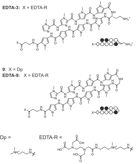
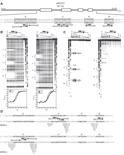
Figure IIB.6 Structures of EDTA-3, and the glycine-linked polyamide 9 and its EDTA conjugate EDTA-9...63
Figure IIB.7 Experiments involving polyamides 3, 9, EDTA-3, and EDTA-9 on plasmid pMFST5...64
Table IIB.2 Binding affinities for parent polyamides 3 and 9 on plasmid pMFST5...65
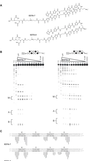
Figure IIB.8 Affinity cleavage experiments for EDTA-7 and EDTA-8...67
Figure IIB.9 Affinity cleavage experiments for EDTA-10 on plasmid pMFST4...68
Figure IIB.10 Structures of polyamide-fluorescein conjugates...70
Chapter IIC
Figure IIC.1 DNA sequence of the coding region of the histone H4C gene and
chemical structures for 1R-Chl and 6R-Chl...83
Table IIC.1 Analysis of a polyamide library...84
Figure IIC.2 Polyamide analysis on pMFST6...85
Figure IIC.3 Alkylation of the H4c gene in vitro...87
Figure IIC.4 Effects of polyamides in cell culture...88
Figure IIC.5 Analysis of inactive polyamide in K562 cells...89
Figure IIC.6 Alkylation of the H4c gene in K562 cells...91
Figure IIC.7 Effect of polyamides on histone H4c and H4k/j transcript expression...92
Figure IIC.8 Real time qRT-PCR of genes...94
Figure IIC.9 Murine K562 xenograft studies...95
Figure IIC.10 Pharmacokinetic parameters and biodistribution of 1R-Chl...97
Table IIC.2 Primers used for real-time qRT-PCR...104
Chapter IIIA Figure IIIA.1 Schematic representation of hairpin polyamides with different turn units...115
Figure IIIA.2 Chemical structures for polyamides 1-16...116
Figure IIIA.3 DNase I footprinting experiments for polyamides 2-4...117
Figure IIIA.4 DNase I footprinting experiments for polyamides 6-8...118
Figure IIIA.5 DNase I footprinting experiments for polyamides 10-12...119
Figure IIIA.6 DNase I footprinting experiments for polyamides 14-16...120
Table IIIA.2 Melting temperatures of DNA/polyamide complexes...123 Figure IIIA.8 Illustrative models of different turn conformations...125 Table IIIA.3 Melting temperatures of DNA/polyamide complexes with
C•G and G•C base pairs at the turn position...127
Figure IIIA.9 Chemical structures of acetylated polyamides...129 Table IIIA.4 Melting temperatures for DNA complexes with acetylated hairpins...129 Figure IIIA.10 Schematic representation of the androgen receptor
transcription complex...130 Figure IIIA.11 Structures of polyamides 20-23, and their effects on gene
expression in cell culture...131 Table IIIA.5 Melting temperatures of polyamides 20-23...132
Chapter IIIB
Figure IIIB.1 Schematic diagram of a six-ring hairpin polyamide targeting DNA...146 Figure IIIB.2 Structures of polyamides targeting 5’-WWGGWW-3’...148
Figure IIIB.3 Structures of polyamides 6-11...148 Table IIIB.1 Melting temperatures of DNA/polyamide complexes targeting
5’-WWGGWW-3’...149
Table IIIB.2 Melting temperatures of DNA/polyamide complexes targeting
5’-WWGWWW-3’ and 5’-WWGGCW-3’...150
Figure IIIB.4 Illustration of the designed inserts for plasmids pCDMF6
and pCDMF4...151 Figure IIIB.5 Quantitative DNase I footprinting experiments for polyamides
targeting 5’-WWGGWW-3’...152
Table IIIB.4 Relative binding affinities for polyamides targeting
5’-WWGGWW-3’...153
Figure IIIB.6 Quantitative DNase I footprinting experiments for polyamides
targeting 5’-WWGWWW-3’...154
Table IIIB.5 Binding affinities for polyamides targeting 5’-WWGWWW-3’...155
Table IIIB.6 Relative binding affinities for polyamides targeting
5’-WWGWWW-3’...155
Figure IIIB.7 Structures of polyamides containing fluoro- and
hydroxyl-substituted hairpin turns...156 Table IIIB.7 Melting temperatures of DNA/polyamide complexes for polyamides
containing fluoro- and hydroxyl-substituted hairpin turns...156
Figure IIIB.8 Quantitative DNase I footprinting experiments for polyamides
containing fluoro- and hydroxyl-substituted hairpin turns...157
Table IIIB.8 Binding affinities for polyamides containing fluoro- and
hydroxyl-substituted hairpin turns...158 Table IIIB.9 Relative binding affinities for polyamides containing fluoro- and
hydroxyl-substituted hairpin turns...158 Figure IIIB.9 Synthesis of fluorine-substituted hairpin turns...161
Figure IIIB.10 Synthesis of hydroxyl-substituted hairpin turns...161
Appendix
Figure A.1 Polyamides targeting the androgen response element...175 Figure A.2 Illustration of chromatin immunoprecipitation versus pull-down
with polyamide-biotin conjugates...177 Figure A.3 Structures of polyamides linked to biotin at the turn subunit...178 Figure A.4 Effects of polyamides 1-3 on the expression of androgen responsive
Table A.2 Melting temperatures of DNA/polyamide complexes...182
Figure A.6 Effects of polyamides 4-9 on PSA expression...183
Figure A.7 Electrophoretic mobility shift assay with polyamides 8 and 9...183
Figure A.8 Structure of polyamide 10...184
Figure A.9 Relative amplification levels of PSA ARE III and FKBP5 following polyamide incubation with sheared chromatin...185
Figure A.10 Relative amplification levels of PSA ARE I, PSA ARE III, and FKBP5 following polyamide treatment in LNCaP cells...186
Chapter I
human genome contains approximately three billion base pairs, which encode 20,000 to 25,000 protein-coding genes.1 Sequences of DNA can not only code for proteins, but are also associated with regulatory regions controlling transcription. In the cell, gene expression is controlled by DNA-binding proteins and the interactions between proteins.2,3
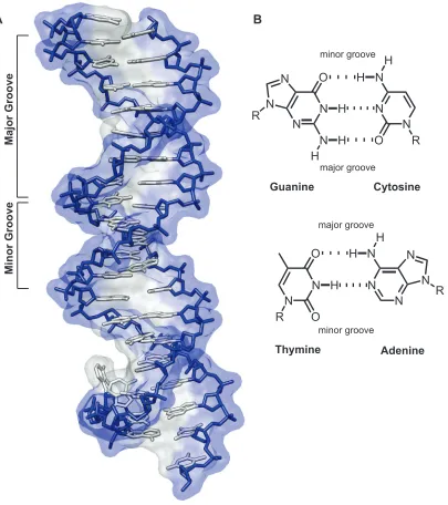
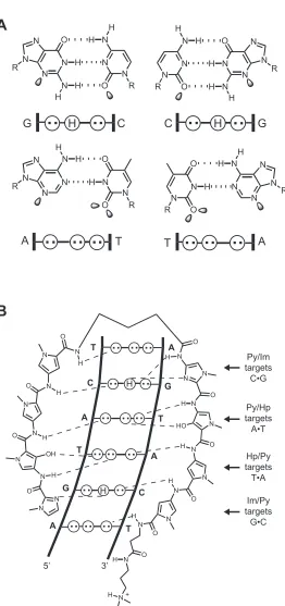
DNA consists of two anti-parallel polydeoxyribonucleotide strands wrapped together in a double helix. Between the two strands, hydrogen bonding of the Watson-Crick heterocyclic base pairs occurs, with adenine (A) pairing with thymine (T), and guanine (G) pairing with cytosine (C). B-form DNA is a right-handed helix containing ten base pairs per turn.4,5 The helix forms two grooves: the major groove, which is wide and shallow, and the minor groove, which is narrow and deep (Figure I.1). The edges of the DNA base pairs present unique molecular surfaces at the major and minor groove floors, providing for the sequence-specific recognition of DNA by transcription factors and other molecules.
DNA-binding small molecules
Figure I.1. The structure of DNA. (A) X-ray crystal structure of B-form DNA. The phosphodiester-linked deoxyribose backbone is shown in blue, and the Watson-Crick base pairs are shown in gray (PDB YSA).
(B) Chemical structures of hydrogen-bonded base pairs. Cytosine (C) is bonded to guanine (G) (top), and thymine (T) is bonded to adenine (A) (bottom). ‘R’ = the sugar phosphosphate backbone of DNA. Dashed lines indicate hydrogen bonds. The major and minor grooves are indicated in both representations.
Major Groove
Minor Groove
N N
N N N
N N O
O
H H
R R
H N
N
N N O
N
N N N
O H H
R
R H
H H
Guanine Cytosine
Thymine Adenine
major groove
minor groove minor groove
major groove
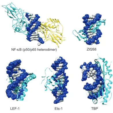
[image:16.612.123.528.117.573.2]Figure I.2. Structures of five protein-DNA complexes: NF-κB (PBD 1VKX),6 Zif68 (PDB ZAA),7 LEF-1
(PDB 2LEF),0 Ets- (PBD STW),8 and TBP (PDB TGH).9 All protein structures are determined by X-ray
crystallography with the exception of Ets-, which was determined by NMR.
NF-
κ
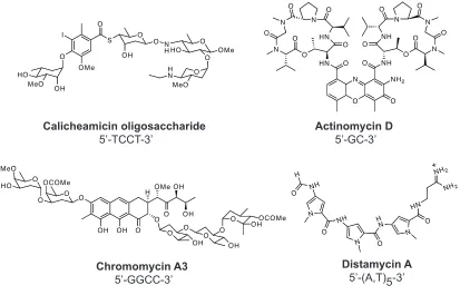
B (p50/p65 heterodimer) Zif268 [image:17.612.123.515.123.514.2]In addition to proteins, a number of small molecule natural products are capable of binding to specific sequences of DNA. Calicheamicin,11 actinomycin D,12 chromomycin13 and distamycin A14 have been shown to bind DNA with high affinity and modest sequence specificity within the minor groove (Figure I.3). Chromomycin targets the sequence 5’-GGCC-3’ and binds in the minor groove in a 2:1 stoichiometry. Actinomycin D intercalates DNA at 5’-GC-3’ sequences in a 1:1 stoichiometry. Calicheamicin oligosaccharide binds the minor groove as a monomer as well, and targets 5’-TCCT-3’. Distamycin A is an A,T-binding oligopeptide whose structure contains three N-methylpyrrole (Py) carboxamide units. It was revealed via x-ray and NMR structural studies that two of these crescent-shaped molecules can bind DNA as a 1:1 or 2:1 complex in an anti-parallel orientation (Figure I.4). This results in expansion of the minor groove relative to the 1:1 ligand-DNA complex.15-17 Chromomycin A3 5’-GGCC-3’ N NH H O O NH N H N O N O HN NH2 NH2 Distamycin A 5’-(A,T)5-3’ Actinomycin D 5’-GC-3’ Calicheamicin oligosaccharide 5’-TCCT-3’ O N NH O NH N N N O O O O O O HN O HN N N N O O O O O O NH2 O
OH OH O O O O O O OCOMe MeO HO H O OMe OH OH O OH O O OH O
O OHOCOMe
I O OMe OH MeO HO S O O OH O N
HHO O OMe
O O MeO H N
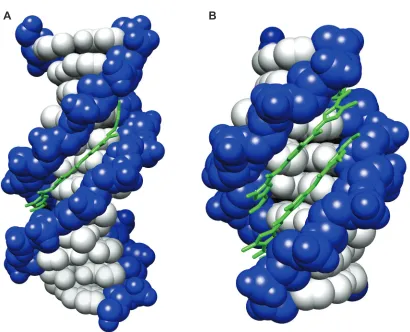
[image:18.612.109.522.397.662.2]Figure I.4. Distamycin bound to DNA. X-ray crystal structures of distamycin bound to DNA in (A) 1:1
(PBD DND) and (B) 2:1 (right, PDB 378D)7 conformations are shown.
[image:19.612.119.529.110.442.2]Minor groove recognition by pyrrole-imidazole polyamides
Figure I.5. Recognition of the DNA minor groove by polyamides. (A) Minor groove hydrogen bonding patterns of Watson-Crick base pairs. Circles with dots represent lone pairs of N() of purines and O() of pyrimidines, and circles containing an H represent the -amino group of guanine. ‘R’ represents the sugar-phosphate backbone of DNA. Shaded orbitals represent electron pairs projecting into the minor groove. (B)
Model of the polyamide ImHpPyPy-γ-ImHpPyPy-β-Dp binding its target sequence, 5’-AGTACT-3’ Putative
hydrogen bonds are shown as dashed lines.
N N N N N N O R R H H H N N N N N N N O O H H R R H
.
G.
.
N N N N N N O R R H H H N N N N N N N O O H H R R H.
..
.
.
.
.
.
.
CG C
T
.
.
AA
.
..
.
T [image:21.612.198.460.68.625.2]Figure I.6. Crystal structures of polyamides bound to DNA. (A) Structure of the 2:1 complex between ImHpPyPy-β-Dp and the sequence 5’-CCAGTACTGG-3’ (PDB 407D). (B) Structure of the cyclic
poly-amide ImImPyPy-(R)α-amine-γ-ImImPyPy-(R)α-amine-γ bound to 5’-CCAGGCCTGG-3’ (PDB 3I5L).6
Polyamides are shown below in ball-and-stick models, where a black circle represents Im, an open circle
represents Py, a circle containing ‘H’ represents Hp, a diamond represents β-alanine, a half-circle with a plus represents Dp, and a half-circle represents γ-turn.
H H
3’ - T C A T G A - 5’
5’ - A G T A C T - 3’
NH3 +
3’ - T C G A - 5’
5’ - A G G C C T - 3’
C G
H N3 +
[image:22.612.119.534.116.499.2]By binding as side-by-side, anti-parallel dimers in the minor groove, the heterocyclic rings of two polyamide strands can recognize DNA via the pairing rules described above.27 Covalent linkage of the carboxy terminus (C) of one strand to the amino terminus (N) of another via an alkyl turn moiety results in a ‘hairpin’ polyamide structure (Figure I.7). Early studies explored a variety of linker lengths and revealed that the use of a γ-aminobutyric acid (γ-turn) is optimal, and resulted in increased affinities (~100-fold) and specificities compared to unlinked subunits.28 The γ-turn also enforces the register of the heterocyclic ring pairings, preventing slipped dimer binding modes.29
T
G
C
A
T
A
5’ 3’
H
A
C
G
T
A
T
Im/Py targets G•C Py/Im targets C•G
β-alanine
targets A•T,T•A
Dp tail targets A•T,T•A
γ-turn
targets A•T,T•A
Py/Py targets A•T,T•A
Ct/Py targets
T•A S
O N H N
O N H
N N O
H N
N N
O
H
Cl
N
N O
H
N N
N O
H
N
N O
H
N N H
O
N O
H
O N
N H
H
NHR
H R = H, Ac
5’ - T
A T
A
C
G T - 3’
3’ - A T
A
T
G
C A - 5’
NHR
Figure I.7. Binding model for the polyamide CtPyPyIm-(R)RHNγ-PyImPyPy-β-Dp targeted to the sequence
’-TATACGT-’. The heterocycles Im and Ct are shown in bold. A ball-and-stick model is shown below, whereby a square represents Ct, a black circle represents Im, an open circle represents Py, a diamond
Eight-ring hairpin polyamides have been shown to bind a wide repertoire of DNA match sequences with subnanomolar affinities, similar to those of DNA-binding proteins (Table I.1).33-35 By using Py-Im polyamides to displace or prevent the binding of transcription factors to their respective promoter DNA sequences, the expression of particular genes can be modulated. Cellular permeability is of vital importance to the successful design of gene regulation studies. Confocal microscopy has been used to determine the nuclear uptake profiles of polyamide-fluorophore conjugates in a variety of cell lines.36-38 The presence of an isophthalic acid (IPA) moiety in the tail region has been shown to yield high affinity conjugates with improved nuclear permeability.39
1 2 3 4 5 6 7 8 9 10 11 12 13 14 15 16 17 18 19 20 21 22 23 24 25 26 27 + + + + + + + + + + + + + + + + + + + + + + + + + + + + + + + + + + + + + + + + + + + + + 3×10⁹ 5×10⁸ 4×10⁹ 9×10⁹ 3×10¹⁰ 2×10⁹ 5×10⁹ 3×10⁸ 1×10¹⁰ 1×10¹⁰ 4×10⁸ 4×10¹⁰ 2×10⁹ 2×10⁹ 2×10⁹ 9×10⁹ 1×10¹⁰ 2×10⁹ 7×10⁸ 2×10⁸ 9×10⁸ 2×10⁸ 1×10⁸ 2×10⁹ 9×10⁹ 3×10⁹ 1×10⁹ WWGWWWW
WWGGWWW WWGWGWW WWGWWGW WWGWWCW WWGWCWW WWGCWWW WWGGGWW WWGGWGW WWGGWCW WWGGCWW WWGWGGW WWGWGCW WWGCGWW WWGCWGW WWGCWCW WWGWCGW WWGWCCW WWGCCWW WWGGGGW WWGCGGW WWGGCGW WWGGGCW WWGCCGW WWGGCCW WWGCGCW WWGCCCW
Polyamide Ka (M-1)
Sequence (5’→3’) Context
5’-TAGTATT-3’ 5’-CTGGTTA-3’ 5’-TAGTGAA-3’ 5’-TAGTAGT-3’ 5’-TAGTACT-3’ 5’-GAGTCTA-3’ 5’-ATGCAAA-3’ 5’-AAGGGAA-3’ 5’-TAGGTGT-3’ 5’-ATGGTCA-3’ 5’-AAGGCAT-3’ 5’-TAGTGGT-3’ 5’-ATGAGCT-3’ 5’-ATGCGTA-3’ 5’-TAGCAGT-3’ 5’-ATGCTCA-3’ 5’-ATGACGT-3’ 5’-TAGACCA-3’ 5’-ATGCCTA-3’ 5’-GAGGGGT-3’ 5’-ATGCGGT-3’ 5’-CAGGCGT-3’ 5’-CTGGGCA-3’ 5’-ATGCCGT-3’ 5’-ATGGCCA-3’ 5’-ATGCGCA-3’ 5’-ATGCCCA-3’
Table I.1. Library of imidazole-capped polyamides. Shown are the targeted sequence, binding association
constant (Ka), and sequence context for binding affinity determination for each polyamide (shown as
Figure I.8. Polyamides as regulators of gene expression in cell culture. (A) Schematic diagram of the VEGF promoter showing inhibition of HRE binding by HIF-1 (shown as HIF-1α/HIF-1β heterodimer), binding
sequence of the HRE enhancer shown with match polyamide 1, and chemical structures and ball-and-stick models of match polyamide 1 and mismatch polyamide 2. (B) Schematic diagram of the androgen receptor transcription complex, binding sequence of the consensus ARE targeted by match polyamide 3, and chemical structures and ball-and-stick models of match polyamide 3 and mismatch polyamide 4.
5’-GGTACANNNTGTTCT-3’
3’-CCATGTNNNACAAGA-5’
3 + 3 +
Consensus ARE
+ IPA
HRE vegf
5‘- C A G T G C AT A C G T GG G C T C -3‘
3‘- G T C A C G T1 A T G C A C C C G A G -5‘
HIF-1 X α β 3 + IPA 4 + N H N O N H N O N N H O N HN N N H N O N H N O N N N H O NH O NH3 N N H O N N H N O O O -O H N H N O N H N O N N H O N HN N N H N O N N H N O N N H O NH O NH3 N N H O N H N O O O -O H + +
NH3+
NH3+ IPA
IPA IPA NH
3+
NH3+
O H N N H N O N H N O N N H N O N H N O NH3+
NH O N N N H O N N H O N NH+ HN O O −O N H O S Cl 1 2 O H N N H N O N H N O N H N O N N H N O NH O N N H O N N H O N N HN O O N H O N N
NH3+
NH+ −O + IPA + IPA
NH3+
NH3+
NH3+
Sequence-specific alkylation of DNA
Polyamides have been used to regulate gene expression by binding to promoter and enhancer elements of DNA,40-42 but do not prevent RNA polymerase elongation when bound to the coding region of genes.43 DNA alkylators typically display limited sequence selectivity. Conjugation of an alkylating moiety to a sequence-specific molecule results in a molecule capable of covalent DNA attachment that arrests transcription.44 An example of this is Tallimustine, which is a conjugate of distamycin A and the nitrogen mustard chlorambucil.45
O N NH O N N H N O N H N H N O O N H N N O N N O N H N H N O H N H N O N H H N O N N O N H N O N H N H O N Cl Cl N NH O N N H N O N H N H N O O N H N N O N O N H N H N O H N H N O O N H N H O O HN CPI N N O N N H Polyamide-Chlorambucil Conjugates N HO O Cl seco-CBI R
Figure I.9. Examples of various polyamide-alkylator conjugates. Shown are seco-CBI and CPI tethered to
a polyamide targeting 5’-WWGGWCW-3’ (top) and chlorambucil attached to a 2-β-2 polyamide targeting
’-WWGCWGCW-’ (bottom).
Polyamide synthesis
Scope of this work
The work described in this thesis relates to the effects of the hairpin turn subunit on Py-Im polyamides, including binding affinity and specificity, and cellular uptake properties. It is divided into two sections: part II describes studies relating to α-2,4-diaminobutyric acid as the hairpin subunit; part III describes effects of 3,4-diaminobutyric acid in the hairpin subunit. The appendix describes methods for genome-wide analysis of polyamide binding using expertise gained in part III.
Chapters IIA and IIB describe the identification and characterization of polyamides and polyamide-Chl conjugates linked by 2,4-diaminobutyric acid (also referred to as α-diaminobutyric acid, or α-DABA), with comparisons to analogous γ-2,4-α-diaminobutyric acid (also referred to as γ-diaminobutyric acid, or γ-DABA) containing molecules. Binding affinities and specificities for small libraries of α- and γ-DABA linked polyamides were determined, and alkylation properties of the Chl conjugates explored. Effects of α- and γ-DABA-linked molecules on colon carcinoma cell growth and on mice were evaluated. In chapter IIC, α-DABA-linked polyamide-Chl conjugates targeting the coding region of histone H4c are studied in vitro and in vivo in an effort to affect chronic myelogenous leukemia.
Chapters IIIA and IIIB characterize polyamides containing various substituents in the γ-turn unit. In Chapter IIIA, polyamides containing (R) and (S)-3,4-diaminobutyric acid (β-amino-γ-turn) in the hairpin turn are compared to analogues containing unsubstituted γ-DABA and γ-2,4-diaminobutyric acid (referred to as α-amino-γ-turn in part III) with respect to binding abilities and in cell culture. In Chapter IIIB, six-ring polyamides are employed to determine the binding affinities and specificities of α-amino-γ-turn, (R) and (S)-β-amino-γ-turn, and fluoro- and hydroxyl-turn polyamides at the turn position.
References
1.Collins, F. S.; Lander, E. S.; Rogers, J.; Waterston, R. H. Nature. 2004. 431, 931-945. 2.Pabo, C. O.; Sauer, R. T. Annu. Rev. Biochem. 1984. 53, 293-321.
3.Ellenberger, T. E.; Brandl, C. J.; Struhl, K.; Harrison, S. C. Cell. 1992. 71, 1223-1237. 4.Wing, R.; Drew, H.; Takano, T.; Broka, C.; Tanaka, S.; Itakura, K.; Dickerson, R. E. Nature. 1980. 287, 755-758.
5.Dickerson, R. E.; Drew, H. R.; Conner, B. N.; Wing, R. M.; Fratini, A. V.; Kopka, M.L. Science. 1982. 216, 475-485.
6.Chen, F. E.; Huang, D. B.; Chen, Y. Q.; Ghosh, G. Nature. 1998. 391, 410-413. 7.Pavletich, N. P.; Pabo, C. O. Science. 1991. 252, 809-817.
8.Werner, M. H.; Clore, G. M.; Fisher, C. L.; Fisher, R. J.; Trinh, L.; Shiloach, J.; Gronenborn, A. M. J. Biomol. NMR. 1997. 10, 317-328.
9.Juo, Z. S.; Chiu, T. K.; Leiberman, P. M.; Baikalov, I.; Berk, A. J.; Dickerson, R. E. J. Mol. Biol. 1996. 261, 239-254.
10.Love, J. J.; Li, X. A.; Case, D. A.; Giese, K.; Grosschedl, R.; Wright, P. E. Nature. 1995. 376, 791-795.
11.Kalben, A.; Pal, S.; Andreotti, A. H.; Walker, S.; Gange, D.; Biswas, K.; Kahne, D. J. Am. Chem. Soc.Chem. Soc. 2000. 122, 8403-8412.
12.Vandyke, M. W.; Dervan, P. B. Biochemistry. 1983. 22, 2373-2377.
13.Hou, M. H.; Robinson, H.; Gao, Y. G.; Wang, A. H. J. Nucleic Acids Res. 2004. 32, 2214-2222.
14.Arcamone, F.; Nicolell.V; Penco, S.; Orezzi, P.; Pirelli, A. Nature. 1964. 203, 1065.
16.Pelton, J. G.; Wemmer, D. E. Proc. Natl. Acad. Sci. U. S. A. 1989. 86, 5723-5727. 17.Mitra, S. N.; Wahl, M. C.; Sundaralingam, M. Acta Crystallogr. Sect. D-Biol. Crystallogr. 1999. 55, 602-609.
18.Dervan, P. B. Bioorg. Med. Chem. 2001. 9, 2215-2235.
19.Dervan, P. B.; Edelson, B. S. Curr. Opin. Struct. Biol. 2003. 13, 284-299.
20.Kielkopf, C. L.; Baird, E. E.; Dervan, P. D.; Rees, D. C. Nat. Struct. Biol. 1998. 5, 104-109.
21.White, S.; Szewczyk, J. W.; Turner, J. M.; Baird, E. E.; Dervan, P. B. Nature. 1998. 391, 468-471.
22.Kielkopf, C. L.; White, S.; Szewczyk, J. W.; Turner, J. M.; Baird, E. E.; Dervan, P. B.; Rees, D. C. Science. 1998. 282, 111-115.
23.Foister, S.; Marques, M. A.; Doss, R. M.; Dervan, P. B. Bioorg. Med. Chem. 2003. 11, 4333-4340.
24.Turner, J. M.; Swalley, S. E.; Baird, E. E.; Dervan, P. B. J. Am. Chem. Soc.Chem. Soc. 1998. 120, 6219-6226.
25.Wang, C. C. C.; Ellervik, U.; Dervan, P. B. Bioorg. Med. Chem.Med. Chem. 2001. 9, 653-657. 26.Chenoweth, D. M.; Dervan, P. B. Proc. Natl. Acad. Sci. U.S.A. 2009. 106, 13175- 13179.
27.Mrksich, M.; Wade, W. S.; Dwyer, T. J.; Geierstanger, B. H.; Wemmer, D. E.; Dervan, P. B. Proc. Natl. Acad. Sci. U. S. A.Natl. Acad. Sci. U. S. A. 1992. 89, 7586-7590.
28.Mrksich, M.; Parks, M. E.; Dervan, P. B. J. Am. Chem. Soc.Chem. Soc. 1994. 116, 7983-7988. 29.deClairac, R. P. L.; Geierstanger, B. H.; Mrksich, M.; Dervan, P. B.; Wemmer, D. E. J. Am. Chem. Soc. 1997. 119, 7909-7916.
Olenyuk, B.; Puckett, J. W.; Wang, C. C.; Dervan, P. B. Tetrahedron. 2007. 63, 6151.
36.Belitsky, J. M.; Leslie, S. J.; Arora, P. S.; Beerman, T. A.; Dervan, P. B. Bioorg. Med.Med. Chem. 2002. 10, 3313-3318.
37.Best, T. P.; Edelson, B. S.; Nickols, N. G.; Dervan, P. B. Proc. Natl. Acad. Sci. U. S. A. 2003. 100, 12063-12068.
38.Edelson, B. S.; Best, T. P.; Olenyuk, B.; Nickols, N. G.; Doss, R. M.; Foister, S.; Heckel, A.; Dervan, P. B. Nucleic Acids Res. 2004. 32, 2802-2818.
39.Nickols, N. G.; Jacobs, C. S.; Farkas, M. E.; Dervan, P. B. Nucleic Acids Res. 2007. 35, 363-370.
40.Olenyuk, B. Z.; Zhang, G. J.; Klco, J. M.; Nickols, N. G.; Kaelin, W. G.; Dervan, P. B. Proc. Natl. Acad. Sci. U. S. A.Natl. Acad. Sci. U. S. A. 2004. 101, 16768-16773.
41.Nickols, N. G.; Jacobs, C. S.; Farkas, M. E.; Dervan, P. B. ACS Chem. Biol. 2007. 2, 561-571.
42.Nickols, N. G.; Dervan, P. B. Proc. Natl. Acad. Sci. U. S. A. 2007. 104, 10418-10423. 43.Dervan, P. B.; Poulin-Kerstien, A. T.; Fechter, E. J.; Edelson, B. S. Top. Curr. Chem. 2005. 253, 1-31.
44.Shinohara, K.; Sasaki, S.; Minoshima, M.; Bando, T.; Sugiyama, H. Nucleic Acids Res. 2006. 34, 1189-1195.
45.Pezzoni, G.; Grandi, M.; Biasoli, G.; Capolongo, L.; Ballinari, D.; Giuliani, F. C.; Barbieri, B.; Pastori, A.; Pesenti, E.; Mongelli, N.; Spreafico, F. Br. J. Cancer. 1991. 64, 1047-1050.
46.Chang, A. Y.; Dervan, P. B. J. Am. Chem. Soc. 2000. 122, 4856-4864.
47.Bando, T.; Narita, A.; Saito, I.; Sugiyama, H. J. Am. Chem. Soc.Chem. Soc. 2003. 125, 3485.
48.Wurtz, N. R.; Dervan, P. B. Chem. Biol.Biol. 2000. 7, 153-161.
50.Wyatt, M. D.; Lee, M.; Hartley, J. A. Anti-Cancer Drug Des. 1997. 12, 49-60. 51.Wang, Y. D.; Dziegielewski, J.; Wurtz, N. R.; Dziegielewska, B.; Dervan, P. B.; Beerman, T. A. Nucleic Acids Res. 2003. 31, 1208-1215.
52.Baird, E. E.; Dervan, P. B. J. Am. Chem. Soc. 1996. 118, 6141-6146.
53.Belitsky, J. M.; Nguyen, D. H.; Wurtz, N. R.; Dervan, P. B. Bioorg. Med. Chem.Med. Chem. 2002. 10, 2767-2774.
Chapter IIA
Unanticipated Differences Between α- and γ-Diaminobutyric
Acid-Linked Hairpin Polyamide-Alkylator Conjugates
The text of this chapter was taken in part from a manuscript co-authored with Sherry M. Tsai,* Peter B. Dervan,* C. James Chou,‡ and Joel M. Gottesfeld ‡ ( * California Institute
of Technology and ‡ The Scripps Research Institute)
(Tsai, S.M.; Farkas, M.E.; Chou, C.J.; Gottesfeld, J.M.; Dervan, P.B. Nucleic Acids Res.
(α-DABA) turn moiety are compared to their constitutional isomers containing the well-characterized γ-DABA turn. Although the DNA-binding properties of unconjugated polyamides are similar, the α-DABA conjugates display increased alkylation specificity and decreased rate of reaction. Treatment of a human colon carcinoma cell line with α-DABA versus γ-α-DABA hairpin conjugates reveals only slight differences in toxicities while
producing similar effects on cell morphology and G2/M stage cell cycle arrest. However,
striking differences in animal toxicity between the two classes are observed. While mice treated with an α-DABA hairpin polyamide do not differ significantly from control mice, the analogous γ-DABA hairpin is lethal. This dramatic difference from a subtle structural
Introduction
The ability to control gene expression through the use of DNA sequence-specific,
cell-permeable molecules holds therapeutic promise. Based on the natural product distamycin A, pyrrole-imidazole polyamides are a class of molecules that can be programmed to bind
a broad repertoire of DNA sequences with specificities comparable to naturally occurring
DNA-binding proteins.1 They have also been shown to permeate living cells, localize to
the nuclei,2,3 and regulate gene expression.4,5
Pairing rules have been established for minor groove recognition, whereby an N-methylpyrrole/N-methylimidazole (Py/Im) pair recognizes C•G, the reverse (Im/Py)
recognizes G•C, and Py/Py specifies A•T or T•A.1,6 The replacement of Py with β-alanine
(β) can enhance binding affinity by allowing the polyamide to reset its curvature with that
of the DNA minor groove.7,8 Polyamides may be linked via a turn moiety to give a hairpin
structure which binds in the “forward” direction N→C with respect to the 5’→3’ direction
of the DNA strand.9 In earlier studies, linkage of antiparallel three-ring subunits via a
γ-aminobutyric acid moiety, resulting in a turn with three methylene units, was shown to
enhance DNA binding affinity ~100-fold over unlinked subunits.10 The γ-aminobutyric
acid turn demonstrates selectivity for A•T or T•A base pairs over G•C and C•G.11 Use of a glycine amino acid, with one methylene turn unit, resulted in a 3-fold reduction in
affinity relative to the γ-aminobutyric acid. Subsequent NMR studies revealed that the glycine amino acid substituted polyamide bound DNA in an extended, rather than a hairpin,
conformation.12,13
Substitution of the γ-aminobutyric acid with the chiral (R)-2,4-diaminobutyric
enhancer elements, but they do not prevent RNA polymerase elongation when bound to the
coding region of genes.1 Conjugation of an alkylating moiety to a sequence-specific
DNA-binding molecule would be expected to result in a molecule capable of covalent DNA
attachment that could arrest transcription.15,16 An early example of this is Tallimustine (FCE 24517), which is a conjugate of distamycin A and the nitrogen mustard chlorambucil.17
When attached to a DNA minor groove binder, chlorambucil is capable of alkylation at the
N3 position of an adenine proximal to the binding site.18,19 Hairpin polyamide-chlorambucil
conjugates have been demonstrated to target specific sequences of the HIV-1 promoter and
simian virus 40.19,20
Recently, a hairpin polyamide-chlorambucil conjugate, synthesized at The
Scripps Research Institute, was found to alkylate within the coding region of the histone
H4c gene of the human colon carcinoma cell line SW620 and to block proliferation of
these cells in culture.21 In addition, this conjugate was found to block tumor growth in a
SW620 xenograft mouse model without apparent toxicity. When we attempted to
scale-up additional conjugate at Caltech, the newly-synthesized molecule was found to be
highly toxic in athymic nude mice, contrary to prior experiments.22 Subsequent studies
showed that the two laboratories had utilized two different turn units in the synthesis of the conjugate: the conjugate synthesis at Scripps used Boc-D-Dab(Fmoc)-OH, while the conjugate synthesis at Caltech used Fmoc-D-Dab(Boc)-OH. The molecules are therefore constitutional isomers differing in the hairpin turn moiety. The original molecule made at Scripps continues to be called 1R-Chl,22 and the new molecule is designated 2R-Chl.
1R-Chl utilizes an α-diaminobutyric acid (α-DABA) turn unit, whereas 2R-Chl employs the
standard γ-DABA turn unit (Figure IIA.1). Linkage via the new α-DABA turn unit results
is lengthened by two methylene groups relative to 2R-Chl.
Polyamides utilizing the α-DABA turn moiety have not been studied previously,
and the reasons for the unique biological properties displayed by 1R-Chl are unknown.
In the current study, we report the chemical and biological characterization of α-DABA polyamides and compare them to the analogous γ-DABA polyamides. A series of four
parent molecules and their corresponding chlorambucil conjugates was synthesized (Figure
IIA.1A). Each molecule contains the heterocyclic sequence: ImImβIm-HT-PyPyPyPy-βDp (where HT = hairpin turn and Dp = 3-(dimethylamino)-propylamine). Molecules
in series 1 contain the new α-DABA turn, and molecules in series 2 utilize the γ-DABA
turn. The R and S enantiomers of the parent polyamides and chlorambucil conjugates
in each series have been generated for comparison. The DNA binding affinities of the parent molecules were measured by DNase I footprinting, and the alkylation profiles (sequence specificity and reactivity) of the polyamide-chlorambucil conjugates were established using thermal cleavage assays. The effects of polyamide treatment on SW620 cell morphology, proliferation, viability, and cell cycle profiles were compared using phase contrast microscopy, cell counting, trypan blue exclusion assay, and fluorescence-assisted cell sorting (FACS) analysis, respectively. Importantly, toxicity effects of the polyamide
constitutional isomers on mice were determined.
Results and Discussion
Polyamide synthesis
Four hairpin polyamides were prepared by solid phase synthesis.23 The hairpin polyamides differed in the turn substituents utilized: Boc-D-Dab(Fmoc)-OH (1R, 1R-Chl), Boc-Dab(Fmoc)-OH (1S, 1S-Chl), Fmoc-D-Dab(Boc)-OH (2R, 2R-Chl), and Fmoc-Dab(Boc)-OH (2S, 2S-Chl) (Figure IIA.1B). Conjugates were generated by coupling
1S 2S X H N O O H N N O N N N O H N O +HN O H N N H N O N H N O N H N O N H N O O N N N H O N H O N N N H O N N H N O NH +HN X X H N N H N N N O N O H N O O O +HN H N N H N N H N O N H N O N H N O NH O N N N H O N H O N N N H O N N H N O O O +HN X X X 1 R/S-Chl X = 2 R/S-Chl 1 R/S 2 R/S HO O NHFmoc NHBoc HO O NHFmoc NHBoc HO O NHBoc NHFmoc HO O NHBoc NHFmoc B NH+ 3 N H O N Cl Cl
Figure IIA.1. Polyamide structures and syntheses. (A) Chemical and ball-and-stick structures for poly-amides 1R, 1S, 2R, 2S, and their Chl conjugates. (B) Chemical structures of the four turn units used in the syntheses of the parent polyamides and polyamide-chlorambucil conjugates: Boc-D-Dab(Fmoc)-OH (top left, used in the syntheses of 1R and 1R-Chl); Boc-Dab(Fmoc)-OH (top right, used in the syntheses of 1S and
1S-Chl); Fmoc-D-Dab(Boc)-OH (bottom left, used in the syntheses of 2R and 2R-Chl); Fmoc-Dab(Boc)-OH (bottom right, used in the syntheses of 2S and 2S-Chl).
reverse-phase HPLC of the free amine and purified similarly.
Plasmid design
The core polyamide structure was constructed to bind the sequence 5’-WWGGWGW-3’, where W = A or T. In order to probe the affinity and sequence specificity of the parent
(‘M’ = 5’-TAGGTGT-3’) and two single base-pair mismatch sites (Figure IIA.2A). The
mismatch sites consist of single G•C to C•G replacements at different locations within
each binding site (‘A’ = 5’-TAGGTCT-3’, ‘B’ = 5’-TAGCTGT-3’). For the purpose of
thermal cleavage assays, each binding site was designed with proximal adenines which
the chlorambucil moiety would be expected to alkylate.19 In addition to the designed sites,
inherent in the plasmid structure is a single base-pair mismatch site (‘C’ = 5’-AGTGGTG
-3’) for a polyamide binding in reverse (Figure IIA.3).
DNA affinity and sequence specificity
Quantitative DNase I footprint titrations to determine binding site affinities and
specificities were conducted for parent polyamides 1R, 1S, 2R, and 2S on the 5’-labeled
280-base-pair PCR product of plasmid pMFST2 (Figure IIA.2 and Table IIA.1). Previous studies have indicated that attachment of the chlorambucil moiety does not significantly
alter the DNA binding affinity of the parent polyamide.19
The α-DABA polyamide 1R bound the match site, M, with Ka = 6.7 x 10⁹ M -1, and
it had a 94-fold specificity over single mismatch site A and 26-fold specificity over single
mismatch site B (Figure IIA.2B). The enantiomer 1S bound the match site M with Ka =
4.8 x 10⁸ M -1, a 14-fold decrease relative to 1R. Binding affinities at the other sites were
too low to be measured, and the polyamide was found to coat DNA at high concentrations
(Figure IIA.2C).
The γ-DABA polyamides 2R and 2S bound DNA in accordance with the expected
orientation of the free amine.14 2R was shown to bind the match and both single base-pair
mismatch binding sites (Figure IIA.2D). The affinity of 2R for site M was Ka = 1.6 x 10¹⁰
M -1, with 15-fold and 6-fold preferences over mismatch sites A and B, respectively. The enantiomer, 2S, bound the single base-pair mismatch reverse site C with Ka = 1.2 x 10⁹ M-1, a 21-fold enhancement over the forward match site M (Figure IIA.2E).
Intact DNase I G
100 nM A 1 pM 1R A B M B Intact
DNase I G
100 nM A 1 pM M 1S C G A Intact DNase I 100 nM 1 pM M A B 2R D Intact
DNase I G
100 nM A 1 pM M C 2S E
1 2 3 4 5 6 7 8 9 10 11 12 13 14 15 1 2 3 4 5 6 7 8 9 10 11 12 13 14 15 1 2 3 4 5 6 7 8 9 10 11 12 13 14 15 1 2 3 4 5 6 7 8 9 10 11 12 13 14 15
M C A B
5'-CGCCCTT TAGGTGT TACGT AGTGGTG CCG TAGGTCT TAGCCG TAGCTGT TGCCGT AAGGGCGAATTCTG-3' 3'-GCGGGAA ATCCACA ATGCA TCACCAC GGC ATCCAGA ATCGGC ATCGACA ACGGCA TTCCCGCTTAAGAC-5'
EcoRI PvuII 32 P 280 bp pMFST2 A
[2R]
-0.4 -0.2 0 0.2 0.4 0.6 0.8 1 1.2
10-1310-1210-11 10-1010-9 10-8 10-7 10-6
M A B
norm
θ
[2S]
-0.4 -0.2 0 0.2 0.4 0.6 0.8 1 1.2
10-1310-1210-1110-1010-910-8 10-7 10-6
M C
norm
θ
[1R]
10-13 10-12 10-11 10-10 10-9 10-8 10-7 10-6 -0.4 -0.2 0 0.2 0.4 0.6 0.8 1 1.2 M A B norm θ
[1S]
10-13 10-12 10-11 10-10 10-9 10-8 10-7 10-6 -0.4 -0.2 0 0.2 0.4 0.6 0.8 1 1.2 M norm θ
Figure IIA.3. Illustration of the parent polyamides (A) 2R and (B) 2S binding DNA.
Polyamide 5’-TAGGTGT-3’ 5’-TAGGTCT-3’ 5’-TAGCTGT-3’ 5’-AGTGGTG-3’
-1 Equilibrium Association Constants: K (M )a
1.1 (± 0.1) x 109 2.9 (± 1.0) x 109
1.6 (± 0.3) x 1010
2R
--6.7 (± 0.1) x 109 7.1 (± 1.8) x 10 7 2.6 (± 0.2) x 108
1R
1.2 (± 0.2) x 109
2S 5.8 (± 2.1) x 107 --
--1S 4.8 (± 0.9) x 108 -- --
--Table 1.
Table IIA.1. Binding affinities (M-1) for parent polyamides. Equilibrium association constants reported are
mean values from three DNase I footprint titration experiments; standard deviations are shown in parentheses.
Previous studies have found a glycine amino acid-linked polyamide is able to bind DNA in an extended dimeric conformation that can be observed via a larger binding site in a DNase
I footprinting gel. We do not observe evidence of an extended conformation for either 1R
or 1S, as the binding sites of both molecules are the same size as those of 2R and 2S. The substituted glycine favors a turn conformation.
The DNase I footprinting experiments reveal that the α-DABA polyamide 1R has
a 2-fold reduced binding affinity relative to the γ-DABA polyamide 2R. A contributing
factor to the decreased binding observed for 1R and 1S could be a reduced ability of the
molecules to fit into the minor groove. The shorter turn linkage between the two subunits in an α-DABA polyamide may reduce its flexibility in conforming to the shape of the groove (Figure IIA.4). The energetics of α-DABA hairpin polyamides appear less sensitive than
the γ-DABA series to the R and S stereochemistry at the turn.
site C reverse binding of 2S
5’ - A G T G G T G - 3’
3’ - T C A C C A C - 5’ H3
+ N
5’ - T A G G T G T - 3’
3’ - A T C C A C A - 5’ NH3
+
match site binding of 2R
1S
NH+ 3
NH+ 3
1R
B
Figure IIA.4. Models of the binding of (A) 1R and (B) 1S to the match site of pMFST2. DNA atoms are indicated with gold; polyamide atoms are indicated as follows: carbon with gray, oxygen with red, nitrogen with blue, and hydrogen with white. Illustrations were rendered using Chimera.
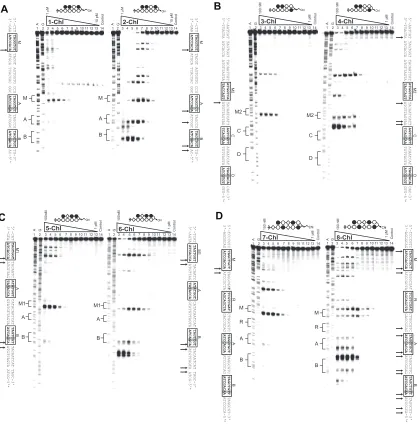
DNA alkylation specificity and time-dependence
To determine the alkylation properties of the polyamide-chlorambucil conjugates,
thermal cleavage assays were performed on the 5’ 32P-labeled 280-base-pair PCR product
of pMFST2 (Figure IIA.5). The alkylation profiles of 1R-Chl and 2R-Chl are dramatically different: 1R-Chl alkylates DNA more specifically than 2R-Chl and appears less reactive.
For the designed sequence, 1R-Chl appears to specifically alkylate the adenine proximal
M
A
B
C
5'-AGCTG CGCCCTT TAGGTGT TACGT AGTGGTG CCG TAGGTCT TAGCCG TAGCTGT TGCCGT AAGGG-3'
3'-C GCGGGAA ATCCACA ATGCA TCACCAC GGC ATCCAGA ATCGGC ATCGACA ACGGCA TTCCC-5'*
Control
G
100 nM
A 1 pM
1 2 3 4 5 6 7 8 9 10 11 12 13 14
B A M
C
1R-Chl G Control
100 nM
A 1 pM
1 2 3 4 5 6 7 8 9 10 11 12 13 14
2R-Chl
B A M
C
Chl Chl
A B
M
A
B
C
5'-AGCTG CGCCCTT TAGGTGT TACGT AGTGGTG CCG TAGGTCT TAGCCG TAGCTGT TGCCGT AAGGG-3'
3'-C GCGGGAA ATCCACA ATGCA TCACCAC GGC ATCCAGA ATCGGC ATCGACA ACGGCA TTCCC-5'*
Control
G
A 1 pM
1 2 3 4 5 6 7 8 9 10 11 12 13 141S-Chl A G Control
1 pM
1 2 3 4 5 6 7 8 9 10 11 12 13 142S-Chl
B A M
C
B A M
C
Chl Chl
C D
100 nM
M
A
B
C
5'-AGCTG CGCCCTT TAGGTGT TACGT AGTGGTG CCG TAGGTCT TAGCCG TAGCTGT TGCCGT AAGGG-3'
3'-C GCGGGAA ATCCACA ATGCA TCACCAC GGC ATCCAGA ATCGGC ATCGACA ACGGCA TTCCC-5'*
M
A
B
C
5'-AGCTG CGCCCTT TAGGTGT TACGT AGTGGTG CCG TAGGTCT TAGCCG TAGCTGT TGCCGT AAGGG-3'
3'-C GCGGGAA ATCCACA ATGCA TCACCAC GGC ATCCAGA ATCGGC ATCGACA ACGGCA TTCCC-5'*
100 nM
Figure IIA.5. Alkylation specificities of the polyamide conjugates. Thermal cleavage assay experiments
promiscuous, alkylating at site M and both mismatch sites, A and B, at concentrations as low as 3 nM (Figure IIA.5B). Only at 1 nM concentration was 2R-Chl observed to
specifically alkylate at site M versus sites A and B.
The S enantiomers of the conjugates are similar to their R analogs in their alkylation
specificities, but they are muted in activity. In contrast to 1R-Chl, 1S-Chl appears to
specifically alkylate the guanine between sites M and C up to a concentration of 30 nM
(Figure IIA.5C). Alkylation at N3 of guanine has been previously observed.18,21,24 Based
on the DNase I footprinting data for 1S, the observed alkylation is probably due to binding
of 1S-Chl at the match site M. The 2S-Chl alkylation profile at 100 nM is similar to that
of 2R-Chl at 10 nM, with the polyamide alkylating at the match site and both single base-pair mismatch sites (Figure IIA.5D). However, at 30 nM, 2S-Chl is specific for the match
site.
Outside of the designed pMFST2 insert, 1R-Chl and 1S-Chl alkylate fewer sites than 2R-Chl and 2S-Chl, respectively, further demonstrating their enhanced alkylation
specificity. There are a number of forward-binding and reverse-binding single base-pair
mismatch sites, as well as one reverse-binding match site, inherent in the labeled fragment of pMFST2 used in these studies. The major sites of alkylation observed for 1R-Chl and 2R-Chl outside of the designed plasmid insert can generally be attributed to forward-binding
single base-pair mismatch sites, although the gel resolution is often too poor to definitively
identify specific alkylation sites. The additional major alkylation sites observed for 2S-Chl
can be attributed to a combination of forward-binding and reverse-binding single base-pair mismatch sites, while the additional major alkylation site observed for 1S-Chl may correspond to a reverse-binding single base-pair mismatch site.
We next studied the time-dependence of alkylation for 1R-Chl and 2R-Chl to
half-life of the labeled DNA (i.e. disappearance of full-length DNA) is 20 h for 1R-Chl and 10 min for 2R-Chl. Thus, 2R-Chl alkylates DNA 120 times faster than 1R-Chl under these conditions. Given the minimal structural differences between 1R-Chl and 2R-Chl and the
similar binding affinities of their parent molecules, the dramatic difference in alkylation specificity and kinetics between hairpins with a common alkylator moiety would not have
been predicted.
M
A
B C
Control
G
A
1R-Chl
24 h 14.5 h 4 h 1 h 30 min 10 min
B A M C
Control
G
A
2R-Chl
24 h 14.5 h 4 h 1 h 30 min 10 min
A
B
1 2 3 4 5 6 7 8 9 1 2 3 4 5 6 7 8 9
change and arrest cell growth in the G2/M phase. In addition, the polyamidewas found
to be a cytostatic agent, decreasing cell proliferation without significantly affecting cell
viability, as determined by trypan blue staining.21 A two-hit mechanism for growth arrest by 1R-Chl was suggested by experiments where a different polyamide-chlorambucil conjugate, which by itself had no effect on SW620 cell proliferation, was found to cause
growth arrest after treatment of these cells with an siRNA directed against H4c mRNA.25
Our model for the action of 1R-Chl involves direct alkylation of the H4c gene, leading to a block in transcription and eventual depletion of H4 protein, opening up the genome for massive alkylation by 1R-Chl and G2/M arrest.
We studied the effects of our polyamide library on SW620 cultured cells to determine
whether the differences between the polyamides observed in vitro would translate to
disparities in cell morphology, proliferation, viability, and cell-cycle profile. As expected,
no effects were observed for the treatment of cells with the four parent polyamides (Figure IIA.7). Cells treated for 3 days with 1R-Chl or 2R-Chl show the same morphological change as previously established (Figure IIA.8A). Each conjugate also decreases cell proliferation
to ~30% relative to control with only a small decrease in cell viability, as measured with trypan blue exclusion staining (Figure IIA.8B). FACS analysis using propidium iodide
staining indicates both molecules arrest cells in the G2/M phase of growth (38% for
1R-Chl and 34% for 2R-Chl, compared to 20% for control; Figure IIA.8C).
Interestingly, a less pronounced cellular effect is observed for cells treated with
1S-Chl and 2S-Chl. Although most cells appear identical to control, some of the cells treated with each of the S enantiomers display a morphology change (Figure IIA.8A). Cell
viability is not significantly reduced, but proliferation decreases slightly (77% for 1S-Chl
and 65% for 2S-Chl relative to control; Figure IIA.8B), indicating a slight cytostatic effect
A
B
C 56% Control
G0/G1
22% S 20%
G2/M 2%
A/N
2S
56% G0/G1
21% S 22%
G2/M 1%
A/N
2R
57% G0/G1
21% S 21%
G2/M 1%
A/N
1R
54% G0/G1
21% S 23%
G2/M 1%
A/N
1S
55% G0/G1
20% S 23%
G2/M 2%
A/N
0 20 40 60 80 100 120
Control 1R 1S 2R 2S
Polyamide
R
el
at
iv
e
Vi
ab
ili
ty
/C
ou
nt
(%
)
Cell Viability Cell Count Control
1R 1S 2R 2S
Figure IIA.8. Effects of polyamide conjugates on cultured cells. Cultured SW620 cells were treated with 200 nM of the indicated polyamide for 3 days prior to analysis. (A) Representative phase microscopy im-ages. (B) Normalized cell viabililty and proliferation values. Data shown are the averages of three samples; error bars indicate standard deviations. (C) Fluorescence-activated cell-sorting analysis of cells. Plots indicate cell numbers versus propidium iodide staining. Percentages of cells in G0/G1, S, G2/M, and apop-totic/necrotic cells (A/N) are indicated.
B C 2S-Chl 0 20 40 60 80 100 120
Control 1R-Chl 1S-Chl 2R-Chl 2S-Chl
Polyamide R el at iv e V ia b ili ty /C o u n t (% ) Cell Viability Cell Count 1R-Chl Control 2R-Chl 1S-Chl Control 56% G0/G1 22% S 20% G2/M 2% A/N Propidium Iodide Counts 0
0 200 400 600
60 120 180 240 1R-Chl 25% G0/G1 18% S 38% G2/M 18% A/N Propidium Iodide Counts 0 0
200 400 600
60 120 180 240 1S-Chl 44% G0/G1 19% S 31% G2/M 4% A/N Propidium Iodide Counts 0
0 200 400 600
60 120 180 240 2S-Chl 48% G0/G1 20% S 24% G2/M 7% A/N Propidium Iodide Counts 0
0 200 400 600
60 120 180 240 2R-Chl 23% G0/G1 19% S 34% G2/M 21% A/N Propidium Iodide Counts 0
0 200 400 600
60
120
180
cells are found to be in the G2/M phase of growth. The greater population of apoptotic/ necrotic cells following 2S-Chl treatment may explain why significant growth arrest is not
observed with these cells. We find that the trend of similar but muted effects of S relative to
R enantiomers observed in the alkylation data appears to correlate to biological effects on
SW620 cells. Previous studies have correlated the DNA interstrand crosslinking ability of
alkylating agents with biological activity.26,27 Since the S isomers are poor DNA alkylators
and have minimal biological activity in SW620 cells, we chose not to investigate them
further.
Additional experiments were performed with longer incubation times to determine
if cells treated with 1R-Chl or 2R-Chl die by apoptosis (Figure IIA.9). SW620 cells were treated with 200 nM polyamide for either 3 or 6 days, with the 6-day experiment involving
replacement of media with fresh media containing 200 nM polyamide on the third day.
Comparison of the FACS analyses of these cells using annexin V-FITC and propidium
iodide staining shows that after 6 days, 2R-Chl treatment leads to greater cell death than
1R-Chl treatment (33.7% versus 20.2%, respectively). However, a significant difference in apoptosis (i.e. cells that are annexin V positive and propidium iodide negative) between
treatments with 1R-Chl and 2R-Chl is not observed (5.28% versus 6.73%, respectively).
In vivo effects of polyamides on BALB/c mice
1R-Chl has been shown to arrest cancer growth in a SW620 xenograft nude mouse
tumor model, whereas 2R-Chl treatment was toxic to the mice.21 The effects of 1R-Chl and 2R-Chl on a normal mouse strain were tested. Groups of five BALB/c mice were
injected with PBS (control), 100 nmol 1R-Chl, or 100 nmol 2R-Chl every other day for up to 5 days. The mice were observed and weighed each day for up to 17 days (Figure IIA.10A). Mice treated with 1R-Chl do not differ significantly from the control group.
Figure IIA.9. Fluorescence-activated cell-sorting analysis of the effects of polyamide conjugates on SW620 cells. Cells were treated with 200 nM of the indicated polyamide or no polyamide (control) for (A) 3 days or (B) 6 days prior to analysis. For 6 day treatments, media was exchanged for fresh media containing 200 nM polyamide after 3 days. Cells were incubated with annexin V-FITC (AV) and propidium iodide (PI) prior to analysis. Percentages of cells that are AV-/PI- (bottom left quadrants; viable cells), AV+/PI- (bottom right quadrants; early apoptotic cells), AV-/PI+ (top left quadrants), and AV+/PI+ (top right quadrants; end-stage apoptotic/dead cells) are indicated.
100 101 102 103 104 100
101
102
103
104
<FL1-H>: AnnexinV-FITC
0.65 13.2
3.67 82.4
100 101 102 103 104 100
101
102
103
104
<FL1-H>: AnnexinV-FITC
0.91 16.3
5.58 77.2
100 101 102 103 104 100
101
102
103
104
<FL1-H>: AnnexinV-FITC
0.23 8.93
1.62 89.2
100 101 102 103 104 100
101
102
103
104
<FL1-H>: AnnexinV-FITC
1.28 6.68
2.25 89.8
100 101 102 103 104 100
101
102
103
104
<FL1-H>: AnnexinV-FITC
2.44 20.2
5.28 72.1
100 101 102 103 104 100
101
102
103
104
<FL1-H>: AnnexinV-FITC
2.81 33.7
6.73 56.8
Control 1R-Chl 2R-Chl
3 days
6 days
A
B
A
0 20 40 60 80 100 120
0 2 4 6 8 10 12 14 16 18
Days After Initial Treatment
N
or
m
al
iz
ed
W
ei
gh
t(
%
)
PBS 1R-Chl 2R-Chl
0 20 40 60 80 100
0 2 4 6 8 10 12 14 16 18
Days After Initial Treatment
M
ou
se
Su
rv
iv
al
R
at
e
(%
) PBS
1R-Chl 2R-Chl
Figure IIA.10. Effects of polyamide conjugates on BALB/c mice. Female BALB/c mice were treated with PBS (control), 100 nmols 1R-Chl, or 100 nmols 2R-Chl at 0, 2, and 4 (PBS and 1R-Chl only) days. Five mice were treated for each experimental condition. (A) Average mouse weights, normalized to weight at day 0, over time. Error bars indicate standard deviations. (B) Survival rates for treated mice.
Conclusion
We have shown that changing the turn unit of a hairpin pyrrole-imidazole polyamide-chlorambucil conjugate from the standard γ-diaminobutyric acid to an α-diaminobutyric
acid can result in dramatically different chemical and biological properties. Although unconjugated parent hairpin polyamides 1R and 2R bind DNA with comparable binding affinities, their corresponding chlorambucil conjugates have different alkylation profiles.
mouse strain with 1R-Chl did not appear to have adverse effects on the mice, whereas treatment with 2R-Chl was lethal. Although it is possible that the difference in toxicities
between the two molecules is due to variance in absorption, distribution, metabolism, or
excretion, given their structural similarities, it is not unreasonable to speculate the origin is the dramatic difference in alkylation specificity and reactivity. These results would not have been predicted a priori and indicate that hairpin polyamides containing the α-diaminobutyric acid turn unit may be an important class of DNA-binding small molecules
with interesting biological properties.
Materials and Methods
Polyamide Synthesis and Characterization
Polyamides were synthesized on solid phase on β-Pam resin using
Boc-protected monomers and dimers according to previously described protocols.23 The following monomers/dimers were used in generating each of the molecules: Boc-Py-OBt
(OBt = benzotriazol-1-yloxy), Boc-β-Im-OH, and Im-Im-OH. Boc deprotection was conducted at room temperature for 30 min using 80% TFA/DCM prior to the first coupling
reaction and after addition of each heterocycle. Carboxylic acids were activated with
N,N-diisopropylethylamine (DIEA) and 2-(1H-benzotriazol-1-yl)-1,1,3,3-tetramethyluronium
hexafluorophosphate (HBTU) for 30 min at 37 °C. Monomer and dimer couplings were
allowed to continue for 1 h (OBt esters) or 2 h (activated carboxylic acids). Polyamides were cleaved from resin with 3-(dimethylamino)-propylamine neat at 55 °C for ~18 h, and subsequently purified by reverse-phase high-performance liquid chromatography (HPLC).
carboxylic acid with HBTU (4 eq) and DIEA (excess) for 30 min at room temperature, after which the activation mixture was added to the polyamide. After allowing the reaction to continue for 1.5-2 h, products were purified by reverse-phase HPLC and immediately
lyophilized.
The purity of all compounds were established by analytical HPLC and matrix-assisted laser desorption ionization-time-of-flight (MALDI-TOF) mass spectrometry.
1R: ImImβIm-(R)H2Nα-PyPyPyPy-Dp Synthesized using Boc-D-Dab(Fmoc)-OH.
MALDI-TOF C54H71N22O10+ calculated [M+H]+: 1188.28, found: 1188.02
1S: ImImβIm-(S)H2Nα-PyPyPyPy-Dp Synthesized using Boc-Dab(Fmoc)-OH.
MALDI-TOF C54H71N22O10+ calculated [M+H]+: 1188.28, found: 1187.99
2R: ImImβIm-(R)H2Nγ-PyPyPyPy-Dp Synthesized using Fmoc-D-Dab(Boc)-OH.
MALDI-TOF C54H71N22O10+ calculated [M+H]+: 1188.28, found: 1187.95
2S: ImImβIm-(S)H2Nγ-PyPyPyPy-Dp Synthesized using Fmoc-Dab(Boc)-OH.
MALDI-TOF C54H71N22O10+ calculated [M+H]+: 1188.28, found: 1187.77
1R-Chl: ImImβIm-(R)Chlα-PyPyPyPy-Dp Synthesized from 1R. MALDI-TOFC
68H88 Cl2N23O10+calculated [M+H]+: 1474.48, found: 1474.98
1S-Chl: ImImβIm-(S)Chlα-PyPyPyPy-Dp Synthesized from 1S. MALDI-TOF
C68H88Cl2N23O10+ calculated [M+H]+: 1474.48, found: 1475.03
2R-Chl: ImImβIm-(R)Chlγ-PyPyPyPy-Dp Synthesized from 2R. MALDI-TOF
C68H88Cl2N23O10+ calculated [M+H]+: 1474.48, found: 1474.73
Construction of Plasmid pMFST2
Oligonucleotides were purchased from Integrated DNA Technologies. The plasmid pMFST2 was constructed by annealing the two oligonucleotides 5’-AGCTGC-
GCCCTTTAGGTGTTACGTAGTGGTGCCGTAGGTCTTAGCCGTAGCTGTTGCC-GTAAGG-GCGAATTCTGC-3’ and 5’-GATCGCAGAATTCGCCCTTACGGCAAC- AGCTACGGCTAAGACCTACGGCACCACTACGTAACACCTAAAGGGCGC-3’, followed by ligation into the BamHI/HindIII restriction fragment of pUC19 using T4 DNA
ligase. The plasmid was then transformed into E. Coli JM109 competent cells.
Ampicillin-resistant white colonies were selected from 25 mL Luria-Bertani agar plates containing 50 mg/mL ampicillin treated with XGAL and IPTG solutions, and grown overnight at 37 °C. Cells were harvested the following day, and purification of the plasmid was performed with a Wizard Plus Midiprep DNA purification kit (Promega). DNA sequencing of the plasmid insert was performed by the Sequence Analysis Facility at the California Institute
of Technology.
Preparation of 5’ 32P-End-Labeled DNA
The primer 5’-GAATTCGAGCTCGGTACCCGGG-3’ was labeled at the 5’-end
and subsequently used with the primer 3’-CAGCCCTTTGGACAGCACGGTC-5’ to
amplify plasmid pMFST2 as previously described.28
DNase I Footprint Titrations
Polyamide equilibrations and DNase I footprint titrations were conducted on the