R E S E A R C H
Open Access
Integrated analyses of DNA methylation and
hydroxymethylation reveal tumor suppressive
roles of
ECM1,
ATF5, and
EOMES
in human
hepatocellular carcinoma
Fei Gao
1*†, Yudong Xia
1†, Junwen Wang
1, Zhilong Lin
1, Ying Ou
2,3, Xing Liu
2,3, Weilong Liu
4,5, Boping Zhou
4,5,
Huijuan Luo
1, Baojin Zhou
1, Bo Wen
1, Xiuqing Zhang
1and Jian Huang
2,3,5*Abstract
Background:Differences in 5-hydroxymethylcytosine, 5hmC, distributions may complicate previous observations
of abnormal cytosine methylation statuses that are used for the identification of new tumor suppressor gene candidates that are relevant to human hepatocarcinogenesis. The simultaneous detection of 5-methylcytosine and 5-hydroxymethylcytosine is likely to stimulate the discovery of aberrantly methylated genes with increased accuracy in human hepatocellular carcinoma.
Results:Here, we performed ultra-performance liquid chromatography/tandem mass spectrometry and single-base high-throughput sequencing, Hydroxymethylation and Methylation Sensitive Tag sequencing, HMST-seq, to synchronously measure these two modifications in human hepatocellular carcinoma samples. After identification of differentially
methylated and hydroxymethylated genes in human hepatocellular carcinoma, we integrate DNA copy-number alterations, as determined using array-based comparative genomic hybridization data, with gene expression to identify genes that are potentially silenced by promoter hypermethylation.
Conclusions:We report a high enrichment of genes with epigenetic aberrations in cancer signaling pathways. Six genes were selected as tumor suppressor gene candidates, among which,ECM1,ATF5andEOMESare confirmed via siRNA experiments to have potential anti-cancer functions.
Background
Hepatocellular carcinoma (HCC), which is frequently caused by hepatitis virus (B and C) infection and alcohol abuse, is the most common type of primary liver cancer and third leading cause of cancer death worldwide [1,2]. Although surgical and chemotherapeutic treatment of HCC is evolving, surgical resection remains the treatment of choice for many patients. Surgical resection for HCC patients is associated with a 5-year survival rate of 50%; however, there is a 70% recurrence rate [3].
The mechanism underlying HCC development remains poorly understood. It is widely accepted that accumulating genetic alterations such as chromosomal alterations, gene amplifications, and mutations are associated with HCC [4,5]. Furthermore, epigenetic alterations, particularly ab-normal DNA methylation at the 5 position of cytosine (5mC), have been extensively studied [6]. DNA hypome-thylation in cancer cells is thought to lead to chromo-somal instability and oncogene activation [7] and has generally been regarded as a highly stable clinical marker for cancer [8]. More importantly, a number of studies have reported that the hypermethylation of tumor suppressor genes (TSGs) contributes to HCC pathogenesis [9-11]. Thus, the accurate detection of DNA methylation may provide powerful mechanistic insight into hepatocarcino-genesis and may have a potential application for the clin-ical diagnosis of HCC.
* Correspondence:[email protected];[email protected]
†Equal contributors
1BGI-Shenzhen, Shenzhen 518083, China 2
National Engineering Center for Biochip at Shanghai, Shanghai 201203, China
Full list of author information is available at the end of the article
However, the differences in 5-hydroxymethylcytosine (5hmC) distributions may complicate previous observa-tions regarding abnormal cytosine methylation status. Previous technologies, such as bisulfite treatment and restriction enzyme-based technologies, are unable to dis-tinguish between 5mC and 5hmC [12,13], and the exist-ence of 5hmC in samples reduces the accuracy of DNA methylation detection [14]. 5hmC is catalyzed by ten-eleven translocation (TET) proteins, which first convert 5mC to 5hmC, then to 5-formylcytosine (5fC) and fi-nally to 5-carboxylcytosine (5caC), thereby may play a role in DNA demethylation [15,16]. 5hmC was found to be abundant in embryonic stem cells and neurons, but it is greatly reduced in tumor cells [15,17-20], including HCC cells [21].
Therefore, there is a renewed interest for the simultan-eous detection of 5mC and 5hmC in the context of gen-omic profiling studies, which may stimulate the discovery of aberrantly methylated genes with increased accuracy in HCC cells. Till now, the number of known aberrantly promoter-methylated genes is fewer for HCC than for colon and gastric cancer [5,6]. Furthermore, a more com-prehensive study of the 5hmC status in HCC is required to determine its role in hepatocarcinogenesis.
To fully examine 5mC and 5hmC status in HCC, we used ultra-performance liquid chromatography/tandem mass spectrometry (UPLC-MS/MS) and a newly devel-oped single-base high-throughput sequencing approach (hydroxymethylation and methylation sensitive tag se-quencing (HMST-seq)) to synchronously measure these two modifications in HCC samples and their adjacent non-cancerous liver tissues (non-HCCs). We report a global loss of 5hmC and key genes containing altered methylation or hydroxymethylation that are enriched for important cancer-relevant signaling pathways. In particular, we identified three new genes (ECM1, ATF5, and EOMES) with potential anti-cancer functions that may promote the understanding of the molecular mechanisms of HCC development and progression and potentiate the future clinical applications of 5hmC detection.
Results
Globally increased 5mC but decreased 5hmC levels at genomic CCGG loci in HCC
We first performed UPLC-MS/MS to investigate global 5mC and 5hmC levels in 16 pairs of HCC and non-HCC samples and two HCC cell lines (97 L and LM6 cells). We found that both 5mC and 5hmC were frequently decreased in 71% and 100% of the HCC specimens, respectively, compared with non-HCC specimens. Cor-respondingly, 5mC and 5hmC levels in the two HCC cell lines remained relatively low (Additional file 1: Tables S2). These results are in accordance with
previous reports of a global loss of 5mC and 5hmC in HCC and other cancer types [18,20,21].
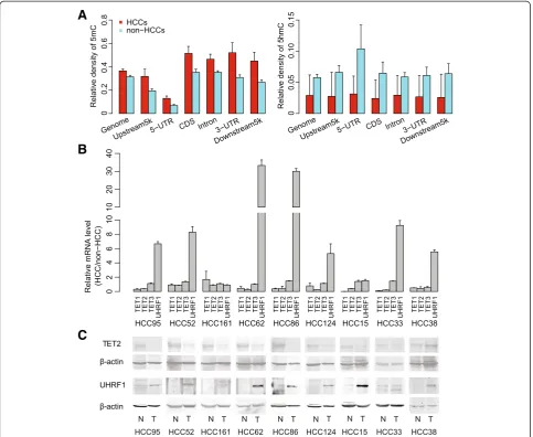
To further examine the distribution of 5mC and 5hmC in HCC specimens and cell lines, we applied a newly de-veloped technology,‘HMST-seq’ [22], on seven of the 16 pairs of HCC specimens and the two cell lines. HMST-seq can detect 5mC and 5hmC at‘CCGG’sites with a single-base resolution, in which three different libraries were constructed for each sample including a library containing unmodified C, mC, and hmC tags throughMspI-mediated digestion of normal genomic DNA, a library containing C and mC tags throughMspI-mediated digestion of glucosy-lated genomic DNA and a library containing only C tags through HpaII-mediated digestion of normal genomic DNA [22]. During manual examination of the tag distribu-tion of these three libraries, we filtered out samples with similar distributions of ‘C + mC’ tags and‘C’tags, which indicate potential insufficient MspI-mediated digestion of glucocylated genomic DNA (Additional file 2: Figure S1). As a result, three out of the seven matched pairs of HCC and non-HCC specimens passed our quality control. We then used these three matched pairs of specimens to infer methylation and hydroxymethylation status across all CCGG sites. On average, 170 million reads for each library were generated, and 147 million (86.58%) reads were aligned to a virtual library of the human genome (see the Methods section). An average of 100 M (58.93%) uniquely aligned reads were then obtained for each library, resulting in a minimum of 1.3 M informative ‘CCGG’ sites with greater than 50 X sequencing depth for each sample (Additional file 1: Table S3). Data normalization for the three libraries was performed, and significantly modified sites with 5mC or 5hmC were identified with our estab-lished methods (sequencing depth >10 X, FDR <0.001) [22]. On average, 433,045 (31.49%) and 72,080 (5.24%) total examined CCGG sites were identified as significant for 5mC and 5hmC, respectively, in non-HCC specimens. In addition, increased 5mC (471,270; 34.72%) but de-creased 5hmC (66,020; 4.89%) was observed in the HCC specimens (Additional file 1: Table S2).
decreased, in consistence with whole-genome 5hmC de-crease tested by UPLC-MS/MS.
As TET proteins are responsible for oxidizing 5mC to 5hmC [15,16], we tested mRNA levels ofTET1,2, and3 in nine pairs of HCC specimens. We found downregula-tion of mRNA levels ofTET1andTET2, but notTET3, in HCCs in comparison with non-HCC samples (Figure 1B). Consistent with its mRNA level, TET2 protein was con-firmed downregulated in HCCs as well (Figure 1C). We also tested UHRF1 as its protein helps to recruit DNMT1 to hemi-methylated DNA to facilitate faithful maintenance of DNA methylation [23]. In contrast to TET genes, both mRNA and protein expression levels ofUHRF1gene were highly upregulated in HCCs (Figure 1B and C), consistent with previous reports in various types of cancers [24]. Based on these results, expression level changes of key
enzymes or cofactors that are involved in DNA methyla-tion/hydroxymethylation regulation might be correlated with 5mC increase and 5hmC decrease in CCGG sites across the HCC genome.
DMRs and DhMRs are revealed by inter-group comparisons
Based on the observation of global methylation and hydroxymethylation alterations, we next aimed to specify differentially methylated regions (DMRs) and differentially hydroxymethylated regions (DhMRs) between HCC and non-HCC samples. A sliding window strategy was used for inter-group comparison of the HCC and non-HCC groups as previously described [22]. To identify differen-tially modified regions containing at least five CCGG sites (Wilcoxon rank-sum test, P <0.05), we began with the
A
B
[image:3.595.57.541.89.485.2]C
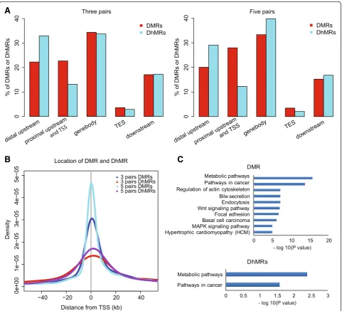
three matched pairs and found 1,851 DMRs and 243 DhMRs (Additional file 1: Table S4, S5). Although both modifications were widely distributed across the genome, DMRs occurred more frequently at proximal regions close to TSSs, while DhMRs were more likely to occur at distal sites upstream of TSSs (Figure 2A and B). Thus, only 10 (4.12%) DhMRs overlapped with DMRs, suggesting that changes in these two modifications rarely co-occur.
Furthermore, we compared the 5mC and 5hmC levels of the DMRs and DhMRs, respectively, among HCC and non-HCC samples and two non-HCC cell lines (97 L and LM6). 97 L
and LM6 were derived from the same HCC patient but ex-hibit highly different metastatic potentials [25]. Similar to the HCC samples, hypermethylation of the DMRs and hypohydroxymethylation of the DhMRs were observed in 97 L and LM6 compared with non-HCC cells. Notably, the LM6 cells, which have a higher metastatic potential, exhib-ited a slightly higher methylation level in the DMRs com-pared with 97 L cells (Additional file 2: Figure S3).
To replicate above results, we re-performed HMST-Seq library construction on two pairs of the samples that first failed our quality control. Similar data quality and
A
[image:4.595.58.540.238.675.2]B
C
5mC and 5hmC levels were achieved (Additional file 1: Table S2, S3). By combining all qualified data, an inter-group comparison of five matched pairs of HCC and non-HCC samples was re-performed, resulting in a total of 5,126 DMRs and 571 DhMRs using the same window-sliding strategy (Additional file 1: Table S4, S5); similar results regarding the distribution of DMR and DhMR were observed (Figure 2A, B).
Enrichment of genes containing DMRs and DhMRs in cancer-related pathways
To further address the functional effects of DNA modifi-cation changes in HCC, we first performed KEGG path-way enrichment analysis (see the Methods section) of 1,373 DMR and 218 DhMR genes from the three matched pairs of HCC and non-HCC samples, respect-ively. A higher enrichment rate for DMR (16.8%; 231 out of 1373) than DhMR genes (8.7%, 19 out 218; FDR <0.05) was found (Additional file 1: Table S6). Although DMR and DhMR genes were enriched in KEGG cancer and metabolic pathways (Figure 2C), these genes rarely overlapped (Additional file 2: Figure S4). In addition, en-richment analysis of 3,283 DMR and 651 DhMR genes from the five pairs of HCC and non-HCC samples, re-spectively, revealed similarly enriched pathways (data not shown). Notably, cancer-related pathways including
‘Pathways in cancer’, ‘Focal adhesion’, ‘Wnt signaling pathway’, and ‘MAPK signaling pathway’ were enriched for DMR genes.
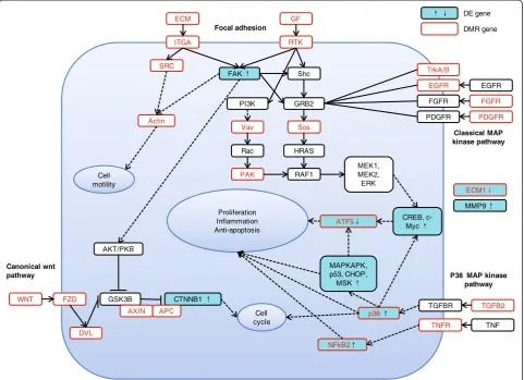
Focal adhesion components are crucial for the cell growth, movement, differentiation, and tailoring of the extracellular microenvironment [26], and of these genes, focal-adhesion kinase (FAK) is an important mediator involved in cancer formation and progression [27]. Mul-tiple genes that encode extracellular proteins and trans-mit signals, including extracellular matrix (ECM), integrin, SRC, and growth factor (GF) genes, were hypermethylated in HCCs. Signaling by the Wnt family of secreted glycolipoproteins via the transcriptional co-activator β-catenin can also determine cell fate and has been implicated in cancer proliferation and survival, in-cluding HCC cells [28]. Aberrant methylation was also observed for key genes of the Wnt signaling pathway in-cluding the Wnt, FZD, and APC genes. Furthermore, multiple key genes in the MAPK signaling pathway were enriched among those containing DMRs, including
TGFB2, EGFR, NFkB2, and p38 mitogen-activated pro-tein kinase (Figure 3, Additional file 1: Table S6).
DMR genes were also enriched in metabolic pathways, which are also relevant. In particular, one gene, PKLR, was aberrantly methylated in HCC; PKLR encodes a pyruvate kinase and is involved in the glycolysis process and important for cancer metabolism and tumor growth, [29,30]. In summary, these results suggest that abnormal
DNA methylation and gene hydroxymethylation might be crucial participants in important cancer-related path-ways in HCC.
Transcriptional changes in key genes in cancer pathways are revealed by integration of multi-omics data
To reveal potential complications resulting from DNA modification changes on gene transcription, we further performed transcriptome profiling of the seven pairs of HCC samples and two HCC cell lines using RNA-Seq and Affymetrix microarray technologies. Based on the high-quality data, we performed pair-wise comparisons to identify differentially expressed genes (DEGs) in the seven pairs of HCC samples. Copy number variation (CNV) analyses were then used to address transcription changes due to gene CNVs (see the Methods section). If significantly upregulated genes were revealed through gene amplification in the HCC samples, we filtered out those DEGs. In addition, significantly downregulated genes resulting from gene deletion in the HCC samples were also removed. As a result, 5,848 upregulated and 1,586 downregulated DEGs in the HCC samples were identified (Additional file 1: Table S7). Furthermore, we examined the transcription level of these DEGs in the two HCC cell lines and found that most of these genes had a similar expression level as in the HCC samples but differed substantially from the non-HCC samples (Additional file 2: Figure S5).
Based on these data, we then examined key genes in the three cancer-related pathways that were enriched in the DMR genes (focal adhesion,Wnt, andMAPK). Not-ably, the FAK (PTK2), β-catenin (CTNNB1), and p38 MAPK (MAPK12) genes were all significantly upregu-lated in HCC. In particular, genes downstream of p38
MAPK including MAPKAPK2, NFkB2, p53, CHOP
(DDIT3), MSK (RPS6KA4), CREB (ATF4), and c-Myc, were all upregulated, indicating activation of these sig-naling pathways in the HCC specimens (Figure 3).
TSG candidate screening
matched pairs. However, only 2 out of the 22 DhMR genes were replicated between the two comparison groups (Additional file 2: Figure S6). These results might indicate that DMRs are more stable than DhMRs in HCC populations. Based on these data and on reports in the literature, which indicated that the biological func-tion of these genes is relevant to liver development or disease, we selected 18 candidate genes for further validation.
Semi-quantitative RT-PCR was then used to detect the mRNA levels of the 18 genes in 20 additional pairs of HCC and non-HCC specimens (Primer sequences listed in Additional file 1: Table S8). Among the 18 genes, six genes including FAM150A, TCF21, EOMES, ATF5, DPT, and ECM1 were frequently downregulated in 85%, 85%, 35%, 60%, 50%, and 60% of the 20 HCC samples, respectively, compared with non-HCC samples (Additional file 2: Figure S7). As all six of these genes only contained DMRs in their promoters, but not DhMRs, bisulfite se-quencing can then efficiently validate DNA methylation
changes for these genes. Therefore, we first performed bi-sulfite sequencing PCR (BSP) on three selected DMR genes (FAM150A,TCF21, andEOMES) in the three pairs of HCC samples for technical validation. The results confirmed sig-nificant differential methylation between HCC and non-HCC samples for each gene, indicating the accuracy of DMR detection by HMST-seq (Additional file 2: Figure S8, Additional file 1: Table S9). Based on these results, we fur-ther validated five selected DMR genes (ATF5, ECM1,
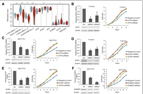
[image:6.595.58.540.90.439.2]EOMES, FAM150A, andTCF21) in 20 additional pairs of HCC and non-HCC samples for biological replication. A high-throughput illumina-sequencing-based BSP method that we developed recently was applied [31] (See Methods). Our results confirmed consistently higher methylation levels of all these genes in HCC samples in comparison with non-HCC samples (Figure 4A, Additional file 1: Table S9). In particular, ECM1andTCF21displayed significant differen-tial methylation between HCC and non-HCC groups (P<0.05). Besides of key hypermethylated gene promoters, we also used this method to test four repetitive elements.
Consistent with whole genome hypomethylation and hypo-hydroxymethylation tested by UPLC-MS/MS, the methyla-tion levels of these repetitive elements tested by bisulfite sequencing were significantly lower in HCC samples (Figure 4A).
Suppression of endogenousATF5,ECM1, andEOMES promotes HCC cell line proliferation
To evaluate the potential contribution of the six target genes to HCC tumorigenesis, we next aimed to perform gene knockdown experiments in HCC cell lines. By evalu-ating the expression level of six target genes in available HCC cell lines using RT-PCR, we selected YY8103 and QGY7701 HCC cell lines, as in whichATF5,EOMES, and
ECM1 were all expressed. However, FAM150A, TCF21, and DPT were hardly expressed in almost all cell lines (Additional file 2: Figure S7). Based on these results, we designed two specific siRNAs against each gene ofATF5,
EOMES, and ECM1 (Additional file 1: Table S10) for knockdown experiments, using siRNA-NC as a negative control. Our data showed that the expression of these three endogenous target genes was efficiently knocked
down in YY-8103 and QGY7701 cells by these siRNAs, as demonstrated through both real-time PCR assays and western blots (Figure 4B, C, D, E, and F). As a result, the transient transfection of siRNA1 and siRNA2 for these three genes enhanced the growth of YY8103 and QGY7701 HCC cells compared with cells transfected with siRNA-NC (P <0.05; Figure 4). Furthermore, analysis of colony formation in soft agar confirmed that among the four target genes, silencingATF5,ECM1, andEOMES sig-nificantly promoted the anchorage-independent growth of YY-8103 and QGY7701 cells compared with the siRNA-NC control cells (P<0.05; Figure 5).
Taken together, these data support the hypothesis that knockdown of ATF5, ECM1, and EOMES consistently promoted the growth of existing HCC cells, suggesting that silencing these three genes may contribute to HCC oncogenesis and progression.
Discussion
Previous studies have shown that hypermethylation of mul-tiple TSGs contributes to HCC pathogenesis and represents a crucial event in HCC progression [6,9,11,32]. However,
A
C
E
F
B
[image:7.595.58.540.89.406.2]D
the recently discovered hydroxymethylation has brought an additional dimension of complexity to the methylation al-terations observed in cancer. Most previous studies have used bisulfite treatment or methylation-sensitive enzyme digestion-based methods to investigate novel DNA methy-lation biomarkers, which cannot distinguish 5-hmC from
5-mC [12,13]. In addition to the heterogeneity of cancer samples or technological differences, inaccurate quantifica-tion of DNA methylaquantifica-tion levels might also lead to incon-sistent or even contradictory results between different studies [33]. Thus, distinguishing between 5hmC and 5mC assists in the identification of DNA methylation biomarkers YY8103
Negative Control ATF5-siRNA1 ATF5-siRNA2
QGY7701
YY8103
Negative Control ECM1-siRNA1 ECM1-siRNA2
QGY7701
Negative Control EOMES-siRNA1 EOMES-siRNA2
QGY7701
A
B
C
0 10 20 30 40 50 60
N
o
. of
sof
t agar
col
oni
es
QGY7701
0 10 20 30 40 50 60 70
N
o
. of
sof
t agar
col
o
ni
es
YY8103
0 10 20 30 40 50 60
N
o
. of
sof
t agar
col
o
ni
es
QGY7701
0 10 20 30 40 50 60 70
N
o
. of
sof
t agar
col
oni
es
YY8103
0 10 20 30 40 50 60
No
.o
f s
o
ft
a
g
a
r
col
oni
es
QGY7701
X100 X100 X100
X100 X100 X100
X100 X100 X100
X100 X100 X100
[image:8.595.57.540.90.602.2]X100 X100 X100
for clinical and commercial use with better sensitivity and specificity.
Here, we applied a multi-omics strategy to vertically in-tegrate epigenomic, genomic, and transcriptomic profiling to screen for novel epigenomic biomarkers and TSG can-didates of HCC. We applied both the commonly used UPLC-MS/MS and the newly developed HMST-Seq tech-nologies to simultaneously examine DNA methylation and hydroxymethylation levels. Globally, 5mC and 5hmC levels were reduced in HCC, as confirmed by UPLC-MS/ MS. As HMST-Seq can only test CCGGMspI sites, an in-creasing amount of 5mC was observed for CCGG sites, in contrast to whole genome 5mC tendency. This result not only reflects the limitation of HMST-Seq in detection of global 5mC pattern, but also indicates an uneven distribu-tion of 5mC in the genome. On the other hand, as 5hmC were evenly distributed across the genome, consistently low levels of 5hmC across the genome-wide CCGGMspI sites were also observed. Furthermore, DMRs and DhMRs rarely co-occurred at the same genomic region. These re-sults might support the hypothesis that 5hmC is an inter-mediate of the demethylation process [15,16], and loss of hydroxymethylation might be also an epigenetic feature of HCCs [21].
We found that a significant number of DMR genes were enriched in several highly interrelated cancer signaling pathways, including the‘Focal adhesion’,‘Wnt’, and‘MAPK’ pathways (Figure 2C). Furthermore, the activation of key signaling pathway genes, includingFAKin‘Focal adhesion’,
β-catenin in ‘Wnt’, and p38 and its downstream genes in
‘MAPK’, indicated the activation of these signaling pathways in HCC, which has been previously reported in HCC. Al-though we currently have no direct evidence that the methylation and hydroxymethylation changes in DMR and DhMR genes may directly contribute to the activation of these pathways, these two types of DNA modifications rep-resent essential participants in heptocarcinogenesis. In addition to gene transcription repression, DNA modifica-tions may also be important for alternative splicing [34], gene mutation [35], and chromatin remodeling [36] in tumorigenesis. Further studies are needed to examine these genes.
In addition to the genes found enriched by KEGG path-way analysis, we confirmed that the suppression of three novel genes by promoter hypermethylation can significantly enhance cell growth and progression in HCC cell lines, in-dicating their potential as TSG candidates. These three genes have never been reported in previous studies on aber-rant DNA methylation of HCCs [6,9,11,32]. Of these three genes, ECM1 has been reported to inhibit the activity of matrix metalloproteinase 9 (MMP9) through high-affinity protein/protein interactions [37]. Due to the significant downregulation ofECM1 transcription, our data also sup-port the significant upregulation ofMMP9transcription in
HCC, which agrees with previous observations of highly up-regulated MMP expression in many solid tumors [38]. MMPs are key modulators of the tumor microenvironment, and it has been shown that MMPs not only regulate ECM turnover but also cell signaling pathways controlling cell growth, inflammation and angiogenesis in a non-proteolytic manner through various non-catalytic domains [38].
ATF5is a highly abundant liver-enriched transcription factor that belongs to the ATF/cAMP response element-binding family [39]. A study has indicated that transcrip-tional expression of ATF5 is directly induced by CHOP and ATF4, which together with ATF5 form the inte-grated stress response (ISR) pathway that is critical for alleviating damage accrued during acute stress [40]. Thus, the proapoptotic functions of ATF5 and CHOP have been revealed [40]. In HCC, although CHOP was activated, ATF5 was hypermethylated in its promoter and exhibited downregulated transcriptional expression. These results are in agreement with previous studies that confirmed downregulation of ATF5 transcription [41] and protein expression levels [42] in HCC. There-fore, ATF5might be blocked in HCC, resulting in anti-apoptotic effects and cancer cell survival.
The T-box transcription factor eomesodermin (EOMES) has been found to have a regulatory role for CD8 T cell ac-tivity in humans [43] and plays a critical role in tumor im-mune surveillance and eradication [44]. The absence of
EOMESin tumor-infiltrating lymphocytes correlates with enhanced lymph node metastasis in colorectal cancer [43].
Conclusions
In summary,ECM1, ATF5, andEOMESmay all serve as novel TSGs that are aberrantly hypermethylated at their promoters leading to their downregulated transcription, which results in HCC tumorigenesis and progression. In addition, we have identified three other genes (FAM150A,
TCF21, and DPT) with promoter hypermethylation and significant transcriptional downregulation in HCCs. These six genes may comprise a gene panel that could be used for the clinical diagnosis and prognosis of HCCs with re-spect to promoter methylation and gene transcription levels.
Materials and methods Sample materials
differentiation grades II to III following the Edmondson-Steiner grading system. The clinical characteristics of the patients and tumors are summarized in Additional file 1: Table S1. This project and protocols involving human and animal tissues were approved by the ethics committee of the Research Ethics Committee of Shenzhen Third Peo-ple’s Hospital (No. 2012-010), and conducted according to the Declaration of Helsinki. The use, for research pur-poses, of tumor tissues from clinically surgical removal was deemed exempt from a requirement for informed consent beyond the consent normally obtained for this clinical procedure.
Normal liver (including LO2 and WRL68) and HCC cell lines (including Hep3B, SK-hep1, Focus, Huh7, SMMC7721, MHCC97L, MHCC97H, MHCC-LM3, MHCC-LM6, PLC, HepG2, YY8103, QGY7701, QGY7703, BEL7402, BEL7404, and BEL7405) were also used in this study. Of these cell lines, Hep3B, SK-hep1, Focus, Huh7, SMMC7721, PLC, HepG2, YY8103, QGY7701, QGY7703, BEL7402, BEL7404, and BEL7405 were obtained from a commercial source (Institute of Chemistry and Cell Biology at Shanghai). In addition, the MHCC97L, MHCC97H, MHCC-LM3, and MHCC-LM6 cell lines were derived from published references [45,46] and were kindly provided by professor Yinkun Liu from the Liver Cancer Institute affiliated with Zhongshan Hospital in Shanghai.
DNA and RNA extraction and cDNA synthesis
Genomic DNA was extracted from all samples using the DNeasy Tissue kit (Qiagen, Valencia, CA, USA), accord-ing to protocols recommended by the manufacturer. Total RNA was extracted from the HCC cell lines and frozen tissue samples, which were pulverized in liquid nitrogen using TRIzol reagent (Invitrogen, Carlsbad, CA, USA). To reduce the risk of genomic DNA contamin-ation, 1 to 2 μg RNA was incubated with 2 U DNase I (Invitrogen, Carlsbad, CA, USA), 1 μL DNase buffer and, 0.4μL RNase Out for 15 min at room temperature. The RNA concentration was determined by spectropho-tometry, and the total RNA integrity was examined by visualization of the 28S and 18S ribosomal RNAs in a 1.2% agarose gel. First-strand cDNA was synthesized using the PrimeScript RT Reagent Kit (TaKaRa, Otsu, Japan) according to the manufacturer’s instructions.
UPLC–MS/MS analysis
Genomic DNA (0.2μg) extracted from all patients and cell lines was digested with 1 U DNase I, 2 U alkaline phosphat-ase, calf intestinal, and 0.005 U snake venom phospho-diesterase I at 37°C for 24 h. A microcon centrifugal filter device with a 3,000 D cutoff membrane was used to remove protein from the digested DNA samples by centrifuging at 12,000 rpm for 60 min. The mobile phase consisted of 0.1% formic acid (solvent A) and methanol containing 0.1%
formic acid (solvent B). The flow rate was set to 500 μL/ min. The enzymatically digested DNA samples (5μL each) were injected for UPLC-MS/MS analysis with a LC gradient as follows: 0.0-4.0 min, 0 to 50% of solvent B; 4.0-6.0 min, 50% solvent B; 6.0-6.1 min, 50% to 5% of solvent B; and 6.1-15 min, 5% of solvent B. The separated analytes were de-tected using a 5500 Qtrap linear ion trap quadrupole mass spectrometer equipped with a Turbo V ion source operated in the ESI mode (AB Sciex) with Analyst software (Version 1.5). The Source and Gas were set as follows: gas 1, nitrogen (45 psi); gas 2, nitrogen (40 psi); ion spray voltage, 5,500 V; ion source temperature, 400°C; and curtain gas, nitrogen (30 psi). The mass spectrometer was operated in the multi-reaction monitoring mode (MRM).
Data analysis for HSMT-Seq
Genomic DNA was extracted from all patients and cells using the QIAamp DNA Blood Mini Kit (Qiagen). HSMT-Seq library construction was performed following our previ-ously reported protocol [22]. The libraries were sequenced using single-end 50 bp sequencing strategy with an Illumina HiSeq2000 sequencing system. The FASTQ sequence reads were mapped to a virtual human reference library (hg19) with no more than one mismatch after adapter removal and low-quality read filtering. The virtual library was constructed as follows. The human reference genome (hg19) was in
silico digested with MspI and NlaIII, and we defined the DNA sequences between the nearest NlaIII sites around eachMspI site in both directions as the virtual library refer-ence for mapping. Unambiguous mapped tags were used for further analysis. The data for three libraries were normalized according to a previous method (Global Rank-invariant Set Normalization, GRSN) [47] based on the normalized tag counts, and the modification abundance of a specific CCGG site was determined as the ratio between the tag counts of two libraries. For instance, the hydroxymethylation level can be defined as the ratio between ‘C + mC + hmC’and‘C + mC’tags. Furthermore, CCGG sites with significantly differ-ent tag counts (sequencing depth >10X, FDR <0.001) based on Poisson distribution with a ratio of tags between two li-braries larger than 1 were determined to be significantly modified sites.
test was performed in the incorporated region; (3) Repeat these steps until the Wilcoxon rank-sum test P value
is ≥0.05; and (4) the incorporated region was defined
as a DMR or DhMR.
Enrichment analysis
Functional enrichment analysis for genes with DMRs or DhMRs was performed using WebGestalt [48,49], which is freely accessible at [50].
Data analysis for differentially expressed genes (DEGs) The gene expression levels based on RNA-seq and Affy-metrix microarray data were measured by the reads per kilobase of transcript per million reads (RPKM) and tags per million reads (TPM) algorithm, respectively. The dif-ferentially expressed genes (DEGs) were identified in the five pair-wise samples with Affymetrix microarray data by Wilcoxon rank-sum test and in each of the two pair-wise samples with RNA-seq data according to a previ-ously published method [51]. Genes that were signifi-cantly expressed (FDR <0.05) in at least one group and regulated similarly in all the groups were further identi-fied as differentially expressed genes (DEGs).
Illumina-sequencing-based BSP
Illumina-sequencing-based BSP was performed based on a previously described strategy with minor adjustment [31]. PCR primers were designed using online software [52] and then concatenated 22 bp common sequences at their 5’ end, respectively, which would mediate high throughput sequencing linker extension during the following PCR procedure. All PCR products were about 500 bp. For the high throughput bisulfite-amplicon library construction using large scale of clinical samples, WaferGen Biosystem (WaferGenBiosystems) was employed. Briefly, 500 ng of genomic DNA extracted from whole blood were converted using EZ DNA Methylation-Gold Kit™(ZYMO). For each sample, bisulfite converted genomic DNA, KAPA2G Ro-bust HotStart ReadyMix (KAPA Biosystems), PCR primers, PCR primers mediated high throughput sequencing linkers with a barcode sequence were dispensed into one individual nanowell of a Smart MyDesign Chip, which contains 5,184 nanowells, by Smart Chip Multisample Nanodispenser. Thirty-six amplicons for each sample and 150 samples could be constructed as one library with barcode sequence classifying samples using one Smart MyDesign Chip. PCR amplification of Smart MyDesign Chip was conducted using the Techne Prime Thermal Cycler. PCR product was purified using the QIAquick Gel Extraction Kit (Qiagen). After analyzed by an Agilent 2100 Bioanalyzer (Agilent Technologies) and quantified by real-time PCR, the library was sequenced with pair end 250 bp using Illumina Miseq sequencer.
Semi-quantitative RT-PCR
Reverse transcription (RT) was performed with 2 μg of total RNA treated with RNase-free DNase I. Semi-quantitative RT-PCR was then performed using primers for target genes (Additional file 1: Table S8). The length of the amplified fragments was 350 bp, and β-actin served as an internal reference. The primers for β-actin
were as follows: 5′-TCACCCACACTGTGCCCATC
TACGA-3′ (forward) and 5′-CAGCGGAACCGCT
CATTGCCAATGG-3′ (reverse). The length of the β -actin amplicon was 295 bp. The PCR products were sep-arated in 2% agarose gels containing ethidium bromide. The PCR reactions were performed in a volume of 20μL using the TaKaRa PCR Kit. The reactions were per-formed at 94°C for 5 min followed by 30 to 35 cycles (for 18 target genes) or 25 cycles (for β-actin) at 94°C for 30 s, 55°C for 30 s, and 72°C for 30 s with a final ex-tension at 70°C for 5 min. The PCR products were brought to 4°C at the end of the reaction, and they were then separated in 2% agarose gels containing ethidium bromide.
Western blot
Cells were collected using a 2× loading lysis buffer (2× concentrations: 50 mmol/L Tris-HCl, pH 6.8, 2% sodium dodecyl sulfate, 10% 2-mercaptoethanol, 10% glycerol, and 0.002% bromophenol blue). Total protein extracts from cultured cells were subjected to protein gel electro-phoresis using 12% SDS-PAGE, transferred to a Hybrid-PVDA membrane (Amersham Life Sciences) via a semi-dry electrophoretic transfer method, and treated with 20% methanol in a Tris-glycine buffer (25 mM Tris-HCl, pH 8.0, 0.2 M glycine, 0.1% SDS). After blocking with PBS containing 5% BSA, the membrane was incubated for immunoblotting analysis with a rabbit anti-myc poly-clonal antibody (1:100; made in our laboratory) or anti-myc antibody (1:1,000) at room temperature for 2 h,
followed by incubation with an IRDye
800DX-conjugated, affinity-purified goat anti-rabbit secondary antibody (1:1,000; Rockland). Signals were detected using the Odyssey Infrared Imaging System (LI-COR Biosciences).β-actin was used as a loading control.
Identification of CNVs
Array-based comparative genomic hybridization (aCGH) was performed using Agilent 244-k arrays as previously described [23]. CNVs that were present in the matching normal sample were filtered, and the remaining CNVs were regarded as somatic variants.
RNA interference
(Shanghai GenePharma Co.) to target the different coding regions of each gene. The siRNA sequences are shown in Additional file 1: Table S6. In addition, siRNA-NC (5′
-GAGUUAAAGUCAAAGUGACTT-3′ and 5′-GUCACU
UUGACUUUAACUCTT-3′) was also synthesized. The siRNAs were transfected into the HCC cell lines, and cell growth was monitored. For siRNA transfection, 3 × 10³ HCC cells per well were seeded into 96-well plates. When the cells reached 30% to 50% confluence, they were trans-fected with synthetic siRNAs at a final concentration of 50 nM using the Lipofectamine 2000 Transfection Reagent (Invitrogen) according to the manufacturer’s instructions.
Cell proliferation analysis
The cells were cultured for 7 days, and cell viability was measured using the ACEA RTCA kit (ACEA Biosciences, San Diego, CA, USA). A microelectronic cell sensor system was used to confirm the number of living cells. NSCLC cells (1 × 10⁴) were seeded into each sensor-containing well (19.6-mm2surface with 150 mL of medium) of the microti-ter plates. The electronic sensors provided a continuous (every 6 h), quantitative measurement of the cell index (reflecting the surface area covered by the cells) in each well. Cell growth was measured every 6 h for 96 to 144 h, and cell indexes were recorded for each well at all time points to assess cell viability. All experiments were inde-pendently repeated at least three times.
Soft agar colony formation
For soft agar colony formation assays in 24-well plates, 2 × 10⁴cells were plated and grown on plates containing 1% base agar and 0.5% top agar. The plated cells were in-cubated at 37°C for 21 days. The plates were stained with 0.005% crystal violet for 1 h, and colonies were counted under a dissecting microscope. All experiments were inde-pendently repeated at least three times.
Data availability
The sequencing and processed data have been deposited in the NCBI SRA and GEO database under the accession number SRA092219 and GSE54141. All the UPLC-MS/MS raw data were deposited in figshare database (http://dx.doi. org/10.6084/m9.figshare.1117723).
Additional files
Additional file 1:All supplemental tables (Tables S1 to S10) and corresponding captions.
Additional file 2:All supplemental figures (Figures S1 to S8) and corresponding legends.
Abbreviations
5fC:5-formylcytosine; 5hmC: 5-hydroxymethylcytosine; 5mC: 5-methylcytosine; CNV: Copy number variation; DEGs: Differentially expressed genes;
DhMRs: Differentially hydroxymethylated regions; DMRs: Differentially methylated
regions; HCC: Hepatocellular carcinoma; HMST-seq: Hydroxymethylation and methylation Sensitive Tag sequencing; non-HCC: Non-cancerous liver tissues; TSG: Tumor suppressor gene; TSS: Transcriptional start sites; UPLC-MS/MS: Ultra-performance liquid chromatography/tandem mass spectrometry.
Competing interests
The authors declare that they have no competing interests.
Authors’contributions
FG, JH, and XQZ conceived the project; YDX conducted the bioinformatic analysis; JWW and HJL performed the HMST-Seq experiments; YO, WL, BPZ, and XL prepared samples and performed the functional experiments; and ZLL, BJZ, and BW performed the UPLC-MS/MS experiment. FG, YDX, and JH interpreted the data and wrote the manuscript. All authors read and approved the final manuscript.
Acknowledgments
We gratefully acknowledge support from the National High Technology Research and Development Program of China (863 Program, 2012AA02A201 and 2012AA02A205), the Chinese National Key Program on Basic Research (973 Program, 2010CB529206 and 2014CB965002), the National Natural Science Foundation of China (81272306), Program of Shanghai Subject Chief Scientist (12XD1421400), Guangdong Innovative Research Team Program (2009010016), and Program of Shenzhen Commission for Science and Technology (JCYJ20130329171031740).
Author details
1BGI-Shenzhen, Shenzhen 518083, China.2National Engineering Center for
Biochip at Shanghai, Shanghai 201203, China.3Shanghai-MOST Key
Laboratory for Disease and Health Genomics, Chinese National Human Genome Center at Shanghai, Shanghai 201203, China.4Shenzhen Key Lab. of
Infection and Immunity, Shenzhen Third People’s Hospital, Guangdong Medical College, Shenzhen 518112, China.5Guangdong Key Lab. Of
Diagnosis & Treatment for Emerging Infectious Disease, Shenzhen Third People’s Hospital, Guangdong Medical college, Shenzhen 518112, China.
Received: 14 April 2014 Accepted: 6 November 2014
References
1. Llovet JM, Burroughs A, Bruix J:Hepatocellular carcinoma.Lancet2003,
362:1907–1917.
2. Ferlay J, Shin HR, Bray F, Forman D, Mathers C, Parkin DM:Estimates of worldwide
burden of cancer in 2008: GLOBOCAN 2008.Int J Cancer2010,127:2893–2917.
3. Sherman M:Recurrence of hepatocellular carcinoma.N Engl J Med2008,
359:2045–2047.
4. Nishida N, Goel A:Genetic and epigenetic signatures in human hepatocellular
carcinoma: a systematic review.Curr Genomics2011,12:130–137.
5. Liu M, Jiang L, Guan XY:The genetic and epigenetic alterations in human
hepatocellular carcinoma: a recent update.Cell Protein2014,5:673–691.
6. Anwar SL, Lehmann U:DNA methylation, microRNAs, and their crosstalk
as potential biomarkers in hepatocellular carcinoma.World J Gastroenterol
2014,20:7894–7913.
7. Eden A, Gaudet F, Waghmare A, Jaenisch R:Chromosomal instability and
tumors promoted by DNA hypomethylation.Science2003,300:455.
8. Chan KC, Jiang P, Chan CW, Sun K, Wong J, Hui EP, Chan SL, Chan WC, Hui DS, Ng SS, Chan HLY, Wong CSC, Ma BBY, Chan ATC, Lai PBS, Sun H, Chiu RWK, Lo
YMD:Noninvasive detection of cancer-associated genome-wide
hypomethylation and copy number aberrations by plasma DNA bisulfite
sequencing.Proc Natl Acad Sci U S A2013,110:18761–18768.
9. Nishida N, Kudo M, Nagasaka T, Ikai I, Goel A:Characteristic patterns of altered DNA methylation predict emergence of human hepatocellular
carcinoma.Hepatology2012,56:994–1003.
10. Umer M, Qureshi SA, Hashmi ZY, Raza A, Ahmad J, Rahman M, Iqbal M: Promoter hypermethylation of Wnt pathway inhibitors in hepatitis C
virus - induced multistep hepatocarcinogenesis.Virol J2014,11:117.
11. Shen J, Wang S, Zhang YJ, Wu HC, Kibriya MG, Jasmine F, Ahsan H, Wu DP, Siegel AB, Remotti H, Santella RM:Exploring genome-wide DNA methylation profiles altered in hepatocellular carcinoma using Infinium HumanMethylation 450
BeadChips.Epigenetics2013,8:34–43.
12. Huang Y, Pastor WA, Shen Y, Tahiliani M, Liu DR, Rao A:The behaviour of
13. Jin SG, Kadam S, Pfeifer GP:Examination of the specificity of DNA methylation profiling techniques towards 5-methylcytosine and
5-hydroxymethylcytosine.Nucleic Acids Res2010,38:e125.
14. Gao F, Xia Y:Hydroxymethylation- and methylation-sensitive tag sequencing: how will this technology change clinical applications of
DNA methylation profiling?Epigenomics2013,5:355–357.
15. Tahiliani M, Koh KP, Shen Y, Pastor WA, Bandukwala H, Brudno Y, Agarwal S, Iyer LM, Liu DR, Aravind L, Rao A:Conversion of 5-methylcytosine to 5-hydroxymethylcytosine in mammalian DNA by MLL partner TET1. Science2009,324:930–935.
16. Ito S, Shen L, Dai Q, Wu SC, Collins LB, Swenberg JA, He C, Zhang Y:Tet proteins can convert 5-methylcytosine to 5-formylcytosine and
5-carboxylcytosine.Science2011,333:1300–1303.
17. Kriaucionis S, Heintz N:The nuclear DNA base 5-hydroxymethylcytosine is
present in Purkinje neurons and the brain.Science2009,324:929–930.
18. Haffner MC, Chaux A, Meeker AK, Esopi DM, Gerber J, Pellakuru LG, Toubaji A, Argani P, Iacobuzio-Donahue C, Nelson WG, Netto GJ, De Marzo AM, Yegnasubramanian S:Global 5-hydroxymethylcytosine content is significantly reduced in tissue stem/progenitor cell compartments and in
human cancers.Oncotarget2011,2:627–637.
19. Yang H, Liu Y, Bai F, Zhang JY, Ma SH, Liu J, Xu ZD, Zhu HG, Ling ZQ, Ye D, Guan KL, Xiong Y:Tumor development is associated with decrease of TET
gene expression and 5-methylcytosine hydroxylation.Oncogene2013,
32:663–669.
20. Lian CG, Xu Y, Ceol C, Wu F, Larson A, Dresser K, Xu W, Tan L, Hu Y, Zhan Q, Lee CW, Hu D, Lian BQ, Kleffel S, Yang Y, Neiswender J, Khorasani AJ, Fang R, Lezcano C, Duncan LM, Scolyer RA, Thompson JF, Kavakand H, Houvras Y, Zon LI, Mihm MC Jr, Kaiser UB, Schatton T, Woda BA, Murphy GF,et al:Loss of 5-hydroxymethylcytosine is an epigenetic hallmark of melanoma. Cell2012,150:1135–1146.
21. Liu C, Liu L, Chen X, Shen J, Shan J, Xu Y, Yang Z, Wu L, Xia F, Bie P, Cui Y, Bian XW, Qian C:Decrease of 5-hydroxymethylcytosine is associated with progression of hepatocellular carcinoma through downregulation of
TET1.PLoS One2013,8:e62828.
22. Gao F, Xia Y, Wang J, Luo H, Gao Z, Han X, Zhang J, Huang X, Yao Y, Lu H, Yi N, Zhou B, Lin Z, Wen B, Zhang X, Yang H, Wang J:Integrated detection of both 5-mC and 5-hmC by high-throughput tag sequencing technology highlights methylation reprogramming of bivalent genes during cellular differentiation. Epigenetics2013,8:421–430.
23. Bostick M, Kim JK, Esteve PO, Clark A, Pradhan S, Jacobsen SE:UHRF1 plays
a role in maintaining DNA methylation in mammalian cells.Science2007,
317:1760–1764.
24. Bronner C, Krifa M, Mousli M:Increasing role of UHRF1 in the reading and inheritance of the epigenetic code as well as in tumorogenesis. Biochem Pharmacol2013,86:1643–1649.
25. Huang J, Deng Q, Wang Q, Li KY, Dai JH, Li N, Zhu ZD, Zhou B, Liu XY, Liu RF, Fei QL, Chen H, Cai B, Zhou B, Xiao HS, Qin LX, Han ZG:Exome sequencing of hepatitis B virus-associated hepatocellular carcinoma. Nat Genet2012,44:1117–1121.
26. Romer LH, Birukov KG, Garcia JG:Focal adhesions: paradigm for a
signaling nexus.Circ Res2006,98:606–616.
27. Zhao J, Guan JL:Signal transduction by focal adhesion kinase in cancer. Cancer Metastasis Rev2009,28:35–49.
28. Fatima S, Lee NP, Luk JM:Dickkopfs and Wnt/beta-catenin signalling in
liver cancer.World J Clin Oncol2011,2:311–325.
29. Christofk HR, Vander Heiden MG, Harris MH, Ramanathan A, Gerszten RE, Wei R, Fleming MD, Schreiber SL, Cantley LC:The M2 splice isoform of pyruvate kinase is important for cancer metabolism and tumour growth. Nature2008,452:230–233.
30. Dong C, Yuan T, Wu Y, Wang Y, Fan TW, Miriyala S, Lin Y, Yao J, Shi J, Kang T, Lorkiewicz P, St Clair D, Hung MC, Evers BM, Zhou BP:Loss of FBP1 by Snail-mediated repression provides metabolic advantages in basal-like
breast cancer.Cancer Cell2013,23:316–331.
31. Gao F, Zhang J, Jiang P, Gong D, Wang JW, Xia Y, Ostergaard MV, Wang J, Sangild PT:Marked methylation changes in intestinal genes during the
perinatal period of preterm neonates.BMC Genomics2014,15:716.
32. Neumann O, Kesselmeier M, Geffers R, Pellegrino R, Radlwimmer B, Hoffmann K, Ehemann V, Schemmer P, Schirmacher P, Lorenzo Bermejo J, Longerich T:Methylome analysis and integrative profiling of human
HCCs identify novel protumorigenic factors.Hepatology2012,
56:1817–1827.
33. Heyn H, Esteller M:DNA methylation profiling in the clinic: applications
and challenges.Nat Rev Genet2012,13:679–692.
34. Maunakea AK, Chepelev I, Cui K, Zhao K:Intragenic DNA methylation modulates alternative splicing by recruiting MeCP2 to promote exon
recognition.Cell Res2013,23:1256–1269.
35. Hitchins MP, Rapkins RW, Kwok CT, Srivastava S, Wong JJ, Khachigian LM, Polly P, Goldblatt J, Ward RL:Dominantly inherited constitutional epigenetic silencing of MLH1 in a cancer-affected family is linked to a
single nucleotide variant within the 5'UTR.Cancer Cell2011,20:200–213.
36. Forn M, Munoz M, Tauriello DV, Merlos-Suarez A, Rodilla V, Bigas A, Batlle E, Jorda M, Peinado MA:Long range epigenetic silencing is a trans-species mechanism that results in cancer specific deregulation by overriding the
chromatin domains of normal cells.Mol Oncol2013,7:1129–1141.
37. Fujimoto N, Terlizzi J, Aho S, Brittingham R, Fertala A, Oyama N, McGrath JA, Uitto J:Extracellular matrix protein 1 inhibits the activity of matrix metalloproteinase 9 through high-affinity protein/protein interactions. Exp Dermatol2006,15:300–307.
38. Kessenbrock K, Plaks V, Werb Z:Matrix metalloproteinases: regulators of
the tumor microenvironment.Cell2010,141:52–67.
39. Pascual M, Gomez-Lechon MJ, Castell JV, Jover R:ATF5 is a highly abundant liver-enriched transcription factor that cooperates with constitutive androstane receptor in the transactivation of CYP2B6: implications in
hepatic stress responses.Drug Metab Dispos2008,36:1063–1072.
40. Teske BF, Fusakio ME, Zhou D, Shan J, McClintick JN, Kilberg MS, Wek RC: CHOP induces activating transcription factor 5 (ATF5) to trigger
apoptosis in response to perturbations in protein homeostasis.Mol Biol
Cell2013,24:2477–2490.
41. Gho JW, Ip WK, Chan KY, Law PT, Lai PB, Wong N:Re-expression of transcription factor ATF5 in hepatocellular carcinoma induces G2-M
arrest.Cancer Res2008,68:6743–6751.
42. Liu X, Liu D, Qian D, Dai J, An Y, Jiang S, Stanley B, Yang J, Wang B, Liu DX: Nucleophosmin (NPM1/B23) interacts with activating transcription factor 5 (ATF5) protein and promotes proteasome- and caspase-dependent
ATF5 degradation in hepatocellular carcinoma cells.J Biol Chem2012,
287:19599–19609.
43. Atreya I, Schimanski CC, Becker C, Wirtz S, Dornhoff H, Schnurer E, Berger MR, Galle PR, Herr W, Neurath MF:The T-box transcription factor eomeso-dermin controls CD8 T cell activity and lymph node metastasis in human
colorectal cancer.Gut2007,56:1572–1578.
44. Zhu Y, Ju S, Chen E, Dai S, Li C, Morel P, Liu L, Zhang X, Lu B:T-bet and eomesodermin are required for T cell-mediated antitumor immune
responses.J Immunol2010,185:3174–3183.
45. Tian J, Tang ZY, Ye SL, Liu YK, Lin ZY, Chen J, Xue Q:New human hepatocellular carcinoma (HCC) cell line with highly metastatic potential (MHCC97) and its expressions of the factors associated with metastasis. Br J Cancer1999,81:814–821.
46. Li Y, Tang Z, Ye S, Liu Y, Chen J, Xue Q, Huang X, Bao W, Yang J, Gao D: [Establishment of human hepatocellular carcinoma cell line with spontaneous pulmonary metastasis through in vivo selection]. Zhonghua Yi Xue Za Zhi2002,82:601–605.
47. Pelz CR, Kulesz-Martin M, Bagby G, Sears RC:Global rank-invariant set normalization (GRSN) to reduce systematic distortions in microarray
data.BMC Bioinformatics2008,9:520.
48. Zhang B, Kirov S, Snoddy J:WebGestalt: an integrated system for
exploring gene sets in various biological contexts.Nucleic Acids Res2005,
33:W741–W748.
49. Wang J, Duncan D, Shi Z, Zhang B:WEB-based GEne SeT AnaLysis Toolkit
(WebGestalt): update 2013.Nucleic Acids Res2013,41:W77–W83.
50. WebGestalt.[http://www.webgestalt.org]
51. Stéphane Audic J-MC:The significance of digital gene expression profiles. Genome Res1997,7:986–995.
52. MethPrimer.[www.urogene.org/methprimer/index.html]
doi:10.1186/s13059-014-0533-9
Cite this article as:Gaoet al.:Integrated analyses of DNA methylation and hydroxymethylation reveal tumor suppressive roles ofECM1,ATF5,
andEOMESin human hepatocellular carcinoma.Genome Biology