http://dx.doi.org/10.4236/aar.2016.52005
How to cite this paper: Wang, Q., Zhou, F.J., Xie, W.M., Zhao, X.R. and Liu, X. (2016) Research Progress on Aging Mechanisms. AdvancesinAgingResearch, 5, 49-57. http://dx.doi.org/10.4236/aar.2016.52005
Research Progress on Aging Mechanisms
Qiang Wang, Fengjuan Zhou, Wenmei Xie, Xiaorong Zhao, Xuan Liu* Gansu Medical College, Pingliang, China
Received 11 May 2015; accepted 14 March 2016; published 17 March 2016
Copyright © 2016 by authors and Scientific Research Publishing Inc.
This work is licensed under the Creative Commons Attribution International License (CC BY). http://creativecommons.org/licenses/by/4.0/
Abstract
Aging refers to a gradual process of functional and organic recession with the age increased after the organism maturing. Many researchers have tried to elucidate aging mechanism which includes the free radical theory: free radicals can become lipofuscin and cause the mutations of mitochon-drial DNA (mtDNA), and the damage of the nuclear DNA. From the view of heredity, aging is the results of the activation and inhibition of a series of genes as well as the products of their interac-tion. From the changes of immune function theory, it points out that aging is attributed to the de-cline of immune responses and the increase of autoimmune reactions.
Keywords
Aging, Free Radical, Lipoprotein, Apoptosis, Mitochondrial DNA, Genes, Immune System
1. Introduction
Aging refers to a gradual process of functional and organic recession with the age increased after the organism maturing, including the decline of the stability of the homeostasis, the decrease of the ability of stress decreased, and the gradual degeneration of structure and components, and finally trend to death [1]. Research on aging has been one of most basic and important parts in life sciences, but the details of the process have been still poorly understood. In the past 20 years, although scientists have made great progress, it is still very limited [2]. Aging is a continuous, dynamic gradual and complex process. The process starts with the end of growing season, which negative impact appears gradually in old age through the system disorders, the decline of organ function, degeneration, and changes in the molecular structure of proteins and enzymes. The main feature is the dysfunc-tions of organs and tissues and the ability to adapt to environmental stimulus reduced, even lost totally. There are many factors that influence ageing, including social factors, economic, disease, nutrition, heredity factors, lifestyle, environment and mental state, etc. Therefore, the aging is an interaction result of many factors [3]. Nowadays, with the progress of the research of the cellular and molecular biology, the study of aging
chromatin aggregation, condensation, fragmentation, dissolution, membrane retraction, cytoplasm vacuolation, mitochondrial number abnormality, Golgi fragmentation, Nissl body disappearance, and membrane fluidity de-creased. Many stimuli factors can induce cells aging, including telomere shortening, DNA damage, high oxida-tive stress, sustained stimulation of mitogen, and other cell stress [4] [5]. telomerase shortening to some extent, can cause the lost of telomere function, and then similar signal of DNA damage is generated, it can be induce aging, but telomere shortening is closely related to cells proliferation, thus this type of aging is known as “rep-licative aging”; Other factors induce aging rapidly and the telomere length has no change apparently, including overactivation of Mitogen bypass, such as overexpression of Ras, Raf, MEK, E2F, etc. [6]. This type of aging is known as “stress-induced aging”. Although research on stress-induced aging mechanism is not so much as rep-licative aging mechanism, phenotypic characteristics and molecular signals from these two types of aging are very similar.
3. The Mechanism of Cells Aging
Exploring the mechanisms of aging is an ancient and new field of scientific research. With in-depth study, the free radical theory, telomere theory, DNA damage theory, immune theory and other theories have been pro-posed.
3.1. Free Radical Theory
In 1956, Harmon D puts forward the free radical theory of aging, which pointed out that the major cause of hu-man aging was constantly produce free radicals in cell metabolism processes [7].
Free Radical (RF), also known as free radicals, is the outer orbit of atoms or special status of molecules con-taining unpaired electrons, because of the natural tendency of electrons in pairs, the unpaired electron has a “tension” to look for “partners”. There are superoxide radical (•O−2), hydroxyl ion free radical (•OH), hydrogen peroxide free radical (•OOH), hydrogen radical (•H), lipid radical (•L), lipid peroxide radicals (LOO•), organic free radical (R•) and organic peroxide radicals (ROO•) produced during body activity (such as cellular respira-tion, the process of oxidation in mitochondria) [8]. Free radical plays an important role in the metabolism of human body. All kinds of free radicals will participate in many physiological processes, such as redox reactions of mitochondria and microsome, and the killing effect of leukocytes to pathogens and tumor cells. Under normal condition, free radicals can be cleared by defense system-antioxidant enzymes and antioxidants in vivo, and there is no toxic effect on cells. With the remove of free radical of enzyme that reduces in activity or decreased in number, which can lead to increase of free radical in vivo, and when other factors such as ultraviolet, X-rays,
γ-rays, cigarette smoke, oxidants, electronic radiation induce abnormality of the production of free radical, these excessive free radicals will cause damage on protein, DNA, and lipid in human body [9] [10], its free radi-cal-induced mechanism involves following aspects.
3.1.1. The Formation of Lipofuscin
(a) (b) (c)
Figure 1. Autofluorescence of lipofuscin granules in neonatal rat cardiac myocytes keeps in cul-ture for 3 months. (a), (b), and (c). Luorescent images use ultraviolet (330 - 380 nm), blue (450 - 490 nm) and green (510 - 560 nm) excitation light, and 420, 520, and 590 nm barrier filters, re-spectively. Asterisks indicate the nuclei. Bar, 10 μM.
Lipofuscin accumulation in brain cells that will be damage the phospholipid membrane, and lead to changes in the sub cellular structure such as the reduction of mitochondria and rough endoplasmic reticulum, it also re-duces the number of neurons cells ,and can result in a series of disorders in memory and function of brain, even leads to Alzheimer’s disease and other age-related disorders [13]; Lipofuscin accumulation in skin cells that will arouse the formation of age pigment causes collagen polymerization, which lead to the skin without tension and elasticity, wrinkles increase and senile bone proliferation; Lipofuscin accumulation in myocardial cells that will result in cardiac dysfunction [14]. All of the basic characteristics of aging mentions above.
3.1.2. Mutations in mtDNA
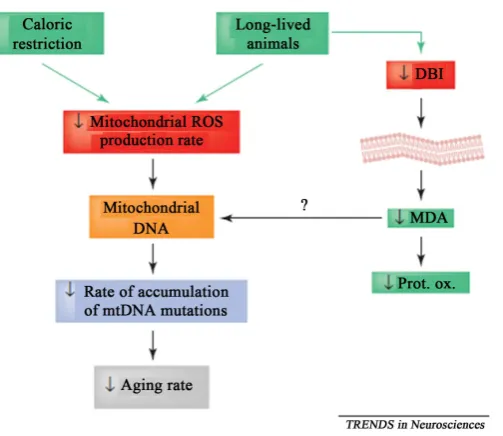
Since at the first time of Harman (1972) has proposed the mitochondrial DNA hypothesis, which is closely re-lated with aging. There are a large number of experimental studies have been confirmed this view. Mitochondri-al DNA (mtDNA) mutations can be detected in severMitochondri-al of degenerative diseases, such as aging and cancer. It in-creases significantly during aging, especially more prominent in the high oxygen consumption of organizations such as brain and muscle. Because of mtDNA nudity, there is no protection of histone and lack of effective re-pair system, the thymine, uracil, guanine in mtDNA molecule to react with the function of •O−2 and •OH, and then causing of base substitution, reorganization or deletions in mtDNA, resulting in germ-line and somatic cell mtDNA mutation rate than nuclear DNA (nDNA) 10 - 100 times, and being accumulated continuously in the cell. Barja, G [15] points out that ROS (reactive oxygen species) can attack many different cellular macromole-cules, including proteins, lipids and DNA. Damage to DNA must be most important for aging, especially in postmitotic cells such as neurons. mtDNA is situated very close to the site of mitochondrial ROS production. Because long-lived vertebrates have low rates of mitochondrial ROS generation, this should affect the level of oxidative damage in their mtDNA (Figure 2).
Figure 2. The summary of available results from mammals and birds is related to aging and oxidative stress. Both long-lived and calorie-restricted animals constitutively have low le-vels of production of mitochondrial reactive oxygen species (ROS), which could be respon-sible for their low rate of accumulation of mitochondrial DNA (mtDNA) mutations, and thus for their low rate of aging.
3.1.3. Nuclear DNA Damaged
The strong oxidation of free radical arouse nucleic acid oxidized, cross-linked, and results in broken and mutant, which has seriously impact on the normal transcription and translation of genetic information, with the protein expression decreased or even to disappeared, the mutant proteins produced, and the protein synthesis reduced, which is the important reason of the memory impairment, mental retardation and muscle atrophy with age.
4. Telomeres and Telomerase Theory
Telomeres are the chromosomal end of special structure of eukaryotic cell and are related intimately to the cells aging. It consists in many simple sequence repeat (Human beings is TTAGGG) and protein. Telomeres have protected the end of chromosomes to prevent DNA repair from the fusion between chromosome, and from telo-meres losing and shortening during DNA replication. It is also the function to maintenance of genome integrity and stability [21]. When DNA replication, small amounts of the end of DNA loses owing to incomplete DNA replication, and the telomeres shorten to an extent after several hundred base of telomere DNA is lost, it cannot maintain the stability of chromosomes, and arouse the cells stop dividing, resulting in ultimately in death, and causing aging [22]. Telomerase is a ribonucleoprotein enzyme that contains primers specific recognition sites, it is own RNA as a template for reverse transcriptase, the telomere DNA synthesis and added to the chromosome ends to make telomere extension, thereby extending the life span of cells even to its immortalization. Calvin B Harley etal. [23] studies have shown that the life span was inverse with the telomere shortening speed and in a direct proportion to the telomerase activity and the intrinsic aging processes almost controlled by the accumula-tion of aging telomere shortening (Figure 3).
Figure 3. Simplified model of human terminal restriction fragment (TRF) structure. (a) An av-erage TRF in cells from a newborn baby may have −4 - 6 kb of pure]q-AGGG repeats at the ter-minus and 3 - 5 kb of degenerate TTAGGG repeats and other simple sequence DNA distal to the first restriction enzyme site (e.g. Rsal). (b) In senescent cells, most of the pure TTAGGG repeats have been lost, leaving the degenerate TTAGGG repeats and non-telomeric sequence. As non- telomeric repeats do not seed new chromosome ends, it is unlikely that the degeneration of TTAGGG repeats will fully function as telomeres. Assuming some heterogeneity in telomere length between chromosomes, the TTAGGG repeats on at least the end of one chromosome may be of insufficient length for telomere function. Thus, the cell could halt proliferation at senes-cence because of a critically short telomere. (c) If the cell has mutations or alterations in the cell cycle checkpoint pathway allows senescence to be bypassed, then telomeres would continue to shorten until most of the pure and degenerion of TTAGGG sequence was lost on essentially all chromosomes. At this point, “crisis” occurs, resulting in massive genomic instability and cell death. (d), (e) Rare cells which survive crisis reactivate telomerase and restore telomere function by adding pure TI-AGGG repeats to chromosome ends. Depending on the balance of telomere loss and telomerase activity, telomeres could be short or long.
5. DNA Damage with Aging
DNA damage can be induced mutations that cause cancer or cell death or senescence, contributing to aging. The type of damage that occurs is important for the type of the outcome. Some lesions are primarily mutagenic, oth-ers mainly cytotoxic or cytostatic (Figure 4) [29].
[image:5.595.166.469.82.417.2]Figure 4. DNA damage can be induced by exogenous physical agents, by endogenous chemical geno-toxic agents that are the products of metabolism, such as reactive oxygen species (ROS), or by spontane-ous chemical reactions, such as hydrolysis. Examples of DNA damage are ultraviolet (UV)-induced pho-toproducts (left), interstrand and intrastrand crosslinks, bulky chemical adducts (purple sphere), a basic sites, and oxidative damage such as 8-oxoguanine (8-oxoG). The consequences of DNA damage are es-sentially twofold. After misrepairing or replication of the damaged template, surviving cells may be sub-ject to permanent changes in the genetic code in the form of mutations or chromosomal aberrations, both of which increase the risk of cancer. Alternatively, damage may interfere with the vital process of tran-scription or induce replication arrest, which may trigger cell death or cellular senescence, contributing to aging. Damage-induced cell death protects the body from cancer. G denotes guanine, and T thymidine.
only associated with higher rates of the free radical production in aging process and the antioxidant levels de-creases, but also closely related to the reduction of DNA repair capacity. the DNA repair capacity of human blood cells, lymphocytes and skin fibroblasts was reduced with age, some progeria have defects in DNA repair, such as Werner syndrome and Cockayne syndrome, and food restriction can increase DNA repair capacity of rodents, Above results of the experiment show that ability of DNA repair can be regarded as the biological marker of aging [30]. The DNA damage repair function is responsible for surveilling the level of gene DNA; thus facilitating or regulating capacity of DNA damage repair function could delay aging.
6. Theory of the Immune System
In 1988, Meites proposed that immune-neuro-endocrine network plays an important role in the process of aging
[image:6.595.140.521.79.364.2]an-tigen-specific T cell immune function decreased, and eventually results in the IL-2 reduction, while IL-2 is from T cells which as a significant index for weigh the immune function, the reduction of IL-2 could accelerates the aging process of body [34]. Under the aging state, B cell maturation process is slowed down, mature cycle ex-tended, and isoforms activity decrease in varying degrees, the antibody production ability and the immune re-sponse decreased with age as well.
Natural killer cells (NK cells) have directly killed activity to virus-infected cells and tumor cells, whereas vir-al infections could induce NK cells to produce interferon, which promote NK cytotoxicity, and enhance the re- destruction activity of infected cells, however, NK cells decrease in the marrow and spleen with aging. In addi-tion, the metabolic activity of macrophages also decreased. Macrophages is the immune cells that performance non-specific immune effectors in vivo, which plays an important role in various stages of specific immune re-sponse, such as swallowed antigens, lymphokines secretion and promoting the proliferation of T and B lympho-cytes.
IL-6 is a multifunction cytokine that produced through mononuclear macrophages, T cells and B cells. IL-6 involved in the regulation of the immune response, hematopoiesis, and inflammation. IL6 was originally identi-fied as a B cell differentiation factor, and thus one of the major functions of IL 6 is antibody induction [35]. In aging and many age-related diseases, the abnormal increase of IL-6 levels, which can lead to a series of immune function and endocrine dysfunction, thus promote the aging process of body [36].
7. Conclusion
Aging is a complicated physiological process, which is the result of comprehensive action of many factors. The research progress in aging is limited to one or more factors, and most of them remain in the hypothesis stage. There is no evidence to support it, and it only responds to a part or a side of the aging process mechanism. Only considering all factors (internal and external) that cause aging can we profoundly reveal the essences of aging.
Acknowledgements
This work was jointly supported by Natural Science Foundation of Gansu Province (Grant No. 1506RJZL326) and Research Program of Higher Education of Gansu Province (Grant No. 2014B-123).
References
[1] Kim, Sh.S.H., Kaminker, P. and Campisi, J. (2002) Telomeres, Aging and Cancer: In Search of a Happy Ending. On-cogene, 21, 503-511. http://dx.doi.org/10.1038/sj.onc.1205077
[2] Reaper, P.M., di Fagagna, F. and Jackson, S.P. (2004) Activation of the DNA Damage Response by Telomere Attrition: A Passage to Cellular Senescence. Cell Cycle, 3, 543-546. http://dx.doi.org/10.4161/cc.3.5.835
[3] Salminen, A. and Kaarniranta, K. (2010) Genetics vs. Entropy: Longevity Factors Suppress the NF-κB-Driven Entropic Aging Process. Ageing Research Reviews, 9, 298-314. http://dx.doi.org/10.1016/j.arr.2009.11.001
[4] Oberdoerffer, P. and Sinclair, D.A. (2007) The Role of Nuclear Architecture in Genomic Instability and Ageing. Na-ture Reviews Molecular Cell Biology, 8, 692-702. http://dx.doi.org/10.1038/nrm2238
[5] Oberdoerffer, P., Michan, S., McVay, M., et al. (2008) SIRT1 Redistribution on Chromatin Promotes Genomic Stabil-ity but Alters Gene Expression during Aging. Cell, 135, 907-918. http://dx.doi.org/10.1016/j.cell.2008.10.025
[6] Frisard, M. and Ravussin, E. (2006) Energy Metabolism and Oxidative Stress—Impact on the Metabolic Syndrome and the Aging Process. Endocrine, 29, 27-32. http://dx.doi.org/10.1385/ENDO:29:1:27
[7] Balaban R.S., Nemoto, S. and Finkel, T. (2005) Mitochondria, Mitochondria, Oxidants, and Aging. Cell, 120, 483-495.
[8] Garinis, G.A., van der Horst, G.T., Vijg, J. and Hoeijmakers, J.H. (2008) DNA Damage and Ageing: New-Age Ideas for an Age-Old Problem. Nature Cell Biology, 10, 1241-1247. http://dx.doi.org/10.1038/ncb1108-1241
[9] Garinis, G.A., Uittenboogaard, L.M., Stachelscheid, H., et al. (2009) Persistent Transcription Blocking DNA Lesions Trigger Somatic Growth Attenuation Associated with Longevity. Nature Cell Biology, 11, 604-615.
http://dx.doi.org/10.1038/ncb1866
[10] Scaffidi, P. and Misteli, T. (2008) Lamin A-Dependent Misregulation of Adult Stem Cells Associated with Accelerated Ageing. Nature Cell Biology, 10, 452-459. http://dx.doi.org/10.1038/ncb1708
[16] Green, D.R. and Reed, J.C. (1998) Mitochondria and Apoptosis. Science, 281, 1309-1312. http://dx.doi.org/10.1126/science.281.5381.1309
[17] Gustafsson, A.B. and Gottlieb, R.A. (2008) Heart Mitochondria: Gates of Life and Death. Cardiovascular Research,
77, 334-343. http://dx.doi.org/10.1093/cvr/cvm005
[18] Reddy, P.H. (2006) Amyloid Precursor Protein-Mediated Free Radicals and Oxidative Damage: Implications for the Development and Progression of Alzheimer’s Disease. Journal of Neurochemistry, 96, 1-13.
http://dx.doi.org/10.1111/j.1471-4159.2005.03530.x
[19] Melov, S, Shoffner, J.M., Kaufman, A., et al. (1995) MtDNA Marked Increase in the Number and Variety of Mito-chondrial DNA Rearrangements in Human Skeletal Muscle. Nucleic Acids Research, 23, 4122-4126.
http://dx.doi.org/10.1093/nar/23.20.4122
[20] Zhang, C., Liu, V.W., Addessi, C.L., et al. (1998) Differential Occurrence of Mutations in Mitochondrial DNA of Human Skeletal Muscle during Aging. Human Mutation, 11, 360-371.
http://dx.doi.org/10.1002/(SICI)1098-1004(1998)11:5<360::AID-HUMU3>3.0.CO;2-U
[21] Blackburn, E.H., Greider, C.W. and Szostak, J.W. (2006) Telomeres and Telomerase. The Path from Maize, Tetrahy-mena and Yeast to Human Cancer and Aging. Nature Medicine, 12, 1133-1138.
http://dx.doi.org/10.1038/nm1006-1133
[22] Hahn, W.C. and Meyerson, M. (2001) Telomerase Activation, Cellular Immortalization and Cancer. Annals of Medi-cine, 33, 123-129. http://dx.doi.org/10.3109/07853890109002067
[23] Harley, C.B. and Villeponteau, B. (1995) Telomeres and Telomerase in Aging and Cancer. Current Opinion in Genet-ics and Development, 5, 249-255. http://dx.doi.org/10.1016/0959-437X(95)80016-6
[24] Sugimoto, M., Yamashita, R. and Ueda, M. (2006) Telomere Length of the Skin in Association with Chronological Aging and Photoaging. Journal of Dermatological Science, 43, 43-47.
http://dx.doi.org/10.1016/j.jdermsci.2006.02.004
[25] Stewart, S.A., Ben-Porath, I., Carey, V.J., et al. (2003) Erosion of the Telomeric Single-Strand Overhang at Replicative Senescence. Nature Genetics, 33, 492-496. http://dx.doi.org/10.1038/ng1127
[26] Feng, R.H., Zhu, Z.G., Li, J.F., et al. (2002) Inhibition of Human Telomerase in MKN-45 Cell Line by Antisense hTR Expression Vector Induces Cell Apoptosis and Growth Arrest. World Journal of Gastroenterology, 8, 436-440. http://dx.doi.org/10.3748/wjg.v8.i3.436
[27] Fleisig, H.B. and Wong, J.M. (2007) Telomerase as a Clinical Target: Current Strategies and Potential Applications. Experimental Gerontology, 42, 102-112. http://dx.doi.org/10.1016/j.exger.2006.05.011
[28] Masutomi, K., Yu, E.Y., Khurts, S., et al. (2003) Telomerase Maintains Telomere Structure in Normal Human Cells. Cell, 114, 241-253. http://dx.doi.org/10.1016/S0092-8674(03)00550-6
[29] Hoeijmakers, J.H.J. (2009) DNA Damage, Aging, and Cancer. New England Journal of Medicine, 361, 1475-148. http://dx.doi.org/10.1056/NEJMra0804615
[30] Harper, J.W. and Elledge, S.J. (2007) The DNA Damage Response: Ten Years after. Molecular Cell, 28, 739-745. http://dx.doi.org/10.1016/j.molcel.2007.11.015
[31] Meites J. (1988) Neuroendocrine Biomarkers of Aging in the Rat. Experimental Gerontology, 23, 349-358. http://dx.doi.org/10.1016/0531-5565(88)90037-X
[32] Dejaco, C., Duftner, C. and Schirme, M. (2006) Are Regulatory T-Cells Linked with Aging? Experimental Gerontolo-gy, 41, 339-345. http://dx.doi.org/10.1016/j.exger.2006.01.008
[33] Malaguarnera, L., Ferlito, L., Imbesi, R.M., et al. (2001) Immunosenescence: A Review. Archives of Gerontology and Geriatrics, 32, 1-14. http://dx.doi.org/10.1016/S0167-4943(00)00086-8
[35] Akira, S., Hirano, T., Taga, T. and Kishimoto, T. (1990) Biology of Multifunctional Cytokines: IL 6 and Related Mo-lecules (IL 1 and TNF). The FASEB Journal, 4, 2860-2867.

![Figure 3. Simplified model of human terminal restriction fragment (TRF) structure. (a) An av-erage TRF in cells from a newborn baby may have −4 - 6 kb of pure]q-AGGG repeats at the ter-minus and 3 - 5 kb of degenerate TTAGGG repeats and other simple sequen](https://thumb-us.123doks.com/thumbv2/123dok_us/7987689.758501/5.595.166.469.82.417/figure-simplified-terminal-restriction-fragment-structure-newborn-degenerate.webp)

