ISSN Online: 2161-7686 ISSN Print: 2161-7678
Systematic Relationship between Sr Content
and the Lattice Constants in Sr Substituted
Hydroxyapatite Thin Films
Hiroaki Nishikawa
1*, Ayaka Saito
2, Akiko Miyake
3, Yuichiro Tashiro
3, Yoshiya Hashimoto
41Faculty of Biology-Oriented Science and Technology, Kindai University, Kinokawa, Japan
2Graduate School of Biology-Oriented Science and Technology, Kindai University, Kinokawa, Japan 3Department of Removable Prosthodontics and Occlusion, Osaka Dental University, Hirakata, Japan
4Department of Biomaterials, Osaka Dental University, Hirakata, Japan
Abstract
To increase the biocompatibility of hydroxyapatite (HA), Ca10(PO4)6(OH)2, the Sr substitution of Ca into the HA structure was effected to yield Ca10−xSrx(PO4)6(OH)2(Sr-HA). For medical and dental applications, it is impor-tant that Sr-HA is prepared as a thin film so that the Sr fully substitutes the Ca sites in the HA structure and does not form segregated impurities consisting of Sr compounds. If the segregated Sr forms different amounts of different impurities, the dissolution of the Sr into the living body will not be reproduci-ble across different samples. To confirm the Sr substitution into the Ca site in the HA structure, the systematic variation in the lattice constants of the Sr-HA with Sr content was evaluated as the first step. The a- and c-axis lengths were found to exhibit a linear relationship with the Sr content for six samples with different Sr contents, indicating that the prepared Sr-HA thin films likely possessed partial Sr substitution into the Ca sites of the HA structure. This result is an important first step in the accurate evaluation of the biological ef-fects of Sr-HA thin films.
Keywords
Sr Substituted Hydroxyapatite, X-Ray Diffraction, Lattice Constant, Pulsed Laser Deposition Technique
1. Introduction
Hydroxyapatite (HA), Ca10(PO4)6(OH)2, is widely applied as a component of medical and dental devices [1][2][3][4][5] owing to its excellent bone repair-ing ability via activatrepair-ing osteoblasts. However, it has been pointed out that the How to cite this paper: Nishikawa, H.,
Saito, A., Miyake, A., Tashiro, Y. and Ha-shimoto, Y. (2017) Systematic Relationship between Sr Content and the Lattice Con-stants in Sr Substituted Hydroxyapatite Thin Films. Journal of Crystallization Process and Technology, 7, 1-10.
https://doi.org/10.4236/jcpt.2017.71001
Received: December 5, 2016 Accepted: January 17, 2017 Published: January 20, 2017
Copyright © 2017 by authors and Scientific Research Publishing Inc. This work is licensed under the Creative Commons Attribution International License (CC BY 4.0).
main component of bones and teeth, which is called biological apatite (BA), is not stoichiometrically-pure HA but variously-substituted HA. Recently, BA and various artificial substituted apatites have been studied from a rapid bone repair point of view [6][7][8][9]. To develop an excellent apatite material for use in medical and dental devices, the most effective dopant concentration for substi-tuted apatite should be investigated, and our attention has been attracted to Sr- substituted HA (Sr-HA), Ca10−xSrx(PO4)6(OH)2. While the amount of Sr in BA is very small, it is understood to have significant effects on the mineralization of bone tissue [10][11] as a treatment material for osteoporosis [12]. These func-tions of Sr likely derive from its excellent ability to activate osteoblasts and deac-tivate osteoclasts. On the basis of this knowledge regarding the function of Sr on bone repair, the preparation of Sr-HA has been studied extensively [8][13][14] [15] [16]. In these studies, it has been found that Sr can be substituted into Ca10−xSrx(PO4)6(OH)2 by up to x = 10 [8][13][14][15], and that the a- and c-axis lengths increase linearly with x [8][13]. Progress in the preparation of bulk Sr- HA, however, has not been sufficient to successfully apply these materials into medical and dental devices. One of the most suitable ways to use Sr-HA in med-ical and dental devices is to coat it as a thin film on the surface of devices, owing to the fact that bulk Sr-HA cannot withstand the variety of stresses in living bo-dies owing to its poor mechanical strength [15].
In this study, we report the systematic variation of the lattice constants with the Sr content for Sr-HA thin films prepared using PLD for a region comprising a relatively low Sr amount of x ≤ 1.0 (the upper limit is 10% of the Ca sites on the HA structure). The lattice constants of the films were determined using X-ray diffraction (XRD) measurements. If the Sr is properly substituted into Ca sites, the lattice constants increase proportionally with x. Verifying a certain amount of the substitution of Sr into Ca sites is an important step towards suc-cessfully applying Sr-HA into medical and dental devices before its biological properties can be examined.
2. Experimental Details
All of the Sr-HA thin films prepared in this work were deposited on a pure Ti substrate disk (Sumitomo Metals Naoetsu, Japan, purity ≥ 99.427%) with a di-ameter and thickness of 15 and 0.5 mm, respectively. The as-received Ti substra- te disk was polished to a mirror finish using the procedure described in a pre-vious paper [18]. Pulsed laser deposition using a KrF excimer laser (Lambda Physik; COMPex 102, λ = 248 nm) was employed to prepare the Sr-HA thin films at a fluence of 2.8 J/cm2 and a repetition rate of 10 Hz. The PLD target was a hand-made pellet prepared using the following method: Commercially-availa- ble powders of CaCO3, Ca3(PO4)2, and SrCO3 were weighed in the appropriate stoichiometric ratio and mixed and crushed. Here, the appropriate stoichiome-tric ratio means that the atomic ratio of Sr/(Ca + Sr) (×10) (i.e., x) was 0, 0.5 and 1.0 while that of (Ca + Sr)/P was maintained at 10/6 (approximately 1.67). To re- present the Sr content, we do not use the value Sr/(Ca + Sr), but instead we use 10 times that value because the value Sr/(Ca + Sr) (×10) is equal to the Sr con-tent (x) in the chemical formula of the Sr-HA, Ca10−xSrx(PO4)6(OH)2. In the case of Ca10−xSrx(PO4)6(OH)2, the atomic ratio of the Ca and the Sr contents are 10 − x and x, respectively. Therefore, the value Sr/(Ca + Sr) itself is not equal to x but to x/[(10 − x) + x] = x/10, and thus the value Sr/(Ca + Sr) (×10) is employed to represent the Sr content in this paper. The mixture was then molded in a φ20
value were considered in this experiment because they occurred easily owing to the slight tilt of the sample surface against the sample holder (the standard of θ = 0˚) of the XRD apparatus. The calibration of the θ was done using the diffraction peak of the Ti (101) plane (located at d = 0.2243 nm) obtained from the sub-strate. The Sr/(Ca + Sr) and (Ca + Sr)/P ratios of the prepared thin film were measured using X-ray photoelectron spectroscopy (XPS, ULVAC-PHI; PHI X- tool), using measurements of Ca 2 p, Sr 3 p, and P 2 s.
[image:4.595.252.493.339.532.2] [image:4.595.206.542.620.734.2]3. Results and Discussion
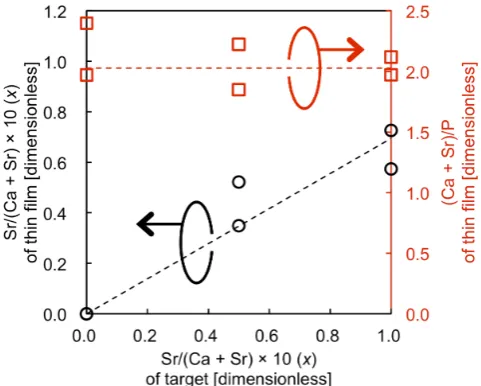
Table 1 lists the XPS results of the Sr/(Ca + Sr) (×10) and (Ca + Sr)/P ratios ob-tained for six Sr-HA thin films prepared with different Sr contents, where the samples A-F were deposited using Sr-HA targets with x = 0.0 (A, B), 0.5 (C, D), and 1.0 (E, F). Figure 1 shows a summary of the XPS results given in Table 1, where the left vertical axis represents the Sr/(Ca + Sr) ratio of the thin films (black circles) and the fitting obtained by the least squares method (black dashed line). Further, the right vertical axis of Figure 1 represents the (Ca + Sr)/P ratio of the thin films (red squares) and the average value of (Ca + Sr)/P for all samples
Figure 1. Relationship between Sr/(Ca + Sr) (black circles, left vertical axis) and (Ca + Sr)/P (red squares, right vertical axis) content in the thin films and the Sr/(Ca + Sr) con-tent in the target.
Table 1. Summary of X-ray photoelectron spectral analysis results for Sr-HA thin films with varying Sr content.
Sample Sr/(Ca + Sr) (× 10) [dimensionless] (Ca + Sr)/P [dimensionless]
A 0.00 2.40
B 0.00 1.97
C 0.35 1.85
D 0.52 2.22
E 0.57 2.12
(red dashed line). The relationship between the Sr content of the targets and the corresponding thin films was found to be roughly proportional, while the value of Sr/(Ca + Sr) of each thin film showed a slight deviation from that of the tar-get. A tendency of Sr defects to exist in PLD-deposited thin films such as these has also been reported in a previous work [16], although the mechanism is not understood. The (Ca + Sr)/P ratios in Figure 1 exhibit some deviation from the stoichiometric value of approximately 1.67, and it can also be observed that the (Ca + Sr)/P ratio of the thin films is not dependent on the Sr content of the tar-get and the thin film. However, considering a previous work where we found that the Sr/(Ca + Sr) and (Ca + Sr)/P of Sr-HA thin films can be highly con-trolled by the energy distribution of the ablation laser on the target [18] [19], these details are now being studied and the control of the chemical composition of Sr-HA and HA thin films will be reported in a subsequent paper. Judging from these results, the constant deviation of the P content independent of the Sr content of the Sr-HA thin film will not affect the present study of the variation of the lattice constants with the Sr content.
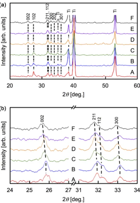
Figure 2. X-ray diffraction patterns of the Sr-HA thin films. (a) Complete 2θ range for measured diffraction spectra. (b) Shift of the (002), (211), (112), and (300) peaks toward lower 2θ is observed with higher Sr content in the Sr-HA thin films.
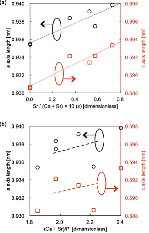
and the other from the (211) and (112) peaks. Figure 3(a) shows that both the a- and c-axis lengths have roughly linear relationships with the Sr/(Ca + Sr) con-tent. The obtained correlation coefficient for the a-axis length was R = 0.911 with p < 0.05 and that for the c-axis length was R = 0.952 with p < 0.005. As de-scribed above, the (Ca + Sr)/P ratio was almost comparable for all samples inde-pendent of their Sr content. Thus, it can be concluded that the variation in the lattice constants was caused by the substitution of Sr into Ca sites in the HA structure. To confirm this finding, the relationships between (Ca + Sr)/P in the thin films and the a- and c-axis lengths are shown in Figure 3(b). Here, the lat-tice constants do not exhibit a clear dependence on the P content of the thin films when considering the least squares method fitting lines (R = 0.494 and 0.370 for the a- and c-axis lengths, respectively). From Figure 3, therefore, the systematic variation in the lattice constants was caused by the variation in the Sr content and not the P content.
Figure 3. Lattice constants as a function of (a) Sr content and (b) P content in the Sr-HA thin films. Both plots show the lengths of the a-axis (black circles, left vertical axis) and the c-axis (red squares, right vertical axis).
with the calibration procedure using the Ti (101) peak of the substrate for all samples. We will consider if this type of dispersion is reduced with an increasing number of samples. Furthermore, statistical processing will be subsequently needed to determine the Sr content for each sample based on the data measured at several positions on the same sample, while in this study they were measured at only a single position for each sample. The spatial distribution of the Sr con-tent should also be evaluated for all samples in subsequent studies. In this ex-panded process, we expect more precise evaluation with a certain error bar for the chemical composition data.
the stoichiometrically-pure HA. However, the experimental result exhibits a sys-tematic relationship between the Sr content and the lattice constants. This result signifies that the Sr must have some effect on the crystal structure of the Sr-HA thin films because the result was not caused by the variation of the P content herein, as shown in Figure 3(b). In this work, the results are considered as a first approximation that a certain ratio of the Ca2+ site was substituted with the larger Sr2+ in the HA structure, while the absolute substitution ratio cannot be esti-mated owing to the defect of the PO4, as described above. This means that the absolute values of the lattice constant sherein cannot be compared directly to the reported values [8][13].
In this study, we have made progress toward eliminating the possibility that the systematic variation of the lattice constants was caused by a variation of the P content, unlike the previous paper [16]. We have three plans to improve these results in subsequent studies.
1) It is necessary to improve the chemical composition of the P content toward the stoichiometric value of (Ca + Sr)/P = 1.67 using the technique we have previously reported [18][19].
2) Fourier transform infrared spectroscopy should be implemented to confirm the CO3 content and to quantify the ratio of the PO4 and the CO3 if the CO3 is present in the samples.
3) The lattice constants and the chemical compositions of the PLD Sr-HA tar-gets were not analyzed in this study. Because the target characteristics can be used as reference data to analyze the results for the Sr-HA thin films, the Sr-HA targets should be analyzed as the bulk Sr-HA.
4. Conclusion
To successfully apply Sr-HA in medical and dental devices, the systematic rela-tionship between the Sr content and the a- and c-axis lengths must be studied for materials with relatively low amounts of Sr. The Sr-HA thin films prepared using PLD in this work exhibited lattice constants that possessed a roughly linear rela-tionship with their Sr content, as evaluated by XPS and XRD. This result is con-sidered as a first approximation showing that the Sr2+ substitutes into the Ca2+ site in the HA structure, where more refinement of the results is needed to real-ize the ultimate goal of the study.
Acknowledgements
This work was supported in part by grants from the Strategic Research Founda-tion Grant-Aided Project for Private Universities from the Ministry of Educa-tion, Culture, Sports, Science, and Technology, Japan, 2013-2017 (No. S1311045) and the Project Research of the Faculty of Biology-Oriented Science and Tech-nology, Kindai University, 2015-2016 (No. 14-I-3) and 2016 (No. 15-III-32).
References
Using Hydroxyl-Apatite Coatings. The Development of a Human Total Hip Pros-thesis for Chemical Fixation to Bone Using Hydroxyl-Apatite Coatings on Titanium Substrates. Clinical Orthopaedics and Related Research, 225, 147-170.
[2] Klein, C.P.A.T., Patka, P., van der Lubbe, H.B.M., Wolke, J.G.C. and de Groot, K. (1991) Plasma-Sprayed Coatings of Tetracalciumphosphate, Hydroxyl-Apatite, and α-TCP on Titanium Alloy: An Interface Study. Journal of Biomedical Materials Re-search, 25, 53-65. https://doi.org/10.1002/jbm.820250105
[3] McPherson, R., Gane, N. and Bastow, T.J. (1995) Structural Characterization of Pla- sma-Sprayed Hydroxylapatite Coatings. Journal of Materials Science: Materials in Medicine, 6, 327-334. https://doi.org/10.1007/bf00120300
[4] Yan, W.Q., Nakamura, T., Kawanabe, K., Nishigochi, S., Oka, M. and Kokubo, T. (1997) Apatite Layer-Coated Titanium for Use as Bone Bonding Implants. Biomate-rials, 18, 1185-1190. https://doi.org/10.1016/S0142-9612(97)00057-4
[5] Stoch, A., Brożek, A., Kmita, G., Stoch, J., Jastrzębski, W. and Rakowska, A. (2001) Electrophoretic Coating of Hydroxyapatite on Titanium Implants. Journal of Mole- cular Structure, 596, 191-200. https://doi.org/10.1016/S0022-2860(01)00716-5
[6] Bigi, A., Falini, G., Foresti, E., Gazzano, M., Ripamonti, A. and Roveri, N. (1993) Magnesium Influence on Hydroxyapatite Crystallization. Journal of Inorganic Bio-chemistry, 49, 69-78. https://doi.org/10.1016/0162-0134(93)80049-f
[7] Kim, T.N., Feng, Q.L., Kim, J.O., Wu, J., Wang, H., Chen, G.C. and Cui, F.Z. (1998) Antimicrobial Effects of Metal Ions (Ag+, Cu2+, Zn2+) in Hydroxyapatite. Journal of
Materials Science: Materials in Medicine, 9, 129-134.
https://doi.org/10.1023/A:1008811501734
[8] O’Donnell, M.D., Fredholm, Y., de Rouffignac, A. and Hill, R.G. (2008) Structural Analysis of a Series of Strontium-Substituted Apatites. Acta Biomaterialia, 4, 1455- 1464. https://doi.org/10.1016/j.actbio.2008.04.018
[9] Ergun, C., Webster, T.J., Bizios, R. and Doremus, R.H. (2002) Hydroxyapatite with Substituted Magnesium, Zinc, Cadmium, and Yttrium. I. Structure and Microstru- cture. Journal of Biomedical Materials Research, 59, 305-311.
https://doi.org/10.1002/jbm.1246
[10] Caverzasio, J. (2008) Strontium Ranelate Promotes Osteoblastic Cell Replication through at Least Two Different Mechanisms. Bone, 42, 1131-1136.
https://doi.org/10.1016/j.bone.2008.02.010
[11] Hurtel-Lemaire, A.S., Mentaverri, R., Caudrillier, A., Cournarie, F., Wattel, A., Ka-mel, S., Terwilliger, E.F., Brown, E.M. and Brazier, M. (2009) The Calcium-Sensing Receptor Is Involved in Strontium Ranelate-Induced Osteoclast Apoptosis. The Journal of Biological Chemistry, 284, 575-584.
https://doi.org/10.1074/jbc.M801668200
[12] Reginster, J.Y. (2009) Strontium Ranelate in Osteoporosis. Current Pharmaceutical Design, 8, 1907-1916. https://doi.org/10.2174/1381612023393639
[13] Bigi, A., Boanini, E., Capuccini, C. and Gazzano, M. (2007) Strontium-Substituted Hydroxyapatite Nanocrystals. Inorganica Chimica Acta, 360, 1009-1016.
https://doi.org/10.1016/j.ica.2006.07.074
[14] Capuccini, C., Torricelli, P., Sima, F., Boanini, E., Ristoscu, C., Bracci, B., Socol, G., Fini, M., Mihailescu, I.N. and Bigi, A. (2008) Strontium-Substituted Hydroxyapatite Coatings Synthesized by Pulsed-Laser Deposition: In Vitro Osteoblast and Osteoc-last Response. Acta Biomaterialia, 4, 1885-1893.
https://doi.org/10.1016/j.actbio.2008.05.005
Hydroxyapatite and Strontium Apatite. Bio-Medical Materials and Engineering, 24, 1447-1456.
[16] Pereiro, I., Rodríguez-Valencia, C., Serra, C., Solla, E.L., Serra, J. and González, P. (2012) Pulsed Laser Deposition of Strontium-Substituted Hydroxyapatite Coatings. Applied Surface Science, 258, 9192-9197.
https://doi.org/10.1016/j.apsusc.2012.04.063
[17] Morohashi, T., Sano, T. and Yamada, S. (1994) Effects of Strontium on Calcium Metabolism in Rats: I. A Distinction between Pharmacologic and Toxic Doses. The Japanese Journal of Pharmacology, 64, 155-162. https://doi.org/10.1254/jjp.64.155
[18] Nishikawa, H. and Yoshikawa, R. (2015) Controlling the Chemical Composition of Hydroxyapatite Thin Films Using Pulsed Laser Deposition. Transactions of the Ma- terials Research Society of Japan, 40, 111-114. https://doi.org/10.14723/tmrsj.40.111
[19] Nishikawa, H., Hasegawa, T., Miyake, A., Tashiro, Y., Hashimoto, Y., Blank, D.H.A. and Rijnders, G. (2016) Relationship Between the Ca/P Ratio of Hydroxyapatite Thin Films and the Spatial Energy Distribution of the Ablation Laser in Pulsed La-ser Deposition. Materials Letters, 165, 95-98.
https://doi.org/10.1016/j.matlet.2015.11.115
Submit or recommend next manuscript to SCIRP and we will provide best service for you:
Accepting pre-submission inquiries through Email, Facebook, LinkedIn, Twitter, etc. A wide selection of journals (inclusive of 9 subjects, more than 200 journals)
Providing 24-hour high-quality service User-friendly online submission system Fair and swift peer-review system
Efficient typesetting and proofreading procedure
Display of the result of downloads and visits, as well as the number of cited articles Maximum dissemination of your research work