Staphylococcus
spp. and
Enterococcus
spp.
April M. Bobenchik,aJanet A. Hindler,bCarmen L. Giltner,aSandra Saeki,bRomney M. Humphriesa
Department of Pathology and Laboratory Medicine, David Geffen School of Medicine at UCLA,aand UCLA Health System,bLos Angeles, California, USA
Vitek 2 (bioMérieux, Inc., Durham, NC) is a widely used commercial antimicrobial susceptibility testing system. We compared MIC results obtained by Vitek 2 to those obtained by the Clinical and Laboratory Standards Institute (CLSI) broth microdilution (BMD) reference method for 134 staphylococcal and 84 enterococcal clinical isolates. Nineteen agents were evaluated, including all those available on Vitek 2 for testing staphylococci and enterococci. The resistance phenotypes tested included methicillin-resistantStaphylococcus aureus(MRSA) (nⴝ58),S. aureuswith inducible clindamycin resistance (ICR) (nⴝ30), trim-ethoprim-sulfamethoxazole-resistant MRSA (nⴝ10), vancomycin-resistantEnterococcus(nⴝ37), high-level gentamicin-resis-tantEnterococcus(nⴝ15), linezolid-resistantEnterococcus(nⴝ5), and daptomycin-nonsusceptibleEnterococcus faecalis(nⴝ 6). For the staphylococci, there was 98.9% categorical agreement (CA). There was one very major error (VME) for gentamicin in aStaphylococcus hominisisolate, six VMEs for inducible clindamycin inS. aureusisolates, and two major errors (ME) for dapto-mycin in anS. aureusand aStaphylococcus epidermidisisolate. For enterococci, there was 97.3% CA. Two VMEs were observed for daptomycin in isolates ofE. faecalisand 2 ME, 1 for high-level gentamicin resistance and 1 for nitrofurantoin, inE. faecium isolates. Overall, there was 98.3% CA and 99% essential agreement for the testing of staphylococci and enterococci by the Vitek 2. With the exception of detecting ICR inS. aureus, Vitek 2 performed reliably for antimicrobial susceptibility testing of staphy-lococci and enterococci.
C
linical microbiology laboratories routinely performantimi-crobial susceptibility testing (AST) on all staphylococci and enterococci isolated from sterile body sites, as well as nonsterile sites, when they are the predominate pathogen. Both these genera are associated with important resistance phenotypes that are fquently encountered in clinical practice, including methicillin
re-sistance inStaphylococcus aureusand vancomycin resistance in
Enterococcus. As these phenotypes pose significant burdens on health care, reliable detection by the laboratory is imperative. In addition, less frequent but equally important resistance
pheno-types, such as elevated vancomycin MICs inS. aureusand linezolid
resistance and daptomycin nonsusceptibility in both staphylo-cocci and enterostaphylo-cocci, must be reliably detected by the laboratory. Prompt and accurate methods for susceptibility testing are needed for the laboratory to identify antimicrobial susceptibilities for these organisms for timely treatment decision making and imple-mentation of infection control practices.
Commercial automated systems for identification and suscep-tibility testing of bacteria are used in most clinical microbiology laboratories in the United States. Due to their ease of use and cost-effectiveness they are often the preferred methods over the more labor-intensive Clinical and Laboratory Standards Institute (CLSI) reference methods of broth microdilution (BMD) and disk diffusion (DD). However, there have been reports that the com-mercial systems can produce inaccurate results for select
antimi-crobial agents when testing either staphylococci (1–3) or
entero-cocci (4, 5). These reports have led to recommendations that
laboratories confirm certain susceptibility phenotypes by a
man-ual method (6–8). We evaluated the performance of the Vitek 2
(bioMérieux, Inc., Durham, NC) AST-GP71 and AST-GP72 cards for staphylococci and enterococci, respectively, compared to the CLSI BMD method.
(This work was presented in part at the 112th General Meeting
of the American Society for Microbiology, San Francisco, CA, 16 to 19 June 2012.)
MATERIALS AND METHODS
Bacterial isolates.A total of 134Staphylococcusand 84Enterococcussingle patient isolates were selected for testing, to represent various resistance phenotypes (Table 1). All isolates were obtained from specimens submit-ted between 2008 and 2012 to the UCLA clinical microbiology laboratory; 151 were freshly isolated (isolated from primary culture within 7 days and never frozen), and the remaining 67 were from frozen stock cultures. Isolates were recovered from a variety of sources, including blood, urine, respiratory secretions, wounds, tissues, fluids, and ocular specimens. Identification was performed with the Vitek 2 (bioMérieux Inc., Durham, NC) system using GP ID cards. Prior to testing, frozen isolates were sub-cultured twice and fresh isolates were subsub-cultured once on tryptic soy agar plates containing 5% sheep blood (blood agar plate [BAP]) and incubated at 35°C for 18 to 24 h. The quality control (QC) strains tested with each run wereS. aureusATCC 29213 andE. faecalisATCC 29212. Upon receipt of a new shipment of Vitek 2 cards or when changing lot numbers of cards, the following QC strains were tested according to the manufacturer’s recommendations:S. aureusATCC 29213 (AST-GP71 and AST-GP72),S.
aureusATCC BAA1026 (AST-GP71),S. aureusATCC BAA976
(AST-GP71),S. aureusATCC BAA977 (AST-GP71),E. faecalisATCC 29212 (AST-GP71 and AST-GP72),E. faecalisATCC 51299 (AST-GP72), and
Escherichia coliATCC 35318 (AST-GP72) (9,10). QC procedures for
in-Received3 September 2013Returned for modification1 October 2013
Accepted5 November 2013
Published ahead of print13 November 2013
Editor:K. C. Carroll
Address correspondence to Romney M. Humphries, [email protected].
Copyright © 2014, American Society for Microbiology. All Rights Reserved.
doi:10.1128/JCM.02432-13
on May 16, 2020 by guest
http://jcm.asm.org/
house BMD panel preparation included testing 19 supplemental QC strains in addition to testing those recommended by CLSI for routine QC.
Antimicrobial susceptibility testing. BMD MIC testing was per-formed according to CLSI guidelines using in-house prepared panels (11). These panels were incubated at 35°C in ambient air, read manually at 16 to 20 h, and then reincubated at 35°C in ambient air to 24 h to obtain final results for oxacillin (staphylococci) and vancomycin (staphylococci and enterococci). For enterococci, high-level gentamicin and streptomycin were read at 24 h, and if high-level streptomycin screening was negative at 24 h, plates were reincubated at 35°C in ambient air and read at 48 h. Susceptibility tests with the Vitek 2 (bioMérieux, Inc., Durham, NC) sys-tem were performed using software version 5.01 and AST-GP71 (staphy-lococci) or AST-GP2 (enterococci) cards according to the manufacturer’s instructions (9,10). Each isolate was tested concurrently with both meth-ods using isolated colonies from a single 18- to 24-h BAP. Purity plates were prepared following inoculation of each test by subculturing an ali-quot of inoculum suspension onto a BAP and incubating for 18 to 24 h.
Testing for inducible clindamycin resistance.BMD panels contained a single well with a combination of 0.5g/ml of clindamycin and 4g/ml of erythromycin. AST-GP71 cards contained two wells for inducible clin-damycin resistance (ICR), one with 0.5g/ml of clindamycin and the other with a combination of 0.25g/ml of clindamycin and 0.5g/ml of erythromycin. For staphylococci that were erythromycin resistant and clindamycin susceptible or intermediate, ICR disk diffusion (D-zone test) was performed according to CLSI recommendations on BAP purity plates by placing a 15-g erythromycin disk and a 2-g clindamycin disk 15 mm apart on a heavily inoculated area of the purity plate. Flattening of the clindamycin zone adjacent to the erythromycin disk after 16 to 18 h of incubation at 35°C ambient air was interpreted as positive for inducible clindamycin resistance (11).
Confirmatory ICR testing.S. aureusisolates that demonstrated dis-crepant results between Vitek 2 and the two CLSI methods (BMD and D-zone test) for ICR were sent to bioMérieux for further testing with two different lots of AST-GP71 cards. In addition, the D-zone test,ermA test-ing by PCR, and strain typtest-ing ustest-ing the DiversiLab System were per-formed at bioMérieux.
Data analysis.Essential agreement (EA), categorical agreement (CA), very major errors (VMEs), major errors (MEs), and minor errors (mEs) were calculated as previously described (12). The EA was defined as an MIC of⫾1 doubling dilution of the reference BMD MIC. The CA was defined as a susceptible, intermediate, resistant, or nonsusceptible result that was the same with both methods. A VME was defined as a false susceptible result with the Vitek 2 system, whereas an ME was a false
resistant or nonsusceptible result with the Vitek 2 system; an mE was identified when one method reported an intermediate result while the other method reported a susceptible or resistant (on nonsusceptible) re-sult.
Discrepant resolution.Isolates with a VME or ME were retested using both methods, as were select isolates with specific drug/organism combi-nations resulting inⱖ10% mEs. Calculations of EA, CA, VMEs, MEs, and mEs were obtained following resolution of discrepant results after repeat testing.
RESULTS
Of 218 isolates tested, 8 (3.7%) terminated due to unacceptable growth in the control well of the Vitek 2 automated susceptibility
testing (AST) card. These included 1 Staphylococcus capitis, 1
Staphylococcusspp. (not identified to species level by the GP ID
card), 3Enterococcus faecium, 2Enterococcus faecalis, and 1
Entero-coccus casseliflavus. OneStaphylococcus capraeisolate was termi-nated because this species is not included in the Vitek 2 system AST database. For the remaining 209 isolates there were 88 dis-crepancies in categorical interpretation between Vitek 2 and BMD, out of a total of 2,950 organism-antimicrobial combina-tions. Upon repeat testing by both BMD and Vitek 2, 45 of the 88 (51.1%) discrepancies were resolved, resulting in EAs of 98.8% and 99.5% and CAs of 98.8% and 97.3% for staphylococci and enterococci, respectively. Of the 45 resolved discrepancies, 38 (84%) were due to BMD and 20 of the 38 (52%) were specifically due to linezolid. The discrepancies with linezolid were attributed to difficulty in manually reading the linezolid endpoint on
BMD (5).
Results for staphylococci.The Vitek 2 failed to identify ICR
for 6 MRSA of 30 (20%)S. aureus (16 MRSA, 14
methicillin-susceptibleS. aureus[MSSA]) isolates that were positive for ICR
by the reference BMD single-well ICR test and the D-zone test (Table 2). The 6 isolates that were ICR negative by Vitek 2 but positive by reference BMD and D-zone tests were retested at bio-Mérieux and were confirmed to be ICR positive by the D-zone test
and by presence ofermAby PCR. Two of 6 isolates gave a positive
ICR reaction on one lot of Vitek AST-GP71 cards (Table 3). Strain
typing was performed using the DiversiLab system to check for clonality of these isolates. The 6 isolates demonstrated 4 pattern types: two isolates belonged to pattern type one (P1) and two isolates belonged to pattern type two (P2). A single band differ-ence was found between P1 and P2, indicating that these isolates were similar, but not indistinguishable. The remaining two
iso-lates belonged to their own pattern types, P3 and P4 (Fig. 1).
A VME was noted for gentamicin in aStaphylococcus hominis
isolate (16.7% of 6 gentamicin-resistant coagulase-negative
Staphy-lococcus). Two MEs were noted for daptomycin, one in anS.
au-reusisolate (1.1% of 92 daptomycin-susceptibleS. aureus) and
one in aStaphylococcus epidermidisisolate (2.7% of 37
daptomy-cin-susceptible coagulase-negativeStaphylococcus) (Tables 2and
4). mEs occurred with ciprofloxacin (n⫽1), clindamycin (n⫽2),
erythromycin (n⫽4), gentamicin (n⫽3), and vancomycin (n⫽
4) (Tables 2and4).
Vancomycin MICs determined by Vitek 2 and BMD for 45 of 58 (78%) MRSA were identical. Of the 13 isolates for which van-comycin MICs were not identical, 4 had vanvan-comycin MICs of 1
g/ml by BMD and 2g/ml by Vitek 2. The remaining 9 MRSA
isolates had a vancomycin MIC of either 0.5g/ml or 1g/ml
[image:2.585.40.288.87.192.2]with either BMD or Vitek 2. No consistent trend toward higher or lower vancomycin MICs was observed for Vitek 2 compared to
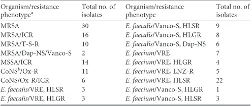
TABLE 1Numbers of isolates tested with various clinically significant resistant phenotypes
Organism/resistance phenotypea
Total no. of isolates
Organism/resistance phenotype
Total no. of isolates
MRSA 30 E. faecalis/Vanco-S, HLSR 9
MRSA/ICR 16 E. faecalis/Vanco-S, HLGR 8 MRSA/T-S-R 10 E. faecalis/Vanco-S, Dap-NS 6 MRSA/Dap-NS/Vanco-S 2 E. faecium/VRE 7 MSSA/ICR 14 E. faecium/VRE, HLGR 4 CoNSb/Ox-R 11 E. faecium/VRE, LNZ-R 5
CoNS/Ox-R/ICR 6 E. faecium/VRE, HLSR 22
E. faecalis/VRE, HLSR 3 E. faecium/Vanco-S, HLGR 1
E. faecalis/VRE, HLGR 3 E. faecium/Vanco-S, HLSR 3
aMRSA, methicillin-resistantS. aureus; ICR, inducible clindamycin resistance; T-S,
trimethoprim-sulfamethoxazole resistance; Dap-NS, daptomycin nonsusceptible; MSSA, methicillin-susceptibleS. aureus; CoNS, coagulase-negativeStaphylococcus; Ox-R, oxacillin resistance; VRE, vancomycin-resistantEnterococcus; HLGR, high-level gentamicin resistance; LNZ-R, linezolid resistance; HLSR, high-level streptomycin resistance; Vanco-S, vancomycin susceptible.
bS. epidermidis(n⫽17),S. lugdunensis(n⫽7),S. haemolyticus(n⫽6),S. capitis, (n⫽
4),S. hominis(n⫽3),S. warneri(n⫽1),S. caprae(n⫽1),Staphylococcusspp. (n⫽1).
on May 16, 2020 by guest
http://jcm.asm.org/
BMD. All MRSA isolates had vancomycin MICs within the
sus-ceptible range ofⱕ2g/ml, but 3 mEs were observed when the
Vitek 2 Advanced Expert System (AES) software edited a vanco-mycin-susceptible interpretation to intermediate for 3 MRSA iso-lates that had daptomycin MICs in the nonsusceptible range (MIC
4g/ml).
Results for enterococci.Two VMEs were observed for
dapto-mycin, both inE. faecalisisolates (33.3% of 6
daptomycin-non-susceptibleE. faecalis). Both VMEs were confirmed to be
dapto-mycin-nonsusceptible by Etest (data not shown). Two MEs
occurred inE. faecium, one for high-level gentamicin (1.6% of 64
high-level gentamicin-susceptibleEnterococcus) and one for
ni-trofurantoin (2.4% of 42 nini-trofurantoin-susceptibleEnterococcus)
(Table 5). mEs amongEnterococcusincluded ciprofloxacin (n⫽
5), erythromycin (n⫽3), linezolid (n⫽2), and nitrofurantoin
(n⫽6).
DISCUSSION
Vitek 2 is a widely used system for identification and antimicrobial susceptibility testing. Recent publications that describe the perfor-mance of Vitek 2 with staphylococci or enterococci focused on an
individual antimicrobial agent such as vancomycin (7), cefoxitin (1),
clindamycin (ICR test) (2,3,8), or linezolid (5). Noteworthy
discrep-ancies occurred inS. aureuswith clindamycin (ICR test), which
had a reported sensitivity of 91 to 95% (2,3,8), and with
vanco-mycin, which had a reported sensitivity of 91% (7). It has been
over 8 years since a comprehensive evaluation of Vitek 2 perfor-mance for staphylococci and enterococci was reported in the
peer-reviewed literature (13). Here, we report the performance of Vitek
[image:3.585.39.549.78.291.2]2 for both staphylococci and enterococci for testing contemporary isolates with currently used antimicrobial agents and the most up-to-date software and AST cards available from bioMérieux.
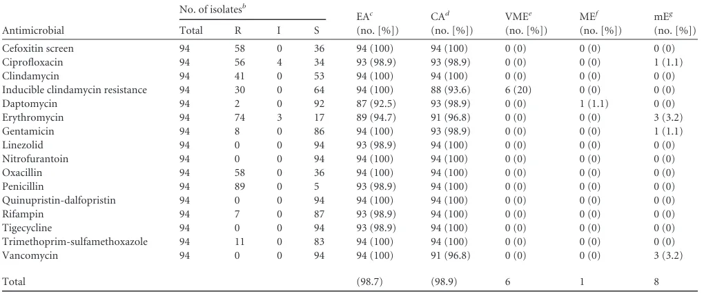
TABLE 2Performance of AST-GP71 card forS. aureusa
Antimicrobial
No. of isolatesb
EAc
(no. [%])
CAd
(no. [%])
VMEe
(no. [%])
MEf
(no. [%])
mEg
(no. [%])
Total R I S
Cefoxitin screen 94 58 0 36 94 (100) 94 (100) 0 (0) 0 (0) 0 (0)
Ciprofloxacin 94 56 4 34 93 (98.9) 93 (98.9) 0 (0) 0 (0) 1 (1.1)
Clindamycin 94 41 0 53 94 (100) 94 (100) 0 (0) 0 (0) 0 (0)
Inducible clindamycin resistance 94 30 0 64 94 (100) 88 (93.6) 6 (20) 0 (0) 0 (0)
Daptomycin 94 2 0 92 87 (92.5) 93 (98.9) 0 (0) 1 (1.1) 0 (0)
Erythromycin 94 74 3 17 89 (94.7) 91 (96.8) 0 (0) 0 (0) 3 (3.2)
Gentamicin 94 8 0 86 94 (100) 93 (98.9) 0 (0) 0 (0) 1 (1.1)
Linezolid 94 0 0 94 93 (98.9) 94 (100) 0 (0) 0 (0) 0 (0)
Nitrofurantoin 94 0 0 94 94 (100) 94 (100) 0 (0) 0 (0) 0 (0)
Oxacillin 94 58 0 36 94 (100) 94 (100) 0 (0) 0 (0) 0 (0)
Penicillin 94 89 0 5 93 (98.9) 94 (100) 0 (0) 0 (0) 0 (0)
Quinupristin-dalfopristin 94 0 0 94 94 (100) 94 (100) 0 (0) 0 (0) 0 (0)
Rifampin 94 7 0 87 93 (98.9) 94 (100) 0 (0) 0 (0) 0 (0)
Tigecycline 94 0 0 94 93 (98.9) 94 (100) 0 (0) 0 (0) 0 (0)
Trimethoprim-sulfamethoxazole 94 11 0 83 94 (100) 94 (100) 0 (0) 0 (0) 0 (0)
Vancomycin 94 0 0 94 94 (100) 91 (96.8) 0 (0) 0 (0) 3 (3.2)
Total (98.7) (98.9) 6 1 8
a
Results calculated following resolution of discrepancies after repeat testing.
bR, resistant; I, intermediate; S, susceptible. c
EA, essential agreement (MIC within⫾1 doubling dilution).
dCA, categorical agreement. e
VME, very major error.
fME, major error. g
mE, minor error.
TABLE 3Discrepant ICR results for MRSA
Isolatea
UCLA bioMérieuxb
AST-GP71 ICR lot 1
BMD ICR test
D-zone test
AST-GP71 ICR lot 1/lot 2
D-zone
test Molecular
MRSA ICR no. 1 Neg Pos Pos Neg/Neg Pos ermA
MRSA ICR no. 2 Neg Pos Pos Neg/Neg Pos ermA
MRSA ICR no. 3 Neg Pos Pos Neg/Pos Pos ermA
MRSA ICR no. 4 Neg Pos Pos Neg/Neg Pos ermA
MRSA ICR no. 5 Neg Pos Pos Neg/Neg Pos ermA
MRSA ICR no. 6 Neg Pos Pos Neg/Pos Pos ermA
a
MRSA, methicillin resistance inS. aureus; ICR, inducible clindamycin resistance.
bNeg, negative for the ICR screen, clindamycin susceptible; Pos, positive for the ICR screen, clindamycin resistant.
on May 16, 2020 by guest
http://jcm.asm.org/
[image:3.585.42.544.603.707.2]With a few exceptions, Vitek 2 performed reliably for staphy-lococci and enterococci for all antimicrobials tested, with overall EAs of 98.8% and 99.5% and CAs of 98.8% and 97.3%,
respec-tively. Categorical error rates for staphylococci (n⫽131) tested
with all antimicrobial agents (n⫽16) were 1.7% VMEs, 0.2%
MEs, and 0.8% mEs. Categorical error rates for enterococci (n⫽
78) and all antimicrobial agents (n⫽11) were 0.7% VMEs, 0.5%
MEs, and 2% mEs. Performances of the Vitek 2 AST-GP71 (staph-ylococci) and AST-GP-72 (enterococci) cards met the criteria sug-gested for acceptable performance of an automated susceptibility test system outlined in Cumitech 31A for AST systems (e.g.,
⬎89.9% EA and CA) (12).
The most notable deficiency of the Vitek 2 performance was
the detection of ICR inS. aureus, for which 20% VME were
ob-served among 30S. aureuswith the ICR phenotype. All 6 isolates
that tested negative for ICR by Vitek 2 were confirmed to harbor
anermAgene by PCR. Strain typing performed by bioMérieux
showed that only 2 of the 6 isolates were clonal (Fig. 1). Several
groups have reported discrepant results for ICR and Vitek 2. Bu-chan and colleagues published a sensitivity of 91% when 51 of 56
ICR-positive isolates were tested (3). Lavallee et al. (2) and
Gar-diner et al. (8) published similar sensitivities of 93% and 95%
when 124 of 134 ICR-positive and 191 of 201 ICR-positive isolates were tested, respectively. All three groups used the D-zone test as confirmation and included a mix of MRSA and MSSA isolates. The CLSI BMD recommendation for detection of ICR involves a
single well containing 4g/ml erythromycin and 0.5g/ml
clin-damycin (11). It has been suggested that a possible reason for the
failed detection of ICR with Vitek 2 is due to an insufficient
con-centration of erythromycin to induce theermgene (8). The
con-centration of erythromycin in the ICR well on Vitek 2 cards is 0.5
g/ml and the concentration of clindamycin is 0.25/ml (9). The
shortened incubation time in the Vitek 2 (⬃8 h) might also
con-tribute to failure to detect ICR in some strains of MRSA. Treatment failures have been reported for infections caused by MRSA isolates with ICR when clindamycin was used for therapy (14–17). A solution to the ICR problem for laboratories using Vitek 2 would be the addition of clindamycin and erythromycin disks placed 15 mm apart on the BAP purity plate inoculated at the
time of AST-GP71 card setup (6, 11). Some investigators have
presented an alternative solution and report all
erythromycin-resistantStaphylococcusspp. as resistant to clindamycin, without
confirming the inducible resistance phenotype (2,6,8). However,
the prevalence of ICR is variable depending upon whether the infection is hospital or community acquired and whether the
or-ganism is MRSA or MSSA (18–21), suggesting this solution may
not be applicable to all settings.
Some discrepancies noted between Vitek 2 and BMD were a result of the Vitek 2 AES. The AES is designed to analyze the
FIG 1Strain typing by DiversiLab showing partial clonality. The results shown were produced from testing at bioMérieux. MRSA,
methicillin-resis-tantS. aureus; ICR, inducible clindamycin resistance. The arrow points to an
[image:4.585.42.286.67.149.2]additional band that differentiates pattern 1 from pattern 2.
TABLE 4Performance of AST-GP71 card for coagulase-negativeStaphylococcusa
Antimicrobial
No. of isolatesb
EAc
(no. [%])
CAd
(no. [%])
VMEe
(no. [%])
MEf
(no. [%])
mEg
(no. [%])
Total R I S
Cefoxitin screen 37 17 0 20 37 (100) 37 (100) 0 (0) 0 (0) 0 (0)
Ciprofloxacin 37 16 0 21 37 (100) 36 (97.3) 0 (0) 0 (0) 1 (2.7)
Clindamycin 37 10 0 27 37 (100) 36 (97.3) 0 (0) 0 (0) 1 (2.7)
Daptomycin 37 0 0 37 34 (91.9) 36 (97.3) 0 (0) 1 (2.7) 0 (0)
Erythromycin 37 20 1 16 36 (97.3) 36 (97.3) 0 (0) 0 (0) 1 (2.7)
Gentamicin 37 6 2 29 35 (94.6) 34 (91.9) 1 (16.7) 0 (0) 2 (5.4)
Inducible clindamycin resistance 37 12 0 25 37 (100) 37 (100) 0 (0) 0 (0) 0 (0)
Linezolid 36 0 0 36 36 (100) 36 (100) 0 (0) 0 (0) 0 (0)
Nitrofurantoin 37 0 0 37 37 (100) 37 (100) 0 (0) 0 (0) 0 (0)
Oxacillin 37 17 0 20 36 (97.3) 37 (100) 0 (0) 0 (0) 0 (0)
Penicillin 35 27 0 8 35 (100) 35 (100) 0 (0) 0 (0) 0 (0)
Quinupristin-dalfopristin 37 0 0 37 37 (100) 37 (100) 0 (0) 0 (0) 0 (0)
Rifampin 37 0 0 37 37 (100) 37 (100) 0 (0) 0 (0) 0 (0)
Tigecycline 37 0 0 37 37 (100) 37 (100) 0 (0) 0 (0) 0 (0)
Trimethoprim-sulfamethoxazole 37 15 0 22 37 (100) 37 (100) 0 (0) 0 (0) 0 (0)
Vancomycin 37 0 0 37 37 (100) 36 (97.3) 0 (0) 0 (0) 1 (2.7)
Total (98.8) (98.7) 1 1 6
aResults calculated following resolution of discrepancies after repeat testing. b
R, resistant; I, intermediate; S, susceptible.
cEA, essential agreement (MIC within⫾1 doubling dilution). d
CA, categorical agreement.
eVME, very major error. f
ME, major error.
gmE, minor error.
on May 16, 2020 by guest
http://jcm.asm.org/
[image:4.585.39.550.449.662.2]antimicrobial susceptibility pattern of each organism to deter-mine biologic validity. Results are reviewed and categorized based on consistency with previously defined wild or resistant pheno-types and inconsistent results are flagged for further review. Anti-microbials are grouped together to create resistant phenotype profiles. The AES groups vancomycin and daptomycin together in
a phenotypic group forS. aureus. When the daptomycin MIC is
ⱖ2g/ml (nonsusceptible) and the vancomycin MIC is 1 or 2
g/ml, the AES edits the vancomycin interpretation from
suscep-tible to intermediate. Three mEs occurred for vancomycin and
MRSA as a result of AES editing a vancomycin MIC of 2g/ml to
“intermediate” when the daptomycin MIC was 4g/ml
(nonsus-ceptible). This AES correction may be misleading because not all daptomycin-nonsusceptible MRSA are vancomycin intermediate
(22,23). Further, reporting vancomycin as intermediate and
dap-tomycin as nonsusceptible leaves very few treatment options, cre-ating an unnecessary challenge for physicians. Laboratories may consider manually overriding this rule in their laboratory infor-mation system.
For enterococci, most errors in this study were for antimicro-bial agents that are generally not considered primary agents for treating enterococcal infections. Two VMEs for daptomycin were
found in vancomycin-susceptibleE. faecalisisolates. Another
ma-jor limitation with the Vitek 2 for daptomycin testing is that re-porting of daptomycin susceptibility results is limited to
vanco-mycin-susceptible E. faecalis, as is required by the FDA to be
consistent with the FDA clinical indications for daptomycin. However, daptomycin is frequently used to treat difficult
infec-tions caused by vancomycin-resistantE. faecalisandE. faecium,
such as endocarditis, as there are very limited FDA-approved agents for the treatment of such infections (e.g., linezolid and quinupristin-dalfopristin). Numerous reports have documented
treatment-emergent daptomycin nonsusceptibility in
Enterococ-cus, primarily in vancomycin-resistantE. faecium(24–26).
Clini-cal laboratories need reliable methods for testing daptomycin for
all enterococcal species, but Vitek 2 is at the present not a viable option for this, due to both limitations in reporting and poor
detection of nonsusceptibility in E. faecalis, as we document
herein. The disk diffusion method is not reliable for daptomycin
(11), leaving laboratories with few options for testing this agent.
This issue is not unique to Vitek 2, Bryant and colleagues recently reported that MicroScan detected only 9 of 30 daptomycin-non-susceptible enterococci with a VME rate of 70% for the MicroScan
prompt inoculation method (27). They attributed this high
per-centage of error to inherent error in the system, as all errors oc-curred within 1 dilution of the susceptible breakpoint; neverthe-less, they too advocate the use of an alternative method for testing enterococci with daptomycin. Although there is a limitation in-cluded in the Vitek 2 package insert, which states that the ability to
detect linezolid resistance inEnterococcusspp. is unknown due to
lack of resistant strains at the time of comparative testing (10),
Vitek 2 detected all 5 linezolid-resistantE. faeciumisolates tested
(Table 5). For the 2 mEs that occurred with linezolid, one oc-curred with an intermediate BMD interpretation and a susceptible Vitek 2 and one was with a susceptible BMD interpretation and an intermediate Vitek 2. Tenover and colleagues published similar results of no VMEs or MEs and only 3 mEs, when 10
linezolid-resistantE. faecium isolates and 5 linezolid-resistantE. faecalis
isolates were tested by Vitek 2 (5).
A limitation of this study was the small sample number of some clinically important phenotypes, including
daptomycin-nonsus-ceptibleE. faecalis(n⫽6), daptomycin-nonsusceptibleS. aureus
(n⫽2), linezolid-resistantE. faecium(n⫽5), and ICR
staphylo-cocci (n⫽30). The percentages of errors for ICR and
daptomy-cin-nonsusceptible isolates were high due to the low number of resistant isolates tested. However, results were consistent with
previously published reports (2,3,8), and worth noting because of
the clinical significance of this observation. No VMEs were
ob-served for linezolid-resistantE. faecium, but the sample size may
[image:5.585.39.551.78.240.2]have been too small to detect any problem. Additionally, no VMEs
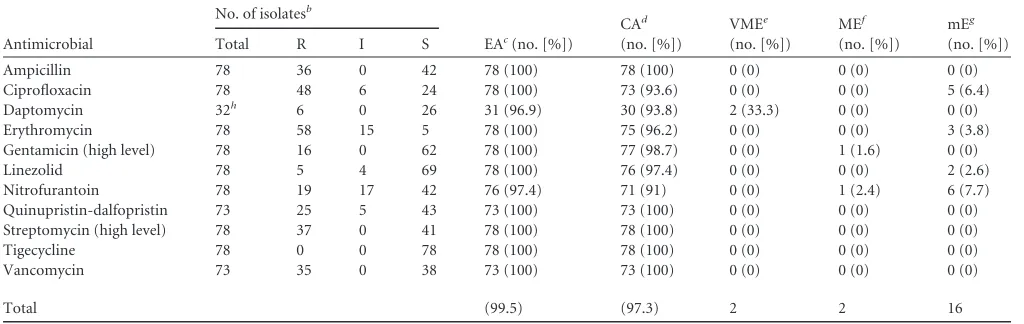
TABLE 5Performance of AST-GP72 card forEnterococcusspp.a
Antimicrobial
No. of isolatesb
EAc(no. [%])
CAd
(no. [%])
VMEe
(no. [%])
MEf
(no. [%])
mEg
(no. [%])
Total R I S
Ampicillin 78 36 0 42 78 (100) 78 (100) 0 (0) 0 (0) 0 (0)
Ciprofloxacin 78 48 6 24 78 (100) 73 (93.6) 0 (0) 0 (0) 5 (6.4)
Daptomycin 32h 6 0 26 31 (96.9) 30 (93.8) 2 (33.3) 0 (0) 0 (0)
Erythromycin 78 58 15 5 78 (100) 75 (96.2) 0 (0) 0 (0) 3 (3.8)
Gentamicin (high level) 78 16 0 62 78 (100) 77 (98.7) 0 (0) 1 (1.6) 0 (0)
Linezolid 78 5 4 69 78 (100) 76 (97.4) 0 (0) 0 (0) 2 (2.6)
Nitrofurantoin 78 19 17 42 76 (97.4) 71 (91) 0 (0) 1 (2.4) 6 (7.7)
Quinupristin-dalfopristin 73 25 5 43 73 (100) 73 (100) 0 (0) 0 (0) 0 (0)
Streptomycin (high level) 78 37 0 41 78 (100) 78 (100) 0 (0) 0 (0) 0 (0)
Tigecycline 78 0 0 78 78 (100) 78 (100) 0 (0) 0 (0) 0 (0)
Vancomycin 73 35 0 38 73 (100) 73 (100) 0 (0) 0 (0) 0 (0)
Total (99.5) (97.3) 2 2 16
a
Results calculated following resolution of discrepancies after repeat testing.
bR, resistant; I, intermediate; S, susceptible. c
EA, essential agreement (within⫾1 doubling dilution).
dCA, categorical agreement. e
VME, very major error.
fME, major error. g
mE, minor error.
hE. faecalisonly.
on May 16, 2020 by guest
http://jcm.asm.org/
were detected for vancomycin and linezolid in staphylococci, which may be misleading because no resistant isolates were tested. Future testing is warranted as the prevalences of these resistant organisms increase.
Overall, the Vitek 2 AST-GP71 and GP72 performed compa-rably to BMD. Performance was reliable for organisms with sig-nificant resistant phenotypes, such as MRSA, high-level
gentami-cin-resistant enterococci, and vancomygentami-cin-resistantEnterococcus.
Discrepancies were observed for ICR inS. aureusand daptomycin
nonsusceptibility inE. faecalis. Based on our findings and others,
we advise supplemental testing forS. aureusICR and for
Entero-coccusspp. and daptomycin. We also urge users to be aware of the
AES correction of daptomycin-nonsusceptibleS. aureusisolates
with vancomycin MICs of 1 or 2g/ml.
ACKNOWLEDGMENTS
This study was funded by bioMérieux, Inc.
We thank Jennifer Freie from the customer response lab at bioMérieux for performing confirmatory AST, PCR, and DiversiLab Systems testing on MRSA with ICR discrepancies. We also thank Farzaneh Sooudipour, Myra Maldonado, Marissa Carvalho, and Maria Tagarao for their techni-cal assistance.
REFERENCES
1.Junkins AD, Lockhart SR, Heilmann KP, Dohrn CL, Von Stein DL, Winokur PL, Doern GV, Richter SS. 2009. BD Phoenix and Vitek 2 detection of mecA-mediated resistance inStaphylococcus aureuswith ce-foxitin. J. Clin. Microbiol.47:2879 –2882.http://dx.doi.org/10.1128/JCM .01109-09.
2.Lavallée C, Rouleau D, Gaudreau C, Roger M, Tsimiklis C, Locas MC, Gagnon S, Delorme J, Labbé A.2010. Performance of an agar dilution method and a Vitek 2 card for detection of inducible clindamycin resis-tance inStaphylococcusspp. J. Clin. Microbiol.48:1354 –1357.http://dx .doi.org/10.1128/JCM.01751-09.
3.Buchan BW, Anderson NW, Ledeboer NA.2012. Comparison of BD Phoenix and bioMérieux Vitek 2 automated systems for the detection of macrolide-lincosamide-streptogramin B resistance among clinical iso-lates ofStaphylococcus. Diagn. Microbiol. Infect. Dis.72:291–294.http: //dx.doi.org/10.1016/j.diagmicrobio.2011.12.003.
4.Raponi G, Ghezzi MC, Gherardi G, Lorino G, Dicuonzo G. 2010. Analysis of methods commonly used for glycopeptide and oxazolidinone susceptibility testing inEnterococcus faeciumisolates. J. Med. Microbiol. 59:672– 678.http://dx.doi.org/10.1099/jmm.0.016444-0.
5.Tenover FC, Williams PP, Stocker S, Thompson A, Clark LA, Limbago B, Carey RB, Poppe SM, Shinabarger D, McGowan JE.2007. Accuracy of six antimicrobial susceptibility methods for testing linezolid against staphylococci and enterococci. J. Clin. Microbiol.45:2917–2922.http://dx .doi.org/10.1128/JCM.00913-07.
6.Jorgensen JH, Crawford SA, McElmeel ML, Fiebelkorn KR.2004. Detec-tion of inducible clindamycin resistance of staphylococci in conjuncDetec-tion with performance of automated broth susceptibility testing. J. Clin. Microbiol.42: 1800 –1802.http://dx.doi.org/10.1128/JCM.42.4.1800-1802.2004. 7.Swenson JM, Anderson KF, Lonsway DR, Thompson A, McAllister SK,
Limbago BM, Carey RB, Tenover FC, Patel JB. 2009. Accuracy of commercial and reference susceptibility testing methods for detecting vancomycin-intermediateStaphylococcus aureus. J. Clin. Microbiol.47: 2013–2017.http://dx.doi.org/10.1128/JCM.00221-09.
8.Gardiner BJ, Grayson ML, Wood GM.2013. Inducible resistance to clinda-mycin inStaphylococcus aureus: validation of Vitek-2 against CLSI D-test. Pathology45:181184.http://dx.doi.org/10.1097/PAT.0b013e32835cccda. 9.bioMérieux.2013. VITEK 2 AST-GP71 product information.
bioMéri-eux, Inc., Durham, NC.
10. bioMérieux.2013. VITEK 2 AST-GP72 product information. bioMéri-eux, Inc., Durham, NC.
11. Clinical Laboratory Standards Institute.2013. Performance standards for antimicrobial susceptibility testing; 23rd informational supplement. CLSI document M100-S23. Clinical Laboratory Standards Institute, Wayne, PA.
12. Clark RB, Lewinski MA, Loeffelholz MJ, Thibbetts RJ.2009. Cumitech 31A, Verification and validation of procedures in the clinical microbiology laboratory. Coordinating ed, Sharp SE. ASM Press, Washington, DC. 13. Eigner U, Schmid A, Wild U, Bertsch D, Fahr AM.2005. Analysis of the
comparative workflow and performance characteristics of the VITEK 2 and Phoenix systems. J. Clin. Microbiol.43:3829 –3834.http://dx.doi.org /10.1128/JCM.43.8.3829-3834.2005.
14. Drinkovic D, Fuller ER, Shore KP, Holland DJ, Ellis-Pegler R.2001. Clindamycin treatment ofStaphylococcus aureusexpressing inducible clindamycin resistance. J. Antimicrob. Chemother.48:315–316.http://dx .doi.org/10.1093/jac/48.2.315.
15. Siberry GK, Tekle T, Carroll K, Dick J.2003. Failure of clindamycin treatment of methicillin-resistantStaphylococcus aureusexpressing induc-ible clindamycin resistance in vitro. Clin. Infect. Dis.37:1257–1260.http: //dx.doi.org/10.1086/377501.
16. Frank AL, Marcinak JF, Mangat PD, Tjhio JT, Kelkar S, Schrecken-berger PC, Quinn P. 2002. Clindamycin treatment of methicillin-resistantStaphylococcus aureusinfections in children. Pediatr. Infect. Dis. J.21:530 –534.http://dx.doi.org/10.1097/00006454-200206000-00010. 17. Patra KP, Vanchiere JA, Bocchini JA.2011. Adherence to CLSI
recommenda-tions for testing ofStaphylococcus aureusisolates in Louisiana hospitals: re-port of a clinical failure and results of a questionnaire study. J. Clin. Mi-crobiol.49:3019 –3020.http://dx.doi.org/10.1128/JCM.00944-11. 18. Fokas S, Tsironi M, Kalkani M, Dionysopouloy M.2005. Prevalence of
inducible clindamycin resistance in macrolide-resistantStaphylococcus
spp. Clin. Microbiol. Infect.11:337–340.http://dx.doi.org/10.1111/j.1469 -0691.2005.01101.x.
19. Patel M, Waites KB, Moser SA, Cloud GA, Hoesley CJ.2006. Prevalence of inducible clindamycin resistance among community- and hospital-associatedStaphylococcus aureusisolates. J. Clin. Microbiol.44:2481– 2484.http://dx.doi.org/10.1128/JCM.02582-05.
20. Schreckenberger PC, Ilendo E, Ristow KL.2004. Incidence of constitu-tive and inducible clindamycin resistance inStaphylococcus aureusand coagulase-negative staphylococci in a community and a tertiary care hos-pital. J. Clin. Microbiol.42:2777–2779.http://dx.doi.org/10.1128/JCM.42 .6.2777-2779.2004.
21. Yilmaz G, Aydin K, Iskender S, Caylan R, Koksal I.2007. Detection and prevalence of inducible clindamycin resistance in staphylococci. J. Med. Microbiol.56:342–345.http://dx.doi.org/10.1099/jmm.0.46761-0. 22. Humphries RM, Hindler JA.2012. Should laboratories test
methicillin-resistantStaphylococcus aureusfor elevated vancomycin minimum inhib-itory concentrations by Etest as a driver of treatment changes? Clin. Infect. Dis.55:612– 613.http://dx.doi.org/10.1093/cid/cis469.
23. Mehta S, Cuirolo AX, Plata KB, Riosa S, Silverman JA, Rubio A, Rosato RR, Rosato AE.2012. VraSR two-component regulatory system contrib-utes to mprF-mediated decreased susceptibility to daptomycin in in vivo-selected clinical strains of methicillin-resistantStaphylococcus aureus. An-timicrob. Agents Chemother.56:92–102.http://dx.doi.org/10.1128/AAC .00432-10.
24. Kelesidis T, Humphries R, Uslan DZ, Pegues DA.2011. Daptomycin nonsusceptible enterococci: an emerging challenge for clinicians. Clin. Infect. Dis.52:228 –234.http://dx.doi.org/10.1093/cid/ciq113.
25. Humphries RM, Kelesidis T, Tewhey R, Rose WE, Schork N, Nizet V, Sakoulas G.2012. Genotypic and phenotypic evaluation of the evolution of high-level daptomycin nonsusceptibility in vancomycin-resistant
En-terococcus faecium. Antimicrob. Agents Chemother.56:6051– 6053.http:
//dx.doi.org/10.1128/AAC.01318-12.
26. Kelesidis T, Tewhey R, Humphries RM.2013. Evolution of high-level dapto-mycin resistance inEnterococcus faeciumduring daptomycin therapy is as-sociated with limited mutations in the bacterial genome. J. Antimicrob. Chemother.68:1926 –1928.http://dx.doi.org/10.1093/jac/dkt117. 27. Bryant KA, Roberts AL, Rupp ME, Anderson JR, Lyden ER, Fey PD,
Van Schooneveld TC.2013. Susceptibility of enterococci to daptomycin is dependent upon testing methodology. Diagn. Microbiol. Infect. Dis. 76:497–501.http://dx.doi.org/10.1016/j.diagmicrobio.2013.04.019.