0095-1137/11/$12.00 doi:10.1128/JCM.00242-11
Copyright © 2011, American Society for Microbiology. All Rights Reserved.
Evaluation of Four Different Diagnostic Tests To
Detect
Clostridium difficile
in Piglets
䌤
E. C. Keessen,
1N. E. M. Hopman,
1L. A. M. G. van Leengoed,
2A. J. A. M. van Asten,
3C. Hermanus,
4E. J. Kuijper,
4and L. J. A. Lipman
1*
Utrecht University, Division of Public Health and Food Safety, Institute for Risk Assessment Sciences, P.O. Box 80175, 3508 TD Utrecht,
Netherlands1; Utrecht University, Faculty of Veterinary Medicine, Department of Farm Animal Health, P.O. Box 80151,
3508 TD Utrecht, Netherlands2; Department of Pathobiology, Faculty of Veterinary Medicine, Utrecht University,
Utrecht, Netherlands3; and Leiden University Medical Center, Department of Medical Microbiology,
P.O. Box 9600, 2300 RC Leiden, Netherlands4
Received 3 February 2011/Returned for modification 18 February 2011/Accepted 10 March 2011
Clostridium difficileis emerging as pathogen in both humans and animals. In 2000 it was described as one of
the causes of neonatal enteritis in piglets, and it is now the most common cause of neonatal diarrhea in the United States. In Europe,C. difficileinfection (CDI) in both neonatal piglets and adult sows has also been reported. Diagnosis of this infection is based on detection of the bacteriumC. difficileor its toxins A and B. Most detection methods, however, are only validated for diagnosing human infections. In this study three commercially available enzyme immunoassays (EIAs) and a commercial real-time-PCR (Becton, Dickinson, and Company) were evaluated by testing 172 pig fecal specimens (139 diarrheic and 33 nondiarrheic piglets). The results of each test were compared to those of cytotoxicity assays (CTAs) and toxigenic culture as the “gold standards.” Compared to CTAs, the sensitivity, specificity, positive predictive value (PPV), and negative predictive value (NPV) were, respectively, as follows: for real-time PCR, 91.6, 37.1, 57.6, and 82.5%; for Premier Toxins A&B (Meridian), 83.1, 31.5, 53.1, and 66.7%; for ImmunoCard Toxins A&B kit (ICTAB; Meridian), 86.6, 56.8, 66.9, and 80.7%; and for VIDAS (bioMe´rieux), 54.8, 92.6, 85.0, and 72.8%. Compared to toxigenic culture, the sensitivity, specificity, PPV, and NPV were, respectively, as follows: for real-time PCR, 93.0, 34.7, 50.0, and 87.5%; for Premier Toxins A&B, 80.3, 27.7, 43.8, and 66.7%; and for ICTAB, 80.0, 46.2, 52.8, and 75.4%; and for VIDAS, 56.4, 89.8, 77.5, and 76.7%. We conclude that all tests had an unacceptably low performance as a single test for the detection ofC. difficile in pig herds and that a two-step algorithm is necessary, similar to that in cases of human CDI. Of all of the assays, the real-time PCR had the highest NPV compared to both reference methods and is therefore the most appropriate test to screen for the absence ofC.
difficilein pigs as a first step in the algorithm. The second step would be a confirmation of the positive results
by toxigenic culture.
Clostridium difficileis reported as the major cause of
diar-rhea in piglets from 0 to 7 days old (22). Nonetheless, some piglets withC. difficile infection (CDI) are nondiarrheic and even constipated or obstipated, although colitis is seen at nec-ropsy (2, 29). CDI affects on average two-thirds of the litters, and within litters the morbidity can be as high as 97 to 100% (2, 21, 27). Mortality attributed to CDI in piglets is usually low, although outbreaks have been reported with mortality rates as high as 16% (2). Piglets recovered from CDI have growth retardation resulting in about half a kilogram lower average weaning weights (21).
Comparative analysis of piglet C. difficileisolates with iso-lates from humans suffering from CDI in the Netherlands showed overlapping antibiotic susceptibility profiles and a high genetic relatedness of the strains (8, 11). This has led to the assumption that transmission of C. difficile from piglets to humans and vice versa is likely to occur (8, 11). SinceC. difficile
is a potential zoonotic pathogen and a major cause of diarrhea
in piglets, it is important to gain insight in the prevalence and transmission ofC. difficilewithin and between pig populations. A prerequisite for these studies are reliable and validated detection methods.
No uniform consensus has been achieved on a “gold stan-dard” to diagnose CDI in humans. Until recently, the cell cytotoxicity assay (CTA) has been used to evaluate the perfor-mance of new diagnostics tests, but now a known positive feces culture with a toxin-producingC. difficilestrain (toxigenic cul-ture) is more frequently used (6, 7).
No guidelines are available for diagnosing CDI in animals, and literature on this topic is scarce. Although commercially available detection methods forC. difficileare extensively evaluated for use to detect human infections, their performance in animal samples is largely unknown. Two commercial enzyme immunoassays (EIAs)—Clostridium difficileTox A/B II (TechLab, Blacksburg, VA) and Gastro-TectClostridium difficileToxin A⫹B (Medical Chemical Corp.)—for the detection ofC. difficiletoxins in hu-mans have been evaluated for use with piglet fecal samples. A sensitivity of 91% was found for the TechLab Tox A/B II test compared to a cytotoxicity assay (19). The results of the Gastro-Tect assay were compared to the results of the TechLab A/B II in a study by Anderson and Songer (2), and a 39% sensitivity was found. Commercially available EIAs for the detection ofC.
diffi-* Corresponding author. Mailing address: Utrecht University, Divi-sion of Public Health and Food Safety, Institute for Risk Assessment Sciences, P.O. Box 80175, 3508 TD Utrecht, Netherlands. Phone: 313 0 2535324. Fax: 313 0 2532365. E-mail: [email protected].
䌤Published ahead of print on 16 March 2011.
1816
on May 16, 2020 by guest
http://jcm.asm.org/
cilein human fecal samples were also described to have lower sensitivity when used in canine and horse fecal specimens com-pared to the use in human fecal samples (3, 5, 16).
Recently, commercially available molecular diagnostics, such as real-time PCR (RT-PCR) methods, for detection of
the C. difficile toxin B gene (tcdB) have been introduced to
diagnose human CDI (7). Except for an in-house-developed semiautomatic PCR method to detect the C. difficile toxin genes A (tcdA) and B (tcdB), and aC. difficilespecific triose phosphate isomerase (tpi) housekeeping gene, real-time PCR methods have not been evaluated to diagnose animal CDI (1). The aim of the present study was to compare the test perfor-mances of three different immunological assays and one mo-lecular test to the CTA and toxigenic culture as the gold stan-dards. The following three EIAs for the detection of both toxins A and B were included: Premier Toxins A&B (Merid-ian), ImmunoCard Toxins A&B (ICTAB; Merid(Merid-ian), VIDAS (bioMe´rieux), and the GeneOhm Cdiff RT-PCR (Becton, Dickinson, and Company) for detection of thetcdBgene.
MATERIALS AND METHODS
Samples.To obtain fecal samples from neonatal piglets, varying from 0 to 7 days in age, 18 pig breeding farms were visited between April 2009 and April 2010. The visited pig breeding farms were characterized by the presence of neonatal diarrhea for longer than 6 months. The cause of diarrhea was diag-nosed, but not further specified asClostridiadiarrhea. In addition, both preven-tive vaccination and therapy with antibiotics were unsuccessful.
At each farm, fecal samples were taken from piglets from at least three different litters. In order to obtain fresh samples, piglets were gently squeezed in the abdomen to make them defecate in sterile 50-ml tubes (Falcon). At least 2.5 ml of feces was needed to perform all assays and culturing of a single sample. If the volume harvested from one piglet was insufficient to perform all of the assays and culturing, the samples from piglets were pooled until a sample volume of 2.5 ml was reached. From piglets with diarrhea 139 samples were investigated, including 89 pooled samples. From piglets without diarrhea 33 samples, which all consisted of pooled samples, were examined. Samples were stored immediately in a cool box. After transport, the samples were split. One part was used for the RT-PCR and stored at⫺20°C until use; the other part, used for culture and the EIA, was stored at 2 to 8°C and processed within 48 h. When processing was delayed, the samples were stored at⫺20°C. The frozen samples for culture and the EIAs were thawed only once, just before processing. Culturing and all of the EIAs were performed within the same day for each batch of samples.
Toxigenic culture, isolation, and identification ofC. difficile.All fecal samples were cultured for presence ofC. difficileusingC. difficileagar (CLO; bioMe´rieux) and Colombia CNA agar (CNA; bioMe´rieux) with ethanol shock pretreatment as described previously (13, 26). Inoculated plates were incubated for 48 h at 37°C in an anaerobic environment. CharacteristicC. difficilecolonies were picked from CLO plates, recultured upon on Schaedler agar (SCS; bioMe´rieux), and anaer-obically incubated for another 48 h at 37°C. In cases where no growth on CLO plates was observed, colonies were picked from CNA plates and inoculated on SCS plates. SuspectedC. difficilecolonies on the SCS plates were selected by their typical horse manure smell, colony morphology, and by Gram staining.
PCR analyses.Colonies were cultured in an anaerobic environment on CLOS plates (Becton, Dickinson). DNA was isolated from singleC. difficilecolonies by using a QIAamp DNA mini-blood kit (Qiagen) according to the manufacturer’s protocol.C. difficileisolates were confirmed to beC. difficileby using an in-house developed PCR (18) for the detection of theC. difficileglutamate dehydrogenase gene (GluD). ConfirmedC. difficileisolates were PCR ribotyped and character-ized for the presence of genes encoding toxin A and toxin B as described previously (4, 18).
CTA.The CTA for the detection ofC. difficiletoxins A and B was performed upon Vero cell monolayers grown in 24-well plates (Greiner) as described earlier (25). Fecal samples were diluted 1:4 in Eagle minimum essential medium (EMEM) supplemented with 10% fetal bovine serum (FBS) and centrifuged at 12,000⫻g. The resulting supernatant was filtered through a 0.45-m-pore-size filter. Subsequently, the filtrate was diluted 1:10 and 1:100 in EMEM with 10% FBS. Undiluted filtrate and both serial dilutions were added onto the Vero cell monolayers. Parallel samples were incubated with specificC. difficileantitoxin,
which was diluted 1:25 (TechLab). The cells were examined both after 24 and 48 h of incubation at 37°C in a 5% CO2incubator. The result of the cytotoxicity
assay was considered positive if cell rounding and detachment of cells was seen only in monolayers without antitoxin (25).
EIAs.In the present study three different types of EIAs that detect both toxins A and B were evaluated: ICTAB (Meridian), which is a membrane-type enzyme-linked immunosorbent assay (ELISA); the Premier Toxins A&B (Meridian) kit, which is a well-type ELISA; and the VIDAS-AB (bioMe´rieux), an enzyme-linked fluorescence immunoassay.
All EIAs were carried out according to the manufacturers’ recommendations. Plates of the Premier Toxins A&B were read at 450/630 nm (EL800 Universal Microplate Reader [Bio-Tek Instruments, Inc.). The optical densities (ODs) were recorded, and results were calculated according to the manufacturers’ instructions. When the OD of a sample was above the cutoff value, the sample was recorded positive. If ODs were equal to the cutoff value, the results were recorded as equivocal. The ICTAB was read by eye according to manufacturer’s protocol. When the entire reaction port was a very light blue, this was recorded as an equivocal result.
The VIDAS-AB assay was performed on the miniVIDAS, provided by bio-Me´rieux. The VIDAS assay used algorithms set by the manufacturer to calculate the results. The results were recorded as positive, negative, or equivocal.
Real-time PCR.The real-time PCR assay (GeneOhm; Becton, Dickinson) for detection of the tcdBgene was performed according to the manufacturers’ instructions on a SmartCycler (Cepheid, United Kingdom; supplied by Becton, Dickinson at the time of the study). The software of the SmartCycler recorded the results of the PCR assay as positive, negative, or unresolved.
Statistical analysis.The sensitivity and specificity were calculated for each kit against both of the gold standard assays (CTA and toxigenic culture). The sensitivity and specificity data were used to calculate the positive predictive value (PPV) and the negative predictive value (NPV). The data were analyzed using SPSS.16 software to determine whether storage conditions influenced the results of the detection methods. Therefore, data were stratified based on whether the samples were fresh or frozen, and Pearson chi-square tests were performed for every detection method compared to the reference methods. The Breslow-Day test was used to examine the homogeneity of the odds ratios of the strata. Furthermore, the data of the frozen samples were stratified based on the storage time of the samples. Pearson chi-square and Breslow-Day tests were used to examine whether the storage time at⫺20°C influenced the results of the detec-tion methods.
Ethical approval.Ethical approval for this study was granted from the Animal Experiments Committee of the University of Utrecht.
RESULTS
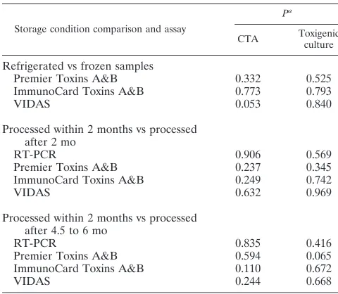
Refrigerated and frozen samples. In all, 172 samples were analyzed using the methods described above. A total of 35 samples were processed within 48 h, and 137 samples were frozen upon arrival and processed after thawing once. Statis-tical analysis showed that the Breslow-Day test was not signif-icant for any of the tests, indicating that the storage conditions did not influence the results of the detection methods. There-fore, it was not necessary to calculate the specificity and sen-sitivity for the fresh and frozen samples separately. The storage times of the frozen samples varied from 1 to 6 months. When the test results of samples processed within 2 months (n⫽38) were compared to test results of samples processed after 2 months (n ⫽99), no significant differences between the test results were found for any of the tests. Also, when the test results of the samples obtained within 2 months (n⫽38) were compared to the test results of the samples obtained between 4.5 to 6 months (n⫽39), no significant differences were found.
Equivocal values. Using the VIDAS assay, 31 samples (18.0%) were recorded as equivocal. The ICTAB yielded 9 (5.2%) equivocal values. These equivocal values were excluded from the analysis of the performance characteristics of both tests.
The manufacturer’s recommendation, in the case of
on May 16, 2020 by guest
http://jcm.asm.org/
ocal results, was not followed in the present study since insuf-ficient substrate was left to repeat the test with the original specimen, and repeated sampling was not possible due to prac-tical considerations.
Isolation and characterization of C. difficile.C. difficilewas isolated from 71 (41.3%) of the 172 fecal samples: 59 (34%) isolated strains were derived from diarrheal piglets (n⫽139), and 12 (36%) isolates were cultured from nondiarrheal piglets (n⫽33). A total of 70 of the 71 isolates were characterized as PCR ribotype 078, and one isolate belonged to PCR ribotype 045. An overview of these results is given in Table 1. The difference in prevalence ofC. difficile between the diarrheal and nondiarrheal piglets was not significant (P⫽0.52).
CTA.The results of the CTA are given in Table 1. In all, 83 (48.3%) of the 172 fecal samples were determined to be positive by CTA. All positive results were observed within 24 h of incubation. Of the fecal samples of 139 diarrheal piglets, 68 (48.9%) were positive with CTA, whereas 15 (45.5%) of the 33 nondiarrheal piglets fecal samples were positive. No difference in prevalence between the diarrheal and nondiarrheal piglets was found (P⫽0.72).
Concordant results. Concordant results with all detection methods, including the reference methods, were observed in 29 (17%) samples. Concordant positive results for all methods were recorded for 24 samples (14%), and concordant negative results were obtained for 5 samples (3%).
A positive result for all of the toxin detection methods, including CTA, was observed in 30 samples (17%).
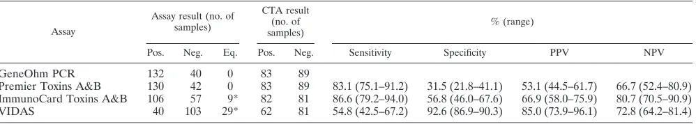
Sensitivity, specificity, PPV, and NPV of the EIAs and the PCR assay compared to toxigenic culture and CTA.The sen-sitivity and specificity data of the EIAs and the RT-PCR assay calculated versus CTA and toxigenic culture are shown in Tables 2 and 3.
The GeneOhm PCR assay was more sensitive than the EIAs
compared to both reference methods. The GeneOhm assay was less specific than the ICTAB or VIDAS assays. The spec-ificity of the tests ranged from 27.7 to 92.6%. The VIDAS assay performed best, but none of the tests demonstrated a high specificity.
The PPVs and NPVs for the EIAs and the GeneOhm com-pared to both reference methods are shown in Tables 2 and 3. The highest NPV was obtained with the GeneOhm assay. The PPVs of the GeneOhm, ICTAB, and Premier Toxins A&B assays were comparable. The highest PPV was obtained with the VIDAS assay.
DISCUSSION
[image:3.585.43.538.82.190.2]We show here that the current commercially available tests for detection ofC. difficile or its toxins in humans with CDI have a much lower sensitivity and specificity when used in porcine fecal samples than in human stool samples. For the evaluation of these tests, 172 fecal samples of piglets were analyzed using three EIAs, one RT-PCR, and two reference methods considered to be gold standards. Even though there was a high prevalence of CDI in the study population, the PPVs and NPVs of the tests were unacceptably low. The concor-dance between the tests and the reference methods was only 16.9%. The lower sensitivities and specificities of the tests than in human samples is in accord with the findings of other stud-ies, wherein the performance of EIAs in fecal samples of pigs, dogs, and horses was also described to be less than in human fecal samples (2, 3, 5, 16). The lower specificity of the detection methods when used to detect CDI in animals may be caused by the presence of an inhibitor in animal feces that reduces the specificity of binding of the toxins (2, 5). However, published data on defined inhibitors in animal fecal specimens are not
TABLE 2. Comparison of various detection methods to CTA (n⫽172)a
Assay
Assay result (no. of samples)
CTA result (no. of samples)
% (range)
Pos. Neg. Eq. Pos. Neg. Sensitivity Specificity PPV NPV
GeneOhm PCR 132 40 0 83 89
Premier Toxins A&B 130 42 0 83 89 83.1 (75.1–91.2) 31.5 (21.8–41.1) 53.1 (44.5–61.7) 66.7 (52.4–80.9) ImmunoCard Toxins A&B 106 57 9* 82 81 86.6 (79.2–94.0) 56.8 (46.0–67.6) 66.9 (58.0–75.9) 80.7 (70.5–90.9) VIDAS 40 103 29* 62 81 54.8 (42.5–67.2) 92.6 (86.9–90.3) 85.0 (73.9–96.1) 72.8 (64.2–81.4)
a
Pos., positive; Neg., negative; Eq., equivocal. Equivocal values were excluded from the analysis of the performance characteristics of the tests.
TABLE 1. Results of CTA and toxigenic culture
Animal group Toxigenic culture PCR ribotype CTA
Result No. of samples (%) Result No. of samples (%)
Diarrheal piglets Positive 59 (42.4) 078/045 Positive 68 (48.9)
Negative 80 (57.6) Negative 71 (41.3)
Nondiarrheal piglets Positive 12 (36.4) 078 Positive 15 (45.5)
Negative 21 (63.6) Negative 18 (54.5)
Total piglets Positive 71 (41.3) 078/045 Positive 83 (48.3)
Negative 101 (58.7) Negative 89 (51.7)
on May 16, 2020 by guest
http://jcm.asm.org/
[image:3.585.44.543.628.717.2]available. This seems to be an important focus for further research.
Discordant results with the reference methods were ob-tained with all of the tests examined here. One test (VIDAS) also had a high number (18.0%) of equivocal results. The samples that yielded discordant results with the reference methods varied widely between the different tests, indicating that it was not a core group of samples repeatedly giving false-positive or false-negative results. This implies that incor-rect results were due neither to the inaccuracy of the inter-preter of the assays nor due to the presence of disturbing substances in a group of samples. Negative toxin tests of feces that harborC. difficilecould indicate colonization with a non-toxigenicC. difficilestrain. Since all of the recovered isolates examined here were toxigenic, this possibility can be excluded for our study. Another reason for the discrepancy between negative toxin tests and positive culture results could be toxin degradation due to inappropriate storage, but all manufactur-ers’ instructions state that freezing of fecal samples at⫺20°C will not affect test results. A study by Weese et al. (28) showed that toxins remain detectable by ELISA (theClostridium diffi-cileTox A/B Test; TechLab) when feces samples from horses inoculated withC. difficileisolates were stored at⫺20°C for 60 days. Nonetheless, contrasting results were reported by Free-man and Wilcox (10), who diluted huFree-man feces 1/20 in prer-educed phosphate-buffered saline (pH 7.4) and inoculated each of these fecal emulsions with one of three differentC.
difficilestrains. Toxin titers of the inoculated fecal emulsions
that were frozen at⫺20°C and measured by CTA were signif-icantly lower than the toxin titers of refrigerated fecal emul-sions (P⬍0.01) by day 56 of the experiment in fecal emulsions inoculated for two of the three strains used. These two studies suggest that storage at⫺20°C might have resulted in a loss of cytotoxicity, but not of immunological recognition of the toxins (10, 28). Nonetheless, in our study no significant differences were found between samples that were frozen for⬍2 months and samples that remained frozen for longer times (Table 4). Furthermore, Chouicha and Marks (5) suggest that the pres-ence of protease activity in animal fecal specimens may also lead to increased toxin degradation, and toxins may not even reach detection levels. Finally, it remains possible that the presence of toxin-producing strains in feces samples and the absence of free toxins A and B reflect an asymptomatic carri-ership, as is known for humans. Another reason for a discrep-ancy between a negative CTA and a positive culture of toxin-producing strain is a lack of a standardized CTA. No standard protocol for the CTA exists. This results in differences in cell
line used for the assay, with different sensitivities, and in dif-ferent methods, such as the dilution of stool specimens (15).
In our study the concordance between the two reference methods was 79.1%. The sensitivity of the CTA compared to toxigenic culture was 83.1%. A low sensitivity of the CTA has previously been reported by several authors, ranging from 56.7% (9) to 61.7% (24) to 74.0% (14). The sensitivity of toxigenic culture compared to CTA was 71.1%. It is difficult to compare reported data on the sensitivity of culture due to the differences in culture methods, e.g., with or without enrich-ment, heat, or alcohol shock and the use of differentC. difficile -specific media. In our study, the samples were pretreated with an alcohol shock, followed by inoculation onto standard com-mercial media to improve the comparability of our results. Reller et al. (20) followed a similar culture method for human samples, although the pretreatment was with heat instead of alcohol, and they reported a sensitivity of 87%.
[image:4.585.43.541.83.171.2]The low sensitivity of the reference methods used in the present study hampers the PPV values of the four studied test methods considerably. The high prevalence of positive samples observed here increases the PPV of the tests. Because the PPV and NPV of a test are codetermined by the prevalence of the pathogen in the sampled population, it is important to evaluate
TABLE 3. Comparison of various detection methods to toxigenic culture (n⫽172)a
Assay
Assay result (no. of samples)
CTA result (no. of samples)
% (range)
Pos. Neg. Eq. Pos. Neg. Sensitivity Specificity PPV NPV
GeneOhm PCR 132 40 0 71 101 93.0 (87.0–98.9) 34.7 (25.4–43.9) 50.0 (41.5–58.5) 87.5 (77.3–97.7) Premier Toxins A&B 130 42 0 71 101 80.3 (71.0–89.5) 27.7 (19.0–36.5) 43.8 (35.3–52.4) 66.7 (52.4–80.9) ImmunoCard Toxins A&B 106 57 9 70 93 80.0 (70.6–89.4) 46.2 (36.1–56.4) 52.8 (43.3–62.3) 75.4 (64.3–86.6) VIDAS 40 103 29 55 88 56.4 (43.3–69.5) 89.8 (83.4–96.1) 77.5 (64.6–90.4) 76.7 (68.5–84.9)
aPos., positive; Neg., negative; Eq., equivocal. Equivocal values were excluded from the analysis of the performance characteristics of the tests.
TABLE 4. Pvalues of the Breslow-Day test for all test results stratified on storage conditions
Storage condition comparison and assay
Pa
CTA Toxigenic
culture
Refrigerated vs frozen samples
Premier Toxins A&B 0.332 0.525
ImmunoCard Toxins A&B 0.773 0.793
VIDAS 0.053 0.840
Processed within 2 months vs processed after 2 mo
RT-PCR 0.906 0.569
Premier Toxins A&B 0.237 0.345
ImmunoCard Toxins A&B 0.249 0.742
VIDAS 0.632 0.969
Processed within 2 months vs processed after 4.5 to 6 mo
RT-PCR 0.835 0.416
Premier Toxins A&B 0.594 0.065
ImmunoCard Toxins A&B 0.110 0.672
VIDAS 0.244 0.668
a
That is, the Breslow-Day test statisticalPvalue.
on May 16, 2020 by guest
http://jcm.asm.org/
[image:4.585.299.539.508.717.2]a test in a population with a prevalence that reflects the normal situation. The high prevalence ofC. difficile(41.3% with toxi-genic culture and 48.3% with CTA) that we observed here is in accord with the prevalences ofC. difficilereported in piglets in other studies in different countries (17, 22, 30).
The main difference between diagnosing CDI in humans and in pigs is the fact that in humans the individual patient is diagnosed, whereas in pigs CDI is diagnosed in groups of pigs or herds. The sensitivity, specificity, and predictive values of the tests are too low for the diagnosis of CDI in an individual piglet; however, the tests can be used to screen for the pres-ence ofC. difficilein pig herds. A test with a high sensitivity ensures that farms whereC. difficileis present can be identified. The fact thatC. difficileis a possible zoonotic pathogen may lead to future surveillance programs forC. difficilein pigs on farms. This underscores the importance of a high NPV of a test to reliably declare a pig herd free ofC. difficile.The GeneOhm PCR assay showed the highest sensitivity and NPV and is therefore the most suitable test for screening a large number of samples forC. difficileon pig farms. However, the PPV of the GeneOhm PCR assay is rather low. Consequently, a two-step algorithm is necessary, with as a second step confirmation of samples that were determined to be positive by the GeneOhm PCR. The confirmatory test could be a reference test, such as toxigenic culture. An additional advantage of culture would be that more information about the isolate, such as the ribotype and antibiotic susceptibility can be obtained. A two-step algo-rithm is also recommended in human medicine for the diag-nosis of CDI (6, 7).
A limitation of our study is that the strains were almost exclusively ribotype 078, which could have influenced the sen-sitivity of the tests. Ribotyping of the isolates that were ob-tained here showed that 70 isolates (99% of all isolates) be-longed to PCR ribotype 078, and one isolate was identified as type 045. The predominance of type 078 could have attributed to the low sensitivity of the assays, because recent research by Tenover et al. (23) shows that the PCR ribotype of strains can have an impact on the sensitivity of molecular diagnostics and EIAs of human feces samples. A lower sensitivity (81.8% with the RT-PCR and 63.6% with EIA) was found for theC. difficile
PCR ribotype 078 than for many other ribotypes (for example, ribotype 027, 100% with the RT-PCR and 78.4% with EIA) using molecular diagnostics and EIAs (23). The high preva-lence of ribotype 078 reflects the current situation in the Neth-erlands, where 078 is the predominant ribotype in piglets (12). Furthermore, it would have been interesting to include another RT-PCR in the study. The strengths of our study are the study design with the use of both gold standards as a reference, and it is the first study to evaluate a commercially available RT-PCR and three EIAs for the use of detection ofC. difficilein pigs.
We conclude that all tests in our study had an unacceptably low performance and that a two-step algorithm is necessary, similar to the situation in human medicine.
ACKNOWLEDGMENTS
We thank N. Promkuntod for help with the cytotoxicity assays, K. M. J. A. van den Brink for help with the sample taking, and I. Sanders for ribotyping the isolates. We also thank all of the suppliers for supplying kits and equipment for the evaluation.
This research was financed by a grant from the ZOnMW (project 50-50800-98-075).
REFERENCES
1.Alvarez-Perez, S., P. Alba, J. L. Blanco, and M. E. Garcia.2009. Detection of toxigenicClostridium difficilein pig faeces by PCR. Veterinarni Medicina
54:360–366.
2.Anderson, M. A., and J. G. Songer.2008. Evaluation of two enzyme immu-noassays for detection ofClostridium difficiletoxins A and B in swine. Vet. Microbiol.128:204–206.
3.Arroyo, L. G., H. Staempfli, and J. S. Weese.2007. Molecular analysis of Clostridium difficileisolates recovered from horses with diarrhea. Vet. Mi-crobiol.120:179–183.
4.Bidet, P., et al.2000. Comparison of PCR-ribotyping, arbitrarily primed PCR, and pulsed-field gel electrophoresis for typingClostridium difficile. J. Clin. Microbiol.38:2484–2487.
5.Chouicha, N., and S. L. Marks.2006. Evaluation of five enzyme immuno-assays compared with the cytotoxicity assay for diagnosis of Clostridium difficile-associated diarrhea in dogs. J. Vet. Diagn. Invest.18:182–188. 6.Cohen, S. H., et al.2010. Clinical practice guidelines forClostridium difficile
infection in adults: 2010 update by the society for health care epidemiology of America (SHEA) and the infectious diseases society of America (IDSA). Infect. Control Hosp. Epidemiol.31:431–455.
7.Crobach, M. J. T., O. M. Dekkers, M. H. Wilcox, and E. J. Kuijper.2009. European Society of Clinical Microbiology and Infectious Diseases (ESCMID): data review and recommendations for diagnosing Clostrid-ium difficile-infection (CDI). Clin. Microbiol. Infect.15:1053–1066. 8.Debast, S. B., et al.2009.Clostridium difficilePCR ribotype 078 toxinotype V
found in diarrhoeal pigs identical to isolates from affected humans. Environ. Microbiol.11:505–511.
9.Delmee, M., J. Van Broeck, A. Simon, M. Janssens, and V. Avesani.2005. Laboratory diagnosis ofClostridium difficile-associated diarrhoea: a plea for culture. J. Med. Microbiol.54:187–191.
10.Freeman, J., and M. H. Wilcox.2003. The effects of storage conditions on viability ofClostridium difficilevegetative cells and spores and toxin activity in human faeces. J. Clin. Pathol.56:126–128.
11.Goorhuis, A., et al.Clostridium difficilePCR ribotype 078: an emerging strain in humans and in pigs? J. Clin. Microbiol.46:1157–1158.
12.Keessen, E. C., et al.2010. Prevalence ofClostridium difficilein swine thought to haveClostridium difficileinfections (CDI) in eleven swine operations in the Netherlands. Tijdschr. Diergeneeskd.135:134–137.
13.Kuijper, E. J., R. J. van den Berg, and S. Debast.2006.Clostridium difficile ribotype 027, toxinotype III, the Netherlands. Emerg. Infect. Dis.12:827– 828.
14.Lozniewski, A., C. Rabaud, E. Dotto, M. Weber, and F. Mory.2001. Labo-ratory diagnosis ofClostridium difficile-associated diarrhea and colitis: use-fulness of Premier Cytoclone A⫹B enzyme immunoassay for combined detection of stool toxins and toxigenic C. difficile strains. J. Clin. Microbiol.
39:1996–1998.
15.Lyerly, D. M., N. M. Sullivan, and T. D. Wilkins. 1983. Enzyme-linked immunosorbent assay forClostridium difficiletoxin A. J. Clin. Microbiol.
17:72–78.
16.Magdesian, K. G., D. C. Hirsh, S. S. Jang, L. M. Hansen, and J. E. Madigan.
2002. Characterization ofClostridium difficileisolates from foals with diar-rhea: 28 cases (1993–1997). J. Am. Vet. Med. Assoc.220:67–73. 17.Norman, K. N., et al.2009. Varied prevalence ofClostridium difficilein an
integrated swine operation. Anaerobe15:256–260.
18.Paltansing, S., et al.2007. Characteristics and incidence of Clostridium difficile-associated disease in The Netherlands, 2005. Clin. Microbiol. Infect.
13:1058–1064.
19.Post, K. W., B. H. Jost, and J. G. Songer.2002. Evaluation of a test for Clostridium difficiletoxins A and B for the diagnosis of neonatal swine enteritis. J. Vet. Diagn. Invest.14:258–259.
20.Reller, M. E., et al.2007. Yield of stool culture with isolate toxin testing versus a two-step algorithm including stool toxin testing for detection of toxigenicClostridium difficile. J. Clin. Microbiol.45:3601–3605.
21.Songer, J. G.2004. The emergence ofClostridium difficileas a pathogen of food animals. Anim. Health Res. Rev.5:321–326.
22.Songer, J. G., and M. A. Anderson.2006.Clostridium difficile: an important pathogen of food animals. Anaerobe12:1–4.
23.Tenover, F. C., et al.2010. Impact of strain type on detection of toxigenic Clostridium difficile: comparison of molecular diagnostic and enzyme immu-noassay approaches. J. Clin. Microbiol.48:3719–3724.
24.Thonnard, J., F. Carreer, V. Avesani, and M. Delmee.1996. Toxin A detec-tion onClostridium difficile colonies from 24-h cultures. Clin. Microbiol. Infect.2:50–54.
25.van den Berg, R. J., et al.2005. Prospective multicenter evaluation of a new immunoassay and real-time PCR for rapid diagnosis ofClostridium difficile-associated diarrhea in hospitalized patients. J. Clin. Microbiol.43:5338– 5340.
26.van den Berg, R. J., et al.2007. Evaluation of real-time PCR and con-ventional diagnostic methods for the detection ofClostridium difficile-associated diarrhoea in a prospective multicentre study. J. Med. Micro-biol.56:36–42.
on May 16, 2020 by guest
http://jcm.asm.org/
27.van Leengoed, L., S. B. Debast, A. A. Bergwerff, and E. J. Kuiper.2008. Neonatal diarrhea in piglets caused byClostridium difficile.Proc. 20th IPVF Cong., p. 134.
28.Weese, J. S., H. R. Staempfli, and J. F. Prescott.2000. Survival ofClostridium difficileand its toxins in equine feces: implications for diagnostic test selec-tion and interpretaselec-tion. J. Vet. Diagn. Invest.12:332–336.
29.Yaeger, M., N. Funk, and L. Hoffman.2002. A survey of agents associated
with neonatal diarrhea in Iowa swine including Clostridium difficileand porcine reproductive and respiratory syndrome virus. J. Vet. Diagn. Invest.
14:281–287.
30.Yaeger, M. J., J. M. Kinyon, and J. Glenn Songer.2007. A prospective, case control study evaluating the association betweenClostridium difficiletoxins in the colon of neonatal swine and gross and microscopic lesions. J. Vet. Diagn. Invest.19:52–59.