0095-1137/10/$12.00 doi:10.1128/JCM.00382-10
Copyright © 2010, American Society for Microbiology. All Rights Reserved.
Clinical Performance of the PreTect HPV-Proofer E6/E7 mRNA
Assay in Comparison with That of the Hybrid Capture 2 Test
for Identification of Women at Risk of Cervical Cancer
䌤
Samuel Ratnam,
1,3* Francois Coutlee,
2Dan Fontaine,
3James Bentley,
4Nicholas Escott,
5Prafull Ghatage,
6Veeresh Gadag,
3Glen Holloway,
5Elias Bartellas,
3Nick Kum,
3Christopher Giede,
7and Adrian Lear
8Public Health Laboratory, St. John’s, Newfoundland and Labrador, Canada1; De´partement de Microbiologie et Immunologie,
Centre Hospitalier de l’Universite´ de Montre´al et Universite´ de Montre´al, Montre´al, Canada2; Faculty of Medicine,
Memorial University, St. John’s, Newfoundland and Labrador, Canada3; Queen Elizabeth II Health Sciences Centre,
Halifax, Nova Scotia, Canada4; Regional Health Sciences Centre, Thunder Bay, Ontario, Canada5;
Tom Baker Cancer Centre, Calgary, Alberta, Canada6; Royal University Hospital, Saskatoon,
Saskatchewan, Canada7; and Dr. H. Bliss Murphy Cancer Centre, St. John’s,
Newfoundland and Labrador, Canada8
Received 24 February 2010/Returned for modification 14 May 2010/Accepted 17 June 2010
Human papillomavirus (HPV) DNA testing has a higher clinical sensitivity than cytology for the detection of high-grade cervical intraepithelial neoplasia or worse (CIN 2ⴙ). However, an improvement in specificity would be desirable. As malignant transformation is induced by HPV E6/E7 oncogenes, detection of E6/E7 oncogene activity may improve specificity and be more predictive of cervical cancer risk. The PreTect HPV-Proofer assay (HPV-Proofer; Norchip) detects E6/E7 mRNA transcripts from HPV types 16, 18, 31, 33, and 45 with simultaneous genotype-specific identification. The clinical performance of this assay was assessed in a cross-sectional study of women referred for colposcopy in comparison with the Hybrid Capture 2 (HC2; Qiagen) test, which detects DNA of 13 high-risk oncogenic HPV types collectively. Cervical specimens were collected in PreservCyt, and cytology was performed using the ThinPrep method (Hologic). The samples were processed for HPV detection with Proofer and HC2 and genotyping with the Linear Array method (Roche Molecular Systems). Histology-confirmed CIN 2ⴙserved as the disease endpoint to assess the clinical performance of the tests. A total of 1,551 women were studied, and of these, 402 (25.9%) were diagnosed with CIN 2ⴙon histology. The Proofer assay showed a sensitivity of 78.1% (95% confidence interval [CI], 74.1 to 82.1) versus 95.8% (95% CI, 93.8 to 97.8) for HC2 (P< 0.05) and a specificity of 75.5% (95% CI, 73.0 to 78.0) versus 39.6% (95% CI, 36.8 to 42.4), respectively (P< 0.05). The lower sensitivity and higher specificity of Proofer for detection of CIN 2ⴙcan be attributed to the fact that this test detects the expression of E6/E7 genes beyond a threshold from a limited number of oncogenic HPV types. In conclusion, Proofer is more specific than HC2 in identifying women with CIN 2ⴙbut has a lower sensitivity.
Numerous nonrandomized studies (19), and more recently randomized clinical trials (3, 24, 31, 34), have clearly estab-lished that testing for human papillomavirus (HPV) DNA is significantly more sensitive than Pap cytology for detection of high-grade cervical intraepithelial neoplasia (CIN 2 and CIN 3) or worse (CIN 2⫹; i.e., CIN 2, CIN 3, squamous cell carci-noma, endocervical adenocarcinomain situ, and endocervical adenocarcinoma). There are also indications that testing for HPV might be the most effective method of cervical cancer screening in developing countries (36). Moreover, HPV testing would be warranted as a primary screening tool in the era of HPV vaccination (14). While the use of HPV testing in primary cervical cancer screening and triage of borderline cytologic abnormalities have been recommended (37, 43, 44), HPV test-ing lacks specificity due to the ubiquitous and transient nature of HPV infection in women, and therefore, the positive
pre-dictive value (PPV) tends to be lower than that of cytology (11, 19). Most studies on primary screening have evaluated HPV DNA detection tests, especially the Hybrid Capture 2 (HC2; Qiagen) test (10, 19). HC2 utilizes a full genomic probe cock-tail for collective detection of DNA of 13 high-risk HPV types (HPV-16, -18, -31, -33, -35, -39, -45, -51, -52, -56, -58, -59, and -68). Although HC2 has been shown to be highly sensitive for the detection of the 13 types targeted by the test (10, 11, 19), it is also known to cross-react with untargeted nononcogenic types, thus potentially contributing to a reduction in the test specificity (4, 32, 35). Nonetheless, HC2 has been extensively validated and approved by the U.S. FDA and hence is recom-mended as a reference test to evaluate any newly developed HPV tests (25).
While infection with high-risk HPV is the necessary biolog-ical factor for cervbiolog-ical cancer, the actual oncogenic process is initiated by persistent high-risk HPV infection and mediated by the upregulation of E6/E7 oncoproteins (39, 45). Thus, overexpression of these oncoproteins is associated with an increased risk of lesion progression (29, 45). On this basis, it would stand to reason that the detection of E6/E7 oncogene * Corresponding author. Mailing address: Public Health Laboratory,
100 Forest Road, St. John’s, NL A1A 3Z9, Canada. Phone: 709 777 6568. Fax: 709 777 7070. E-mail: [email protected].
䌤Published ahead of print on 23 June 2010.
2779
on May 16, 2020 by guest
http://jcm.asm.org/
activity should be more specific and be a better predictor of cervical cancer risk than HPV DNA detection methods that do not differentiate between persistent and transient HPV infec-tions (9, 22, 23, 26, 28). Detection of E6/E7 oncogene activity can be achieved by testing for E6/E7 mRNA transcripts (12, 21, 27).
The PreTect HPV-Proofer assay (Proofer; Norchip AS, Nor-way) is a type-specific E6/E7 mRNA-based test for oncogenic types 16, 18, 31, 33, and 45, with both HPV detection and genotyping performed in the same reaction (27). These five types have been shown to account for about 82% of cervical cancer worldwide, but their prevalence varies between differ-ent geographical regions (2, 5). Furthermore, the lesion pro-gression is more strongly associated with types 16 and 18 (13), and there are indications that an HPV test that distinguishes types 16 and 18 from other oncogenic types may be more useful as it could identify women at greater risk of cervical cancer (18). A recent study of the APTIMA HPV assay, an-other E6/E7 mRNA-based HPV test which targets 14 onco-genic types, has shown the same level of clinical sensitivity as the HC2 test but with a higher clinical specificity (12). Several studies on the relative performance of Proofer have been con-ducted in Europe with an indication that this test is more specific than other tests, including the APTIMA and HC2 assays to identify CIN 2⫹ (9, 21, 23, 26, 28, 41). However, Proofer has a reduced clinical sensitivity for the detection of CIN 2⫹as this test targets only five oncogenic HPV types, and therefore, this limitation is of concern. The usefulness of the Proofer test targeting the five most prevalent genotypes in cervical cancer in a North American setting is unknown.
The aim of the present study was to assess the clinical per-formance of Proofer for detection of CIN 2⫹ in comparison with HC2, supplemented with HPV genotypic analysis using a standardized commercially available genotyping kit. This study was carried out as part of a multicenter study in Canada which is assessing the clinical usefulness of testing for E6/E7 mRNA and other molecular biomarkers in cervical cancer screening in comparison with HPV DNA testing and cytology. This study accrued women with a history of abnormal cytology referred for colposcopic assessment. The data in this paper present the relative sensitivity, specificity, and predictive values of Proofer in comparison with those of HC2 for detection of CIN 2⫹ based on cross-sectional data obtained at enrollment of study participants.
MATERIALS AND METHODS
Study population.The study population consisted of women either newly diagnosed with abnormal Pap cytology of any grade who were referred to col-poscopy or with a history of abnormal cytology who were being followed up in colposcopy clinics as per the routine standard of care. Women 15 years of age or older who had any grade of cytological abnormality within the previous 2 years and who had not received treatment or had not had a hysterectomy were eligible. Participants were enrolled from five tertiary care referral centers in five prov-inces across Canada. The prompting Pap test had been performed using con-ventional cytology at various sites served by the referral centers. Those consent-ing to participate in the study were enrolled sequentially with written informed consent. The study was approved by institutional ethics review board of all participating study centers.
A total of 1,571 women were enrolled, with all having cervical specimens collected. The mean age of participants was 31.0 years (standard deviation [SD], 10.6 years; range, 15 to 80 years; median, 28.0 years). The proportion of women
⬍30 years of age was 56.5%. The interval of time between the initial cytological
diagnosis and enrollment at the colposcopy referral visit ranged from 1 to 3 months for new cases and up to 2 years for colposcopy follow-up cases with a history of abnormal cytology.
Study procedures and testing methods.Upon enrollment, a single cervical specimen was collected immediately prior to colposcopic examination using the Cervex broom-type brush (Rovers Medical Devices, Oss, Netherlands) and sus-pended in PreservCyt collection medium (Hologic, Inc., Marlborough, MA) as per the manufacturer’s instructions. Liquid-based cytology (LBC) was performed using the ThinPrep (Hologic, Inc.) method in a central laboratory in accordance with the manufacturer’s instructions, and the results were reported according to the 2001 Bethesda System (38a). Only the LBC results obtained at the time of enrollment were used for study purposes.
Residual PreservCyt samples were tested simultaneously with Proofer and HC2, and HPV genotyping was performed with the Linear Array (LA) HPV genotyping test (Roche Molecular Diagnostics, Laval, Canada). The Proofer and HC2 tests were carried out using fresh specimens within 2 weeks of collection, and LA genotyping was performed with frozen aliquots. The procedures used for these tests are briefly summarized below. Technologists performing these tests were blinded to results obtained in the other tests and also cytology, colposcopy, and histology results.
Proofer test. Proofer is a real-time multiplex nucleic acid sequence-based amplification assay (NASBA) for isothermal amplification and detection of E6/E7 mRNA from high-risk oncogenic types 16, 18, 31, 33, and 45 using molecular beacon probes. Five milliliters of cervical specimen in PreservCyt was processed for the extraction of HPV RNA using Magnapure (Roche), and HPV E6/E7 mRNA was detected according to the manufacturer’s instructions and as previously described (21, 27). An FLx900I fluorescence reader (Bio-Tek, Wi-noosky, VT) was used for the detection of the accumulated mRNA product with the PreTect Analysis software (Norchip) for analyzing the fluorescence profiles. To verify the integrity of RNA in the specimen, the test includes a primer set and a probe directed against the human U1 small nuclear ribonucleoprotein-specific mRNA. Standardized artificial oligonucleotides corresponding to the respective viral sequences are provided in the test kit, and these were included as positive controls for each of the five HPV types included in the test. Water was used as the negative control.
HC2 assay.HC2 assay is a signal amplification test based on the hybridization of a RNA probe cocktail for 13 high-risk oncogenic types with the target DNA, and capture and detection of the DNA-RNA hybrid by chemiluminescence. This test was performed with 4 ml of PreservCyt samples according to the manufac-turer’s instructions. Specimens with relative light unit/cutoff (RLU/CO) values of
ⱖ1 were considered positive.
LA genotyping test.The LA assay is an L1 consensus primer-based PCR test with reverse line blot hybridization for the detection of 36 mucosal HPV geno-types (HPV geno-types 6, 11, 16, 18, 26, 31, 33, 34 [formerly known as type 64], 35, 39, 40, 42, 44 [formerly known as type 55], 45, 51, 52, 53, 54, 56, 58, 59, 61, 62, 66, 67, 68, 69, 70, 71, 72, 73, 81, 82 [including subtype IS39], 83, 84, and 89 [formerly known as CP6108]). The LA panel thus covers all known oncogenic types and includes those targeted by both the Proofer and HC2 tests. HPV DNA was extracted from 250l of sample in PreservCyt using the AmpliLute liquid medium extraction kit (Roche) as per the manufacturer’s instructions. Extracted DNA was then tested using the LA assay as previously described (8). Because the HPV 52 probe cross-reacts with HPV-33, -35, and -58, samples positive with the HPV-52 probe were further tested with a validated real-time PCR assay specific for type 52 (7). Only the samples reactive in the HPV-52 real-time PCR assay were considered as HPV-52-positive.
Histology.Participating obstetrics and gynecology (OB/GYN) specialists in the five study centers carried out colposcopy. Cervical biopsies were taken only from those women with abnormal colposcopy, and this was performed at the time of cervical cytology specimen collection, at patient enrollment, as per the standard of care. In some cases, biopsies were taken in subsequent follow-up visits, and in such instances, histology results for biopsies taken no later than 6 months fol-lowing enrollment were included in the study analysis. Cervical biopsy results read by one or more pathologists were obtained from participating centers and accepted as the disease endpoint for the study purposes. Pathologists were blinded to HPV results.
Data analysis.The clinical performance of the HPV tests was assessed based on a histological diagnosis, with CIN 2⫹serving as the disease endpoint and “gold standard.” The cross-sectional HPV data based on a single cervical spec-imen collected at the time of enrollment and histology results on cervical biopsies obtained either at enrollment or during follow-up for up to 6 months were utilized in this evaluation. Sensitivity, specificity, and predictive values were calculated using the conventional contingency tables, and 95% confidence inter-vals (95% CIs) were computed using exact binomial methods. TheZ-scores were
on May 16, 2020 by guest
http://jcm.asm.org/
used to test the differences between sensitivity and specificity values across the Proofer and HC2 tests. Accuracy of HPV detection was calculated as the per-centage of the correct results by the respective HPV tests compared to histology. Differences between the Proofer and HC2 tests were tested for statistical signif-icance using McNemar’s chi-square test. The HPV results were also studied analytically with LA genotyping results. A significance level of 0.05 was used in comparing performance characteristics.
RESULTS
From the total of 1,571 cervical specimens representing unique patients tested in the study, 10 (0.6%) failed the inter-nal control of Proofer and were considered invalid as per the manufacturer’s protocol. A diagnosis of CIN 2 or CIN 3 was made based on histology in 3 of these 10 cases, and all 10 were excluded from the study. In another 10 cases, while the Proofer assay identified one or more of the five genotypes targeted by the assay, the results were below the test’s cutoff and deemed indeterminate as per the test protocol. In such instances, the manufacturer recommends reextracting 5 ml of specimen and repeating the test. This, however, could not be carried out due to the lack of specimen. In this subset, Proofer results were confirmed by LA genotyping in 9 of the 10 cases, in that, at least one of the five types targeted by Proofer was detected by LA. All of these 10 cases tested positive by HC2, five of which were diagnosed as CIN 2 or CIN 3 by histology. These 10 cases were also excluded from further analysis, and the remaining 1,551 women served as the study population for the main data analysis.
The concurrent LBC taken on the day of colposcopy was unsatisfactory in 79 of the 1,551 women. The distribution of cytology results for the remainder was as follows: normal cy-tology, 551; atypical squamous cells of undetermined signifi-cance (ASCUS), 341; ASCUS—favor high grade (ASC-H), 26; atypical glandular cells (AGC), 5; low-grade squamous intra-epithelial lesions (LSILs), 409; and high-grade squamous in-traepithelial lesions (HSILs), 140. Histology identified a total of 402 cases with a diagnosis of CIN 2⫹, comprising 97 cases of CIN 2, 293 of CIN 3, and 12 of invasive cervical cancer (CIN 2⫹prevalence in the study population, 25.9%). This included 16 cases of CIN 2 or CIN 3 and 2 cases of invasive cervical cancer among the 79 women having unsatisfactory cytology (18/79 [22.8%]). There were 401 cases with CIN 1, along with 748 women having either a normal colposcopy with no biopsy or negative histology, for a total of 1,149 cases representing a diagnosis of CIN 1 or less (ⱕCIN 1). The sensitivity of Proofer
and HC2 was assessed based on the 402 CIN 2⫹ cases and specificity on 1,149 women withⱕCIN 1 who were considered to represent the group without high-grade cervical lesions at baseline.
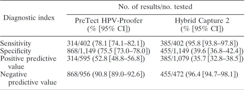
Proofer showed a sensitivity of 78.1% for detection of CIN 2⫹ compared with a sensitivity of 95.8% for HC2 (Table 1;P ⬍0.05). The specificity was 75.5% for Proofer compared with 39.6% for HC2 (P ⬍ 0.05). Analysis of the sensitivity of the tests for the detection of CIN 2⫹stratified by histological grades indicated improved sensitivity of Proofer with lesion severity and cancer (Table 2). The comparative performance of Proofer and HC2 in the subset of 402 CIN 2⫹ cases showed a concordance of 80.3% (Table 3). Both tests were negative in 13 (3.2%) CIN 2⫹cases. Of these, LA geno-typing detected type 31 or 33 in two cases, oncogenic types included in HC2 but not in HPV-Proofer in four cases, types other than the 13 genotypes included in HC2 in three cases, and failed to detect HPV DNA in four cases. Among 79 dis-cordant results, there were 75 Proofer-negative, HC2-positive specimens; 22 of these (29.3%) contained the Proofer-targeted oncogenic types 16 and 31 either alone or in coinfections, with or without other HPV types. Of the remaining 53, 48 contained at least one of the eight other oncogenic types targeted by HC2. There were four Proofer-positive, HC2-negative speci-mens, and these contained types 16, 18, or 45.
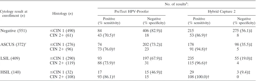
The performance of both HPV tests was further evaluated according to cytology results at enrollment. Histology-con-firmed CIN 2⫹ was found in 61 (11.1%) of 551 women with normal cytology (Table 4). The proportion of CIN 2⫹cases at histology, as expected, increased with the sequential higher grade of cytologic abnormalities: 25.8% (96/372) among those with ASCUS/ASC-H/AGC cytology, 29.1% (119/409) in cases with LSIL, and 77.1% (108/140) in cases with HSIL. The com-parison of sensitivities and specificities between Proofer and HC2 for detection of CIN 2⫹in each of the cytologic catego-TABLE 1. Diagnostic indices of the PreTect HPV-Proofer and
Hybrid Capture 2 assays for detection of CIN 2⫹a
Diagnostic index
No. of results/no. tested
PreTect HPV-Proofer (%关95% CI兴)
Hybrid Capture 2 (%关95% CI兴)
Sensitivity 314/402 (78.1关74.1–82.1兴) 385/402 (95.8关93.8–97.8兴) Specificity 868/1,149 (75.5关73.0–78.0兴) 455/1,149 (39.6关36.8–42.4兴) Positive predictive
value
314/595 (52.8关48.8–56.8兴) 385/1,079 (35.7关32.8–38.5兴)
Negative predictive value
868/956 (90.8关89.0–92.6兴) 455/472 (96.4关94.7–98.1兴)
a
[image:3.585.43.284.90.179.2]n⫽1,551. CIN 2⫹, cervical intraepithelial neoplasia grade 2 or worse (CIN 2, CIN 3, squamous cell carcinoma, endocervical adenocarcinomain situ, and endocervical adenocarcinoma).
TABLE 2. Sensitivity of PreTect HPV-Proofer and Hybrid Capture2 assays for detection of CIN 2⫹by histological gradesa
Histology gradeb No. of
cases
% sensitivity (95% CI) of:
PreTect
HPV-Proofer Hybrid Capture 2
CIN 2 97 62.9 (51.4–71.5) 91.8 (84.4–96.4) CIN 3c
293 82.9 (78.1–87.1) 97.3 (94.7–98.8) Invasive cervical cancer 12 91.7 (61.5–99.8) 91.7 (61.5–99.8)
an⫽402.
[image:3.585.299.543.90.157.2]bCIN 2 and CIN 3, cervical intraepithelial neoplasia grades 2 and 3. cIncludes 12 cases of adenocarcinomain situ.
TABLE 3. Comparative performance of PreTect HPV-Proofer and Hybrid Capture 2 assays for detection of CIN 2⫹a
PreTect HPV-Proofer result
No. of Hybrid Capture 2 resultsb
:
⫹ ⫺ Total
⫹ 310 4 314
⫺ 75 13 88
Total 385 17 402
a
n⫽402. CIN 2⫹, cervical intraepithelial neoplasia grade 2 or worse.
b
Concordance, 80.3%; McNemar’s chi-square, 62.03 (P⬍0.001).
on May 16, 2020 by guest
http://jcm.asm.org/
[image:3.585.300.543.636.708.2]ries showed significant differences (P⬍0.05 for each compar-ison). The sensitivity of Proofer for detecting CIN 2⫹ in ⱕLSIL ranged from 70.5% to 76.0%; the corresponding value for HSIL was 86.1%, which was significantly higher than the previous values (P⬍0.05 for each comparison). The results of both HPV tests according to cytologic categories were further analyzed to compare the overall positivity for HPV infection. While Proofer detected a much lower proportion of women as having HPV infection than HC2 in each of the cytologic cat-egories, this test also detected fewer cases of CIN 2⫹ than HC2 in each of these categories.
LA testing detected one or more of the 36 genotypes tar-geted by the LA assay in 1,312 (84.6%) of the total of 1,551 samples tested; 838 (63.9%) of these were multiple-type infec-tions containing a variety of genotypes, including those tar-geted by Proofer and HC2. The predominant type was 16, followed by types 18 and 31, and as expected, their relative prevalence increased with increasing lesion severity, as ascer-tained by histology (data not shown). Of the total of 1,079 samples testing positive by HC2 (Table 1), 70 (6.5%) did not have any of the 13 types targeted by HC2 but contained a variety of other genotypes, including types 53, 66, 42, 70, 62, 61, 54, 67, 89, 73, 6, 55, 82, 40, 84, 83, 81, and 72, according to LA genotyping. Among these, HPV-53 and -66 were the most common types. The above were considered to be cross-reac-tive. In this subset, there were four cases with CIN 2 or CIN 3 whose specimens contained types 55, 66, 67, 70, or 82, and all were identified as positive by HC2. Further, in seven HC2-positive cases, LA testing failed to detect any of the 36 types included in the assay. In contrast, there was an excellent agree-ment between the genotypes identified by Proofer and LA testing, in that, of the total of 595 specimens testing positive by Proofer (Table 1), at least one of the five types targeted by the assay was detected by LA in 574 (97.3%) of 590 specimens tested; in the remaining 16, LA detected other oncogenic types in 14 and was negative in 2. In particular, Proofer identified type 16 in 365 (61.3%) of the 595 specimens, and 13 (13/365
[3.6%]) of these were multiple-type infections with one or more of the other four genotypes targeted by Proofer. For the 365 specimens with type 16, LA results were available for 363; LA testing detected type 16 alone or in coinfections with other types in 360 of these with a 99.2% concordance with Proofer. All 12 cases of invasive cervical cancer contained at least one of the five types targeted by Proofer, as determined by LA genotyping, and 11 of the same cases tested positive by both Proofer and HC2 (Table 2). The five types targeted by Proofer were detected in 330 (82.1%) of the 402 CIN 2⫹cases by LA genotyping. Of the 330 cases, Proofer was positive in 314 (95.2%) and HC2 in 326 (98.8%), with both testing positive in 300 (90.9%).
The LA genotyping data were also analyzed to determine the prevalent genotypes in the 88 CIN 2⫹cases testing negative by Proofer (Table 3). Of this, 24 (27.3%) contained at least one of the five genotypes targeted by Proofer, and a profile of these cases is summarized in Table 5. Furthermore, 52 (59.1%) were found to have the eight additional oncogenic types targeted by HC2. The most common of these types detected, in the order of frequency, were 52, 39, 51, 35, and 58. In the remaining 12 cases, LA geno-typing identified HPV types other than the 13 oncogenic types targeted by the HC2 assay or was negative. To better characterize the specificity of HPV assays, genotyping results obtained with LA were considered in specimens from the 1,149 women with ⱕCIN 1 who tested negative by Proofer or HC2. This indicated that, among the 868 cases testing negative by Proofer (Table 1), the five genotypes targeted by Proofer were found in 168 (19.4%) and the eight additional genotypes included in HC2 were found in 278 (32.0%). For the 455 cases testing negative by HC2 (Table 1), the corresponding figures were 52 (11.4%) and 60 (13.2%), re-spectively.
DISCUSSION
[image:4.585.44.542.80.238.2]This study is one of the largest cross-sectional studies to date to assess and compare the clinical performance of the Proofer TABLE 4. Performance of PreTect HPV-Proofer and Hybrid Capture 2 according to cytology and histology resultsa
Cytology result at
enrollment (n) Histology (n)
No. of resultsb:
PreTect HPV-Proofer Hybrid Capture 2
Positive (% sensitivity)
Negative (% specificity)
Positive (% sensitivity)
Negative (% specificity)
Negative (551) ⱕCIN 1 (490) 84 406 (82.9)‡ 215 275 (56.1)‡
CIN 2⫹(61) 43 (70.5)† 18 53 (86.9)† 8
ASCUS (372)c ⱕCIN 1 (276) 74 202 (73.2)‡ 178 98 (35.5)‡
CIN 2⫹(96) 73 (76.0)† 23 91 (94.8)† 5
LSIL (409) ⱕCIN 1 (290) 93 197 (67.9)‡ 235 55 (19.0)‡
CIN 2⫹(119) 88 (73.9)† 31 115 (96.6)† 4
HSIL (140) ⱕCIN 1 (32) 17 15 (46.9)‡ 29 3 (9.4)‡
CIN 2⫹(108) 93 (86.1)† 15 108 (100.0)† 0
a
n⫽1,472. Seventy-nine cases with unsatisfactory cytology were excluded from the total of 1,551 cases. ASCUS, atypical squamous cells of undetermined significance; LSIL, low-grade squamous intraepithelial lesion; HSIL, high-grade squamous intraepithelial lesion;ⱕCIN 1, cervical intraepithelial neoplasia of grade 1 or better; CIN 2⫹, cervical intraepithelial neoplasia of grade 2 or worse.
b
†, sensitivities for detection of CIN 2⫹between Proofer and HC2 for each cytologic category significant atP⬍0.05; ‡, specificities for detection of CIN 2⫹between Proofer and HC2 for each cytologic category significantly different atP⬍0.05.
c
Includes 26 cases of ASC-H and 5 cases of AGC.
on May 16, 2020 by guest
http://jcm.asm.org/
assay with the HC2 test. The study also included HPV geno-typing with a standardized assay to assess the analytical per-formance of the two tests. We chose a population of women with abnormal cytology referred for colposcopy for the study because of high disease prevalence in this population so as to allow for accurate assessment of the performance of the tests with a larger number of women with cervical precancerous lesions and also to accrue as many cases of cervical cancer as possible.
Based on pooled data generated from cervical cancer case control studies, at least 15 HPV types have been classified as high-risk oncogenic (30). There are indications that an HPV test should be capable of detecting at least 13 of these onco-genic types (25, 40). Accordingly, the currently available DNA-and RNA-based HPV tests target 13 to 14 of these oncogenic types (12, 15, 16, 33). The rationale for limiting the probes to five types in the Proofer assay was that these are the most common types identified in invasive cervical cancer worldwide (2, 6, 27). A concern, however, is that this test will miss CIN 2⫹ lesions caused by the other oncogenic types not targeted by the test. Moreover, the distribution of the oncogenic HPV types detected in CIN 2⫹varies geographically.
Our study demonstrates a concordance between Proofer and HC2 of 80.3%, despite the difference in the number of onco-genic HPV types targeted by these tests. In terms of clinical performance, the Proofer assay scored significantly fewer HPV-positive samples than HC2, thus yielding a much higher clinical specificity but a lower sensitivity at detecting CIN 2⫹.
The higher specificity could be attributed in part to the fact that this assay detects RNA transcripts of integrated HPV genes involved in the oncogenic process. Therefore, the test is less often positive in benign and low-grade lesions or in their absence than is the case with HPV DNA testing (23, 28). Regardless, our analysis indicates that the higher specificity of Proofer to a greater extent is due to the smaller number of oncogenic types targeted by the assay. While Proofer certainly will significantly reduce the number of women without CIN 2⫹ referred for colpocopy or requiring further assessment, the increased specificity is attained with concomitant loss in sensi-tivity for detecting CIN 2⫹.
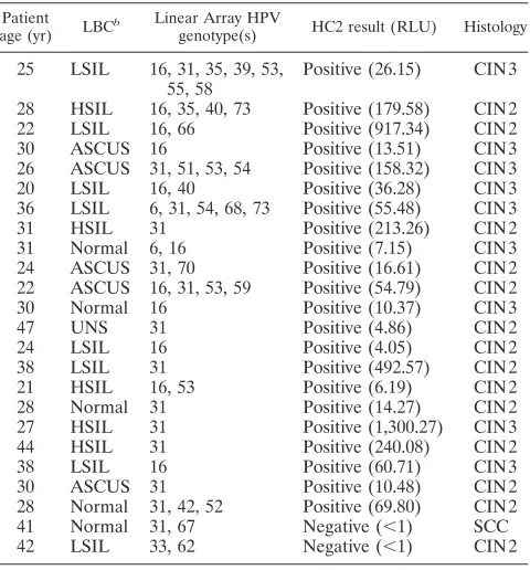
The five oncogenic HPV types targeted by Proofer were found in 82.1% of CIN 2⫹cases, as determined by LA geno-typing. The global average attributable fraction of these five types in CIN 2 and CIN 3 is 70.4%, with a range of 50.9% in Asia to 79.7% in Europe, and about 73% for North America (5, 38). The sensitivity of Proofer in our study was 78.1%, and this is line with the above prevalence rates. There were 88 (21.9%) CIN 2⫹ cases testing negative by Proofer, and the majority contained oncogenic types not targeted by the assay. When analysis was restricted to only the five types targeted by Proofer, the sensitivity of the assay for detecting CIN 2⫹was found to be equivalent to that of HC2. This indicates that the lower sensitivity of Proofer for the detection of CIN 2⫹ is largely the result of the limited number of genotypes targeted by the test. Our study data also appear to indicate that this may in part be due to the targeted ability of the test to detect E6/E7 mRNA expression only beyond a certain threshold. For in-stance, there were 24 Proofer-negative CIN 2⫹specimens, all of which contained at least one of the types targeted by the test (Table 5). Furthermore, all of the 10 Proofer-indeterminate specimens excluded from the study were found to have one of the five types, and five of these were CIN 2⫹. A Proofer-negative result in samples positive for DNA of the five types in CIN 2⫹cases could be explained by HPV genomes remaining episomal, low transcriptional activity of integrated genomes, or the occurrence of mutations in the regions covered by the primers or probes (21). It is also likely to depend on the test’s cutoff for detection, as mentioned above, and the difference in targets between the assays.
[image:5.585.44.284.98.356.2]While the reduced clinical sensitivity of Proofer to detect CIN 2⫹is of concern, it is recognized that a proportion of CIN 2 and some CIN 3 lesions, especially those not associated with oncogenic types targeted by Proofer, will regress spontane-ously (5, 13, 38). In this context, the Proofer assay designed to detect transcriptionally active infection with the five most com-mon oncogenic HPV types might still identify the majority of women whose lesions are likely to progress. There is an indi-cation that Proofer is more suited for predicting CIN 2⫹than DNA-based PCR, and the test has a higher sensitivity for detecting cervical cancer than precancerous lesions (20, 21, 23, 28). This may be attributable to the potential for progression of lesions associated with the five targeted genotypes, especially types 16 and 18, and consequently the higher prevalence of these two types in cervical cancer than precancerous lesions (13). Although there were only 12 cases of cervical cancer in our study, all of them contained one of the five types targeted by Proofer, and 11 of these tested positive by Proofer. It is worth noting that in a related study carried out exclusively on TABLE 5. CIN 2⫹cases testing negative by PreTect HPV-Proofer
but found to have the five oncogenic types targeted by the assay as determined by Linear Array genotypinga
Patient age (yr) LBC
b Linear Array HPV
genotype(s) HC2 result (RLU) Histology
25 LSIL 16, 31, 35, 39, 53, 55, 58
Positive (26.15) CIN 3
28 HSIL 16, 35, 40, 73 Positive (179.58) CIN 2 22 LSIL 16, 66 Positive (917.34) CIN 2
30 ASCUS 16 Positive (13.51) CIN 3
26 ASCUS 31, 51, 53, 54 Positive (158.32) CIN 3
20 LSIL 16, 40 Positive (36.28) CIN 3
36 LSIL 6, 31, 54, 68, 73 Positive (55.48) CIN 3
31 HSIL 31 Positive (213.26) CIN 2
31 Normal 6, 16 Positive (7.15) CIN 3
24 ASCUS 31, 70 Positive (16.61) CIN 2 22 ASCUS 16, 31, 53, 59 Positive (54.79) CIN 2
30 Normal 16 Positive (10.37) CIN 3
47 UNS 31 Positive (4.86) CIN 2
24 LSIL 16 Positive (4.05) CIN 2
38 LSIL 31 Positive (492.57) CIN 2
21 HSIL 16, 53 Positive (6.19) CIN 2
28 Normal 31 Positive (14.27) CIN 2
27 HSIL 31 Positive (1,300.27) CIN 3
44 HSIL 31 Positive (240.08) CIN 2
38 LSIL 16 Positive (60.71) CIN 3
30 ASCUS 31 Positive (10.48) CIN 2
28 Normal 31, 42, 52 Positive (69.80) CIN 2
41 Normal 31, 67 Negative (⬍1) SCC
42 LSIL 33, 62 Negative (⬍1) CIN 2
a
n⫽24.
b
LBC, liquid-based cytology; ASCUS, atypical squamous cells of undeter-mined significance; LSIL, low-grade squamous intraepithelial lesion; HSIL, high-grade squamous intraepithelial lesion; UNS, unsatisfactory; CIN, cervical intra-epithelial neoplasia (grade 1, 2, or 3); SCC, squamous cell carcinoma.
on May 16, 2020 by guest
http://jcm.asm.org/
278 cervical cancer cases in India, the five targeted types were found in 242 (87.1%) cases, and Proofer detected 237 (85.9%) of the 276 cases tested (1). The value of Proofer for detecting cervical cancer may thus be different from that of detecting precancerous lesions, and this could vary geographically.
In a study similar to ours conducted in the United Kingdom and comprising 953 colposcopy referral cases, Proofer showed a sensitivity of 73.6%, a specificity of 73.1%, and a PPV of 52.0% for detecting CIN 2⫹(41). These values are comparable to our findings in a Canadian setting. Our results are also similar to those obtained in studies conducted in Norway and Ireland (17, 23). The HC2 results in our population were consistent with those previously established (10, 25, 41).
Although the study population was comprised of colposcopy referral patients with a history of abnormal cytology, a large number had normal cytology at the time of enrollment. This could be due to many factors, including lesion regression, in-terpretation, etc. A point worth noting is that 61 (15.9%) of 384 CIN 2⫹cases were found in women with normal cytology at the time of enrollment, and 70.5% and 86.9% of these were positive by Proofer and HC2, respectively (Table 4). This inforces the importance of incorporating HPV testing or re-peat cytology in cervical cancer screening. Also, a separate analysis of unsatisfactory cytology indicated an enriched pop-ulation of CIN 2⫹. This reflects the inherent limitation of cytology-based evaluation. Regardless, the positivity with Proofer or HC2 was dependent on the number of genotypes covered by the respective tests and their prevalence in different grades of cytologic and histologic lesions.
We used the LA genotyping assay to gain further insight into the analytical performance of Proofer and HC2 tests. It should be noted that the failure of LA to detect some or all of the HPV types in a sample has been reported (42) and cannot be ruled out in our study. In 77 HC2-positive cases, LA failed to detect any of the 13 oncogenic types included in the HC2 assay in 70 cases and was negative in the remaining 7. The cross-reactivity rate of 6.5% we observed with HC2 is similar to the 7.9% reported in a larger study based on line blot and LA methods (4). The above data thus appear to indicate that the extent of cross-reactivity with untargeted nononcogenic types is one of the reasons for the reduced analytic as well as clinical specificity of HC2. In contrast, there was an excellent agree-ment of the LA-based genotype data with those of Proofer for the five genotypes detected, indicating a high analytic specific-ity of Proofer to the five types targeted as reported previously (27). This indicates that the Proofer assay can be used reliably to obtain simultaneous type-specific information for the five genotypes targeted by the test. This is an appealing feature given the indication for the identification of types 16 and 18 in risk stratification and better clinical management of women with a positive HPV test (18).
It is worth noting that studies assessing the clinical perfor-mance of HPV tests traditionally utilize CIN 2⫹as the disease endpoint, but a proportion of these lesions will regress spon-taneously, and this needs to be taken into account. Further-more, we did not assess the clinical performance of Proofer in a routine screen population. Therefore, longitudinal, popula-tion-based studies are needed to fully determine the predictive values and the clinical utility of the Proofer assay in cervical cancer screening. In conclusion, the Proofer assay has the
potential to serve as a more specific test than HC2, albeit with limited sensitivity, for identification of women with CIN 2⫹.
ACKNOWLEDGMENTS
This study was supported by a research grant from Merck Frosst Canada, Ltd.
We thank James Mansi for his support and enthusiasm.
The clinical collaborators included Justice Arthur, Burin Peninsula Health Care Centre, Burin; Carol Greene, Medical West Clinic, St. John’s; Andrea Singleton, Churchill Square Medical Clinic, St. John’s; Lesa Dawson, Catherine Popadiuk, and Patti Power, Dr. H. Bliss Murphy Cancer Centre, St. John’s; Thomas Baskett, Catherine Craig, Isabelle Delisle, Jeffery Dempster, Robert Grimshaw, Katharina Kieser, Winifred Lee, Barbara Parish, and Khalid Sait, Queen II Health Sciences Centre, Halifax; and Cheryl Algers, Nisrin Anfinan, Pam Chu, Jennifer Hilton, Jalene Mannerfeldt, Jill Nation, and Gregg Nelson, Tom Baker Cancer Centre, Calgary.
The research staff included Bettina Bentley, Queen Elizabeth II Health Sciences Centre, Halifax; Carol Blady and Bonnie Kozak, Re-gional Health Sciences Centre, Thunder Bay; Erin Breit and Danielle Arseneault, Tom Baker Cancer Centre, Calgary; Pierre Forest, Centre Hospitalier de l’Universite´ de Montre´al, Montreal; Elizabeth Oates, Adam Byrne, Samantha Ratnam, Laura Gilbert, Claire Press, Elyse Bruce, and Debbie McGrath, Public Health Laboratory, St. John’s; and Patsy Francis and Mary Paul, Regional Cytology Laboratory, St. John’s.
REFERENCES
1.Basu, P., S. Roychowdhury, U. D. Bafna, S. Chaudhury, S. Kothari, R. Sekhon, D. Saranath, S. Biswas, P. Gronn, I. Silva, M. Siddiqi, and S. Ratnam.2009. Human papillomavirus genotype distribution in cervical can-cer in India: results from a multi-center study. Asian Pac. J. Cancan-cer Prev. 10:27–34.
2.Bosch, F. X., A. N. Burchell, M. Schiffman, A. R. Giuliano, S. de Sanjose, L. Bruni, G. Tortolero-Luna, S. Kruger Kjaer, and N. Mun˜oz.2008. Epidemi-ology and natural history of human papillomavirus infections and type-specific implications in cervical neoplasia. Vaccine26S:K1–K16.
3.Bulkmans, N. W. J., J. Berkhof, L. Rozendaal, F. J. van Kemenade, A. J. P. Boeke, S. Bulk, F. J. Voorhorst, R. H. M. Verheijen, K. van Groningen, M. E. Boon, W. Ruitinga, M. van Ballegooijen, P. J. F. Snijders, and C. J. L. M. Meijer.2007. Human papillomavirus DNA testing for the detection of cer-vical intraepithelial neoplasia grade 3 and cancer: 5-year follow-up of a randomised controlled implementation trial. Lancet370:1764–1772. 4.Castle, P. E., D. Solomon, C. M. Wheeler, P. E. Gravitt, S. Wacholder, and
M. Schiffman.2008. Human papillomavirus genotype specificity of Hybrid Capture 2. J. Clin. Microbiol.46:2595–2604.
5.Clifford, G. M., J. S. Smith, T. Aguado, and S. Franceschi.2003. Comparison of HPV type distribution in high-grade cervical lesions and cervical cancer: a meta-analysis. Br. J. Cancer89:101–105.
6.Clifford, G. M., J. S. Smith, M. Plummer, N. Mun˜oz, and S. Franceschi. 2003. Human papillomavirus types in invasive cervical cancer worldwide: a meta-analysis. Br. J. Cancer88:63–73.
7.Coutle´e, F., D. Rouleau, G. Ghattas, C. Hankins, S. Ve´zina, P. Cote´, J. Macleod, A. de Pokomandy, D. Money, S. Walmsley, H. Voyer, P. Brassard, and E. Franco.2007. Confirmatory real-time PCR assay for human papillo-mavirus (HPV) type 52 infection in anogenital specimens screened for HPV infection with the Linear Array HPV genotyping test. J. Clin. Microbiol. 45:3821–3823.
8.Coutle´e, F., D. Rouleau, P. Petignat, G. Ghattas, J. R. Kornegay, P. Schlag, S. Boyle, C. Hankins, S. Ve´zina, P. Cote´, J. Macleod, H. Voyer, P. Forest, S. Walmsley, The Canadian Women’s HIV Study Group, and E. L. Franco. 2006. Enhanced detection and typing of human papillomavirus (HPV) DNA in anogenital samples with PGMY primers and the linear array HPV geno-typing test. J. Clin. Microbiol.44:1998–2006.
9.Cuschieri, K. S., M. J. Whitley, and H. A. Cubie.2004. Human papilloma-virus type specific DNA and RNA persistence—implications for cervical disease progression and monitoring. J. Med. Virol.73:65–70.
10.Cuzick, J., M. Arbyn, R. Sankaranarayanan, V. Tsu, G. Ronco, M. H. Mayrand, J. Dillner, and C. J. L. M. Meijer.2008. Overview of human papillomavirus-based and other novel options for cervical cancer screening in developed and developing countries. Vaccine26(Suppl. 10):K29–K41. 11.Cuzick, J., C. Clavel, K. U. Petry, C. J. L. M. Meijer, H. Hoyer, S. Ratnam,
A. Szarewski, P. Birembaut, S. Kulasingam, P. Sasieni, and T. Iftner.2006. Overview of the European and North American studies on HPV testing in primary cervical cancer screening. Int. J. Cancer.119:1095–1101. 12.Dockter, J., A. Schroder, C. Hill, L. Guzenski, J. Monsonego, and C.
Gia-chetti.2009. Clinical performance of the APTIMA®
HPV Assay for the
on May 16, 2020 by guest
http://jcm.asm.org/
detection of high-risk HPV and high-grade cervical lesions. J. Clin. Virol. 45:S55–S61.
13.Franceschi, S., and G. M. Clifford.2005. Re: a study of the impact of adding HPV types to cervical cancer screening and triage tests. J. Natl. Cancer Inst. 97:938–939.
14.Franco, E. L., J. Cuzick, A. Hildesheim, and S. de Sanjose´.2006. Chapter 20: issues in planning cervical cancer screening in the era of HPV vaccination. Vaccine24(Suppl. 3):S3/171–177.
15.Ginocchio, C. C., D. Barth, and F. Zhang.2008. Comparison of the third wave invader human papillomavirus (HPV) assay and the Digene HPV Hybrid Capture 2 assay for detection of high-risk HPV DNA. J. Clin. Mi-crobiol.46:1641–1646.
16.Huang, S., B. Erickson, N. Tang, W. B. Mak, J. Salituro, J. Robinson, and K. Abravaya.2009. Clinical performance of Abbott RealTime high risk HPV test for detection of high-grade cervical intraepithelial neoplasia in women with abnormal cytology. J. Clin. Virol.45:S19–S23.
17.Keegan, H., J. McInerney, L. Pilkington, P. Grønn, I. Silva, F. Karlsen, N. Bolger, C. Logan, L. Furuberg, J. O’Leary, and C. Martin.2009. Comparison of HPV detection technologies: Hybrid capture 2, PreTect™ HPV-Proofer and analysis of HPV DNA viral load in HPV16, HPV18 and HPV33 E6/E7 mRNA positive specimens. J. Virol. Methods155:61–66.
18.Khan, M. J., P. E. Castle, A. T. Lorincz, S. Wacholder, M. Sherman, D. R. Scott, B. B. Rush, A. G. Glass, and M. Schiffman.2005. The elevated 10-year risk of cervical precancer and cancer in women with human papillomavirus (HPV) type 16 or 18 and the possible utility of type-specific HPV testing in clinical practice. J. Natl. Cancer Inst.97:1072–1079.
19.Koliopoulos, G., M. Arbyn, P. Martin-Hirsch, M. Kyrgiou, W. Prendiville, and E. Paraskevaidis.2007. Diagnostic accuracy of human papillomavirus testing in primary cervical screening: a systematic review and meta-analysis of non-randomized studies. Gynecol. Oncol.104:232–246.
20.Kraus, I., T. Molden, L. E. Ernø, H. Skomedal, F. Karlsen, and B. Hagmar. 2004. Human papillomavirus oncogenic expression in the dysplastic portio; an investigation of biopsies from 190 cervical cones. Br. J. Cancer90:1407– 1413.
21.Kraus, I., T. Molden, R. Holm, A. K. Lie, F. Karlsen, G. B. Kristensen, and H. Skomedal.2006. Presence of E6 and E7 mRNA from human papilloma-virus types 16, 18, 31, 33, and 45 in the majority of cervical carcinomas. J. Clin. Microbiol.44:1310–1317.
22.Lie, A. K., and G. Kristensen.2008. Human papillomavirus E6/E7 mRNA testing as a predictive marker for cervical carcinoma. Expert Rev. Mol. Diagn.8:405–415.
23.Lie, A. K., B. Risberg, B. Borge, B. Sandstad, J. Delabie, R. Rimala, M. Onsrud, and S. Thoresen. 2005. DNA- versus RNA-based methods for human papillomavirus detection in cervical neoplasia. Gynecol. Oncol.97: 908–915.
24.Mayrand, M. H., E. Duarte-Franco, I. Rodrigues, S. D. Walter, J. Hanley, A. Ferenczy, S. Ratnam, F. Coutle´e, and E. L. Franco for the Canadian Cervical Cancer Screening Trial Study Group.2007. Human papillomavirus DNA versus Papanicolaou screening tests for cervical cancer. N. Engl. J. Med. 357:1579–1588.
25.Meijer, C. J. L. M., J. Berkhof, P. E. Castle, A. T. Hesselink, E. L. Franco, G. Ronco, M. Arbyn, F. X. Bosch, J. Cuzick, J. Dillner, D. A. M. Heideman, and P. J. F. Snijders.2009. Guidelines for human papillomavirus DNA test requirements for primary cervical cancer screening in women 30 years and older. Int. J. Cancer124:516–520.
26.Molden, T., I. Kraus, F. Karlsen, H. Skomedal, J. F. Nygård, and B. Hagmar. 2005. Comparison of human papillomavirus messenger RNA and DNA detection: a cross sectional study of 4,136 women⬎30 years of age with a 2-year follow-up of high-grade squamous intraepithelial lesion. Cancer Epi-demiol. Biomarkers Prev.14:367–372.
27.Molden, T., I. Kraus, H. Skomedal, T. Nordstrøm, and F. Karlsen.2007. PreTect™ HPV-Proofer: real-time detection and typing of E6/E7 mRNA from carcinogenic human papillomaviruses. J. Virol. Methods142:204–212. 28.Molden, T., J. F. Nygård, I. Kraus, F. Karlsen, M. Nygård, G. B. Skare, H. Skomedal, S. Ø. Thoresen, and B. Hagmar.2005. Predicting CIN2⫹when detecting HPV mRNA and DNA by PreTect HPV-Proofer and consensus PCR: a 2-year follow-up of women with ASCUS or LSIL Pap smear. Int. J. Cancer114:973–976.
29.Mu¨nger, K., A. Baldwin, K. M. Edwards, H. Hayakawa, C. L. Nguyen, M. Owens, M. Grace, and K. W. Huh.2004. Mechanisms of human papilloma-virus-induced oncogenesis. J. Virol.78:11451–11460.
30.Mun˜oz, N., F. X. Bosch, S. de Sanjose´, R. Herrero, X. Castellsague´, K. V. Shah, P. J. F. Snijders, and C. J. L. M. Meijer for the International Agency
for Research on Cancer Multicenter Cervical Cancer Study Group.2003. Epidemiologic classification of human papillomavirus types associated with cervical cancer. N. Engl. J. Med.348:518–527.
31.Naucler, P., W. Ryd, S. To¨rnberg, A. Strand, G. Wadell, K. Elfgren, T. Rådberg, B. Strander, O. Forslund, B. G. Hansson, E. Rylander, and J. Dillner.2007. Human papillomavirus and Papanicolaou tests to screen for cervical cancer. N. Engl. J. Med.357:1589–1597.
32.Poljak, M., I. J. Marin, K. Seme, and A. Vince.2002. Hybrid Capture II HPV test detects at least 15 human papillomavirus genotypes not included in its current high-risk probe cocktail. J. Clin. Virol.25:S89–S97.
33.Ratnam, S., E. L. Franco, and A. Ferenczy.2000. Human papillomavirus testing for primary screening of cervical cancer precursors. Cancer Epide-miol. Biomarkers Prev.9:945–951.
34.Ronco, G., P. Giorgi-Rossi, F. Carozzi, M. Confortini, P. D. Palma, A. Del Mistro, A. Gillio-Tos, D. Minucci, C. Naldoni, R. Rizzolo, P. Schincaglia, R. Volante, M. Zappa, M. Zorzi, J. Cuzick, and N. Segnan.2008. Results at recruitment from a randomized controlled trial comparing human papillo-mavirus testing alone with conventional cytology as the primary cervical cancer screening test. J. Natl. Cancer Inst.100:492–501.
35.Safaeian, M., R. Herrero, A. Hildesheim, W. Quint, E. Freer, L. J. van Doorn, C. Porras, S. Silva, P. Gonza´lez, M. C. Bratti, A. C. Rodriguez, and P. Castle for the Costa Rican Vaccine Trial Group.2007. Comparison of the SPF10-LiPA system to the Hybrid Capture 2 assay for detection of
carcino-genic human papillomavirus genotypes among 5,683 young women in Guanacaste, Costa Rica. J. Clin. Microbiol.45:1447–1454.
36.Sankaranarayanan, R., B. M. Nene, S. S. Shastri, K. Jayant, R. Muwonge, A. M. Budukh, S. Hingmire, S. G. Malvi, R. Thorat, A. Kothari, R. Chinoy, R. Kelkar, S. Kane, S. Desai, V. R. Keskar, R. Rajeshwarkar, N. Panse, and K. A. Dinshaw.2009. HPV screening for cervical cancer in rural India. N. Engl. J. Med.360:1385–1394.
37.Saslow, D., C. D. Runowicz, D. Solomon, A. B. Moscicki, R. A. Smith, H. J. Eyre, and C. Cohen.2002. American Cancer Society guideline for the early detection of cervical neoplasia and cancer. CA Cancer J. Clin.52:342–362. 38.Smith, J. S., L. Lindsay, B. Hoots, J. Keys, S. Franceschi, R. Winer, and G. M. Clifford.2007. Human papillomavirus type distribution in invasive cervical cancer and high-grade cervical lesions: a meta-analysis update. Int. J. Cancer121:621–632.
38a.Solomon, D., D. Davey, R. Kurman, A. Moriarty, D. O’Connor, M. Prey, S. Raab, M. Sherman, D. Wilbur, T. Wright, Jr., and N. Young for the Forum Group Members and Bethesda 2001 Workshop.2002. The 2001 Bethesda System: terminology for reporting results of cervical cytology. JAMA287: 2114–2119.
39.Sotlar, K., A. Stubner, D. Diemer, S. Menton, M. Menton, K. Dietz, D. Wallwiener, R. Kandolf, and B. Bu¨ltmann.2004. Detection of high-risk human papillomavirus E6 and E7 oncogene transcripts in cervical scrapes by nested RT-polymerase chain reaction. J. Med. Virol.74:107–116. 40.Stoler, M. H., P. E. Castle, D. Solomon, and M. Schiffman.2007. The
expanded use of HPV testing in gynecologic practice per ASCCP-guided management requires the use of well-validated assays. Am. J. Clin. Pathol. 127:335–337.
41.Szarewski, A., L. Ambroisine, L. Cadman, J. Austin, L. Ho, G. Terry, S. Liddle, R. Dina, J. McCarthy, H. Buckley, C. Bergeron, P. Soutter, D. Lyons, and J. Cuzick.2008. Comparison of predictors for high-grade cervical intra-epithelial neoplasia in women with abnormal smears. Cancer Epidemiol. Biomarkers Prev.17:3033–3042.
42.van Hamont, D., M. A. P. C. van Ham, J. M. J. E. Bakkers, L. F. A. G. Massuger, and W. J. G. Melchers.2006. Evaluation of the SPF10-INNO
LiPA human papillomavirus (HPV) genotyping test and the Roche Linear Array HPV genotyping test. J. Clin. Microbiol.44:3122–3129.
43.Wright, T. C., L. S. Massad, C. J. Dunton, M. Spitzer, E. J. Wilkinson, and D. Solomon for the American Society for Colposcopy and Cervical Pathol-ogy-sponsored Consensus Conference.2007. 2006 consensus guidelines for the management of women with abnormal cervical cancer screening tests. Am. J. Obstet. Gynecol.197:346–355.
44.Wright, T. C., L. S. Massad, C. J. Dunton, M. Spitzer, E. J. Wilkinson, and D. Solomon for the American Society for Colposcopy and Cervical Pathol-ogy-sponsored Consensus Conference.2007. 2006 consensus guidelines for the management of women with cervical intraepithelial neoplasia or adeno-cercinoma in situ. Am. J. Obstet. Gynecol.197:340–345.
45.zur Hausen, H.1994. Molecular pathogenesis of cancer of the cervix and its causation by specific human papillomavirus types. Curr. Top. Microbiol. Immunol.186:131–156.