R E S E A R C H
Open Access
Characterization of immortalized human
dermal microvascular endothelial cells
(HMEC-1) for the study of HDL functionality
Mónica Muñoz-Vega
1, Felipe Massó
2, Araceli Páez
2, Elizabeth Carreón-Torres
1, Hector A. Cabrera-Fuentes
3,4,5,
José Manuel Fragoso
1, Nonanzit Pérez-Hernández
1, Laurent O. Martinez
6, Souad Najib
6,
Gilberto Vargas-Alarcón
1and Óscar Pérez-Méndez
1*Abstract
Background:Primary cultures endothelial cells have been used as models of endothelial related diseases such atherosclerosis. Biological behavior of primary cultures is donor-dependent and data could not be easily reproducible; endothelial cell lines are emerging options, particularly, human dermal microvascular endothelial cells (HMEC-1), that should be validated to substitute primary cultures for the study of HDL functions.
Methods: Morphology, size and granularity of cells were assessed by phase contrast microscopy and flow cytometry of HMEC-1. The adhesion molecules, ICAM-1and VCAM-1 after TNF-α stimulation, and endothelial markers CD105 endoglin, as well as HDL receptor SR-BI were determined by flow cytometry. Internalization of HDL protein was demonstrated by confocal microscopy using HDL labeled with Alexa Fluor 488. HUVECs were used as reference to compared the characteristics with HMEC-1.
Results: HMEC-1 and HUVEC had similar morphologies, size and granularity. HMEC-1 expressed endothelial markers as HUVECs, as well as functional SR-B1 receptor since the cell line was able to internalize HDL particles. HMEC-1 effectively increased ICAM-1 and VCAM-1 expression after TNF-α stimulation. HUVECs showed more sensibility to TNF-α stimulus but the range of ICAM-1 and VCAM-1 expression was less homogeneous than in HMEC-1, probably due to biological variation of the former. Finally, the expression of adhesion molecules in HMEC-1 was attenuated by co-incubation with HDL.
Conclusion: HMEC-1 possess characteristics of endothelial cells, similar to HUVECs, being a cell line suitable to evaluate the functionality of HDL vis-à-vis the endothelium.
Keywords: HDL, HUVEC, Adhesion molecules, Inflammation, Endoglin, Atherosclerosis
Background
Endothelium has been focused as the site of initiation of atherosclerosis [1–6]; endothelial cells perform important inflammatory, apoptotic and thrombotic activities in order to maintain vascular homeostasis [7–10]. To elucidate the cellular and molecular mechanisms of pathologies related with the endothelium such as atherosclerosis, primary cul-tures of bovine aortic endothelial cells (BAECs) or human umbilical vein endothelial cells (HUVECs) have been used
as models. However, the biological responses of endothelial cells to different stimulus are donor-dependent [11–15], thus the achievement of reproducible results becomes challenging. This is one of the major disadvantages of these primary cultures and stresses the validity of conclusions ob-tained with HUVEC of BAECs. In addition, endothelial cell conservation, isolation, as well as a nutritional requirements make these primary cultures technically demanding [15]. Besides the biological variability, and the economic and technical disadvantages, ethical considerations and legisla-tions in some countries make difficult the donation of um-bilical cords to isolate HUVECs. However, HUVECs are
* Correspondence:[email protected]
1Molecular Biology Department, Instituto Nacional de Cardiología“Ignacio
Chávez”, Juan Badiano 1, Sección XVI, 14080 Mexico City, Mexico Full list of author information is available at the end of the article
still considered the reference model in almost several endothelial-based studies.
Some endothelial cell lines have been developed as al-ternative to primary cell culture with advantages in life span and growth requirements [15]. An example of these alternatives are HMEC-1 cells, a microvasculature endo-thelial cell line developed from human foreskins and transformed with a vector designated as pSVT. This construct is based in PBR322 containing the sequences encoding the transforming protein SV40 large T, and its expression is driven by the Rous sarcoma virus long ter-minal repeat [16].
HMEC-1 has a life span 10 times longer than primary cul-ture and their nutritional exigencies are lower. Additionally, HMEC-1 cell line retains endothelial phenotypical character-istics like expression of von Willebrand factor, uptake of acetylated-LDL, and expresses several endothelial markers and adhesion molecules [16]. These characteristics suggest that HMEC-1 would be a suitable model to study lipoproteins-endothelium interactions studies, specifically with lipoproteins, an approach that has not been explored yet. Therefore, in the present study we analyzed the feasibil-ity of using HMEC-1 cell line as alternative for the study of some HDL propertiesvis-à-visthe endothelial cells, i.e. regu-lation of adhesion molecules and HDL internalization.
Methods
Reagents
Fetal calf serum were from GE heathcare (Logan, Utah) and Corning (New York, NY) L-glutamine, N-[2-hydroxyethylpi-perazine-N0-[ethanesulfonic acid] (HEPES), endothelial cell
growth supplement and porcine heparine were purchased from Sigma Chemical Co. (St. Louis,MO). M-199 medium with phenol red, MCDB-131 medium with phenol red, type II collagenase, liquid trypsin EDTA were from Gibco Laboratories (Grand Island, NY). Recombinant TNF-α was from Boehringer-Mannheim Bioquímica (Mexico City). APC conjugated anti-CD105, anti-VCAM-1 labeled with PE and anti-ICAM-1 associated with FITC were pur-chased from BioLegend (San Diego, CA) and anti- SR-B1 from Novus Biologicals (Littleton, CO). Goat anti-mouse IgG secondary antibody conjugated with PE from Santa Cruz Biotechnology (Dallas, TX). Protein labeling kit mo-lecular probes Alexa 488 was purchased from Life tech-nologies (Eugene, OR).
Cell culture
HUVECs were isolated by treatment with 0.2% type II col-lagenase and cultured using M-199 medium with phenol red supplemented as previously described [12]. Briefly, HUVECs were cultured at 37 °C in a 7% CO2humidified
atmosphere, in medium M-199 with phenol red and 20% fetal calf serum, penicillin, streptomycin, L-glutamine 10 mM, hydrocortisone 1μg/mL, endothelial cell growth
supplement (40 μg/mL) and heparin. The experiments were performed using pools composed of three different umbilical cords from healthy donators without personal and familiar history of cardiovascular diseases.
HMEC-1 (ATCC CRL-3243) were cultured at 37 °C in a 7% CO2humidified atmosphere using MCDB-131 medium
with phenol red and supplemented with 15% fetal calf serum, penicillin, streptomycin, L-glutamine 10 mM, hydrocortisone 1μg/mL, endothelial cell growth supplement (20μg/mL).
Morphology and granularity were assessed using cells without markers or stimuli using flow cytometry in a BD FACS Calibur equipment (Singapore).
Expression of adhesion molecules and endothelial markers
To induce the expression of vascular cell adhesion molecule-1 (VCAM-1) and intercellular adhesion molecule-1 (ICAM-1), cells were recovered using PBS solution using 0.5% trypsin/ EDTA. After incubation, medium was changed by MCDB-131 or M-199 supplemented with 7% lipid poor serum prepared by ultracentrifugation (starvation medium) [17]. Then, cells were incubated during 5 h with TNF-αat different concentrations.
After treatment with TNF-α, cells were recovered using collagenase, washed and suspended. Cells were fixed with 3.7% paraformaldehyde in PBS and then la-beled by incubation for 1 h with fluorophore-conjugated anti-ICAM-1, anti-VCAM-1, anti-CD105 antibodies. Alternatively, anti-scavenger receptor class B member 1 (SR-B1) and the corresponding secondary anti-mouse phycoerythrin (PE) conjugated antibody were used to de-termine this receptor. Antibodies were washed and then cells were analyzed by flow cytometry.
HDL isolation and labeling
We obtained plasma of 81 voluntary healthy donors from“Instituto Nacional de Cardiologia Ignacio Chávez” who agreed to participate in our study trough signing the correspondent informed consent approved by the in-stitutional research committee. Subjects were excluded if they had personal history of diabetes, hypertension, chronic kidney disease, liver disease, anemia, thyroid abnormalities, if they were taking any medication or if they present any dyslipidemia. Samples were divided by day of obtaining in 7 pools each with an average of 11 samples. HDL were isolated by sequential ultracentrifu-gation as reported before [18]. Cell stimulation with HDL was performed using a final concentration of 40 mg/dL of cholesterol for each condition.
HDL internalization assay
assays were performed as described before [19] with slight modifications. Briefly, cells were starved and incu-bated in all steps of the assay with medium MCDB or M-199, accordingly with cell type, containing 7% of lipid poor fetal calf serum, cells were washed with PBS and images were obtained by confocal microscopy using a LSM-700 Zeiss equipment (Baden-Württemberg).
Statistical analysis
Results were expressed as mean fluorescence intensity obtained after analysis of 5000 events. Comparison be-tween groups was performed using Kruskall-Wallis non-parametric test using Graph Pad Prism 5.0 software.
Results
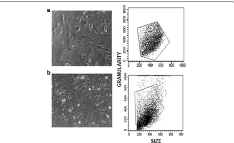
Morphology, size and granularity were similar for both, HMEC-1 and HUVECs (Fig.1). We determined the opti-mal concentration of TNF-α to induce ICAM-1 and VCAM-1, by a dose-response curve (Fig.2); we measured the response trough dot plots in terms of quantity of both, ICAM-1 and VCAM-1 expressed as double positive cells (right up quadrant) for HMEC-1 (Fig. 2A) and HUVEC (Fig.2B). The optimal response of HMEC-1 to TNF-αwas reached with a concentration of 15 ng/mL (Fig. 2C);
higher doses of TNF-αdid not induce a greater expression of adhesion molecules. Therefore we used the concentra-tion of 15 ng/mL of TNF-αin further experiments.
Concerning HUVECs, the dot plots showed a wider range of TNF-α-induced expression of ICAM-1 and VCAM-1 (Fig.2B); responses were observed from doses of 0.75 ng/mL of TNF-α but the dose-response effect was not as regular as for HMEC-1. We used 0.75 ng/mL of TNF-α concentration for the subsequent experiments with HUVECs.
We further search for the expression of VCAM-1 and CD105, also named endoglin, characteristic of endothe-lial cells. We observed a co-expression of VCAM-1 and endoglin in both types of endothelial cells after TNF-α stimulation. Endoglin was expressed in the same extent in both types of cells and histograms were very similar for a constitutive marker (Fig. 3). In contrast, VCAM-1 was expressed in a broad range in HUVEC pool of 3 healthy donors whereas such expression was more homogeneous in HMEC-1 cells (Fig.3).
We further quantified membrane SR-B1 (Fig.3); both, HUVEC and HMEC-1 were positive for this receptor at similar levels of expression (Fig. 3). To explore func-tional aspects of HDL on endothelium function, we
Fig. 2Induction of ICAM-1 and VCAM-1 in HMEC-1 (a) and HUVECs (b) by increasing concentrations of TNF-α. ICAM-1 and VCAM-1 presence in cell membranes was determined by flow cytometry; ICAM-1 and VCAM-1 antibodies were labeled with FITC and PE, respectively. Lower panel,
dose-response curves of HMEC-1 (c) and HUVECs (d)
Fig. 3Co-expression of endothelial markers and SR-B1. Membrane levels of endoglin (CD-105), VCAM-1 and SR-B1 were measured by flow cytometry
in (a) HMEC-1 and (b) HUVEC after we treated them with TNF-αat final concentrations of 15 and 0.75 ng/mL respectively. Endoglin (CD-105) labeled
incubated HMEC-1 with 7 different HDL pools, TNF-α, or both. ICAM-1 and VCAM-1 tended to be expressed below the basal levels (constitutive expression) when cells were co-incubated with HDL, but the differences did not reach statistical significance. In contrast, HDL significantly attenuated the expression of TNF-α-induced VCAM-1, whereas ICAM-1 HDL inhibition did not reach statistical significance (Fig.4).
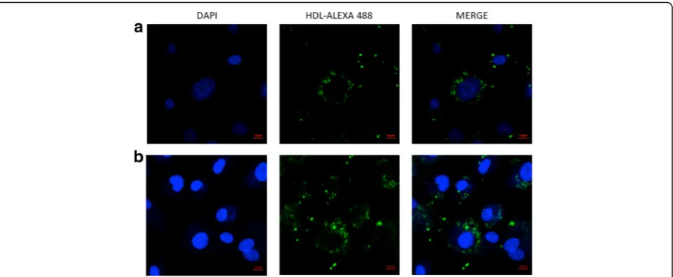
SR-B1 expression in HMEC-1 is relevant in terms of HDL endothelial functionality; previous studies [20, 21] using primary cultures demonstrated that HDL are inter-nalized by endothelial cells. Therefore, we performed HDL internalization assays in HMEC-1 cells and HUVECs using HDL-protein labeled with Alexa Flour 488. Confocal microscopy showed that HMEC-1 and HUVEC are able to internalize HDL (Fig.5). Interestingly, both endothelial models showed that HDL protein was located in discrete granules inside the cytoplasm.
Discussion
In this study we demonstrated that HMEC1 possess similar properties than HUVECs vis-à-vis HDL inter-action. HUVECs have been extensively used for the study of lipoprotein properties with regard to endothelial cells. However, the use of HUVECs represents some dis-advantages, particularly related with reproducibility, due to the inter-individual biological variability [11–14] and technical complexity. For this, in the present work we propose the use of HMEC-1 a cell line of endothelium as alternative of HUVECs.
We first demonstrated that both cell types have similar size and granularity; size and granularity are suitable pa-rameters to identify cell types. Granularity is a measure of cell complexity and depends of nucleus size and the
presence of cytoplasmic organelles and vesicles. There-fore, the structure and complexity of HMEC-1 and HUVECs are comparable. Interestingly, HMEC-1 were more homogeneous than HUVECs in terms of size and granularity, suggesting less variability of experimental data obtained with these cells.
ICAM-1 and VCAM-1 are cell adhesion molecules expressed by the endothelium with important roles in cell migration during inflammation. ICAM-1 is expressed con-stitutively and strongly induced by stimulus like TNF-α, whereas VCAM-1 is mainly expressed after the pro-inflammatory stimulus [22, 23]. In this context, HMEC-1 reached a maximum expression of ICAM-1 and VCAM-1 with 15 ng/mL TNF-α, and such expression remained stable with higher concentrations of TNF-α. In contrast, HUVECs showed a variable expression at increasing doses of the stimulus. The inter-individual variability of HUVECs may be the cause of the less homogeneous dose-response of HUVECs in these experiments, even if we use a pool of umbilical cords from three different donors to obtain more representative results than those obtained with single donor samples. These observations support the idea that HMEC-1 cultures are helpful to obtain more reproducible results. However, it should be emphasized that the HMEC-1 are less sensitive to stimulus, the amount of TNF-αto reach a maximum response in was about 20 times the concentra-tion required for HUVECs. These results should be consid-ered when using HMEC-1 to evaluate endothelial response to inflammatory stimulus.
One of the aims of this study is to determine whether HMEC-1 are suitable for evaluating some HDL properties with regard to endothelial cells; to the best of our knowledge, there are not previous re-ports with this purpose. Interaction of HDL with
Fig. 4ICAM-1 and VCAM-1 inhibition by HDL in HMEC-1. Measures were performed by flow cytometry using a FITC-conjugated ICAM-1 antibody
cells is often mediated by SR-B1 also named CLA-1. This is the main known receptor for HDL expressed by the liver, steroidogenic tissues and recently, it has been reported in endothelium [24, 25]. For this rea-son, we first look for the expression of such receptor in HUVEC and HMEC-1; our results clearly showed that both type of cells expressed SR-B1 in a similar extent, supporting again the idea that HMEC-1 are useful for the study of HDL properties. In addition, the endothelial marker CD105, also known as endoglin, was expressed on the membrane of both types of endothelial cells. CD105 is a transforming growth factor-beta (TGF-beta) co-receptor expressed mainly on endothelial cells and involved in cardio-vascular development, angiogenesis, and cardio-vascular re-modeling [26].
Once we demonstrated that HMEC-1 express key markers of endothelium and the HDL receptor, SR-B1, we further analyzed the usefulness of this cell line to evaluate the anti-inflammatory property of HDL related with the expression of adhesion molecules induced by TNF-α, [27, 28]. Previous studies have demonstrated that this property of HDL is impaired in some individ-uals and may be associated with increased risk of coronary heart disease [29–31]. We performed these experiments using pools of plasma obtained from at least 12 different donors in order reduce the heterogen-eity of the samples in terms of the regulation of adhesion molecules. We observed that HDL clearly inhibited VCAM-1 expression when incubated with TNF-α as expected, whereas ICAM-1 only showed a tendency to a lower expression. Interestingly, the incubation of HDL inhibited expression of adhesion molecules below control levels. This experiment demonstrated HMEC-1
are suitable for HDL anti-inflammatory function studies as well.
A potential mechanism involved in the regulation of endothelial cell function by HDL, may be the internaliza-tion of these lipoproteins as previously demonstrated in HUVECs and bovine aortic endothelial cells [21,32–34]. Therefore, we look for the capacity of HMEC-1 to internalize HDL particles by labeling the protein moiety; our data clearly showed that HMEC-1 were able to internalize HDL particles. Interestingly, HDL is likely to be inside vesicles in perinuclear area, similar to previous reports [20, 21]. These previous studies have demon-strated that HDL vesicles did not present any typical marker of organelles from secretory pathway, suggesting an additional mechanism for HDL; nevertheless, internal-ization process at the moment is not totally understood [35] and requires further investigation.
Conclusion
In this study we demonstrated that HMEC-1 possess characteristics of endothelial cells, in some cases more homogeneous than HUVECs, supporting the idea that this cell line is suitable to evaluate the functionality of HDL vis-à-vis the endothelium.
Abbreviations
BAECs:bovine aortic endothelial cells; CD105: cluster of differentiation 105; HDL: high-density lipoproteins; HMEC-1: human dermal microvascular endothelial cells-1; HUVECs: human umbilical vein endothelial cells; ICAM-1: intercellular adhesion molecule-1; SR-BICAM-1: scavenger receptor class b type 1;
TNF-α: transforming necrosis factor alpha; VCAM-1: vascular cell adhesion
molecule-1
Acknowledgments
None declared.
Funding
Mónica Muñoz-Vega is a doctoral student from Programa de Doctorado en Ciencias Biomédicas, Universidad Nacional Autónoma de México (UNAM) and received fellowship 261915 from CONACYT.
Availability of data and materials
The datasets supporting the conclusions of this article are included within the article.
Authors’contributions
Conception and design of the study: ÓP-M, MM-V, FM, AP, LOM. Collection of the samples and generation of data: MM-V, FM, AP, EC-T, SN. Analysis, interpretation of data, critical review of the manuscript: ÓP-M, FM, HAC-F, JMF, NP-H, GV-A. Drafting and revision of the manuscript: MM-V, ÓP-M, GV-A, HAC-F., LOM. All authors read and approved the final version of the manuscript.
Ethics approval and consent to participate
This study was approved by the Scientific and Ethics Committees of the
National Institute of Cardiology“Ignacio Chávez”. Healthy volunteers were
informed about the objectives of the study and those who agreed to participate signed a letter of informed consent.
Consent for publication
Not Applicable
Competing interests
The authors declare that they have no competing interests.
Publisher’s Note
Springer Nature remains neutral with regard to jurisdictional claims in published maps and institutional affiliations.
Author details
1
Molecular Biology Department, Instituto Nacional de Cardiología“Ignacio Chávez”, Juan Badiano 1, Sección XVI, 14080 Mexico City, Mexico.2Physiology
Departments, Instituto Nacional de Cardiología“Ignacio Chávez”, Mexico City, Mexico.3Cardiovascular and Metabolic Disorders Program, Duke-NUS
Graduate Medical School, Singapore, Singapore.4Institute of Biochemistry, Medical School, Justus-Liebig-University, Giessen, Germany.5National Heart
Centre Singapore, National Heart Research Institute Singapore, Singapore, Singapore.6Institute of Metabolic and Cardiovascular Diseases, I2MC, Inserm,
UMR, 1048 Toulouse, France.
Received: 14 September 2017 Accepted: 2 March 2018
References
1. Lopez AD, Mathers CD, Ezzati M, Jamison DT, Murray CJ. Global and
regional burden of disease and risk factors, 2001: systematic analysis of
population health data. Lancet. 2006;367:1747–57.
2. Murray CJ, Lopez AD. Global mortality, disability, and the contribution of risk
factors: global burden of disease study. Lancet. 1997;349:1436–42.
3. Davies MJ, Woolf N, Rowles PM, Pepper J. Morphology of the
endothelium over atherosclerotic plaques in human coronary arteries.
Br Heart J. 1988;60:459–64.
4. Faggiotto A, Ross R, Harker L. Studies of hypercholesterolemia in the
nonhuman primate. I. Changes that lead to fatty streak formation. Arteriosclerosis. 1984;4:323–40.
5. Gerrity RG. The role of the monocyte in atherogenesis: I. Transition of
blood-borne monocytes into foam cells in fatty lesions. Am J Pathol.
1981;103:181–90.
6. Nakashima Y, Raines EW, Plump AS, Breslow JL, Ross R. Upregulation of
VCAM-1 and ICAM-1 at atherosclerosis-prone sites on the endothelium in
the ApoE-deficient mouse. Arterioscler Thromb Vasc Biol. 1998;18:842–51.
7. Deanfield JE, Halcox JP, Rabelink TJ. Endothelial function and dysfunction:
testing and clinical relevance. Circulation. 2007;115:1285–95.
8. Michiels C. Endothelial cell functions. J Cell Physiol. 2003;196:430–43.
9. Hadi HA, Carr CS, Al Suwaidi J. Endothelial dysfunction: cardiovascular risk
factors, therapy, and outcome. Vasc Health Risk Manag. 2005;1:183–98.
10. Rajendran P, Rengarajan T, Thangavel J, Nishigaki Y, Sakthisekaran D, Sethi
G, Nishigaki I. The vascular endothelium and human diseases. Int J Biol Sci. 2013;9:1057–69.
11. Mendez-Cruz AR, Paez A, Jimenez-Flores R, Reyes-Reali J, Varela E,
Cerbulo-Vazquez A, Rodriguez E, Lopez-Marure R, Masso FA, Flores-Romo L, et al. Increased expression of inflammation-related co-stimulatory molecules by HUVECs from newborns with a strong family history of myocardial infarction stimulated with TNF-alpha and
oxLDL. Immunol Lett. 2007;111:116–23.
12. Paez A, Rodriguez E, Rendon E, Varela E, Fortoul T, Espinosa B, Masso F,
Guevara J, Montano LF. Altered detection of molecules associated with leukocyte traffic in HUVECs derived from newborns with a strong family
history of myocardial infarction. Acta Histochem. 2008;110:42–52.
13. Alvarado-Vasquez N, Paez A, Zapata E, Alcazar-Leyva S, Zenteno E, Masso F,
Montano LF. HUVECs from newborns with a strong family history of diabetes show diminished ROS synthesis in the presence of high glucose
concentrations. Diabetes Metab Res Rev. 2007;23:71–80.
14. Lockmann A, Schon MP. TNF alpha-induced leukocyte-endothelial cell
interactions show marked interindividual differences independent of the
clinical response to adalimumab. Exp Dermatol. 2014;23:133–4.
15. Bouis D, Hospers GA, Meijer C, Molema G, Mulder NH. Endothelium in vitro:
a review of human vascular endothelial cell lines for blood vessel-related
research. Angiogenesis. 2001;4:91–102.
16. Ades EW, Candal FJ, Swerlick RA, George VG, Summers S, Bosse DC, Lawley TJ.
HMEC-1: establishment of an immortalized human microvascular endothelial cell line. J Invest Dermatol. 1992;99:683–90.
17. Renaud JF, Scanu AM, Kazazoglou T, Lombet A, Romey G, Lazdunski M.
Normal serum and lipoprotein-deficient serum give different expressions of excitability, corresponding to different stages of differentiation, in chicken
cardiac cells in culture. Proc Natl Acad Sci U S A. 1982;79:7768–72.
18. Havel RJ, Eder HA, Bragdon JH. The distribution and chemical composition
of ultracentrifugally separated lipoproteins in human serum. J Clin Invest.
1955;34:1345–53.
19. Cardouat G, Duparc T, Fried S, Perret B, Najib S, Martinez LO. Ectopic
adenine nucleotide translocase activity controls extracellular ADP levels and regulates the F1-ATPase-mediated HDL endocytosis pathway on
hepatocytes. Biochim Biophys Acta. 1862;2017:832–41.
20. Perisa D, Rohrer L, Kaech A, von Eckardstein A. Itinerary of high density
lipoproteins in endothelial cells. Biochim Biophys Acta. 1861;2016:98–107.
21. Fruhwurth S, Pavelka M, Bittman R, Kovacs WJ, Walter KM, Rohrl C, Stangl H.
High-density lipoprotein endocytosis in endothelial cells. World J Biol Chem. 2013;4:131–40.
22. Wildner O, Lipkow T, Knop J. Increased expression of ICAM-1, E-selectin, and
VCAM-1 by cultured human endothelial cells upon exposure to haptens.
Exp Dermatol. 1992;1:191–8.
23. Wong D, Dorovini-Zis K. Expression of vascular cell adhesion molecule-1
(VCAM-1) by human brain microvessel endothelial cells in primary culture.
Microvasc Res. 1995;49:325–39.
24. Yeh YC, Hwang GY, Liu IP, Yang VC. Identification and expression of
scavenger receptor SR-BI in endothelial cells and smooth muscle cells of rat
aorta in vitro and in vivo. Atherosclerosis. 2002;161:95–103.
25. Acton SL, Scherer PE, Lodish HF, Krieger M. Expression cloning of SR-BI, a
CD36-related class B scavenger receptor. J Biol Chem. 1994;269:21003–9.
26. Sanchez-Elsner T, Botella LM, Velasco B, Langa C, Bernabeu C. Endoglin
expression is regulated by transcriptional cooperation between the hypoxia and
transforming growth factor-beta pathways. J Biol Chem. 2002;277:43799–808.
27. Cockerill GW, Rye KA, Gamble JR, Vadas MA, Barter PJ. High-density
lipoproteins inhibit cytokine-induced expression of endothelial cell adhesion
molecules. Arterioscler Thromb Vasc Biol. 1995;15:1987–94.
28. Barter PJ. Inhibition of endothelial cell adhesion molecule expression by
high density lipoproteins. Clin Exp Pharmacol Physiol. 1997;24:286–7.
29. Balstad TR, Holven KB, Ottestad IO, Otterdal K, Halvorsen B, Myhre AM, Ose L,
Nenseter MS. Altered composition of HDL3 in FH subjects causing a HDL subfraction with less atheroprotective function. Clin Chim Acta. 2005;359:171–8.
30. Huang X, He D, Ming J, He Y, Zhou C, Ren H, He X, Wang C, Jin J, Ji L, et al.
High-density lipoprotein of patients with breast cancer complicated with type 2 diabetes mellitus promotes cancer cells adhesion to vascular endothelium via ICAM-1 and VCAM-1 upregulation. Breast Cancer Res Treat.
2016;155:441–55.
31. Luc G, Arveiler D, Evans A, Amouyel P, Ferrieres J, Bard JM, Elkhalil L,
molecules ICAM-1 and VCAM-1 and incident coronary heart disease: the
PRIME study. Atherosclerosis. 2003;170:169–76.
32. Rohrer L, Ohnsorg PM, Lehner M, Landolt F, Rinninger F, von Eckardstein A.
High-density lipoprotein transport through aortic endothelial cells involves scavenger receptor BI and ATP-binding cassette transporter G1. Circ Res.
2009;104:1142–50.
33. von Eckardstein A, Rohrer L. Transendothelial lipoprotein transport and
regulation of endothelial permeability and integrity by lipoproteins. Curr
Opin Lipidol. 2009;20:197–205.
34. Cavelier C, Ohnsorg PM, Rohrer L, von Eckardstein A. The beta-chain of cell
surface F(0)F(1) ATPase modulates apoA-I and HDL transcytosis through
aortic endothelial cells. Arterioscler Thromb Vasc Biol. 2012;32:131–9.
35. Rohrl C, Stangl H. HDL endocytosis and resecretion. Biochim Biophys Acta.
1831;2013:1626–33.
• We accept pre-submission inquiries
• Our selector tool helps you to find the most relevant journal
• We provide round the clock customer support
• Convenient online submission
• Thorough peer review
• Inclusion in PubMed and all major indexing services
• Maximum visibility for your research
Submit your manuscript at www.biomedcentral.com/submit