R E S E A R C H
Open Access
Effects of canola and corn oil mimetic on
Jurkat cells
Gabriela Ion
1*, Kayla Fazio
1,2, Juliana A Akinsete
1and W Elaine Hardman
1Abstract
Background:The Western diet is high in omega-6 fatty acids and low in omega-3 fatty acids. Canola oil contains a healthier omega 3 to omega 6 ratio than corn oil. Jurkat T leukemia cells were treated with free fatty acids mixtures in ratios mimicking that found in commercially available canola oil (7%a-linolenic, 30% linoleic, 54% oleic) or corn oil (59% linoleic, 24% oleic) to determine the cell survival or cell death and changes in expression levels of inflammatory cytokines and receptors following oil treatment.
Methods:Fatty acid uptake was assessed by gas chromatography. Cell survival and cell death were evaluated by cell cycle analyses, propidium-iodide staining, trypan blue exclusion and phosphatidylserine externalization. mRNA levels of inflammatory cytokines and receptors were assessed by RT-PCR.
Results:There was a significant difference in the lipid profiles of the cells after treatment. Differential action of the oils on inflammatory molecules, following treatment at non-cytotoxic levels, indicated that canola oil mimetic was anti-inflammatory whereas corn oil mimetic was pro-inflammatory.
Significance:These results indicate that use of canola oil in the diet instead of corn oil might be beneficial for diseases promoted by inflammation.
Keywords:Lymphocytes, Canola oil mimetic, Corn oil mimetic, Apoptosis, Inflammation
Background
The ratio of omega-3 to omega-6 in the average western diet is heavily weighted in favor of omega-6 [1]. When tested as single fatty acids, omega 6 fatty acids tend to be pro-inflammatory but omega-3 fatty acids tend to be anti-inflammatory. Therefore, omega-3 deficiencies have been implicated in inflammatory diseases, cancer, cardi-ovascular diseases, dyslipidaemia and metabolic syn-drome [1,2].
The human diet is very complex and foods provide a mixture of fatty acids in different ratios not just one sin-gle fatty acid at a time. Food is the source of two essen-tial fatty acids, linoleic (omega-6) and a-linolenic acid (omega-3), which cannot be synthesized de novoin ani-mal cells and, therefore, must be obtained from the diet. A good dietary source of omega-3 with an omega-6 to omega-3 ratio of 3:1 is canola oil. We hypothesize that
consuming canola oil in the diet instead of corn oil could decrease pro-inflammatory stimuli.
There is a lack of data aimed at exploring the effect of complex combinations of food fats inin vitromodels. In general, manyin vitromodels focus on only single fatty acids at different concentrations [3-6]. Therefore, to be more relevant to human health, it might be beneficial to consider an experimental design closer to the ratios of the components found in the food which might be consumed.
There is a body of evidence demonstrating that fatty acids affect T lymphocyte functions. In vitro and in vivo studies have shown that fatty acids modulate cytokine release, proliferation, cell death, activation by antigens, surface proteins expression and signaling proteins [7-14]. Single free fatty acids have been shown to induce cell death when used at various concentrations in differ-ent cellular models [4,5,8,15,16]. To study the pro- or anti-inflammatory effects of fatty acids combinations on cytokine production by lymphocytes it is important to explore the effects of fatty acids at non cytotoxic doses. * Correspondence: [email protected]
1
Department of Biochemistry and Microbiology, Marshall University School of Medicine, Huntington, WV, USA
Full list of author information is available at the end of the article
These data would be more relevant to a typical diet where food ingested does not have a cytotoxic effect and could demonstrate alterations in inflammatory cytokines.
In spite of the well-recognized beneficial effects of omega-3 fatty acids for human health, there is a lack of data regarding the effect of canola oil, a common food source rich ina-linolenic acid (omega-3 fatty acid) ver-sus corn oil rich in linoleic acid (omega-6 fatty acid), on lymphocytes. In this study, Jurkat T leukemia cells were treated with free fatty acids mixtures in ratios mimick-ing that found in commercially available canola oil (7% a-linolenic, 30% linoleic, 54% oleic) or corn oil (59% linoleic, 24% oleic) at non cytotoxic dose to determine changes in expression levels of inflammatory cytokines and receptors following oil treatment.
Methods Reagents
The following reagents were used: propidium iodide, Tri-Reagent, 2-propanol, 1 bromo-3-chloro propane, RNase A, ethanol, 3-sodium citrate, butylated hydroxy-toluene (BHT) from Sigma-Aldrich; a-linolenic acid (Cayman Chemical Company), linoleic acid and oleic acid (MP-Biomedicals, LLC); Triton-X100 (IBI Shelton Scientific, Inc.); Chloroform and Hexane (Honeywell, Burdick & Jackson™), Methanol (Fisher Scientific), Iso-octane (EMD).
Cell Lines
Jurkat, Clone E6-1 cells (gift from Dr. Pyali Dasgupta, MU) were maintained in 10% FBS (Hyclone) in RPMI-1640 (ATCC) supplemented with 100 units/ml penicillin and 0.1 mg/ml streptomycin (Sigma-Aldrich). The cells were kept in a humidified atmosphere, at 37°C, contain-ing 5% CO2. The cells were seeded at a cell density of
3 × 10⁵ per ml for all experimental designs.
Fatty acid treatment
The free fatty acids, in ratios mimicking that found in commercially available canola oil (7% a-linolenic, 30% linoleic, 54% oleic) or corn oil (59% linoleic, 24% oleic) were dissolved in ethanol. Cells were treated with an oil concentration of 75μM, 100μM or 150μM for 48 or 72 hours. The final concentration of ethanol in culture media did not exceed 0.15%.
Cell viability and membrane integrity
Cell viability and membrane integrity were assessed by Trypan Blue exclusion and propidium iodide staining, respectively. After treatment, the cells were washed with PBS and stained with propidium iodide (20μg/ml) for 15 min in the dark, at room temperature. The cells were analyzed on a FACSAria flow cytometer (Becton
Dickinson) using DIVA software (Becton Dickinson) and the propidium iodide positive population was evaluated. The Trypan Blue exclusion assay was used to determine cell viability, and the live cells (negative for staining) and dead cells (positive for staining) were enumerated using a hemocytometer.
Annexin V labeling
Jurkat cells were treated as indicated, then washed twice with PBS and resuspended in Annexin V binding buffer (0.01 M HEPES, 0.14 M NaCl and 2.5 mM CaCl2).
Annexin V-Pacific Blue™ conjugate (Invitorgen, Mole-cular Probes) and propidium iodide (20 μg/ml) were added to the cells for 15 min in the dark, at room tem-perature. Cells were analyzed on a FACSAria flow cyt-ometer using DIVA software and the Annexin V positive/propidium iodide negative population was con-sidered early apoptotic.
Cell cycle and DNA fragmentation
Treated cells were subjected to DNA content analysis. Briefly, the cells were harvested and washed two times with PBS and fixed with cold 70% ethanol for at least 24 hours. The ethanol was removed and followed by two PBS washes. Cells were stained in the following solution: PBS supplemented with 0.1% Triton X-100, 0.1% Na3
-citrate, 30 μg/ml RNase and 20μg/ml propidium iodide. After incubation in the dark for 30 minutes at room temperature the cells were analyzed on a FACSAria flow cytometer. DNA fragmentation was determined by cell cycle analysis using DIVA software.
Gas chromatography
Gene expression assay
Human Inflammatory Cytokines and Receptors RT2 Pro-filer™ PCR Array, RT2 First Strand Kit and SuperArray RT2 qPCR Master Mix (SuperArray Bioscience Corpora-tion, Frederick, MD) were used to analyze the expres-sion of a panel of genes in cells treated at 75 μM oil concentration for 72 hours. After treatment, cells were homogenized in Tri Reagent following the protocol of the manufacturer to isolate the RNA. RNA quality con-trol was performed for all samples. The gene expression assay followed the protocol provided by SuperArray. The relative fold differences in gene expression and sta-tistical analyses were calculated on SuperArray software.
Results
Lipid composition of treated cells
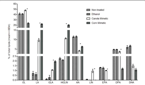
Gas chromatography was performed to investigate whether the cells were able to uptake the canola and corn oil mimetic. Jurkat cells were treated with 150μM canola or corn oil mimetic for 72 hours. When compar-ing the canola oil mimetic treatment with the corn oil mimetic treatment, the canola oil mimetic treated cells had significantly morea-linolenic (LIN) acid and more of the biosynthetic omega-3 fatty acids products (EPA,
DPA) (n = 4, p < 0.005) (Figure 1). The corn oil mimetic treated cells showed increased levels of linoleic acid (LA) and the biosynthetic omega-6 fatty acids pro-ducts (GLA, HGLIN, AA) at a higher fraction than canola oil mimetic (n = 4, p < 0.005) (Figure 1).
Ethanol was used as a carrier for the fatty acids and addition to cell culture did not induce any change in lipid content compared to non-treated cells. As was expected, comparison of either fatty acid treatment (canola or corn oil mimetic) to ethanol and non-treated controls demonstrates a change in the lipid content. Both the canola and corn oil mimetic increased the per-centage of linoleic acid (an omega-6 fatty acid) when compared to ethanol and non-treated cells. Although the canola mimetic contains more omega-3 fatty acids than corn oil mimetic, it is essential to note that it also contains a percentage (30%) of linoleic acid, although less than the corn oil mimetic (59%). Therefore, the increase in linoleic acid for both treatments is expected. In contrast, when comparing the two oil mimetics, it is apparent that the canola oil mimetic (containing just 7% a-linolenic acid) is generating a higher fraction of omega-3 fatty acids than the corn oil mimetic. Despite the comparable percentages of EPA or DPA between
controls (non treated and ethanol treated cells) and the canola oil mimetic, noteworthy is the decrease in omega-3 fatty acids in the presence of the corn oil mimetic.
Membrane Integrity
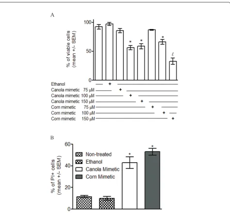
Trypan Blue exclusion (Figure 2A) showed that treat-ment with 100 and 150 μM canola or corn oil mimetic treatment for 72 hours significantly decreased the per-centage of viable cells when compared to the controls
(non-treated and ethanol treated cells) (n = 6, p < 0.05).
Among the treatments, 150 μM corn oil mimetic
showed the highest decrease of cell viability by Trypan blue (n = 6, p < 0.05) (Figure 2A). Propidium-iodide staining (Figure 2B) showed a significant increase in the percentage of cells that lost of membrane integrity in the treated cells (150 μM canola or corn oil mimetic for 48 hours) when compared to the controls (non-treated and ethanol treated cell) (n = 3 for non-treated, n = 4 for all other samples, p < 0.05).
A
B
Apoptosis
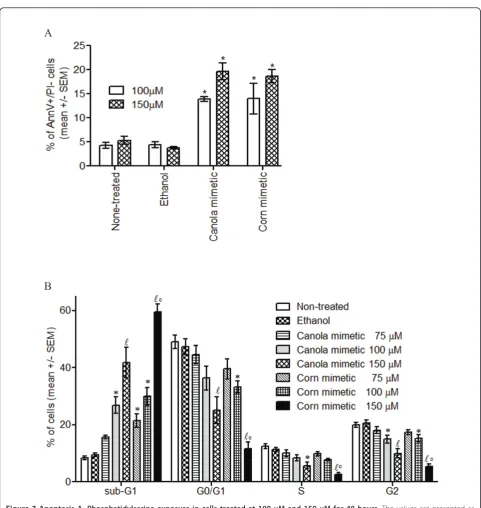
The loss of membrane asymmetry and the exposure of phosphatidylserine on the outer surface of the cell mem-brane as an early apoptotic marker was detected with Pacific Blue labeled AnnexinV. Jurkat cells treated with canola or corn oil mimetic at 100 μM and 150 μM for 48 hours showed a significant increase in Annexin V
+
/propidium iodide- population compared to the con-trols (non-treated and ethanol treated cells) (n = 3 for 150 μM non-treated, n = 4 for all other samples, p < 0.05) (Figure 3A).
DNA fragmentation
The degradation of the nuclear DNA as a late apoptotic marker was assessed by the formation of the ‘sub-G1’ population [17]. Cell cycle analysis of Jurkat cells treated with 75, 100 or 150 μM canola or corn oil mimetic for 72 hours was evaluated (Figure 3B). When comparing the treatments to the controls (non-treated and ethanol treated cells), there was a significant increase in the ‘sub-G1’population for the following treatments: 100 and 150 μM canola oil mimetic and 75, 100 and 150
μM corn oil mimetic (n = 6, p < 0.05) (Figure 3B). When comparing the 150 μM corn oil mimetic treat-ment with the 150 μM canola oil mimetic treatment, the‘sub-G1’population was significantly higher in the corn oil mimetic treated cells.
The exposure of early apoptotic marker phosphatidyl-serine at 48 hours followed by loss of membrane integ-rity and DNA fragmentation at 72 hours indicates that treatment with ≥ 100 μM canola or corn oil mimetic induced apoptosis in Jurkat cells.
Cell cycle
There was a significant decrease in the percentage of Jurkat cells in the G0/G1 phase for cells treated with 100 μM corn oil mimetic compared to the controls (non-treated and ethanol treated cells) (n = 6, p < 0.05) (Figure 3B). There was a significant decrease in the per-centage of Jurkat cells in the G0/G1 phase for cells trea-ted with 150 μM canola or corn oil mimetic when compared to the controls (non-treated and ethanol trea-ted cells) or to the other treatments (n = 6, p < 0.05) (Figure 3B). Moreover, when comparing 150μM corn oil mimetic to 150 μM canola oil mimetic treatment there was a statistically significant difference in the G0/ G1 phase between the treatments.
There was a significant decrease in the percentage of cells in the S phase of both 150μM canola and corn oil mimetic treated cells when compared to the controls (non-treated and ethanol treated cells) (n = 6, p < 0.05) (Figure 3B). The percentage of cells in the S phase was significantly decreased by 150 μM corn oil mimetic when compared to all other treatments.
The percentage of cells in G2 exhibited a significant decrease for 100 and 150μM oil treatment when com-pared to the controls. Cells treated with 150 μM oil mimetic exhibited a significant decreased fraction in the G2 phase when compared to 75 and 100 μM canola or corn oil mimetic. Also, there was a significant difference in the G2 phase fraction between corn and canola oil mimetic at 150μM concentration.
Inflammatory cytokines and receptors
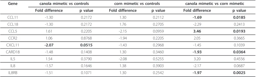
The highest concentration of oil not inducing significant changes in the cell cycle of Jurkat cells was 75μM. This dose was utilized to investigate the effect of canola and corn oil mimetic on expression of inflammatory cyto-kines and receptors. Jurkat cells treated with 75 μM canola oil mimetic for 72 hours showed a 3.46 fold up-regulation (n = 3, p = 0.0193) for CCL5 (RANTES), compared to corn oil mimetic treated cells (Table 1). Moreover, gene expression analysis following canola oil mimetic treatment showed a trend towards down-regu-lating expression of CCL11, CARD18, IL8 and IL8RB when compared to the controls (non-treated and etha-nol treated cells). Whereas, corn oil mimetic treatment showed a trend towards up-regulating the expression for the same genes (CCL11, CARD18, IL8, IL8RB) as com-pared to the controls. To better assess the differences in the gene profiling as a response to the oil mimetic treat-ments, the oil treatment groups were compared. Table 1 shows a significant down-regulation of gene expression in canola oil mimetic treated cells when compared to corn oil mimetic treated cells. A low oil concentration (75 μM) did not have a significant effect on cell cycle but was able to induce differences in gene expression.
Discussion
acids (eicosapentaenoic acid, docosapentaenoic acid, docosahexaenoic acid) than corn oil mimetic treated cells.
Both oil mimetic treatments at a concentration≥100
μM increased the DNA fragmentation (’sub-G1 popula-tion’). The DNA fragmentation was associated with a
decreased in the percentage of cells in the other phases of cell cycle. Previous studies using individual fatty acids showed that linoleic acid was cytotoxic at 100μM anda -linolenic acid had an anti-proliferative effect at 60μM [11]. Cury-Boaventura et al. [8] showed that 50 and 100μM linoleic acid induced phosphatidylserine exposure, an early
A
B
marker for apoptosis, on human lymphocytes. The authors suggested mitochondrial depolarization and ROS produc-tion as a mechanism for cell death induced by 200μM linoleic acid. ROS represent key molecules involved in mul-tiple cellular functions like cell adhesion, apoptosis, regula-tion of immune responses [19]. On Jurkat cells 130μM linoleic acid or 60μMa-linolenic acid had a prooxidant-induced antiproliferatve effect that was negatively corre-lated with caspase 3 activation [11]. In addition, the pro-apoptotic activity ofa-linolenic acid has been associated with up regulation of Bax expression and cytochrome c translocation [18]. In support of these previous studies, the present study demonstrates that canola and corn oil mimetic induced apoptosis and significant changes in cell cycle at a concentration of 100μM. Further investigations are required to establish the mechanism involved in linoleic anda-linolenic fatty acids modulation of cell cycle progres-sion. Many studies have used single long chain omega 3 or 6 fatty acids to modulate cell cycle progression in different cancer cell lines [20-23]. For example, arachidonic acid increased expression of cyclin D1 mRNA and the percen-tage of cells in S phase [20]. Docosahexaenoic acid reduced cyclin D1, E, and A-associated kinase activity and pre-vented the entry of cells in S phase [21]. Eicosapentaenoic acid inhibited synthesis and expression of cyclin D1 and E and blocked cell cycle in G1 [22].Trans-10,cis-12 conju-gated linoleic acid increased the levels of p21cip1/waf1
and blocked the cells in G0/G1 [23]. However, there is a lack of data regarding the effect of the two essential fatty acids, linoleic anda-linolenic acid present in the most commonly cook oils, corn and canola oil, respectively.
The association between inflammation and cancer is thought to be a critical component for cancer develop-ment [24]. The polyunsaturated fatty acids (n-3, n-6) are responsible for the production of families of anti- and pro-inflammmatory bioactive lipid mediators [25]. In vivostudies showed that omega 3 fatty acids decreased
chemoattractant protein-1 (MCP-1), interleukin (IL)-6, interferon (IFN)-gamma mRNA expression [26], and TNF-a level [27]. In vitrolinoleic acid and a-linolenic acid inhibited IL-2 production [11]. In this work, a low oil concentration (75 μM) did not have a significant effect on cell cycle but was able to induce differences in gene expression. One such gene, CCL5 (Regulated upon Activation, Normal T-cell Expressed, and Secreted, abbreviated RANTES) has a dual role regarding tumori-genesis. CCL5 can mediate tumor cell survival, cell growth and metastasis in a number of malignances [28-31]. CCL5 is also proposed as a natural adjuvant to boost anti-tumor immunity [32]. In our experimental design, canola oil mimetic increased CCL5 expression compared to corn oil mimetic treatment. Moreover, the proinflammatory molecules, IL8 (interleukin8), IL8RB (interleukin8 receptor, beta known as CXCR2), CARD18 (caspase recruitment domain family, member 18; ICE-BERG) and CCL11 (chemokine (C-C motif) ligand 11) were slightly down-regulated in canola oil mimetic Jur-kat treated cells compared to slightly up-regulated in corn oil mimetic treatment. IL-8, a chemotactic factor for leukocytes, has been shown to contribute to human cancer progression through its potential functions as a mitogenic and angiogenic factor [33]. CARD18 (ICE-BERG), induced by pro-inflammatory stimuli, inhibits generation of IL-1b by interacting with caspase-1 and preventing its association with RIP2 [34]. CCL11 (eotaxin-1) displays chemotactic activity for eosinophils [35] and is a key player in the angiogenic cascade [36]. Taken together, suppression of these chemokines would be expected to slow cancer progression.
Conclusion
This study was designed to explore the effects of oil mimetics in ratios found in two common cooking oils (canola and corn) on Jurkat T leukemia cells. At high Table 1 Gene expression in Jurkat cells treated with canola or corn oil mimetic
Gene canola mimetic vs controls corn mimetic vs controls canola mimetic vs corn mimetic
Fold difference p value Fold difference p value Fold difference p value
CCL11 -1.30 0.2172 1.30 0.2112 -1.69 0.0185
CCL18 -1.30 0.2172 1.76 0.2705 -2.29 0.2413
CCL5 1.61 0.2205 -2.15 0.0959 3.46 0.0193
CCR2 1.06 0.8768 -1.94 0.2205 2.05 0.3665
CXCL11 -2.07 0.0515 -1.43 0.2968 -1.45 0.1039
CARD18 -1.48 0.1408 1.30 0.3460 -1.93 0.0364
IL5 1.54 0.3790 -2.08 0.5255 3.20 0.4556
IL8 -1.57 0.1646 1.38 0.3903 -2.17 0.0687
IL8RB -1.51 0.1071 1.30 0.2542 -1.97 0.0025
concentrations (100 and 150 μM) both types of oils induced apoptosis. At a non-toxic dose (75μM) the dif-ferent oil mimetics displayed differences in their action on pro-inflammatory molecules with canola oil being anti-inflammatory whereas corn oil was pro- inflamma-tory. Findings from this study emphasize the need to investigate the effect of dietary fat within complex mix-tures at non-cytotoxic doses when evaluating the inflam-matory response. Oil mimetic could be enough to induce differences in fatty acid and immune modulator profiles. This is critical in regards to the importance of examining conventional diet sources in human health and disease. Oil mixtures are more physiologically rele-vant than single fatty acids since humans must consume both omega-3 and omega-6 fatty acids. In this respect canola oil may have a more favorable fatty acid profile for decreasing the chance of inflammation that is pro-motional for development of chronic diseases.
Acknowledgements
Financial support by NCI grant CA114018-01 and Cancer Research and Prevention Foundation is gratefully acknowledged. We thank Dr. Melissa J. Seelbach and Margaret Putt for reading the manuscript and helpful discussion.
Author details
1Department of Biochemistry and Microbiology, Marshall University School of
Medicine, Huntington, WV, USA.2Bluefield State College, Bluefield, WV, USA.
Authors’contributions
All authors have read and approve the final manuscript. GI designed the study, analyzed, interpreted the data, and drafted the manuscript. KF carried out the apoptosis assays and gas chromatography. JAA carried out the gene array. WEH gave the final approval of the version to be published.
Competing interests
The authors declare that they have no competing interests.
Received: 13 April 2011 Accepted: 1 June 2011 Published: 1 June 2011
References
1. Simopoulos AP:Evolutionary aspects of diet, the omega-6/omega-3 ratio and genetic variation: nutritional implications for chronic diseases. Biomed Pharmacother2006,60:502-507.
2. Yashodhara BM, Umakanth S, Pappachan JM, Bhat SK, Kamath R, Choo BH:
Omega-3 fatty acids: a comprehensive review of their role in health and disease.Postgrad Med J2009,85:84-90.
3. Denys A, Hichami A, Khan NA:n-3 PUFAs modulate T-cell activation via protein kinase C-alpha and -epsilon and the NF-kappaB signaling pathway.J Lipid Res2005,46:752-758.
4. Siddiqui RA, Jenski LJ, Neff K, Harvey K, Kovacs RJ, Stillwell W:
Docosahexaenoic acid induces apoptosis in Jurkat cells by a protein phosphatase-mediated process.Biochim Biophys Acta2001,1499:265-275. 5. Verlengia R, Gorjao R, Kanunfre CC, Bordin S, de Lima TM, Curi R:Effect of arachidonic acid on proliferation, cytokines production and pleiotropic genes expression in Jurkat cells–a comparison with oleic acid.Life Sci 2003,73:2939-2951.
6. Takahashi HK, Cambiaghi TD, Luchessi AD, Hirabara SM, Vinolo MA, Newsholme P, Curi R:Activation of survival and apoptotic signaling pathways in lymphocytes exposed to palmitic acid.J Cell Physiol2011. 7. Costabile M, Hii CS, Melino M, Easton C, Ferrante A:The
immunomodulatory effects of novel beta-oxa, beta-thia, and gamma-thia polyunsaturated fatty acids on human T lymphocyte proliferation,
cytokine production, and activation of protein kinase C and MAPKs.J Immunol2005,174:233-243.
8. Cury-Boaventura MF, Gorjao R, de Lima TM, Newsholme P, Curi R:
Comparative toxicity of oleic and linoleic acid on human lymphocytes. Life Sci2006,78:1448-1456.
9. Vedin I, Cederholm T, Freund LY, Basun H, Garlind A, Faxen IG, Jonhagen ME, Vessby B, Wahlund LO, Palmblad J:Effects of
docosahexaenoic acid-rich n-3 fatty acid supplementation on cytokine release from blood mononuclear leukocytes: the OmegAD study.Am J Clin Nutr2008,87:1616-1622.
10. Kim W, Fan YY, Barhoumi R, Smith R, McMurray DN, Chapkin RS:n-3 polyunsaturated fatty acids suppress the localization and activation of signaling proteins at the immunological synapse in murine CD4+ T cells by affecting lipid raft formation.J Immunol2008,181:6236-6243. 11. Bergamo P, Luongo D, Maurano F, Rossi M:Butterfat fatty acids
differentially regulate growth and differentiation in Jurkat T-cells.J Cell Biochem2005,96:349-360.
12. Peck MD, Li Z, Han T, Wang W, Jy W, Ahn YS, Ziboh VA, Chu AJ, Bourguignon LY:Fatty acid unsaturation increases expression and capping of murine lymphocyte CD44 and CD45.Nutrition1996,
12:616-622.
13. Pompos LJ, Fritsche KL:Antigen-driven murine CD4+ T lymphocyte proliferation and interleukin-2 production are diminished by dietary (n-3) polyunsaturated fatty acids.J Nutr2002,132:3293-3300.
14. Shaikh SR, Edidin M:Immunosuppressive effects of polyunsaturated fatty acids on antigen presentation by human leukocyte antigen class I molecules.J Lipid Res2007,48:127-138.
15. Lu X, Yu H, Ma Q, Shen S, Das UN:Linoleic acid suppresses colorectal cancer cell growth by inducing oxidant stress and mitochondrial dysfunction.Lipids Health Dis2010,9:106.
16. Yuan H, Zhang X, Huang X, Lu Y, Tang W, Man Y, Wang S, Xi J, Li J:NADPH oxidase 2-derived reactive oxygen species mediate FFAs-induced dysfunction and apoptosis of beta-cells via JNK, p38 MAPK and p53 pathways.PLoS One2010,5:e15726.
17. Nicoletti I, Migliorati G, Pagliacci MC, Grignani F, Riccardi C:A rapid and simple method for measuring thymocyte apoptosis by propidium iodide staining and flow cytometry.J Immunol Methods1991,139:271-279. 18. Kim JY, Park HD, Park E, Chon JW, Park YK:Growth-inhibitory and
proapoptotic effects of alpha-linolenic acid on estrogen-positive breast cancer cells.Ann N Y Acad Sci2009,1171:190-195.
19. Droge W:Free radicals in the physiological control of cell function. Physiol Rev2002,82:47-95.
20. Razanamahefa L, Prouff S, Bardon S:Stimulatory effect of arachidonic acid on T-47D human breast cancer cell growth is associated with enhancement of cyclin D1 mRNA expression.Nutr Cancer2000,
38:274-280.
21. Chen ZY, Istfan NW:Docosahexaenoic acid, a major constituent of fish oil diets, prevents activation of cyclin-dependent kinases and S-phase entry by serum stimulation in HT-29 cells.Prostaglandins Leukot Essent Fatty Acids2001,64:67-73.
22. Palakurthi SS, Fluckiger R, Aktas H, Changolkar AK, Shahsafaei A, Harneit S, Kilic E, Halperin JA:Inhibition of translation initiation mediates the anticancer effect of the n-3 polyunsaturated fatty acid eicosapentaenoic acid.Cancer Res2000,60:2919-2925.
23. Cho HJ, Kim EJ, Lim SS, Kim MK, Sung MK, Kim JS, Park JH:Trans-10, cis-12, not cis-9, trans-11, conjugated linoleic acid inhibits G1-S progression in HT-29 human colon cancer cells.J Nutr2006,136:893-898.
24. Coussens LM, Werb Z:Inflammation and cancer.Nature2002,
420:860-867.
25. Serhan CN, Savill J:Resolution of inflammation: the beginning programs the end.Nat Immunol2005,6:1191-1197.
26. Matsunaga H, Hokari R, Kurihara C, Okada Y, Takebayashi K, Okudaira K, Watanabe C, Komoto S, Nakamura M, Tsuzuki Y, Kawaguchi A, Nagao S, Miura S:Omega-3 polyunsaturated fatty acids ameliorate the severity of ileitis in the senescence accelerated mice (SAM)P1/Yit mice model.Clin Exp Immunol2009,158:325-333.
28. Wilcox RA, Wada DA, Ziesmer SC, Elsawa SF, Comfere NI, Dietz AB, Novak AJ, Witzig TE, Feldman AL, Pittelkow MR, Ansell SM:Monocytes promote tumor cell survival in T-cell lymphoproliferative disorders and are impaired in their ability to differentiate into mature dendritic cells. Blood2009,114:2936-2944.
29. Vaday GG, Peehl DM, Kadam PA, Lawrence DM:Expression of CCL5 (RANTES) and CCR5 in prostate cancer.Prostate2006,66:124-134. 30. Yaal-Hahoshen N, Shina S, Leider-Trejo L, Barnea I, Shabtai EL, Azenshtein E,
Greenberg I, Keydar I, Ben-Baruch A:The chemokine CCL5 as a potential prognostic factor predicting disease progression in stage II breast cancer patients.Clin Cancer Res2006,12:4474-4480.
31. Aldinucci D, Lorenzon D, Cattaruzza L, Pinto A, Gloghini A, Carbone A, Colombatti A:Expression of CCR5 receptors on Reed-Sternberg cells and Hodgkin lymphoma cell lines: involvement of CCL5/Rantes in tumor cell growth and microenvironmental interactions.Int J Cancer2008,
122:769-776.
32. Lapteva N, Huang XF:CCL5 as an adjuvant for cancer immunotherapy. Expert Opin Biol Ther2010,10:725-733.
33. Xie K:Interleukin-8 and human cancer biology.Cytokine Growth Factor Rev 2001,12:375-391.
34. Humke EW, Shriver SK, Starovasnik MA, Fairbrother WJ, Dixit VM:ICEBERG: a novel inhibitor of interleukin-1beta generation.Cell2000,103:99-111. 35. Garcia-Zepeda EA, Rothenberg ME, Ownbey RT, Celestin J, Leder P,
Luster AD:Human eotaxin is a specific chemoattractant for eosinophil cells and provides a new mechanism to explain tissue eosinophilia.Nat Med1996,2:449-456.
36. Salcedo R, Young HA, Ponce ML, Ward JM, Kleinman HK, Murphy WJ, Oppenheim JJ:Eotaxin (CCL11) induces in vivo angiogenic responses by human CCR3+ endothelial cells.J Immunol2001,166:7571-7578.
doi:10.1186/1476-511X-10-90
Cite this article as:Ionet al.:Effects of canola and corn oil mimetic on Jurkat cells.Lipids in Health and Disease201110:90.
Submit your next manuscript to BioMed Central and take full advantage of:
• Convenient online submission • Thorough peer review
• No space constraints or color figure charges • Immediate publication on acceptance
• Inclusion in PubMed, CAS, Scopus and Google Scholar • Research which is freely available for redistribution