Contralateral artery enlargement
predicts carotid plaque progression based
on machine learning algorithm models
in apoE
−
/
−
mice
Bing Li
1†, Yun Jiao
2†, Cong Fu
1, Bo Xie
2, Genshan Ma
1, Gaojun Teng
2and Yuyu Yao
1*Abstract
Background: This study specifically focused on anatomical MRI characterization of the low shear stress-induced atherosclerotic plaque in mice. We used machine learn-ing algorithms to analyze multiple correlation factors of plaque to generate predictive models and to find the predictive factor for vulnerable plaque.
Methods: Branches of the left carotid artery in apoE−/− and C57BL/6J mice were ligated to produce the partial left carotid artery model. Before surgery, and 7, 14, and 28 days after surgery, in vivo serial MRI measurements of carotid artery diameter were obtained. Meanwhile, proximal blood flow was evaluated. After image acquisition and animal sacrifice, carotid arteries were harvested for histological analysis. Support vec-tor machine (SVM) and decision tree (DT) were used to select features and generate predictive models of vulnerable plaque progression.
Result: Seven days after surgery, neointima formation was visualized on micro-MRI in both apoE−/− and C57BL/6J mice. Ultrasonography showed that blood flow had significantly decreased compared to that in the contralateral artery. Partial ligation of the carotid artery for 4 weeks in apoE−/− mice induced vulnerable plaque; however, in C57BL/6J mice this same technique performed for 4 weeks induced arterial stenosis. Contralateral carotid artery diameter at 7 days after surgery was the most reliable pre-dictive factor in plaque progression. We achieved over 87.5% accuracy, 80% sensitivity, and 95% specificity with SVM. The accuracy, sensitivity, and specificity for the DT classi-fier were 90, 90, and 90%, respectively.
Conclusions: This study is the first to demonstrate that SVM and DT methods could be suitable models for identifying vulnerable plaque progression in mice. And con-tralateral artery enlargement can predict the vulnerable plaque in carotid artery at the very early stage. It may be a valuable tool which helps to optimize the clinical work flow process by providing more decision in selecting patients for treatment. Keywords: Low shear stress, Atherosclerosis, Machine learning algorithms, Animal model
Open Access
© The Author(s) 2016. This article is distributed under the terms of the Creative Commons Attribution 4.0 International License (http://creativecommons.org/licenses/by/4.0/), which permits unrestricted use, distribution, and reproduction in any medium, provided you give appropriate credit to the original author(s) and the source, provide a link to the Creative Commons license, and indicate if changes were made. The Creative Commons Public Domain Dedication waiver ( http://creativecommons.org/publicdo-main/zero/1.0/) applies to the data made available in this article, unless otherwise stated.
RESEARCH
*Correspondence: [email protected] †Bing Li and Yun Jiao contributed equally to this work
1 Department of Cardiology, Zhongda Hospital, Medical School of Southeast University, 87 Dingjiaqiao, Nanjing 210009, Jiangsu, China
Background
Atherosclerosis, a complex multifactorial disease, is the leading cause of death world-wide. It is characterized as patchy intimal plaques that encroach on the lumen of medium-sized and large arteries; the plaques comprise lipids, inflammatory cells, smooth muscle cells, and collagen matrix [1]. The etiology of atherosclerosis is com-plex, poorly understood, and remains controversial [2]. Extensive evidence indicates that blood flow-induced shear stress plays a critical role in atherogenesis in humans and animals [3, 4]. Shear stress is biomechanical force acting on the endothelium, which is determined by blood flow, vessel geometry, and fluid viscosity [5]. Researchers have found that low and oscillatory endothelial shear stress-induced atherosclerotic lesions form at specific arterial regions [6].
Two common animal models are used to mimic the low shear stress that, in humans, contributes to carotid atherosclerosis. One is carotid constriction, using a constrictive cuff [7] or a conical cast [8]. The other is ligation of the left external and internal carotid arterial branches [9]. Merino et al. [10] used well-examined apoE−/− mice to
gener-ate atherosclerosis by ligating the carotid artery, inducing low blood flow velocity, and generating shear stress, which is evidenced by plaque formation in the carotid artery, infiltration of monocytes and macrophages in the plaque, increase in pro-inflammatory cytokines, and decrease in blood velocity. This model shows that this could be a poten-tial animal model to study human cardiovascular disease.
Generally, animal models have the advantage of a shorter time to lesion development and allow the investigation of pathology in a large number of individuals [11]. Tradi-tionally, animal research has resulted in great benefits for humans, but it requires that researchers keep sacrificing animals to gather the beneficial information. This situation changed with the advent of molecular imaging techniques and bioinformation. High-resolution magnetic resonance imaging (MRI) [12], ultrasound [13], and positron emis-sion tomography (PET) [14] are noninvasive and permit longitudinal studies of plaque initiation, growth, and regression in the same animal. Micro-MRI plays a significant role in the study of experimental atherosclerosis. MRI can effectively assess luminal narrow-ing, plaque size, and morphology [15]. Despite these advantages, MRI still suffers from low spatial resolution and motion artifacts in the artery, and thus remains limited in its ability to identify the vulnerable components of plaque [16]. The features seen on the MRI images are not exactly the same with those found in the histological examination. In order to improve the accuracy of MRI in the diagnosis of plaque, there is a need for extracting discriminate features on the MRI images [17, 18].
In this study, we focused on establishing a predictive model to evaluate vulnerable ath-erosclerotic plaque formation and progress. We established low shear stress model of atherosclerosis in apoE−/− mice and C57BL/6J mice. MRI was used to collect the data of
plaque formation and progress. The plaques in the C57BL/6J group were “stable” and the plaques in the apoE−/− group were “vulnerable” according the ultima pathology
detec-tion and vulnerability index formula. And we used two machine learning algorithms to analyze the multiple correlation factors of low shear stress induced plaque in apoE−/−
and C57BL/6J mice, and to try to find the predictive factor for vulnerable plaque pro-gression. Analysis shows that the proposed feature is clinically significant.
Methods Animal model
All procedures in the present study were conducted in accordance with the National Instituted of Health Guide for the Care and Use of Laboratory Animals and approved by the Care of Experimental Animals Committee of the Southeast University (Approval ID: SYXK-2012.3921). Male apoE−/− mice (n = 20), 9 weeks old, and a control group of
male C57BL/6J mice (n = 20), 9 weeks old, were obtained from the Changzhou Cavens Laboratory Animal Ltd. (Changzhou, China, approval ID:SCXK2011-0003). The ani-mals received a western-type diet (high levels of fat and cholesterol added to grain-based chow diets) for 2 weeks prior to surgery. After all the mice had eaten this diet for 2 weeks, their left external and internal carotid arterial branches were isolated and ligated with 6-0 silk suture. The contralateral right carotid artery was sham-operated to serve as an intra-animal control.
In vivo serial MRI studies
After the surgery, all mice underwent MRI examinations to measure the diameter of the lumen wall at 7, 14, and 28 days. All scanning was performed on a Micro-MRI animal scanner (7.0T Bruker PharmaScans, Bruker Biospin, Ettlingen, Germany). Isoflurane anesthesia (2%) in each mouse was induced and maintained in medical-grade air and monitored using small animal instrument monitoring (reduced respiratory rate to 40 breath/min) to eliminate the influence of respiratory movement on MRI image qual-ity. All animals were placed in the supine position. Eighteen contiguous, 500-μm thick axial slices spanning from the neck region of the mouse were acquired using a spin echo sequence. MRI images were obtained using black-blood T2 to proton density (PD)-weighted multi-spin multi-echo (MSME) sequence and a dedicated mouse volume coil. Imaging parameters were as follows: TR 1206.9 ms, TE 12.8/34.2 ms, FOV 2.5 cm, FA was 180°, matrix size 256 × 256, and in-plane resolution 141 × 141 × 800 μm3; slice thickness of 0.5 mm, and four excitations. Fat suppression was performed for proton density and T2-weighted imaging. The total MR scanning procedure required approxi-mately 50 min [23].
Ultrasonographic imaging and pulse‑wave Doppler measurement
The blood flow velocity was measured in both carotids using an ultrasonic instrument (Vevo 2100, VisualSonices Inc, Toronto, Canada) at 7, 14, and 28 days after the surgery. Each mouse was anesthetized and maintained under isoflurane anesthesia (2%) and laid on a platform in the supine position with all legs taped to ECG electrodes for heart rate monitoring. An MS 400 (30 MHz centerline frequency) probe was used to measure the frequency of the common carotid artery blood flow to the midpoint spectrum and to measure the blood flow velocity, as previously described [24]. Shear stress was calculated using the following formula: SS = 4 μVm/Ds [25], where μ is the blood viscosity (taken as 0.035 poise), Vm is the mean flow velocity (mm/s), and Ds is the lumen diameter of the targeted carotid artery (mm).
Serum lipid measurement
After MRI and ultrasound examination at 28 days, blood samples were taken while the mice were still anesthetized by angular vein puncture at the time of sacrifice. Serum was separated and serum total cholesterol (TC) and low-density lipoprotein (LDL) were determined by using an automatic biochemistry analyzer.
Histological examination
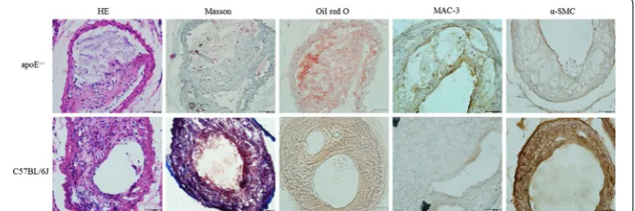
At the end of the procedure, carotid arteries were collected from sacrificed mice, imme-diately embedding OCT compound, and rapidly frozen using liquid nitrogen. Serial cryosections of 8 μm were cut on a Lecia CM1950 Cryostat (Lecia Instruments, Heidel-berg, Germany). The serial cryosections were stained with hematoxylin-eosin, Oil red O, Masson’s trichrome, respectively. For immunohistochemical analyses, α-smooth mus-cle actin (α-SMC, Abcam, Cambridge, UK) and MAC-3 (BD Biosciences Pharmingen, San Diego, CA) stains were used for detection of smooth muscle cells and macrophages, respectively. ImageJ 1.41 software was used to measure the content of collagen, lipid, macrophages and vascular smooth muscle cells. The vulnerability index was calculated using the following formula:positive staining area of (macrophages + lipid)/positive staining area of (SMCs + collagen) [26].
Machine learning algorithms
We selected SVM because it has been shown empirically to achieve good generaliza-tion performance on a wide variety of classificageneraliza-tion problems. The attribute weight, w, describes the separating hyper plane of a liner SVM [27]. Parameter w of SVM indicated the contribution of the feature to SVM. In this report, the absolute value for each weight means the importance for prediction of carotid vulnerable plaque progress. Positive w means increased in the normal control (decreased in apoE−/− group), while negative
value means decreased activities in the normal control (increased in apoE−/− group).
We selected ten features as follows: the lumen diameter of left carotid artery (LCA) and right carotid artery (RCA), the lumen area of LCA and RCA, the blood flow velocity of LCA and RCA, the ratio of the lumen diameter of LCA to RCA, the ratio of the lumen area of LCA to RCA, the ratio of the blood flow velocity of LCA to RCA and the plaque volume of LCA at 7 days separately.
Predictive model evaluation
SVM and DT were applied by use of WEKA (Version 3.6, The University of Waikato, Hamilton, New Zealand). We evaluated the predictive models generated by these two machine-learning algorithms based on tenfold cross-validation; we randomly divided the data into ten groups of four subjects each (i.e., one-tenth of subjects were placed in each group), with controls being mixed with vulnerable plaque subjects. We ran ten iterations, using 90% of subjects for classifier generation (i.e., training) and the remain-ing 10% for testremain-ing. After cyclremain-ing through all ten partitions for classification, every group was used for testing, and each group appeared in a training set every time except when it was used for testing [29].
We evaluated the performance of each predictive model based on the following met-rics: true-positive rate, false-positive rate, accuracy, and area under the receiver oper-ating characteristic (ROC) curve (AUC). Let NTP denote the number of mice with vulnerable plaque (28-day pathology confirmed as vulnerable plaque) correctly pre-dicted as having vulnerable plaque, let NFP denote the number of control subjects incor-rectly identified as having vulnerable plaque, let NTN denote the number of control subjects correctly identified, and let NFN denote mice with vulnerable plaque incorrectly identified as control subjects. Accuracy (ACC) is the proportion of correctly labeled instances in the study, defined as ACC = (NTP + NTN)/(NTP + NTN + NFP + NFN). The true-positive rate (TPR), or sensitivity, is the proportion of positive instances that were correctly reported as being positive (e.g., TPR = NTP/[NTP + NFN]). The false-positive rate (FPR), or (1 − specificity), is the proportion of negative instances that were erro-neously reported as being positive (FPR = NFP/[NFP + NTN]). For a diagnostic system with probabilistic output, the ROC curve is a graphical plot of TPR (y-axis) against FPR (x-axis), because the discrimination threshold is varied. We used AUC as an additional measure of the performance for each model.
Statistics
Data were evaluated by use of SPSS 19 (SPSS, Chicago, IL, USA). Differences for two groups were compared by t test. Differences were considered statistically significant at a value of P < 0.05. Data are expressed as mean ± standard deviation of the mean (SD).
Results
In vivo MRI of the vessel wall morphology
increased in the apoE−/− group. There is a significant reduction in the diameter of
the left common carotid artery 0.42 ± 0.01 mm at 7 days, 0.29 ± 0.02 mm at 14 days, 0.23 ± 0.01 mm at 28 days, compared to the diameter of the left carotid artery before surgery (0.55 ± 0.02 mm, Fig. 1). We also observed a significant expansion in the
diam-eter of the right common carotid artery 0.64 ± 0.02 mm at 7 days, 0.66 ± 0.03 mm at 14 days and 0.67 ± 0.01 mm at 28 days compared to the diameter of the right carotid artery 0.56 ± 0.01 mm before surgery. In the C57BL/6J mice group, ligation also induced
arterial stenosis. But we observed a mild reduction in the diameter of the left common carotid artery 0.44 ± 0.02 mm at 7 days and significantly reduced diameter of the left carotid artery 0.30 ± 0.03 mm at 14 days and 0.27 ± 0.01 mm at 28 days, compared to the diameter of the left carotid artery at baseline (0.55 ± 0.02 mm, Fig. 2). We observed
a slight expansion in the diameter of the right common carotid artery 0.61 ± 0.01 mm at 7 days, 0.62 ± 0.02 mm at 14 days, and 0.63 ± 0.01 mm at 28 days compared to the diameter of the right carotid artery 0.55 ± 0.03 mm at baseline. We also calculated the plaque volume using MRI data, as shown in Table 1. A significantly larger plaque volume is found in apoE−/− compared to C57BL/6J mice (P < 0.05).
Blood velocity and shear stress
In both apoE−/− and C57BL/6J mice, the blood flow velocity in the surgery side
decreased significantly compared with the contralateral side as showed in Table 2. And according to the formula SS = 4 μVm/Ds, the shear stress in the surgery side also decreased significantly compared with that in the contralateral side. As time went on, the blood flow velocity and shear stress declined gradually, and no statistically significant difference between apoE−/− and control mice was found during the study.
Serum lipid levels
During the experiment, there was no significant difference in body weight between apoE−/− (28 ± 1.28 g) and C57BL/6J mice (27.6 ± 1.46 g, P > 0.05). The serum levels of
total cholesterol and LDL in the apoE−/− group (13.37 ± 6.24 and 3.98 ± 1.24 mmol/L)
were significantly higher compared with that in the C57BL/6J group (2.96 ± 0.28 and 1.42 ± 0.49 mmol/L, P < 0.05).
Table 1 Mean and standard deviation for monitoring variables of apoE−/− and C57BL/6J
mice group at 7 days
Data are presented as mean ± SD. Predictive power is the parameter w of SVM indicated the contribution of the feature to SVM. It means the importance for prediction of carotid vulnerable plaque progress. Positive means increased in the normal control (decreased in apoE−/− group), while negative is contrast. P value indicated the result of t tests between two groups. * P = 1.29 × 10⁻⁷, ** P = 3.38 × 10⁻⁷, *** P = 2.38 × 10⁻⁶, **** P = 1.24 × 10⁻⁶
Predictive power ApoE−/− C57BL/6J P value
Lumen diameter of LCA (mm) −0.21 0.42 ± 0.01 0.44 ± 0.02 0.015 Lumen diameter of RCA (mm) −1.48 0.65 ± 0.02 0.61 ± 0.01 <0.001* Lumen area of LCA (mm2) 0.09 0.14 ± 0.01 0.15 ± 0.02 0.030 Lumen area of RCA (mm2) −1.31 0.34 ± 0.02 0.30 ± 0.02 <0.001** Blood flow velocity of LCA (mm/s) 0.47 152.42 ± 43.67 162.17 ± 33.41 0.438 Blood flow velocity of RCA (mm/s) 0.24 532.84 ± 104.35 487.82 ± 76.34 0.128 The ratio of the lumen diameter of LCA to
RCA 0.63 0.64 ± 0.03 0.72 ± 0.05 <0.001*** The ratio of the lumen area of LCA to RCA 0.72 0.41 ± 0.05 0.53 ± 0.06 <0.001**** The ratio of the blood flow velocity of LCA
to RCA 1.09 0.29 ± 0.08 0.34 ± 0.09 1.011 Plaque volume of LCA (mm3) 0.56 0.77 ± 0.14 0.53 ± 0.15 0.001
Table 2 Changes of shear stress in low velocity regions between two groups detected by MRI and US measurement before and after surgery
Data are mean ± SD
Vm mean velocity of carotid artery, Ds end-systolic diameters of carotid artery, SS shear stress of carotid artery, μ blood viscosity of mice, SS 4 μVm/Ds
* P < 0.05, comparing the shear stress of LCA with the shear stress of RCA. # P < 0.05, comparing the shear stress at 14 days with the shear stress at 7 days
ApoE−/− C57BL/6J
7 days 14 days 7 days 14 days
LCA RCA LCA RCA LCA RCA LCA RCA
Vm (mm/s)
152.4 ± 43.7 532.8 ± 104.4 71.3 ± 16.6 505.3 ± 98.7 162.2 ± 33.4 487.8 ± 76.3 79.1 ± 9.9 458.9 ± 55.8
Dr (mm) 0.42 ± 0.01 0.64 ± 0.02 0.29 ± 0.02 0.66 ± 0.03 0.44 ± 0.02 0.61 ± 0.01 0.3 ± 0.03 0.62 ± 0.02 SS (dyn/
cm2) 50.4 ± 14.6* 113.1 ± 22.1 34.3 ± 6.0*
Morphometric analyses
The composition of plaque including macrophages, SMCs, collagen, and lipids was measured by histological and immunohistochemical staining (Table 3).
The plaque in the apoE−/− group had a higher content of lipids and macrophages and
a lower content of collagen than the C57BL/6J group (Fig. 3). The stenosis in C57BL/6J mice at 4 weeks contained smooth muscle cells. The vulnerability index of the apoE−/−
group (2.81 ± 0.21) was higher than that of the C57BL/6J group (0.42 ± 0.13, P < 0.05). According to the diagnostic criteria of vulnerable plaque established by Naghavi et al. [30], the apoE−/− group had a higher accumulation of lipids and macrophages, a lower
content of collagen, thinner fibrous caps, and an increased vulnerability index. So we recognized that all the plaque in apoE−/− mice was vulnerable; however, the plaque in
C57BL/6J mice was stable, because the plaques contained lots of smooth muscle cells.
Results of the predictive models
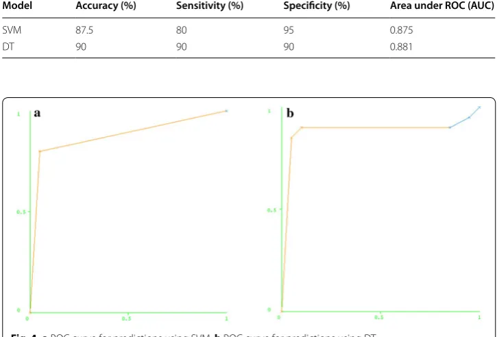
The performance of the SVM-based model was good with a sensitivity of 95%, specific-ity of 80%, and accuracy of 87.5% (Table 4). The predictive value (weight) of each fea-ture was estimated in predicting the final result by SVM (Table 1). We found that lumen diameter of RCA at 7 days was the most important feature in the SVM-based model with the highest absolute value (w = −1.48).
The DT only selected the lumen diameter of RCA at 7 days with sensitivity of 90%, specificity of 90% and accuracy of 90% (Table 4). Figure 4 shows the ROC for predictions using SVM and DT.
Table 3 Comparison of plaque morphological parameters after 28 days
Data are presented as mean ± SD
* P < 0.05, comparing the C57BL/6J group with the apoE−/− group
ApoE−/− C57BL/6J
Macrophages (%) 29.4 ± 8.7 18.9 ± 6.5*
SMCs (%) 6.8 ± 2.3 26.2 ± 9.8*
Lipids (%) 42 ± 10.6 10.9 ± 2.4*
Collagen (%) 15.8 ± 3.5 44.6 ± 13.7*
The vulnerability index 2.81 ± 0.21 0.42 ± 0.13*
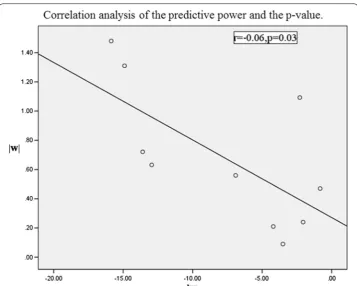
We also applied two sample t tests between groups to find statistically significant parameters. The significant features, their respective range (mean ± standard deviation) for both classes, and the P values were shown in the Table 1. It was evident that four fea-tures have a P value less than 0.01, including the lumen diameter of RCA at 7 days, these features can be considered significant enough for classification. The lumen diameter of RCA at 7 days was both selected by the machine learning method and the traditional sta-tistical methods as one of the significant features. Because some P values are too small, we chose a natural logarithm of P values. The absolute value of predictive power and P value using the natural logarithm determined that every feature was associated through statistical correlation analysis (r = −0.66, P = 0.03, Fig. 5). It showed that our predictive method was correlated with the traditional method.
Discussion
We applied the feature selection method based on serial MRI and ultrasound data to search the optimized discriminative predictive factor for vulnerable plaque progression in the early stage of atherosclerosis. The SVM classifier and DT classifier were imple-mented for the classification of mice with plaque progression. Our study first dem-onstrates that contralateral artery enlargement can predict carotid plaque progress in apoE−/− mice in the very early stage.
For centuries, researchers have made much progress in understanding the anatomic initiation and progression of atherosclerosis. But how to identify patients at high risk for an acute cardiovascular event and assessing the total atherosclerotic burden are clinically important. New methods have constantly emerged for assessment of athero-sclerotic plaque vulnerability, such as computational modeling, molecular imaging, and
Table 4 The statistical features of SVM and DT
Model Accuracy (%) Sensitivity (%) Specificity (%) Area under ROC (AUC)
SVM 87.5 80 95 0.875
DT 90 90 90 0.881
mechanical analysis [31–33]. However, methods to identify the best predictors and to predict plaque rupture are currently lacking [22].
In this study, we ligated the left external and internal carotid arterial branches to cause plaque in atherosclerosis-prone apoE−/− mice and athero-resistant C57BL/6J mice. In
apoE−/− mice, this process bears much resemblance to spontaneous atherosclerosis
in terms of both etiology and pathogenesis. From 1 to 4 weeks, the left carotid artery displayed progressive stenosis based on Micro-MRI. At 4 weeks, the results of the left carotid artery’s pathologic examination showed a higher content of lipids and mac-rophages and a lower content of collagen, indicating that the plaque was vulnerable. But in C57BL/6J mice, although the left carotid artery also displayed progressive stenosis on Micro-MRI, the immunohistochemical staining showed the stenosis in C57BL/6J mice at 4 weeks contained a number of smooth muscle cells. We therefore presumed that the plaque was stable. This is consistent with previous reports that indicate that liga-tion of C57BL/6J left external and internal carotid branches causes vascular remodeling in a mouse model [9]. We used ultrasound to measure flow velocity from 1 to 4 weeks and found that partial ligation induced low blood velocity. Both apoE−/− and C57BL/6J
mice showed significantly decreased flow velocity in the left carotid artery, but in the contralateral artery the flow velocity is increased. Using values of flow velocity and vessel dimensions, we calculated the shear stress. Low shear stress in the left carotid artery caused vulnerable plaque in apoE−/− mice, but caused stable plaque in C57BL/6J
mice. In accord with previous reports, plasma cholesterol levels and LDL lipoprotein in
apoE−/− mice were significantly higher than that in C57BL/6J mice. In addition,
histol-ogy results confirmed the different plaque composition between two strains. Hoving et al. [34] showed carotid arteries of atherosclerosis-prone apoE−/− mice and C57BL/6J
mice respond very differently to irradiation. This might be partly explained by higher base-line expressions of inflammatory VCAM-1 and thrombotic mediator thrombo-modulin. Low shear stress, high plasma cholesterol levels and different gene expression may be involved in the initiation of partial ligation-induced vulnerable atherosclerotic plaque formation in apoE−/− mice. As a matter of fact, the plaque volume of apoE−/−
mice is larger than that in C57BL/6J mice.
In this study, we focused on establishing a predictive model to evaluate vulnerable atherosclerotic plaque formation and progress. Using MRI, we serially detected left and right carotid artery lumen diameter, lumen area, and plaque volume from 7 days to 4 weeks. The SVM is widely used in the prediction model [35, 36]. Our results dem-onstrate that the lumen diameter of RCA at 7 days was the most important feature by SVM. DT has its own feature selection, which is based on the result of SVM, and is per-formed within the DT training step. The lumen diameter of RCA at 7 days was selected as the only feature by DT in predicting vulnerable plaque and proved to be efficient. The regular statistical method gave the significance of the features, but it could not verify whether the significance feature could reflect the results of plaque vulnerability. Our models not only provided the significance of the features but also selected one of the most important features. The reliability of our model is proven by its accuracy, sensi-tivity, and specificity. To the best of our knowledge, this is the first study to use SVM and DT as a predictive model to predict plaque progression in an animal model at the early stage of the disease. Plaque identification from images is very difficult; plaque char-acterization has been carried out by different researchers and physicians. It is difficult to accurately capture and differentiate plaque objectively. With the current model, our system can predict two types of plaque formation with a high accuracy, sensitivity, and specificity. We used the diameter of the right artery of apoE−/− mice at 7 days to
accu-rately predict the vulnerable plaque in the left carotid artery. This method is fairly objec-tive and simple; the researcher can easily measure the diameter with Micro-MRI.
Conclusion
This study demonstrates that with MRI data, the SVM and DT methods could be suit-able models for identifying vulnersuit-able plaque progression in mice. Contralateral artery enlargement can predict carotid plaque progression in apoE−/− mice in the very early
stage.
Declarations Authors’ contributions
YY were responsible for the design, data collection and overall investigation. BL, CF, BX and were responsible for estab-lishment of animal model, MRI and histology data acquisition and image analysis. YJ and GT were responsible for predic-tive model evaluation. GM was responsible for the statistical analysis part. All authors (1) have made substantial contribu-tions to conception and design, or acquisition of data, or analysis and interpretation of data; (2) have been involved in drafting the manuscript or revising it critically for important intellectual content; and (3) have given final approval of the version to be published. Each author has participated sufficiently in the work to take public responsibility for appropriate portions of the content. All authors read and approved the final manuscript.
Authors’ information
Author details
1 Department of Cardiology, Zhongda Hospital, Medical School of Southeast University, 87 Dingjiaqiao, Nanjing 210009, Jiangsu, China. 2 Jiangsu Key Lab of Molecular and Function Imaging, Department of Radiology, Zhongda Hospital, Medi-cal School of Southeast University, Nanjing 210009, China.
Acknowledgements
We would like to acknowledge grant support for our laboratory from National Natural Science Foundation of China (81470401, 81271637 to Yuyu Yao).
Competing interests
The authors declare that they have no competing interests.
About this supplement
This article has been published as part of BioMedical Engineering OnLine Volume 15 Supplement 2, 2016. Compu-tational and experimental methods for biological research: cardiovascular diseases and beyond. The full contents of the supplement are available online http://biomedical-engineering-online.biomedcentral.com/articles/supplements/ volume-15-supplement-2.
Availability of data and materials
All data and materials are fully available without restriction. Ethics approval and consent to participate
All procedures in the present study were conducted in accordance with the National Instituted of Health Guide for the Care and Use of Laboratory Animals and approved by the Care of Experimental Animals Committee of the Southeast University (Approval ID: SYXK-2012.3921).
Funding
Publication charges for this article have been funded by National Natural Science Foundation of China (81470401, 81271637).
Published: 28 December 2016
References
1. Davies MJ, Richardson PD, Woolf N, Katz DR, Mann J. Risk of thrombosis in human atherosclerotic plaques: role of extracellular lipid, macrophage, and smooth muscle cell content. Br Heart J. 1993;69(5):377–81.
2. Traish AM, Abdou R, Kypreos KE. Androgen deficiency and atherosclerosis: the lipid link. Vasc Pharmacol. 2009;51(5–6):303–13.
3. Malek AM, Alper SL, Izumo S. Hemodynamic shear stress and its role in atherosclerosis. JAMA. 1999;282(21):2035–42. 4. Zarins CK, Giddens DP, Bharadvaj BK, Sottiurai VS, Mabon RF, Glagov S. Carotid bifurcation atherosclerosis.
Quantita-tive correlation of plaque localization with flow velocity profiles and wall shear stress. Circ Res. 1983;53(4):502–14. 5. Cunningham KS, Gotlieb AI. The role of shear stress in the pathogenesis of atherosclerosis. Lab Investig.
2005;85(1):9–23.
6. Chatzizisis YS, Coskun AU, Jonas M, Edelman ER, Feldman CL, Stone PH. Role of endothelial shear stress in the natural history of coronary atherosclerosis and vascular remodeling: molecular, cellular, and vascular behavior. J Am Coll Cardiol. 2007;49(25):2379–93.
7. von der Thüsen JH, van Berkel TJ, Biessen EA. Induction of rapid atherogenesis by perivascular carotid col-lar placement in apolipoprotein E-deficient and low-density lipoprotein receptor-deficient mice. Circulation. 2001;103(8):1164–70.
8. Cheng C, van Haperen R, de Waard M, van Damme LC, Tempel D, Hanemaaijer L, et al. Shear stress affects the intra-cellular distribution of eNOS: direct demonstration by a novel in vivo technique. Blood. 2005;106(12):3691–8. 9. Korshunov VA, Berk BC. Flow-induced vascular remodeling in the mouse: a model for carotid intima-media
thicken-ing. Arterioscler Thromb Vasc Biol. 2003;23(12):2185–91.
10. Merino H, Parthasarathy S, Singla DK. Partial ligation-induced carotid artery occlusion induces leukocyte recruitment and lipid accumulation—a shear stress model of atherosclerosis. Mol Cell Biochem. 2013;372(1–2):267–73. 11. Cullen P, Baetta R, Bellosta S, Bernini F, Chinetti G, Cignarella A, MAFAPS consortium, et al. Rupture of the
atheroscle-rotic plaque: does a good animal model exist? Arterioscler Thromb Vasc Biol. 2003;23(4):535–42.
12. Itskovich VV, Choudhury RP, Aguinaldo JG, Fallon JT, Omerhodzic S, Fisher EA, et al. Characterization of aortic root atherosclerosis in ApoE knockout mice: high-resolution in vivo and ex vivo MRM with histological correlation. Magn Reson Med. 2003;49(2):381–5.
13. Ni M, Zhang M, Ding S, Chen W, Zhang Y. Micro-ultrasound imaging assessment of carotid plaque characteristics in apolipoprotein-E knockout mice. Atherosclerosis. 2008;197(1):64–71.
14. Conway RG, Chernet E, De Rosa DC, Benschop RJ, Need AB, Collins EC, et al. Glucose metabolic trapping in mouse arteries: nonradioactive assay of atherosclerotic plaque inflammation applicable to drug discovery. PLoS ONE. 2012;7(11):e50349. doi:10.1371/journal.pone.0050349.
15. van Bochove GS, Straathof R, Krams R, Nicolay K, Strijkers GJ. MRI-determined carotid artery flow velocities and wall shear stress in a mouse model of vulnerable and stable atherosclerotic plaque. MAGMA. 2010;23(2):77–84. 16. Hyafil F, Laissy JP, Mazighi M, Tchétché D, Louedec L, Adle-Biassette H, et al. Ferumoxtran-10-enhanced MRI of
• We accept pre-submission inquiries
• Our selector tool helps you to find the most relevant journal
• We provide round the clock customer support
• Convenient online submission
• Thorough peer review
• Inclusion in PubMed and all major indexing services
• Maximum visibility for your research Submit your manuscript at
www.biomedcentral.com/submit
Submit your next manuscript to BioMed Central
and we will help you at every step:
17. Kerwin W, Xu D, Liu F, Saam T, Underhill H, Takaya N, et al. Magnetic resonance imaging of carotid atherosclerosis: plaque analysis. Top Magn Reson Imaging. 2007;18(5):371–8.
18. Wu Z, Yang C, Tang D. In vivo serial MRI-based models and statistical methods to quantify sensitivity and specific-ity of mechanical predictors for carotid plaque rupture: location and beyond. J Biomech Eng. 2011;133(6):064503. doi:10.1115/1.4004189.
19. Vapnik VN. Statistical learning theory. New York: Wiley; 1988. p. 339–71.
20. Qian M, Yang W, Meng L, Xiao Y, Wong KK, Abbott D, et al. Surface roughness detection of arteries via texture analy-sis of ultrasound images for early diagnoanaly-sis of atheroscleroanaly-sis. PLoS ONE. 2013;8(10):e76880. doi:10.1371/journal. pone.0076880.
21. Furey TS, Cristianini N, Duffy N, Bednarski DW, Schummer M, Haussler D. Support vector machine classification and validation of cancer tissue samples using microarray expression data. Bioinformatics. 2000;16(10):906–14. 22. Ozşen S, Kara S, Latifoğlu F, Güneş S. A new supervised classification algorithm in artificial immune systems with
its application to carotid artery Doppler signals to diagnose atherosclerosis. Comput Methods Programs Biomed. 2007;88(3):246–55.
23. Yao Y, Jiang Y, Sheng Z, Zhang Y, An Y, Yan F, et al. Analysis of in situ and ex vivo αVβ3 integrin expression during experimental carotid atherogenesis. Int J Nanomedicine. 2012;7:641–9.
24. Anea CB, Ali MI, Osmond JM, Sullivan JC, Stepp DW, Merloiu AM, et al. Matrix metalloproteinase 2 and 9 dysfunction underlie vascular stiffness in circadian clock mutant mice. Arterioscler Thromb Vasc Biol. 2010;30(12):2535–43. 25. Ding S, Ni M, Liu X, Qi L, Zhang M, Liu C, et al. A causal relationship between shear stress and atherosclerotic
lesions in apolipoprotein E knockout mice assessed by ultrasound biomicroscopy. Am J Physiol Heart Circ Physiol. 2010;298(6):H2121–9.
26. Shiomi M, Ito T, Hirouchi Y, Enomoto M. Fibromuscular cap composition is important for the stability of established atherosclerotic plaques in mature WHHL rabbits treated with statins. Atherosclerosis. 2001;157(1):75–84. 27. Wang X, Jiao Y, Tang T, Wang H, Lu Z. Altered regional homogeneity patterns in adults with attention-deficit
hyper-activity disorder. Eur J Radiol. 2013;82(9):1552–7.
28. Quinlan JR. Simplifying decision trees. Int J Man Mach Stud. 1987;27:221.
29. Jiao Y, Chen R, Ke X, Chu K, Lu Z, Herskovits EH. Predictive models of autism spectrum disorder based on brain regional cortical thickness. Neuroimage. 2010;50(2):589–99.
30. Naghavi M, Libby P, Falk E, Casscells SW, Litovsky S, Rumberger J, et al. From vulnerable plaque to vulnerable patient: a call for new definitions and risk assessment strategies: part I. Circulation. 2003;108(14):1664–72.
31. Tang D, Teng Z, Canton G, Yang C, Ferguson M, Huang X, et al. Sites of rupture in human atherosclerotic carotid plaques are associated with high structural stresses an in vivo MRI-based 3D fluid-structure interaction study. Stroke. 2009;40(10):3258–63.
32. Bluestein D, Alemu Y, Avrahami I, Gharib M, Dumont K, Ricotta JJ, et al. Influence of microcalcifications on vulnerable plaque mechanics using FSI modeling. J Biomech. 2008;41(5):1111–8.
33. Choudhury RP, Fisher EA. Molecular imaging in atherosclerosis, thrombosis, and vascular inflammation. Arterioscler Thromb Vasc Biol. 2009;29(7):983–91.
34. Hoving S, Heeneman S, Gijbels MJ, Te Poele JA, Visser N, Cleutjens J, et al. Irradiation induces different inflamma-tory and thrombotic responses in carotid arteries of wildtype C57BL/6J and atherosclerosis-prone apoE(−/−) mice. Radiother Oncol. 2012;105(3):365–70.
35. Platt JC. Fast training of support vector machines using sequential minimal optimization. In: Advances in kernel methods. Cambridge: MIT Press; 1999. p. 185–208.