Novel Biomimetic Adhesives Based on
Algae Glue
Ronit Bitton, Havazelet Bianco-Peled
*
Introduction
The development of biomimicking adhesives has received increasing attention in the last decade. The research in this area is largely motivated by the clinical demand for improved tissue adhesives. Using an adhesive for tissue reattachments or repair procedures usually requires the adhesive to be applied to a hydrated tissue surface. Moreover, biomedical adhesives have to overcome contact with physiological fluids such as blood or saline in order to
form contacts with the underlying tissue. The success of synthetic adhesives in a hydrated environment is limited, and typically requires certain treatments in order to clean the contact surface by using chemical means and/or performing partial dehydration of the contact surface in certain cases.[1]Therefore, synthetic adhesives are rarely
used for medical applications. In contrast, many natural adhesives perform well in highly hydrated conditions or even under complete submersion in water. These adhe-sives are secreted by marine sessile organisms, such as mussels, barnacles, and tube worms, and effectively stick to almost any hydrated underwater surface.[2] For example, mussels attach to wet surfaces by creating a byssus, an extracorporeal bundle of tiny tendons that are attached distally to a foreign substratum and proximally by insertion of the stem root into the byssal retractor muscles. All proteins isolated from the mussel foot contain the redox functional group 3,4-dihydroxyphenyl-L-alanine
Inspired by the remarkable adhesive capabilities to wet surfaces of the secretes of the brown
alga
Fucus serratus
, novel glues have been designed and characterized. Formulations
com-posed of phloroglucinol, alginate, and calcium ions are capable of adhering to a variety of
surfaces. Rheological data show that the
pre-sence of phloroglucinol lowers the amount
of Ca
2þions required for sol-gel transition, which
indicates interactions between the alginate and
the phloroglucinol. SAXS data support this
claim. The phloroglucinol adhesive binds porcine
tissues together with an adhesive strength of
17–25 kPa, which indicates appropriate
mechani-cal properties for application as a soft tissue
adhesive.
R. Bitton
Inter-Departmental Program for Biotechnology, Technion-Israel Institute of Technology, Israel
H. Bianco-Peled
Department of Chemical Engineering, Technion-Israel Institute of Technology, Israel
(DOPA).[3]The DOPA residues are thought to be responsible for forming weak molecular interactions that appear to dominate the surface behavior and adhesion strength. In addition, DOPA residues mediate covalent cross-links among byssal proteins and thus increase the cohesion strength of the material.[3] ‘Mussel glues’ have been proposed to be
suitable for medical applications because of their high adhesion strength and their ability to adhere to wet surfaces. However, it is clear that the commercial production of such glues is currently not practical, since the extraction of 1 kg of the naturally existing adhesive raw material (proteins and polypeptides) would require the processing of five to ten million mussels.[4]An alternative and more practical method is based on taking a ‘biomimetic’ approach, which entails the construction of artificial materials that mimic natural forms. Polymeric analogs may be synthesized from amino acids that have been identified as being functional to the naturally existing adhesive proteins.[1] Much effort has been made to
synthesize random block copolymers whose chemical structure approximates naturally existing adhesive pro-teins and polypeptides. These attempts include peptide synthesis of amino acid sequences identified in adhesive proteins of mussels,[5] co-polypeptides that contain DOPA,[1]DOPA-modified polyethylene glycol hydrogels,[6] and DOPA-modified pluronics.[7]
Another effective natural adhesion mechanism exists in red and brown algae, which produce phenolic compounds that exhibit adhesive properties, and extraordinarily high cohesive strength. These adhesive phenolic compounds bind non-specifically to both hydrophobic and hydrophilic surfaces in aqueous conditions.[8]Vreeland et al.[9] postu-lated that the initial substratum adhesion by zygotes of the brown algaFucus gardenri involves the secretion of polyphenols. After their secretion, the polyphenols are activated by a vanadate-peroxidase type of enzyme catalyst that enables the cross-linking of the polyphenol to the extracellular carbohydrate fibers, which eventually leads to formation of an algal adhesive. Vreeland et al.[8] disclosed various formulations of a water-resistant, aqueous, phenolic adhesive or glue derived from algal raw materials. Berglin et al.[10] studied the enzymatic cross-linking of a phenolic polymer extracted from the alga
Fucus serratususing a quartz crystal microbalance with dissipation monitoring methodology (QCM-D). Their results showed that addition of a vanadium-dependent haloperoxidase enzyme, in particular, bromoperoxidase (BPO), along with potassium bromide, and hydrogen peroxide to the phenolic polymer, caused a decrease in dissipation, which indicated that a cross-linking process may have occurred. Recently, the nanostructure of adhesive materials extracted from the brown algaFucus serratus has been investigated by our group.[11] These adhesive materials are composed of phenolic polymer, alginate, and CaCl2. The phenolic polymer was used either
in its native form or following oxidation in the presence of bromoperoxidase, KI, and H2O2. We have shown that the
phenolic polymer self-assembles and forms flexible chain-like objects. Oxidation or adding alginate does not alter this structure. However, once calcium ions are added, a rigid network is formed. This network is presumably responsible for the cohesive strength of the glue. The alga-born adhesives were found to have good adhesion to both hydrophobic surfaces such as plastic, and to hydrophilic surfaces such as collagen sheets.[12]
[image:2.595.104.507.603.704.2]Inspired by the remarkable ability of algae to attach to wet solid surfaces, we aimed towards finding a glue that would have similar capabilities, yet the production of which would not rely on the complex and time-consuming process of extracting natural polyphenols from algae. Following the biomimetic approach, we suggest replacing the natural phenolic polymers with a low-molecular-weight synthetic analog whose chemical structure resembles that of the natural compound. Our current study examines the feasibility of the biomimetic approach. We have prepared and characterized a novel glue in which the polyphenol has been replaced by its monomeric unit, phloroglucinol (Figure 1).
Experimental Part
Materials
Hydrogen peroxide, 30 wt.-%, was purchased from Merck. Potassium iodide was purchased from Spectrum Chemical Mfg. Corp. Calcium chloride (1Mstandard solution) andD(þ)-gluconic
acidd-lactone (GDL) were purchased from Fluka. Phloroglucinol, calcium carbonate, and bromoperoxidase were purchased from Sigma. Alginate (LF 200S) was supplied by FMC Biopolymers, Drammen, Norway. Mylar (a trademark of DuPont for polyester) was obtained from Pronat Company (Israel). Collagen films were kindly supplied to us by Yechiam corp. (Israel). Porcine skeletal muscle tissues were donated by the ‘White Meilia’ slaughterhouse (Meilia, Israel). Prior to testing, the tissue was sliced to a thickness of about 2 mm and stored at 48C in Krebs solution treated with gentamycin antibiotic.[13]
Adhesive Preparation
Formulations for the adhesives were prepared by dissolving a mixture of phloroglucino, H2O2, KI, bromoperoxidase, alginate,
and calcium ions in milli-Q water. Calcium ions were added either as CaCl2or by blending the mixture with the insoluble salt CaCO3
followed by addition of the slowly hydrolyzingD-gluco-D-lactone
(GDL). The stoichiometric ratio,f, is defined as the molar ratio of calcium ions to residues of alginate. It is assumed that f is proportional to the fraction of cross-links formed.[14]
Unless otherwise stated, a composition containing 5 mgmL1 phloroglucino, 0.75 UmL1BPO, 0.44% H
2O2, 4.4 mgmL1KI,
15 mgmL1alginate, and 4103Mcalcium ions was used.
Methods
Adhesion Properties
Characterization of the adhesion properties was performed using tensile tests.[15]
Specimens were prepared by attaching two Mylar rectangular strips, having dimension of 25 mm length and 25 mm width, to stainless steel sample holders using commercial cyanoacrylate glue. Adhesive (42 mL) was then applied onto one strip and immediately covered with the second strip. The overlapped samples were immediately clamped together for a desired period of time to prevent motion. Unless otherwise mentioned, tensile tests were performed 20 min after preparation using a Lloyd tensile machine equipped with a 50 N load cell. The force necessary to separate the two adhered strips was determined at a crosshead speed of 5 mmmin1. Seven to ten samples were
measured for each experiment, and the average of these values is reported. For all measurements, a confidence level of 95% was determined using a statistical two-sided student’s t-test. The ranges of the data points are plotted as error bars on the graphs. The adhesion to glass, Teflon, collagen, polystyrene, and porcine tissue was also characterized. Collagen adherents were prepared by attaching collagen films (kindly supplied to us by Yechiam corp.) to a Mylar sheet using commercial cyanoacrylate glue. Porcine tissue adherents were prepared in the same manner.
Teflon and polystyrene adherents were prepared by gluing them to the stainless steel sample holders and then rinsing them with acetone. Glass slides were treated with sulfochromic acid at 75–858C for 20 min before being attached to the stainless steel sample holders. All experiments were performed at room
temperature. No attempt was made to maintain hydration of the samples. However, since no effort was made to drive off all of the water, the adhesives were likely to retain some moisture of hydration.[1]
Rheological Measurements
Rheological measurements were conducted using a Rheometric Scientific ARES strain-controlled rheometer fitted with a 50 mm cone-and-plate fixture. The sample was applied to the plate and to prevent dehydration of the solution an anti-evaporator cover was used. The values of the strain amplitude were checked to ensure that all oscillatory shear experiments were performed within the linear viscoelastic regime, where the dynamic storage modulus (G0) and loss modulus (G00) are independent of the strain amplitude.
Calculations and processing of the results were performed using the RSI Orchestrator 6.5.1 software.
1H NMR Spectroscopy
Proton NMR spectra were acquired on a Bruker Avance 500 spectro-meter operating at 500.13 MHz and equipped with a Bruker bbo-z-gradient probe. Typical acquisition parameters were: 5 500 Hz spectroscopic width, 32k real points, 21 kHz B1 field, and signal averaging of 64 transients. Spectra were typically processed with zero-filling and without window functions. All samples were prepared with a 1: 9 ratio of D2O/H2O. To suppress
the large water signal, the standard Bruker 3-9-19 watergate pulse sequence[16,17]withz-gradients was used with the water signal on
resonance. The measurements were taken at 298 K.
Small-Angle X-Ray Scattering (SAXS) Experiments
SAXS measurements were performed using a slit-collimated compact Kratky camera (A. Paar Co.). The entrance slit to the collimating block was 40mm, and the slit length delimiters were set at 15 mm. Ni-filtered Cu Ka (l¼1.542 A˚ ) radiation was generated by a sealed tube (Philips). Samples were placed in cylindrical quartz cells (A. Paar Co., 2 mm path length), and their temperature was kept constant by means of a temperature controller (A. Paar Co.) at 258C. The sample-to-detector distance was 26.4 cm, and the flight path was kept under vacuum. Scattering was measured with a linear position sensitive detector system (Raytech, gold-coated tungsten wire in a stream of 90% Arþ10% CH4gas at 3 bar), with pulse-height discrimination and a
multichannel analyzer (Nucleus). A total of 3 000 or more counts for each channel were collected in order to obtain a high signal-to-noise ratio. Primary beam intensities were determined using the moving slit method of Stabinger and Kratky,[18]followed
by using a thin quartz monitor as a secondary standard. The scattering curves, as a function of the scattering vector q¼4p sinu/l (where 2u and l are the scattering angle and the wavelength, respectively), were corrected for counting time and for sample absorption. The constant background was determined from a Porod plot and subtracted from the scattering curve. To rectify the effects of the beam dimensions, a desmearing procedure was performed according to the indirect transformation method[19]using the ITP program. Data analysis was based on
Results and Discussion
In a previous study[12] we have demonstrated that formulations composed of oxidizedFucus serratus poly-phenol, alginate and calcium ions are capable of adhering to a variety of surfaces. Therefore, our first step was to compare the adherence capability of alga-born and biomimetic glue formulations. The alga-born glue is composed of components extracted from the brown alga Fucus Serratus, i.e., alginate and polyphenol, which were oxidized in the presence of bromoperoxidase, KI, H2O2, and
calcium ions. The biomimetic glue contains the same ingredients, but the polyphenol has been replaced by phloroglucinol. As can be seen in Figure 2, the adhesive strength of the biomimetic glue is comparable to that of the alga-born one. Moreover, both glues seem to adhere better to the more hydrophobic surfaces. Given the fact that tissue adhesives are usually required to be applied onto a hydrated tissue surface, the adherence of the phloroglucinol glue to hydrophilic surfaces needed to be improved.
Although marine invertebrates and algae have different environmental needs and subsequent uses for the adhesive they produce, it was suggested that oxidase-mediated polymerization of quinones[9] is a common theme in the adhesion strategies of blue mussel and algae. It has been shown that the adhesion strength of the blue mussel is generally greater on polar surfaces such as glass or slate than on non-polar surfaces such as wax or poly(tetrafluoroethylene).[20]In order to clarify the role of DOPA in the mussels’ adhesive process, Yu et al.[21] measured the bonding capability of a DOPA-lysine copolymer in both the absence and the presence of the
oxidant H2O2. They observed that rapid oxidation of the
copolymer solutions caused a considerable decrease in its adhesive-forming ability. Furthermore, the rapidly oxi-dized adhesives formed bonds that failed at the interface while a cohesive failure was detected for slowly oxidized samples, which illustrates that catechol is the active form of DOPA in surface adhesion.
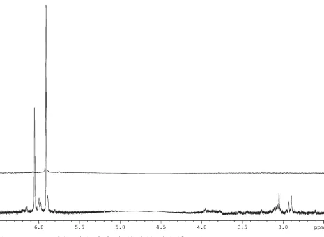
In a previous study of the polyphenol extracted from Fucus Serratus we have detected changes in high-resolution 1H NMR spectra upon addition of oxidizing agents. The signal from the aromatic protons was reduced and a larger variety of methoxy-methyl group protons were seen, which suggests the creation of cross-links during the oxidation process.[11]Since fast cross-linking of mussel protein was associated with a decrease in the adhesion ability, we speculated that a similar mechanism may be involved in alga adhesion and, therefore, the glue’s adherence to polar surfaces may be improved by replacing the oxidized polyphenol with a non-oxidized one. Along this line, we further speculated that the oxidation of the phloroglucinol affects its adhesion properties. This hypoth-esis arises from1H NMR spectra (Figure 3A) which show that upon oxidation, phloroglucinol undergoes chemical changes in a similar manner to the algal polyphenol. The chemical shift of the phloroglucinol’s protons is located in the aromatic zone (dH¼5.5–6). The presence of the hydroxy
group caused a shift in the resonance towards higher magnetic field (dH¼4.5–5). As in the case of Fucus
polyphenol, the signal from the aromatic protons was decreased in the presence of oxidizing agents (Figure 3B). We note that an additional incentive for using a non-oxidized phloroglucinol originates from toxicity con-siderations. It has been shown that oxidative cross-linking of phenolics involves a one-electron oxidation and subsequent deprotonation; the formed phenoxyl radical species can combine with other radicals.[10]In the presence of thiols and molecular oxygen, the reactive phenoxyl radicals can stimulate an oxidative stress and cause oxidative damage to biomolecules.[22]
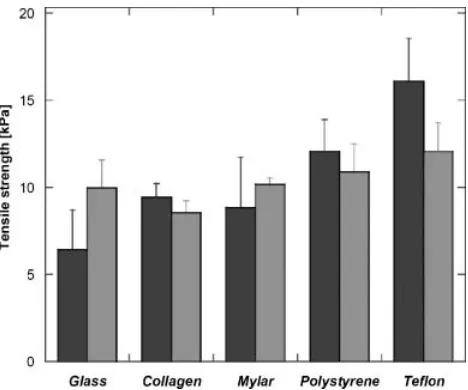
The adhesive strength of oxidized and non-oxidized fucusglue is presented in Figure 4. The differences in the adhesion strengths of the glues are particularly noticeable when the adherents were glass (hydrophilic) and Teflon (hydrophobic). As suspected, the oxidized glue adhered much better to the hydrophobic surface. In the case of Mylar and polystyrene the non-oxidized glue performed slightly better but the differences between the two formulations are less pronounced.
[image:4.595.64.298.502.697.2]Similar results were obtained for the biomimetic glue (Figure 5). As with the algal-born glue, the oxidized formula adhered better to the Teflon while the non-oxidized formula adhered better to the glass and Mylar. Although the non-oxidized glue’s superiority in adhering hydrophilic surfaces is not as clear as it was in theFucus
glue, the non-oxidized formula seems to be better suited for use on hydrated tissues.
In order to examine the feasibility of the biomimetic glue to perform as a tissue adhesive, and to optimize its
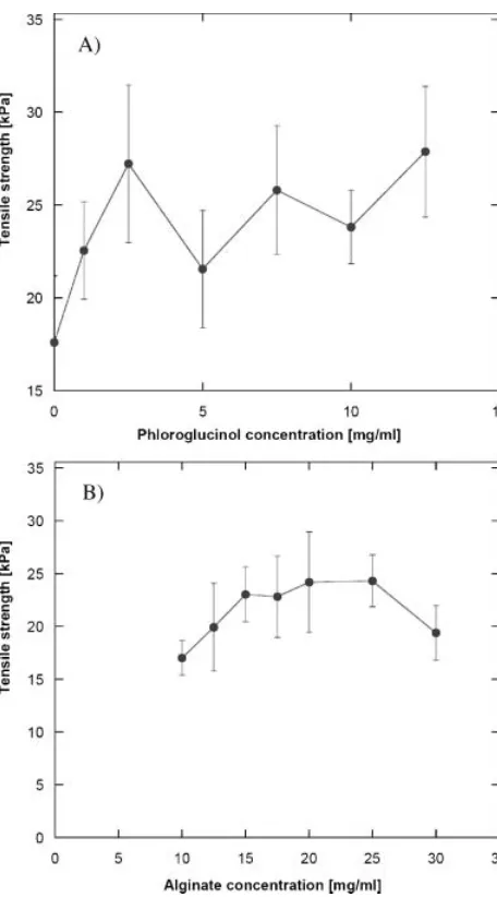
[image:5.595.58.523.73.414.2]composition for maximum performance, the adhesive strengths of various biomimetic glue formulations were studied using a porcine tissue. Figure 6A shows the effect of the phloroglucinol concentration on the glue’s adhesive
[image:5.595.53.532.495.694.2]Figure 3.NMR spectra of phloroglucinol (top) and oxidized phloroglucinol (bottom).
Figure 4.Tensile strength of KI-oxidizedFucusglue (dark gray) and non-oxidizedFucusglue (light gray) to various substrates.
strength. As can be seen, above a concentration of 2 mgmL1there are no major differences in the adhesion strength. It should be noted that phloroglucinol concen-trations higher than 12.5 mgmL1 were not studied because of its limited solubility in water. The influence of the alginate concentration, presented in Figure 6B, shows an optimal range of alginate concentrations (15–25 mgmL1). A possible explanation for the decrease in the adhesion strength at alginate concentrations above 25 mgmL1is that the high viscosity causes formation
of defects at the glue/tissue interface.
The adhesive strength dependence on the calcium ion concentration is shown in Figure 7. As can be seen, there is a distinct increase in the adhesion strength at a calcium concentration of 4103 M. We hypothesized that the
increment in the adhesive strength can be attributed to a sol-gel transition of the glue.
A better understanding of the influence of the calcium concentration was obtained by characterization of the glue’s rheological behavior. Liu et al.[23]characterized the sol–gel transition of alginate by viscoelasticity. The gel point was determined according to the appearance of a power law of the dynamic moduli at the critical gel, as proposed by Winter and Chambon.[24,25] We have employed the same approach in order to determine the glue’s gel point. The relaxation modulus at the gel point can be described as
GðtÞ ¼Stn (1)
whereSis the strength of the network at the gel point and n is an exponent characteristic of the relaxation of the shear stress modulus.
The tangent of the loss angle d can be derived from Equation (1) as
tand¼G00ðvÞ=G0ðvÞ ¼tanðnp=2Þ (2)
The tand independence of v provides a convenient criterion for determining the gel point.
Figure 8 shows the f dependence of tand at several frequencies. The intersecting point gives the values offgel
and n as 0.041 and 0.78, respectively. The fgel value
correlates well with the increased adhesive strength, which indicates that the improved adhesion is a result of a sol-gel transition of the glue. In a similar fashion thefgel
[image:6.595.64.292.74.485.2]andnof Ca/alginate combinations were determined to be 0.072 and 0.67 (Figure 8b). This is in good agreement with
[image:6.595.315.549.76.277.2]Figure 6.Tensile strength required to separate two porcine tissue strips adhered by a biomimetic glue as a function of A) phloro-glucinol concentration, and B) alginate concentration.
values reported in the literature for various Ca/alginate systems.[26]
The lowerfgelvalue obtained for the glue suggests that
the presence of the phloroglucinol decreases the amount of Ca2þions required to coordinate with alginate chains in order to form a three-dimensional network, which indicates possible interactions between the alginate and the phloroglucinol.
Although not extensively studied, polyphenol/polysac-charide interactions are known phenomena.[27–29]
Hydro-phobic interactions are believed to be the major driving force for macromolecular association of this type, with direct hydrogen bonding contributing little binding energy but enhancing specify.[27]Usinga- andb-cyclodextrin as model polysaccharides, Cai et al.[27] proposed that the phenyl ring penetrates into the cyclodextrin cavity. The
molecular weight and the conformational flexibility of the polyphenol, as well as the conformation of the polysaccharide, influence the affinity between the two molecules. Possibly, alginate and phloroglucinol interact in a similar manner, i.e., phloroglucinul units are encapsu-lated within the guluronic acid cavities. However, further investigation is required in order to support this hypothesis. Another support to alginate/phloroglucinol interactions is apparent from SAXS measurements of the biomimetic glue shown in Figure 9. As can be seen, the overall structure of the glue resembles that of the alginate gel. Yet, since the scattering from phloroglucinol by itself was very weak and a scattering curve could not be obtained, the differences between the two curves can be attributed to interactions between the alginate and the phloroglucinol. As in the case of algal-born glue,[11]both the biomimetic glue and alginate gel curves are well fitted by the ‘broken rod linked by a flexible chain’ model:[30,31]
IðqÞ /SðqÞPðqÞ
/ 1
[image:7.595.297.532.74.275.2] [image:7.595.46.279.78.472.2]1þCexpðj2q2Þ |fflfflfflfflfflfflfflfflfflfflfflfflfflffl{zfflfflfflfflfflfflfflfflfflfflfflfflfflffl}
SðqÞ
1 q2
X
i kiq
J1ðqRiÞ qRi
2
|fflfflfflfflfflfflfflfflfflfflfflfflfflfflfflfflffl{zfflfflfflfflfflfflfflfflfflfflfflfflfflfflfflfflffl}
PðqÞ
(3)
Where P(q) is the form factor of cylindrical elements specified by cross-sectional radiiRiand relative weights of
ki. The electrostatic interactions are taken into account by
the structure factor S(q) which assumes a Gaussian-type interaction potential specified by the correlation lengthj.C is a an adjustable parameter that represents the strength
Figure 9.SAXS curve from alginate and calcium (*) and biomi-metic glue (^). The solid lines represent the fit results of the ‘broken rod linked by a flexible chain’ model. Best-fit parameters are summarized in Table 1.
Figure 8.Tandat indicatedvplotted againstffor A) alginates and B) biomimetic glues to determine the gel pointfgeland critical
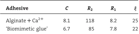
of interaction, which depends on the second virial coefficient and the polymer concentration.[30,31]The best fit parameters for the alginate gel and the biomimetic glue are presented in Table 1.
The analysis of the SAXS data shows that the two samples differ by the correlation lengthj, the adjustable parameter C, and R2, which represents the alginates’
tendency to form large aggregates.[32]A smaller correlation length and adjustable parameter were obtained for the biomimetic glue, thus indicating weaker electrostatic repulsion and lower hydrophilicity compared to the alginate gel.[33]
Conclusion
Natural algal-born polyphenol can be successfully replaced by phloroglucinol, a low-molecular-weight synthetic analogue, without considerable changes in the adhesion properties. Formulations composed of phloroglucinol, alginate, and calcium ions are capable of adhering to a variety of surfaces including porcine tissue. Rheology measurements show that the presence of phloroglucinol lowers the amount of Ca2þ ions required to coordinate with alginate chains in order to form a three-dimensional network, which indicates interactions between the alginate and the phloroglucinol. SAXS experiments sup-port this claim.
Acknowledgements: The financial support by the Yeshaya Horowitz Associationis gratefully acknowledged. This research was supported in part byRubin Scientific and Medical Research Fund. We thank Anna Seredovaya, Irena Shimshilashvily, and
Elinor Joseph for their assistance in performing the adhesion assays, andMaya Davidovich-Pinhasfor her help in obtaining the NMR spectra.
Received: September 19, 2007; Revised: November 5, 2007; Accepted: November 5, 2007; DOI: 10.1002/mabi.200700239
Keywords: adhesion; biomimetic; small angle X-ray scattering (SAXS); water-soluble polymers
[1] M. Yu, T. J. Deming,Macromolecules1998,31,4739. [2] J. H. Waite,Int. J. Adhesion Adhes.1987,7,9. [3] J. H. Waite,Ann. N. Y. Acad. Sci.1999,875,301.
[4] I. Webster, P. J. West, ‘‘Adhesives for Medical Applications’’, in: Polymeric Biomaterials, 2nd edition, S. Dumitrion, Ed., Marcel Dekker, New York 2002, p. 703.
[5] H. Yamamoto, Y. Sakai, K. Ohkawa,Biomacromolecules2000,
1,543.
[6] M. Ishihara, K. Ono, Y. Saito, H. Yura, H. Hattori, T. Matsui, A. Kurita,Int. Congress Ser.2001,1223,251.
[7] K. Huang, B. Lee, P. B. Messersmith,Polym. Prepr. (Am. Chem. Soc., Div. Polym. Chem.)2001,42,147.
[8] V. Vreeland, L. Epstein,Modern Meth. Plant Anal.1996,17,95. [9] V. Vreeland, J. H. Waite, L. Epstein,J. Phycol.1998,34,1. [10] M. Berglin, L. Delage, P. Potin, H. Vilter, H. Elwing,
Biomacro-molecules2004,5,2376.
[11] R. Bitton, M. Ben-Yehuda, M. Davidovich, Y. Balazs, P. Potin, L. Delage, C. Colin, H. Bianco-Peled,Macromol. Biosci.2006,6, 737.
[12] R. Bitton, M. Berglin, H. Elwing, C. Colin, L. Delage, P. Potin, H. Bianco-Peled,Macromol. Biosci.2007,7,1280.
[13] L. Ninan, J. Monahan, R. L. Stroshine, J. J. Wilker, R. Shi,
Biomaterials2003,24,4091.
[14] Z.-Y. Wang, J. W. White, M. Konno, S. Saito, T. Nozawa,
Biopolymers1995,35,227.
[15] I. Shehter-Harkavyk, H. Bianco-Peled,J. Adhes. Sci. Technol. 2004,18,1415.
[16] V. Sklenar, M. Piotto, R. Leppik, V. Saudek,J. Magn. Reson., Ser. A1993,102,241.
[17] M. Piotto, V. Saudek, V. Sklenar,J. Biomol. NMR1992,2,661. [18] H. Stabinger, O. Kratky,Makromol. Chem.1978,179,1655. [19] O. Glatter,J. Appl. Cryst.1977,10,415.
[20] M. Wiegemann,Aquatic Sci.2005,67,166.
[21] M. Yu, J. Hwang, T. J. Deming,J. Am. Chem. Soc.1999,121, 5825.
[22] R. A. Manderville,Can. J. Chem.2005,83,1261. [23] X. Liu, L. Qian, T. Shu, Z. Tong,Polymer2002,44,407. [24] F. Chambon, H. H. Winter,J. Rheol.1987,31,683. [25] H. H. Winter, F. Chambon,J. Rheol.1986,30,367.
[26] L. Lu, X. Liu, L. Dai, Z. Tong,Biomacromolecules2005,6,2150. [27] Y. Cai, S. H. Gaffney, T. H. Lilley, D. Magnolato, R. Martin, C. M. Spencer, E. Haslam,J. Chem. Soc., Perkin Trans. 21990, 2197.
[28] S. H. Gaffney, R. Martin, T. H. Lilley, E. Haslam, D. Magnolato,
J. Chem. Soc., Chem. Commun.1986, 107.
[29] J. P. McManus, K. G. Davis, J. E. Beart, S. H. Gaffney, T. H. Lilley, E. Haslam,J. Chem. Soc., Perkin Trans. 21985, 1429. [30] Y. Yuguchi, H. Urakawa, K. Kajiwara, K. I. Draget, B. T. Stokke,
J. Mol. Struct.2000,554,21.
[31] Y. Yuguchi, M. Mimura, H. Urakawa, S. Kitamura, S. Ohno, K. Kajiwara,Carbohydr. Polym.1996,30,83.
[32] T. Windhues, W. Borchard,Carbohydr. Polym.2003,52,47. [33] M. Shimode, M. Mimura, H. Urakawa, S. Yamanaka,
[image:8.595.62.299.112.170.2]K. Kajiwara,Sen’i Gakkaishi1996,52,301.
Table 1.Fitted parameters obtained from alginate and calcium and the biomimetic glue to the ‘broken rod linked by a flexible chain’ model.
Adhesive C R2 R1 j
AlginateRCa2R 8.1 118 8.2 25