Sex-independent expression of chloride/formate exchanger
Cfex (Slc26a6) in rat pancreas, small intestine, and liver,
and male-dominant expression in kidneys
Dean Karaica
1, Davorka Breljak
1, Jovica Lončar
2, Mila Lovrić
3, Vedran Micek
1,
Ivana Vrhovac Madunić
1, Hrvoje Brzica
1, Carol M. Herak-Kramberger
1, Jana Ivković Dupor
1,
Marija Ljubojević
1, Tvrtko Smital
2, Željka Vogrinc
3, Gerhard Burckhardt
4, Birgitta C. Burckhardt
4,
and Ivan Sabolić
1Molecular Toxicology Unit, Institute for Medical Research and Occupational Health1, Laboratory for Molecular Ecotoxicology, Ruđer Bošković Institute2, Clinical Institute of Laboratory Diagnosis, University Hospital Center3,
Zagreb, Croatia, Institute for Systemic Physiology and Pathophysiology, University Medical Center Göttingen, Göttingen, Germany4
[Received in June 2018; Similarity Check in June 2018; Accepted in November 2018]
Chloride/formate exchanger (CFEX; SLC26A6) mediates oxalate transport in various mammalian organs. Studies in Cfex knockout mice indicated its possible role in development of male-dominant hyperoxaluria and oxalate urolithiasis. Rats provide an important model for studying this pathophysiological condition, but data on Cfex (rCfex) localisation and regulation in their organs are limited. Here we applied the RT-PCR and immunochemical methods to investigate rCfex mRNA and protein expression and regulation by sex hormones in the pancreas, small intestine, liver, and kidneys from intact prepubertal and adult as well as gonadectomised adult rats treated with sex hormones. rCfex cDNA-transfected HEK293 cells were used to confirm the specificity of the commercial anti-CFEX antibody. Various biochemical parameters
were measured in 24-h urine collected in metabolic cages. rCfex mRNA and related protein expression varied in all tested organs. Sex-independent expression of the rCfex protein was detected in pancreatic intercalated ducts (apical domain), small intestinal enterocytes (brush-border membrane; duodenum > jejunum > ileum), and hepatocytes (canalicular membrane). In kidneys, the rCfex protein was immunolocalised to the proximal tubule brush-border with segment-specific pattern (S1=S2<S3), and both rCfex mRNA and protein expression exhibited male-dominant sex differences driven by stimulatory effects of androgens after puberty. However, urinary oxalate excretion was unrelated to renal rCfex protein expression. While the effect of male-dominant expression of rCfex in renal proximal tubules on urine oxalate excretion remains unknown, its expression in the hepatocyte canalicular membrane may be a pathway of oxalate elimination via
bile.
KEY WORDS: anion exchanger; immunolocalisation; nephrolithiasis; oxaluria; RT-PCR; sex differences; urolithiasis
Chloride/formate exchanger (CFEX), alias putative anion transporter 1 (PAT-1), is the 6th member of the SLC26
transporter family (SLC26A6 in humans, Slc26a6 in rodents) and is located in the plasma membrane of various cells, where it operates as an exchanger of chloride, bicarbonate, oxalate, formate, sulphate, and hydroxyl ions, involving pairs of these anions (1-4). In several human and animal organs (pancreas, gastrointestinal tract, kidneys) the transporter largely mediates Cl-/HCO
3- and/or Cl-/oxalate
exchange, thus contributing to Cl- (re)absorption and HCO 3
-or oxalate secretion (4-11).
Studies in human organs have revealed abundant
expression of CFEX mRNA in the kidneys and pancreas, less abundant in the placenta, skeletal muscle, small
Correspondence to: Ivan Sabolić, MD, PhD, Molecular Toxicology Unit, Institute for Medical Research and Occupational Health, Ksaverska cesta 2, 10000 Zagreb, Croatia, E-mail: [email protected]
intestine, heart, and liver, and very low in the lungs, brain, and ovaries (12-15). In mice, mCfex mRNA was detected
in the heart, stomach, small intestine, liver, kidneys, and skeletal muscle and less in the brain, lungs, colon, and testes (16-18). In rats, rCfex mRNA was detected in the uterus (19) and kidneys (10), the pattern in kidneys being cortex
> outer medulla > inner medulla. However, tissue
localisation of the CFEX/Cfex protein is controversial. In the human pancreas, the protein was immunolocalised to the apical surface of duct cells (13), which supports its proposed role as a luminal Cl-/HCO
3- exchanger in
brush-border membrane (BBM) of cortical proximal tubules (11, 16), apical membrane of villous and crypt cells in
duodenum (18), glandular cells in the stomach (22), and the membranes of atrial and ventricular myocytes in the heart (23, 24). These localisations are in accordance with mCfex-mediated anion exchange demonstrated in functional studies (5–7, 18, 22–29). In rats, the functional
rCfex protein was demonstrated in microperfusion experiments, and immunolocalised to the proximal tubule
BBM (10).
Most characteristics of this transporter have been
obtained from studying orthologues in human and mouse
organs; much less is known about other species, including rats [reviewed in (1)]. Human CFEX (759 amino acid
residues) has been described as a variably glycosylated
protein with an apparent Mr ranging from ~84 kDa to ~200 kDa (14, 15, 30). The mouse protein (mCfex; 758 amino acid residues; Mr ~83 kDa) is 79 % homologous
with the human (16). In various mCfex-transfected cell lines
and in mouse organs (pancreas, intestine, kidneys, and heart) the Mr of mCfex protein ranges between ~80 kDa and ~120 kDa (5, 16, 18, 23, 29–32). One study reported Mr of ~90 kDa for a rat Cfex protein (rCfex; 758 amino acid residues; Mr~83 kDa) in isolated renal BBM (10).
Based on the studies in knockout mice, two oxalate
transporters, sulphate anion transporter 1 (Sat-1/Slc26a1) and Cfex have been proposed to play a role in the development of hyperoxalaemia, hyperoxaluria, and oxalate
urolithiasis, a disease which in men occurs 2-3 times more
often than in women (2, 25, 33–36). Possible contribution of Sat-1 in generating male-dominant hyperoxaluria and nephrolithiasis was further suggested by a finding of strong,
androgen hormone-driven sex differences (males > females)
in the expression of Sat-1 protein and associated oxalate transport in the hepatocyte sinusoidal membrane (liver being the major oxalate-producing organ) and renal proximal tubule basolateral membrane (kidney being the
oxalate-secreting organ) (37). However, later studies in
mice (38) and rats (39, 40) could not confirm a significant contribution of hepatic and renal Sat-1 in this male-dominant pathophysiological condition. Other studies using mCfex knockout mice have shown that the Cfex transporter
has a major constitutive role in oxalate secretion across the
small intestine, which limits the existing paracellular net intestinal absorption of dietary oxalate (25, 28, 32). Apart from exhibiting a greatly reduced secretory flux, which led to hyperoxalaemia and hyperoxaluria, Cfex-deficient mice
exhibited strong sex-related oxalate urolithiasis, with 3-4
times higher prevalence in males than in females (25, 34). However, the cause of this phenomenon has not been identified. We do not know if there are sex differences in CFEX/Cfex mRNA and/or protein expression in major
oxalate-handling organs (intestine, liver, and kidneys) of adult humans and animals and if these sex differences affect the oxalate secretory function of these organs in health and
performed in rats, aiming to: a) detect rCfex mRNA expression in pancreas, gastrointestinal tract, kidneys, and liver, b) immunolocalise in detail the rCfex protein in these organs using an antibody with tested specificity, c) detect possible sex-related differences in the expression of rCfex in these organs at mRNA and protein levels, and d) to link the renal expression of rCfex with urine excretion of oxalate
in male and female rats.
MATERIALS AND METHODS
Animals
In this study, we used prepubertal (three weeks old; N=4) and adult (12-13 weeks old; N=3–10 per experimental group) male and female Wistar rats bred at the Institute of Medical Research and Occupational Health (IMROH) in
Zagreb, Croatia. The animals were handled in accordance
with the Directive 2010/63/EU on the protection of animals used for scientific purposes and their use was approved by the Ethics Committee of IMROH [Approval No. 100-21/14-7 of 11-06-2014] and by the Ministry of Agriculture, Zagreb [Approval No. 525-10/0255-15-4 of 08-09-2015]. Before and during experiments, the animals received standard pelleted food (4RF21, Mucedola, Italy) and had free access to tap water.
Gonadectomy and sex hormone treatment
Prepubertal rats were not treated and were sacrificed at
three weeks of age. Adult rats were either untreated,
sham-operated, or gonadectomised and were sacrificed at 12-13 weeks of age. Gonadectomy was performed on six-week
old males by the scrotal route (castration) and females
(ovariectomy) by the dorsal (lumbal) approach under general intraperitoneal anaesthesia (ketamine hydrochloride,
80 mg kg-1 bm + xylazine hydrochloride, 12 mg kg-1 bm).
Sham-operated animals underwent the same procedure, but
without removing gonads. Gonadectomised and
sham-operated rats were left to recover for six to seven weeks before sacrifice. Four castrated animals in each treated group received subcutaneous injections of either sunflower
oil (vehicle) or sex hormones (testosterone enanthate,
oestradiol dipropionate, or progesterone; each (2.5 mg kg-1
bm per day for 14 days) dissolved in the vehicle. Four
control animals received an equivalent amount of oil [0.5 mL kg-1 bm per day for 14 days].
Sacrificed animals were harvested for kidneys, liver, pancreas, and various intestinal segments (duodenum,
jejunum, ileum, cecum, and colon), which were then used
Three days before sacrifice, each rat was placed in its
own metabolic cage with free access to water but without food. Over the next 24 h their urine was collected and its
volume measured together with their body mass. Urine was
centrifuged at 2,500 g for 15 min to remove debris, and creatinine, osmolality, citrate, calcium, and oxalate were
determined in the supernatant as described elsewhere (40).
Antibodies
An affinity purified goat polyclonal antibody for C-terminal peptide of the human CFEX [CFEX-Ab, #sc-26728 (C-17)] with possible cross-reactivity to rat and mouse Cfex proteins, the related immunising peptide (#sc-26728 P), and monoclonal antibody against Na+/K+-ATPase
α1-subunit (Na+/K+-ATPase-Ab, #sc-48345) were
purchased from Santa Cruz Biotechnology, Inc. (Santa Cruz, CA, USA). Monoclonal pan-actin antibody (actin-Ab) was purchased from Millipore (Tamecula, CA, USA, #MAB1501R). The mouse monoclonal 6xHis antibody (6xHis-Ab) was purchased from Clontech Labs/TakaRa Bio USA, Inc. (Mountain View, CA, USA, #631212). Secondary antibodies were purchased from Jackson ImmunoResearch (West Grove, PA, USA) or Kirkegaard and Perry (Gaithersburg, MD, USA) and included the
CY3-labelled donkey anti-goat IgG (DAG-CY3), FITC-CY3-labelled goat anti-mouse IgG (GAM-FITC), and alkaline
phosphatase-labelled rabbit anti-goat (RAG-AP) and goat anti-mouse IgG (GAM-AP).
Chemicals and other material
Anaesthetics ketamine hydrochloride and xylazine
hydrochloride (Narketan and Xylapan, respectively) were purchased from Chassot (Bern, Switzerland), sunflower oil solutions of testosterone enanthate, oestradiol dipropionate, and progesterone from RotexMedica (Trittau, Germany)
and Galenika (Belgrade, Serbia), the Immobilon membrane
from Millipore (Bedford, MA, USA), the markers of protein
molecular mass from Fermentas Int. (Ontario, Canada), and
Superfrost® Plus microscope slides from Thermo Scientific
(Braunschweig, Germany). Other chemicals and reagents
used in this study were of analytical grade and purchased from Sigma (St. Louis, MO, USA) or Fisher Scientific (Pittsburgh, PA, USA).
RNA isolation, cDNA synthesis, end-point and real-time RT-PCR
The abdomen of anaesthetised rats was opened, their
large blood vessels cut and exsanguinated under a stream
of cold water, organs removed, shortly blotted on a filter paper, and ~1 mm thick tissue slices of each kidney (middle transversal slice), liver, pancreas, and intestinal segments
(duodenum, jejunum, ileum, cecum, and colon) cut off, and
immediately immersed in a RNAlater solution (Sigma, St.
extracted using Trizol (Invitrogen, Karlsruhe, Germany)
and subsequently purified using the RNeasy Mini Kit (Qiagen, Venlo, The Netherlands) respecting the manufacturer’s instructions. RNA concentration and purity were estimated spectrophotometrically (BioSpec Nano, Shimadzu, Japan), and the integrity of RNAs was verified under ultraviolet light after agarose gel electrophoresis and ethidium bromide staining. Isolated RNA was stored at -70 °C until further use. First strand cDNA synthesis was performed using the High-Capacity cDNA RT Kit (Applied Biosystems, Foster City, CA, USA) following the manufacturer’s instructions. cDNA was stored at -20 °C
until further use.
The end-point PCR was performed on a 25 µL total volume sample with 100 ng of the first-strand cDNA template, 0.4 µmol L-1 of specific primers, 0.2 mmol L-1 of
dNTP mixture (Applied Biosystems, USA), 1x AmpliTaq®
DNA polymerase buffer (Applied Biosystems), 0.025 U µL-1
of AmpliTaq® DNA polymerase (Applied Biosystems), and
14.9 µL nuclease-free water. The primers for rCfex, rβ-actin, and rHprt1 genes were custom-designed with a
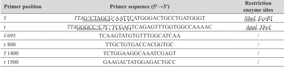
Primer 3 freeware (41, 42), and synthesised and purchased from Invitrogen (Carlsbad, California, USA). Intron over-spanning primers were used to avoid amplification of genomic DNA. The exact sequence of these primers and their predicted size are listed in Table 1.
Reaction conditions during PCR were as follows: initial DNA denaturation at 94 °C for 3 min, DNA denaturation
at 95 °C for 30 sec, annealing at 57 °C for 30 sec, and
elongation at 72 °C for 45 sec. PCR products were separated with electrophoresis in 1.5 % agarose gel stained with
ethidium bromide and visualised under ultraviolet light.
Non-template control reactions, where cDNA was
substituted with nuclease-free water, did not result in
detectable products indicating contamination-free PCR reactions (data not shown). cDNA input variations were controlled in different tissues with two housekeeping genes
(rβ-actin and rHprt1). The number of PCR cycles within the exponential phase of the PCR reaction was 30 cycles
for rCfex in the kidney, liver, and pancreas, 23 cycles for rCfex in the intestinal segments, 25 cycles for rβ-actin in the kidney and liver, 30 cycles for rHprt1 in the pancreas,
and 27 cycles for rHprt1 in the intestinal segments.
Quantitative real-time RT-PCR (qRT-PCR) of independent renal RNA isolated from four intact rats of each sex was performed on a 25 µL total volume sample with 100 ng of the first-strand cDNA template, 12.5 µL of
2x TaqMan® Universal PCR Master Mix, and 1.25 µL of
20x TaqMan® Gene Expression Assays mix (all from
Applied Biosystems). Primers and probes were designed by Applied Biosystems and supplied as a TaqMan® Gene
Expression Assay mix containing a 20x mix of unlabelled PCR forward and reverse primers and a TaqMan® MGB
probe. Assay IDs for the rat genes were Rn01445892_m1
quantified with TaqMan® Assays (Applied Biosystems)
according to the manufacturer’s instructions. Amplification and detection were performed using the 7500 RT-PCR System (Applied Biosystems). Thermal cycling conditions
were 50 °C for 2 min and 95 °C for 10 min, followed by
40 two-step cycles of denaturation (95 °C for 15 sec) and annealing/extension (60 °C for 1 min). NTC reactions were included in each run to check for possible contamination. The housekeeping gene rβ-actin was quantified for endogenous control, and the results were normalised to
these values. mRNA expression was quantified with the comparative CT method using the Relative Quantification Study Software supplied by Applied Biosystems (43). Each reaction was performed in duplicate.
Cloning and expression of rCfex in HEK293 cells
The rCfex gene coding sequence (GeneBank accession
number NP_001137289.1) was amplified from the previously synthesised rat kidney cDNA using forward and reverse primer pairs (Table 1) with Phusion High-Fidelity DNA polymerase (Finnzymes, Vantaa, Finland). The amplicon was cloned into pJET 1.2 vector (ThermoFisher Scientific, Waltham, MA, USA) following the previously described procedure (44). Recombinant plasmid carrying
the cloned rCfex sequence was purified from two transformed (PCR-screened) competent bacterial DH5α cells (clones) (ThermoFisher Scientific) using the plasmid isolation kit QIAprep Spin Miniprep Kit (Qiagen). The rCfex gene sequence was verified by sequencing of both purified plasmids on an ABI PRISM® 3100-Avant Genetic
analyser with the primers listed in Table 2. Using the
introduced sites for NheI, EcoRI, and ApaI, and restriction
enzyme digestion, it was possible to subclone rCfex cDNA from the pJET 1.2 multiple cloning site into pcDNA3.1/
HisC (Invitrogen) expression vector with and without the polyHis tag (Invitrogen). Both pcDNA vectors were used in parallel to transiently transfect HEK293 cells using the PEI (polyethyleneimine) reagent method (45). An additional
HEK293 cell culture was used as negative control, which
underwent the same transfection procedure, except that the plasmid did not contain the rCfex gene sequence (vector-transfected cells). For immunocytochemical studies the
cells were seeded 48 h before transfection in coverslip 24-well plates at cell density of 1.85x10⁵ cells per mL.
Transfection efficiency was evaluated 24 h after transfection on a separate HEK293 cell culture with pcDNA3.1/His/ LacZ plasmid following the LacZ staining protocol (46).
Immunocytochemistry
When transfection efficiency exceeded 70 % (24 h after
transfection), rCfex-transfected and vector-transfected
HEK293 cells on coverslips were fixed in 4 % p-formaldehyde
(PFA) for 30 min, extensively rinsed in PBS, and then stored in PBS at 4 °C until further use. Removed tissue pieces (kidney, liver, pancreas, duodenum, jejunum, ileum, cecum, and colon) were fixed overnight in 4 % PFA, extensively rinsed in PBS, and stored in PBS (+0.02 % NaN3) at 4 °C.
When needed, they were infiltrated with 30 % sucrose (in PBS) overnight, frozen at –25 °C, cut into 4-µm thick
cryosections (cryostat Leica CM 1850, Leica Instruments,
Nussloch, Germany), and placed on Superfrost® Plus
microscope slides (Fischer Scientific).
Optimal steps for immunostaining with fixed HEK293
cells have been described in detail earlier (37, 40) and
included microwave heating in citrate buffer (pH 6),
treatment with Triton X-100 containing buffers, incubation
in primary CFEX-Ab, Na+/K+-ATPase-Ab or 6xHis-Ab
(each diluted in PBS, 1:100) at 4 °C overnight, rinsing in Gene Forward (f) / reverse (r) primers (5ꞌ→3ꞌ) Accession No. Gene Bank Location PCR product size (bp)
rCfex r:AGGTGACCACAAAACCGAAGf:GGCTCTTGGGTGACCTGTTA NM_001143817.1 302-321 639-658 357
rβ-actin r:AGGAAGGAAGGCTGGAAGAGf:GTCGTACCACTGGCATTGTG NM_031144.3 515-534 859-878 364
rHprt1 f:TGCTCGAGATGTCATGAAGG r:AGAGGTCCTTTTCACCAGCA NM_012583.2 213-232 554-573 361
Table 2 Forward (f) and reverse (r) primer sequences used for rCfex gene cloning and sequencing
Primer position Primer sequence (5ꞌ→3ꞌ) enzyme sitesRestriction
f TTAG^CTAGCG^AATTCATGGGACTGCCTGATGGGT NheI, EcoRI
r TTAGGGCC^CTC^TCGAGTCAGAGTTTGGTGGCCAAAAC ApaI, XhoI
f 695 TCAAGTATGTGTTTGGCATCAA /
r 800 TTGCTGTGACCACGGTGC /
f 1400 TCTGGAAGGCAAATCGAGT /
r 1500 GAAGACTATGGAGACTGCC /
antibodies DAG-CY3 or GAM-FITC at room temperature for 1 h, and extensive rinsing in PBS between all incubations. In some experiments, CFEX-Ab was pre-incubated with its immunising peptide (0.5 mg mL-1) at
room temperature for 4 h before immunocytochemistry. In double staining experiments (CFEX-Ab + Na+/K+
-ATPase-Ab), tissue cryosections were first incubated with CFEX-Ab overnight, then with DAG-CY3 for 1 h, then with Na+/
K+-ATPase-Ab overnight and GAM-FITC for 1 h, applying
all other steps as described above. The samples were finally overlaid with fluorescence fading retardant Vectashield
(Vector Laboratories, Burlingame, CA), closed with a
coverslip, and the resulting immunostaining was examined with an Opton III RS fluorescence microscope (Opton
Feintechnik, Oberkochen, Germany). Images were taken
with the computer-guided Spot RT slider digital camera and
software (Diagnostic Instruments, Sterling Heights, MI,
USA). All images were taken using the identical microscope and camera settings and imported into Adobe Photoshop 6.0 (Adobe Systems, San Jose, CA, USA) for processing,
assembling, and labelling.
Pixel intensity of CY3 fluorescence was measured in
the original images with Image-J software, version 1.46r
(NIH, Bethesda, MD, USA). In one cryosection of each organ stained with CFEX-Ab, three to four representative images were captured using the 25x magnification objective.
In each image, six to ten randomly-chosen regions of
interest (ROI) of the staining-positive membranes were
encircled. Fluorescence intensity in ROI was measured,
averaged, and subtracted for background fluorescence intensity of non-stained cytoplasm. The average value of all measurements in respective cryosections was used as
one datum in further calculations.
Western blotting
The removed kidneys were decapsulated and used in toto, or the tissue was manually dissected into cortex and
outer stripe and processed further as separate tissue pools. The main liver lobe and the pancreas were used in toto. The intestinal segments (duodenum, jejunum, ileum, cecum,
and colon) were separated, and their mucosa scraped and processed further. Tissue samples were homogenised, and total cell membranes (TCM; pellet between 6,000 g and 150,000 g) were isolated from each homogenate by
differential centrifugation, as previously described in detail elsewhere (37). Proteins were measured with the Bradford
assay (47).
For PAGE, isolated TCM were prepared in reducing conditions (with 5 % β-mercaptoethanol) at 65 °C for 15 min, their proteins (80 μg lane-1) separated through 10 %
SDS-PAGE mini gels, and wet-transferred by electrophoresis to an Immobilon membrane (Millipore, Bedford, MA, USA)
as described in detail elsewhere (37). The Immobilon membrane was then incubated in the buffer containing
rinsed, incubated in RAG-AP (1:500) or GAM-AP (1:1000) at room temperature for 1 hour, and rinsed again. Protein bands were visualised at pH 9 via alkaline phosphatase activity-mediated reaction using the BCP/NBT method (37). To verify the specificity of CFEX-Ab for its immunising peptide, the antibody was pre-incubated with the peptide
(0.5 mg mL-1) at room temperature for 4 h and used in the
blotting assay. Labelled protein bands were evaluated by densitometry using Image-J software. The broadest protein
band of rCfex or actin was encircled, an equal area was then
applied to other related bands on the same membrane, and the area density of each band was expressed in arbitrary units, relative to the broadest band density (=1 unit).
Statistical analysis
Immunochemical data represent the findings for three to four animals from each experimental group. RT-PCR was done with cDNA obtained from independent RNA preparations from individual organs of three to four animals from each experimental group. Data shown as mean±standard
deviation (SD) were statistically analysed with either Student’s t-test or ANOVA/Duncan test at 5 % level of significance using Statistica 10 (StatSoft, Tulsa, OK, USA).
RESULTS
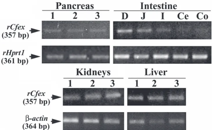
Expression of rCfex mRNA in rat organs
Figure 1 shows that the expression of rCfex mRNA measured at 30 PCR cycles varied between the pancreatic, renal, and liver tissues. In the intestine, its expression was segment-dependent. Measured at 23 PCR cycles, the expression pattern was as follows: duodenum > jejunum >
ileum. In the stomach (not shown), cecum, and colon it was
negligible, and barely increased at 30 PCR cycles (data not
shown).
Immunochemical characterisation of CFEX-Ab in
rCfex-transfected HEK293 cells
In preliminary immunocytochemical experiments, we have tested the CFEX-Ab in cryosections and paraffin
sections of human, mouse, and rat kidneys and liver by
applying different antigen-unmasking techniques (48), and
found immunoreactivity only in rat organs (data not shown).
To verify the specificity and efficiency of this antibody in detecting the rCfex protein, CFEX-Ab was tested in vector-
and rCfex-transfected HEK293 cells. Figure 2A shows that
CFEX-Ab stained the plasma membrane of rCfex
-transfected, but not of vector-transfected cells, whereas Na+/
K+-ATPase-Ab stained the plasma membrane of both kinds
of transfected cells. In addition, rCfex-transfected cells were
immunocytochemical analysis. In order to obtain a visible
rCfex-related protein band for SDS-PAGE and Western blotting, we isolated the plasma membranes from the rCfex -transfected and vector--transfected cells and incubated them
with or without reducing agent (5 % β-mercaptoethanol) at different temperatures (37 °C for 30 min, 65 °C for 15 min,
95 °C for 5 min). However, for unknown reason, we
obtained none (data not shown), as opposed to rat tissues. Expression of the rCfex protein in pancreas
In the Western blots of the TCM isolated from the pancreas of male rats, CFEX-Ab labelled a single protein band of ~120 kDa (Figure 3A, -P), which was absent when the antibody was blocked with the immunising peptide (+P).
Similarly, CFEX-Ab stained the luminal domain of acini
(Fig. 3B, -P, arrowhead) in tissue cryosections, but there
was no staining when the antibody was blocked with the
immunising peptide (+P) (Figure 3B). In double staining experiments (Figures 3C, X, Y, and W), CFEX-Ab stained
the luminal domain of acini in red-yellow (arrows), whereas
Na+/K+-ATPase-Ab stained the cell basolateral membrane
in green (arrowheads). CFEX-Ab also stained the luminal membrane of initial (intercalated) ducts (Y, double
arrowhead), but not the Langerhans’ islet cells (W, LI) and large pancreatic ducts (W, asterisk). Because of the low
image resolution we could not discern if the luminal staining
in the acini was present only in the membranes of initial
(intercalated) ducts or also in the luminal membrane of acinar cells. However, numerous discrete red-stained organelles inside the acinar cells (Figures 3C, Y, and inset
in Figure 3F, Male) indicate that rCfex may also be present inside the acinar cells. In the Western blots of TCM with
CFEX-Ab and actin-Ab (Figure 3D) and in the densitometric
evaluation of the protein bands (Figure 3E) the related band density of each protein was similar in both sexes. This result was confirmed in tissue cryosections from male and female
organs, where CFEX-Ab-related immunostaining in
pancreatic ducts was similar in both sexes (Figure 3F).
Expression of the rCfex protein in intestine
In the Western blots of TCM isolated from duodenal mucosa, CFEX-Ab labelled a protein band of ~120 kDa (Figure 4A, -P) but the staining was absent when the antibody was blocked with the immunising peptide (+P).
The same was true for the cryosections of the duodenal tissue; CFEX-Ab strongly stained the BBM of enterocytes
(Figure 4B, -P), but the staining was absent when the immunising peptide-blocked antibody was used (+P). In Western blots of TCM from various intestinal segments, CFEX-Ab and actin-Ab labelled the rCfex protein band in
the membranes from the duodenum and jejunum (duodenum > jejunum), but not in ileum, cecum, and colon (Figures 4C
and D). Accordingly, in tissue cryosections the rCfex protein
was immunolocalised to the BBM of enterocytes in the duodenum and jejunum (duodenum > jejunum) but not in the ileum, cecum, and colon (Figures 4E and F). The staining intensity in the duodenal villi increased about six
times towards the tip compared to the basal domain (Figures
4G and H). The staining distribution and intensity was similar between males and females (data not shown).
Segmental, zonal, and sex differences in kidney rCfex
expression
In Western blots of the TCM from the whole male kidneys, CFEX-Ab labelled a single protein band of Figure 1rCfex mRNA expression in rat organs as determined by end-point RT-PCR. Housekeeping genes rHprt1 and β-actin were
~120 kDa, which was absent when the same antibody was pre-incubated with its immunising peptide (Figure 5A). In
tissue cryosections, the antibody staining was weak in the
BBM of the S1/S2 cortex segments of the proximal tubules,
stronger in the S3 segments of medullary rays, and the
strongest in the S3 segments of the outer stripe (-P) (Figures
5B and C), exhibiting in this way zonal differences (cortex
< outer stripe). Other nephron segments remained unstained. No staining was observed with the antibody pre-incubated with its immunising peptide (Figure 5B, +P).
Zonal differences in immunostaining were confirmed in the Western blots of isolated TCM from the kidney cortex and outer stripe; the rCfex-related protein band of ~120 kDa was stronger in the outer stripe (Figure 5D). Its density was
7.3 times higher than in the cortex, whereas the density of
the actin band (where the housekeeping protein was used
as loading control) was similar in TCM from both zones (Figure 5E).
The expression of rCfex in the rat kidneys also exhibited sex differences (Figure 6). The relative expression of rCfex mRNA in the whole kidney, established with qRT-PCR, was ~20 % higher in males than in females (P<0.050) (Figure 6A). This observation was confirmed on the protein
level in TCM isolated from the whole male and female
kidneys; the rCfex-related protein band (~120 kDa) was
~50 % stronger in males than in females, whereas the actin
band had similar density in both sexes (Figure 6B and C).
Sex-dependent expression of the rCfex protein was also confirmed by immunostaining the kidney cryosections
(Figure 6D); CFEX-Ab-related staining intensity in the
cortical and outer stripe tubules was ~30 % and ~4.3 times
stronger in males than in females (Figure 6E).
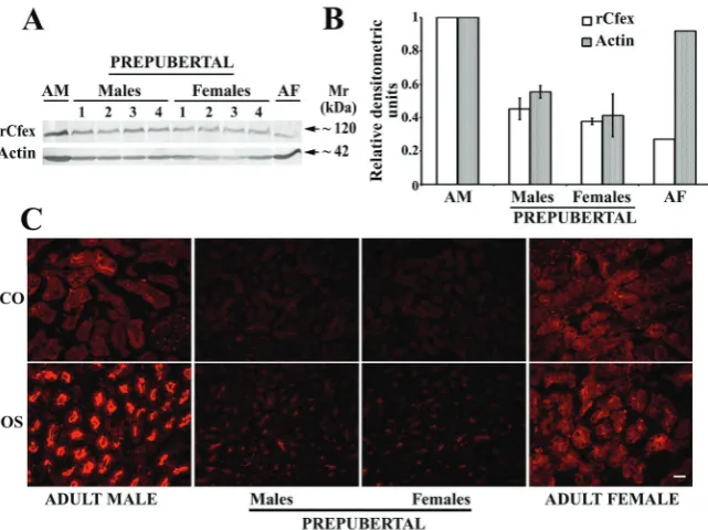
Renal expression of the rCfex protein in prepubertal rats Western blotting with CFEX-Ab and actin-Ab of the
whole kidney TCM from four male and four female
prepubertal rats showed ~60 % lower rCfex protein band density than in the adult male and ~100 % higher than in the adult female rats, one of each, that served as comparators.
However, we found no sex differences. The density of the
actin protein band was also similar between the sexes, but ~55 % lower than in adult animals (Figures 7A and B).
Western blotting was confirmed by the CFEX-Ab-related immunostaining in the kidney cortex and outer stripe in adult and prepubertal male and female rats (Figure 7C).
In adult animals it exhibited male-dominant sex differences
(stronger staining in the outer stripe), and in prepubertal
animals the immunostaining in both tissue zones was weak and similar between the sexes.
Figure 2 Testing the specificity of anti-CFEX antibody. A Immunocytochemical testing of CFEX-Ab and Na+/K+-ATPase-Ab in vector-
(-rCfex cDNA) and rCfex-transfected (+rCfex cDNA) HEK293 cells. Arrows denote immunoreactivity in the plasma membrane.
B Immunostaining of rCfex-transfected cells with CFEX-Ab (-Peptide) and its absence when blocked with the immunising peptide
on the rCfex protein expression in kidneys
In male rats, the rCfex protein band density in Western blots decreased 60 % after castration, whereas actin band
density remained unchanged (Figures 8A and B). In tissue cryosections, the CFEX-Ab-related immunostaining was not significantly affected by castration in the cortical
tubules, but in the outer stripe tubules staining intensity decreased ~65 % (Figures 8C and D). In female animals, however, the density of protein bands in the whole kidney TCM, and the staining intensity of proximal tubules in tissue
cryosections remained unchanged after ovariectomy (data not shown), which clearly suggests that female sex
hormones are not responsible for the observed phenomenon. To confirm male sex hormones as being responsible for sex differences in the renal rCfex expression, castrated males were treated with sunflower oil or oil-solutions of testosterone, oestradiol, or progesterone for two weeks (Figure 9). Western blotting with CFEX-Ab (Figure 9A)
and rCfex-related band densitometry (Figure 9B) showed
that Cfex protein expression was upregulated by testosterone,
effect. CFEX-Ab-related immunostaining in the cortical
tubules showed no changes, but in the outer stripe,
testosterone treatment resulted in stronger staining, while
oestradiol and progesterone had no effect (Figure 9C).
Urine parameters in sham-operated and gonadectomised male and female rats
In order to estimate if the renal expression of Cfex in sham-operated and gonadectomised male and female rats
has any relation to urine excretion of oxalate and a few
other parameters known to stimulate (calcium; Ca) or inhibit (citrate) formation of oxalate stones (2), these parameters
were determined in urine collected in metabolic cages for
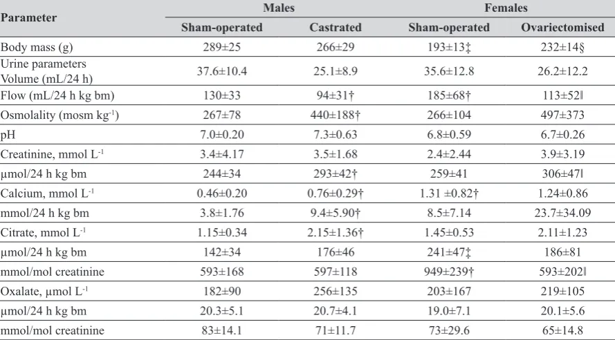
24 h. Table 3 shows that sham-operated females had a significantly lower body mass (P<0.001) and higher urine Ca (P<0.001) and citrate excretion per day (P<0.002) than sham-operated males, when expressed per creatinine.
Castrated males had lower urine volume excretion
(P<0.002) and higher urine osmolality (P<0.021), creatinine (P<0.012), and Ca (P<0.021) than sham-operated males.
Figure 3 Immunolocalisation and sex-independent expression of the rCfex protein in the pancreas. A Western blot of total cell membranes
(TCM) from the whole male pancreas with CFEX-Ab (-P) and with CFEX-Ab blocked by the immunising peptide (+P). Each lane contained
80 µg of protein. Mr – relative molecular mass. B Tissue cryosection staining with CFEX-Ab (-P), and with CFEX-Ab blocked by its immunising
peptide (+P). The arrows point to the staining of the luminal domain in the acini. C (X, Y, W) Double staining with CFEX-Ab (arrows) and
Na+/K+-ATPase-Ab (arrowheads). Double arrowhead points to the initial (intercalated) duct. LI – Langerhans islet; * – larger (intralobular)
pancreatic duct. D Western blots of TCM from male and female rat pancreata with CFEX-Ab and actin-Ab. Each lane contained 80 µg of
protein from membrane preparations from different rats. E Densitometry (means ±SD; n=3 in each bar) of the protein bands shown in D.
Differences in rCfex or actin between males and females were not significant (P>0.05). F CFEX-Ab immunostaining in cryosections of male
Ovariectomised females had higher body mass (P<0.001) and creatinine excretion (P<0.030), but lower urine flow (P<0.019) and citrate excretion (P=0.002), when expressed per creatinine than sham-operated females. However, the
concentration of oxalate in urine, as well as the daily
excretion of oxalate were similar in sham-operated animals
of either sex and were not affected by gonadectomy.
rCfex protein expression in the liver
In the liver of adult rats, the CFEX-Ab-related,
immunising peptide-blockable immunoreactivity was
localised in the membrane of bile canaliculi (Figure 10A,
-P, arrows). This was confirmed by double staining with CFEX-Ab and Na+/K+-ATPase-Ab, where the rCfex protein
was stained red in the bile canaliculi (arrows), while the
Na+/K+-ATPase α-subunit protein was stained green in the
Figure 4 Expression of the rCfex protein in the intestine. A Western blot of total cell membranes (TCM) from duodenal mucosa with
CFEX-Ab (-P) and with the same antibody preincubated with its immunising peptide (+P). Each lane contained 80 µg protein. Mr,
relative molecular mass. B Immunostaining in duodenum with CFEX-Ab (-P) and with the antibody preincubated with its immunising
peptide (+P). C Representative (out of 3 similar) Western blots of TCM from the intestinal segments with CFEX-Ab and actin-Ab. Du,
duodenum; J, jejunum; I, ileum; Ce, cecum; Co, colon. Each lane contained 80 µg protein. D Densitometric evaluation of the rCfex-related protein bands shown in B, collected from 3 independent experiments. Shown are means ±SD (n=3 in each bar). *J vs. Du,
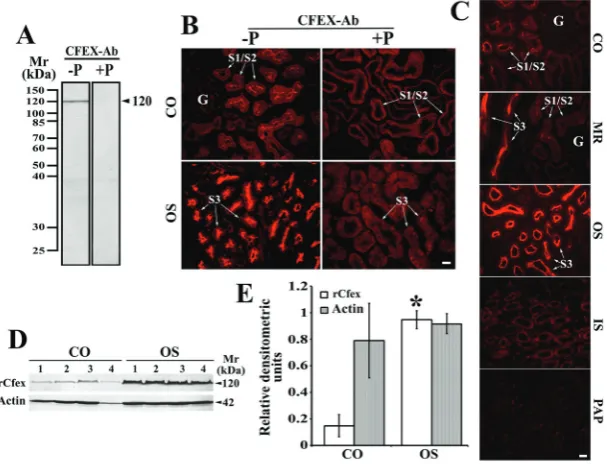
Figure 5 Immunolocalisation of rCfex in male kidneys; zonal and segmental differences. A Western blot of total cell membranes (TCM)
from the whole kidney with CFEX-Ab (-P) and with the same antibody preincubated with its immunising peptide (+P). Each lane
contained 80 µg protein. Mr, relative molecular mass. B and C Zonal and segmental differences in immunostaining with CFEX-Ab
(-P) and with the same, peptide-blocked antibody (+P). CO, cortex; MR, medullary rays; OS, outer stripe; IS, inner stripe; PAP, papilla (inner medulla); G, glomeruli. S1/S2, proximal tubule convoluted segments. S3, proximal tubule straight segment. Bar (for all images in the respective panel), 20 µm. Data are representative for similar findings in tissue cryosections from 3 male rats. D Western blots
of TCM from the kidney cortex and outer stripe with CFEX-Ab and actin-Ab. Actin (~42 kDa protein) was used as a loading control. Each lane contained 80 µg protein from different membrane preparation. E Densitometric evaluation of the protein bands shown in D. Bars show mean ±SD (n=4 in each group). *vs. rCfex in CO, P<0.001
Figure 6 Sex differences in the expression of rCfex mRNA and protein in the kidneys. A qRT-PCR. Relative expression of rCfex mRNA
from the whole male and female kidneys. Shown are means ±SD (n=4 in each group; *vs. Males, P<0.05). B Western blots of TCM,
isolated from the whole male and female kidneys, with CFEX-Ab and actin-Ab. Each lane contained 80 µg protein from different
membrane preparation. C Densitometric evaluation of the protein bands shown in B. Bars show mean ±SD (n=4 in each group). *vs.
rCfex in Males, P<0.008). D Representative (for four animals of each sex), CFEX-Ab-related immunostaining in proximal tubules in the cortex (CO) and outer stripe (OS) of male and female kidneys. G, glomerulus. S1/S2 and S3, respective proximal tubule segments. Bar (for all images), 20 µm. E Intensity of CFEX-Ab-related staining in the proximal tubule brush-border membrane of cortical (CO)
Immunoblotting of TCM isolated from the liver tissue with
CFEX-Ab labelled a protein band of ~120 kDa, which was blocked by the immunising peptide (Figure 10B).
Furthermore, blots of isolated TCM from male and female livers with CFEX-Ab and actin-Ab (Figure 10C) and
densitometry of their protein bands (Figure 10D) showed no sex differences in the abundance. This was supported
by similar CFEX-Ab-related staining intensity in tissue cryosections from both sexes (Figure 10E).
DISCUSSION
In this study we established the expression and
distribution of rCfex mRNA and the rCfex protein in the pancreas, gastrointestinal tract, kidneys, and liver of male and female rats. End-point RT-PCR detected rCfex mRNA in the tissue of all tested organs, which confirms earlier findings in humans (12–15), mice (16–18), and rats (10).
In our initial immunocytochemical studies in the rCfex
-transfected cells, the CFEX-Ab was found efficient in immunostaining the rCfex protein in the cell plasma
membranes. It also exhibited good efficiency in immunostaining rCfex in tissue cryosections, whereas in the membranes isolated from the investigated rat organs it
labelled a single, immunising peptide-blockable protein band of ~120 kDa. This size of the Cfex protein falls within
the range detected in various mammalian tissues and cell
expression systems (~84 kDa to ~200 kDa) and is attributed to variable posttranslational modifications such as
glycosylation (5, 14–16, 18, 23, 29–32). In various
band and CFEX-ab-related staining distribution and
intensity in the respective organs matched, indicating their protein-related origin. Sex differences (males > females) in the expression of both rCfex mRNA and the protein were
detected only in the kidneys.
Our finding of the rCfex (PAT-1) protein in the rat pancreas confirms previous reports for human and animal pancreas (4, 8, 9, 13, 20, 21). Studies on microperfused pancreatic ducts and ductal cell lines, suggest that Cfex (regulated by CFTR) is the major apical Cl-/HCO
3
-exchanger in mammalian pancreatic ducts and that it
contributes to the secretion of HCO3--rich fluid (4, 6, 21,
49). However, in contrast to previous immunocytochemical and functional findings of Cfex in the luminal membrane of interlobular duct cells of human (13), guinea pig (9), and mouse (6) pancreas, our study demonstrated: a) no
CFEX-Ab-related immunoreactivity in the cells of intralobular and larger ducts, b) weak immunoreactivity in the cell luminal domain of initial (intercalated) ducts, and c) strong immunoreactivity in the luminal domain of acinar cells. In many cells we also observed numerous weakly
rCfex-positive intracellular organelles, which suggests that the level of the rCfex protein in the apical cell membrane may be controlled by vesicle recycling and possibly regulated
by endo- and exocytosis.
As detected by qRT-PCR, rCfex mRNA in various intestinal segments revealed the highest expression was in
the duodenum, lower in the jejunum, and even lower in the ileum, whereas in other segments (stomach, cecum, and
proximal colon) the expression was negligible. These results
Table 3 Body mass, urine excretion, and urine biochemical parameters in the sham-operated and gonadectomised male and female rats
Parameter Males Females
Sham-operated Castrated Sham-operated Ovariectomised
Body mass (g) 289±25 266±29 193±13‡ 232±14§
Urine parameters
Volume (mL/24 h) 37.6±10.4 25.1±8.9 35.6±12.8 26.2±12.2
Flow (mL/24 hkg bm) 130±33 94±31† 185±68† 113±52‖
Osmolality (mosm kg-1) 267±78 440±188† 266±104 497±373
pH 7.0±0.20 7.3±0.63 6.8±0.59 6.7±0.26
Creatinine, mmol L-1 3.4±4.17 3.5±1.68 2.4±2.44 3.9±3.19
µmol/24 hkg bm 244±34 293±42† 259±41 306±47‖
Calcium, mmol L-1 0.46±0.20 0.76±0.29† 1.31 ±0.82† 1.24±0.86
mmol/24 h kg bm 3.8±1.76 9.4±5.90† 8.5±7.14 23.7±34.09
Citrate, mmol L-1 1.15±0.34 2.15±1.36† 1.45±0.53 2.11±1.23
µmol/24 hkg bm 142±34 176±46 241±47‡ 186±81
mmol/mol creatinine 593±168 597±118 949±239† 593±202‖
Oxalate, µmol L-1 182±90 256±135 203±167 219±105
µmol/24 h kg bm 20.3±5.1 20.7±4.1 19.0±7.1 20.1±5.6
mmol/mol creatinine 83±14.1 71±11.7 73±29.6 65±14.8
The results are presented as means ±SD of body mass and biochemical parameters in 24-h urine collected from 9 to 10 animals in each experimental group. †P=0.001-0.047 vs. sham-operated males; ‡P<0.001 vs. sham-operated males; §P<0.001 vs. sham-operated
Figure 7 Immunolocalisation of the rCfex protein in the kidneys of prepubertal male and female rats; comparison with adult animals. A Western blots of total cell membranes (80 µg protein/lane), isolated from the whole kidneys from 4 male and 4 female prepubertal
rats, and from one adult male (AM) and female (AF) rat, blotted with CFEX-Ab and actin-Ab. B Densitometric evaluation of the protein
bands shown in A. Data for adult rats arefrom a single animal, whereas the data for prepubertal animals are means ±SD (n=4 in each
group). Statistics for the rCfex and actin data in prepubertal rats: Males vs. Females, N.S. (P>0.05). C Representative (out of 3 similar)
rCfex-Ab-related immunostaining in the kidney cortex (CO) and outer stripe (OS) in adult and prepubertal male and female rats. Bar (for all images), 20 µm
differ from the Northern hybridisation data in mouse intestine, showing this sequence of expression: duodenum=jejunum > ileum > proximal colon > cecum >
distal colon (17, 18). Our immunochemical data of the
intestinal rCfex matched the mRNA expression, showing the rCfex protein in the BBM of duodenal and jejunal
enterocytes (duodenum > jejunum). In other segments,
including the stomach, the rCfex protein could not been visualised. In the mouse stomach, the mCfex protein was previously detected with a different antibody and found
colocalised with gastric H+-K+-ATPase in parietalcells (22).
This suggests that intestinal Cfex mRNA and protein expression implies species differences between rats and
mice.
The role of Cfex in the murine small intestine has been
functionally defined in studies on wild type and mCfex
knockout mice, in Cfex-transfected cell lines and in Xenopus
oocytes (5, 7, 18, 26, 28, 29, 31). Two of them also described
species differences in kinetic properties between the mouse and human Cfex/CFEX proteins. The mCfex-mediated Cl-/
oxalate2- exchange was electrogenic with high affinity for
Cl-, whereas the human exchanger was electroneutral (2Cl-/
oxalate2-), with low affinity for Cl- (5, 50). Previously
published data indicates that duodenal and ileal Cfex could mediate absorption of luminal Cl- or SO
42- in exchange for
secreted HCO3- or oxalate (7, 26, 28, 29). With the latter
function, the exchanger should significantly contribute in eliminating oxalate from the body (26, 34), thus preventing
the development of hyperoxalemia, which can lead to subsequent hyperoxaluria and Ca-oxalate nephrolithiasis,
as has been demonstrated in the mCfex knockout mouse model (32, 34, 51).
The segment-related expression of Cfex protein along the renal proximal tubule (S1/S2 in the cortex << S3 in medullary rays and outer stripe) and sex differences in the expression of Cfex mRNA and protein (males > females) found in our study in rats have not been previously reported for other species. In a study using northern hybridisation
(10), rCfex/PAT-1 mRNA was highly expressed in the
kidney cortex and weakly in the outer and inner medulla, whereas immunocytochemical staining localised the rCfex
protein in the proximal tubule BBM. The mCfex protein was also stained in the BBM of the proximal tubules (11,
16), but in humans, CFEX was detected in the distal
segments of the proximal tubules and in several more distal nephron parts (12), which all points to species differences in the nephron localisation of this transporter between rodents and humans. In various transport studies, performed in microperfused proximal tubules from wild type and mCfex knockout mice (10, 11, 25, 29), isolated BBM (10, 11, 25, 52), and mCfex- and CFEX-transfected Xenopus
oocytes (5, 50), the renal Cfex/CFEX was shown to function
predominantly as Cl-/HCO
3- or Cl-/oxalate exchanger, thus
serving in reabsorption of Cl- and secretion of HCO 3- and
oxalate. As shown in the mouse kidney, a strongly
affinity of renal mCfex for oxalate than for HCO3- (25, 52)
indicate Cl-/oxalate exchange as the primary role of the
renal transporter.
Two phenomena related to our findings of male-dominant testosterone-induced rCfex protein expression in the kidney deserve particular attention. One is the segment-dependent abundance of rCfex, which is in S3 much higher than in S1/S2. Previous studies assumed that oxalate elimination via proximal tubule cells was mediated by the concerted action of the basolateral Sat-1, which pumps
SO42- out and oxalate into the cell, and luminal Cfex, which
transports oxalate into the tubule lumen in exchange for
Cl- [reviewed in (1, 2, 35)]. In the rat kidney, this pattern
may be valid for S1/S2 segments, which exhibit
male-dominant expression of both rSat-1 (37, 40) and rCfex (this
study), but is not entirely true for the S3 segments, where
apical rCfex expression is higher (this study) and basolateral
rSat-1 lower (37, 53). It remains for the future studies to
resolve the physiological role of the male-dominant rCfex expression in the S3 segment in secretion or reabsorption
of oxalate and other related anions.
The other phenomenon are strong male-dominant sex differences in rCfex expression along the entire proximal
tubule, which may be relevant to the well-known
male-dominant prevalence of urolithiasis in humans and animals [reviewed in (1, 2, 35)]. According to the urine parameters
in Table 3, similar urine concentrations and 24-h excretion
of oxalate in the sham-operated and gonadectomised male and female rats suggests no apparent relation between oxalate excretion and renal rCfex expression, which is in contradiction with some previously reported studies (26,
29, 34).
Male-dominant expression of renal rCfex in our study may play a role in hyperoxalaemic conditions such as chronic renal failure (54, 55), inflammatory bowel disease,
small bowel resection (28), and urolithiasis induced by ethylene glycol (EG) in rats (40). Several studies have
shown that male rats have a predilection toward kidney
stones and that testosterone enhances while oestrogen inhibits urinary stone formation in normal and EG-treated
animals (56–58), but the expression and the role of Cfex
was not investigated in those studies. However, higher excretion of citrate in healthy females than males [(40) and
this study] may point to a major protective mechanism
against the formation of Ca-oxalate crystals. An in vitro
study by Ohana et al. (32) showed that mCfex can interact with and inhibit the activity of sodium-dicarboxylate
cotransporter mNaDC-1/Slc13a2 located in the proximal tubule BBM, which reabsorbs filtrated citrate. In vivo, such inhibition could result in citrate retention in the tubular fluid, which would bind Ca2+ and ensure safe oxalate
elimination through urine (32). The 24-h urine in our
previous (40) and present study consistently showed significant sex differences (males<females) in the excreted citrate but not oxalate. These differences disappeared with
hormones regulate (stimulate) citrate excretion by
modulating the activity and/or expression of NaDC-1, while
the Cfex remains unaffected. EG-induced urolithiasis,
hyperoxaluria, and hypocitraturia in rats (40) generated
conditions for the formation of Ca-oxalate crystals in male but not female animals. Since male rats exhibit higher renal
expression of the rCfex protein than females (this study), it is possible that this and several other conditions with hyperoxaluria and Ca-oxalate urolithiasis may involve an interaction between Cfex and NaDC-1, such as inflammatory
bowel disease and conditions resulting from small bowel resection (28, 35).
Earlier Northern blotting and/or RT-PCR studies have
detected CFEX/Cfex mRNA in the liver of humans (13–15) and mice (16), but no CFEX/Cfex protein has been localised to hepatocytes in these species so far. Following up on our preliminary report in the rat liver of a few years ago (2), we have now confirmed its exclusive presence in the bile canaliculi, where it may play a role in elimination of oxalate from hepatocytes into bile. Judging by the CFEX-Ab-related immunostaining intensity its expression was sex-independent. Liver is the organ with the largest capacity of producing oxalate from various precursors [reviewed in (2, 35)]. This is particularly clear in hepatectomised rats, whose production of oxalate from the precursor glycolate dropped more than 80 % (59). Being an end-product of metabolism
and toxic for the cell, most of oxalate is removed from the
hepatocytes via Sat-1-mediated SO42-/oxalate exchange
located in the sinusoidal membrane [reviewed in (1, 2, 35)]. In an experimental rat model of EG-induced hyperoxaluria, the expression of rSat-1 mRNA and rSat-1 protein in the
liver (and kidneys) exhibited only limited changes (39, 40), whereas in Sat-1 knockout mice with hyperoxalaemia, hyperoxaluria, and nephrolithiasis (33) the mode of oxalate elimination from hepatocytes has not been explained. Previous transport studies by Meier et al. (60) in canalicular plasma membrane vesicles isolated from the rat liver showed the presence of HCO3-/SO42-, HCO3-/oxalate and
SO42-/oxalate exchange. According to Boyer (61), HCO 3- is
secreted into the bile in exchange for Cl- by anion exchanger
2 (AE2/SLC4A2) located in the bile canalicular membrane, and the out-to-in gradient of bicarbonate may drive SO4
2-and oxalate exit by another exchanger in the same
membrane, possibly Cfex. We therefore propose that oxalate produced in the liver can leave the hepatocyte via sinusoidal
Sat-1 (2, 37) and canalicular Cfex. The Cfex-mediated
excretion of oxalate may be low under physiological conditions and may increase in case of impaired oxalate
clearance, as shown in rats with kidney failure (54). For
Figure 8 Effect of gonadectomy on the rCfex protein expression in male kidneys. A Western blots of the whole kidney total cell membranes, isolated from 4 sham-operated and 4 castrated animals, with CFEX-Ab and actin-Ab. Each lane contained 80 µg protein. B Densitometric evaluation of the protein bands shown in A. Shown are means ±SD (n=4 in each bar). *vs. rCfex in sham-operated
males, P<0.002. The actin band had similar density in the membranes from both groups (P>0.05). C rCfex-Ab-related immunostaining
in the cortical (CO) and outer stripe (OS) proximal tubule segments (S1/S2 and S3) from sham-operated and castrated animals. G, glomeruli. Bar (for all images), 20 µm. Data are representative for 3 animals in each group. D Staining intensity in the brush-border
membrane of cortical (CO, S1/S2) and outer stripe (OS, S3) proximal tubule segments. Shown are relative fluorescence intensities
(means ±SD; n=3 in each bar). Statistics: In CO, a vs. c, N.S. (P>0.05); In OS, b vs. d, P<0.005
Figure 9 Effect of sex hormone treatment in castrated rats on renal rCfex protein expression. A Western blots (representative for 2
different sets of membrane preparations) of total cell membranes, isolated from the outer stripe of sham-operated and castrated rats
treated with sunflower oil or sex hormones (Test, testosterone; Estr, oestrogen; Prog, progesterone) for two weeks, with CFEX-Ab and actin-Ab. Each lane contained 80 µg protein. B Densitometric evaluation of the bands shown in A. Shown are means ±SD of the data
collected from two independent experiments (n=4 in each group); Statistics: a vs. b, d or e; b vs. c, and c vs. d or e, P<0.001; Other
relationships, N.S. (P>0.05). The actin protein band was not changed by the treatments (A). C The CFEX-Ab-related immunostaining
In conclusion, by applying the commercial polyclonal anti-CFEX antibody (sc-26728) in rat tissues, the putative rCfex protein was detected in a few known localizations (duodenal and jejunal BBM, luminal domain of pancreatic intercalated ducts, BBM in proximal tubules), and a few previously unknown localizations (bile canalicular membrane in hepatocytes, and apical membrane and intracellular organelles in pancreatic acinar cells). The expression of rCfex was found sex-dependent only in kidneys, exhibiting a strong male-dominant expression driven by androgen stimulation after the puberty. The described localizations may be important in handling oxalate and related anions in physiological and pathophysiological conditions. However, while the applied
antibody worked well by immunocytochemistry in tissue
cryosection, by immunoblotting it labelled no protein in the
membranes isolated from transfected cells, but labelled the
protein band of ~120 kDa in the membranes isolated from various organs. In our preliminary studies, we have also
tested the two other commercially-available anti-Cfex
antibodies, one polyclonal (sc-26735) and one monoclonal (sc-515230), which are supposed to label Cfex in rodents; both were ineffective in our immunochemical experiments, and thus, we could not prove with an independent approach if the ~120 kDa protein was strictly related to rCfex. Yet, the CFEX-Ab-related density pattern of ~120 kDa protein fully matched the pattern of immunostaining in various
tissues, indicating the possibility that both, the protein band and immunostaining reflect the same protein expression.
Acknowledgements
The authors wish to thank Mrs Ljiljana Babić and late
Mrs Eva Heršak for technical assistance. This work was
supported by grants from the Croatian Science Foundation (IP-11-2013-1481) and Deutsche Forschungsgemeinschaft (BU 998/4-2).
Author contributions
DK and DB contributed equally to this work. DB, TS,
GB, BCB and IS designed the concept of study. DK, DB, JL, ML, VM, IVM, HB, CMH-K, JID, MLJ, TS, and ŽV performed experiments. DK, DB, and IS analysed and interpreted the data and prepared and approved the final version of the manuscript. All authors read and approved the manuscript.
Conflict of interest None to declare.
Abbreviations
bm – body mass; BBM – brush border membrane; BCP – 5-bromo-4-chloro-3-indolyl-phosphate; cDNA – complementary deoxyribonucleic acid; CFEX (Cfex)/ Figure 10 Immunolocalisation and sex-independent expression of the rCfex protein in the liver. A CFEX-Ab-related immunoreactivity in hepatocytes. The immunostaining with CFEX-Ab (-P) and with the immunising peptide-blocked antibody (+P). The colour figure (and insert) shows a representative, double-stained cryosection with CFEX-Ab (red fluorescence) and Na+/K+-ATPase-Ab (green fluorescence). Arrows, rCfex in bile canaliculi; arrowheads, Na+/K+-ATPase in sinusoidal membrane; CV, central vein; PV, portal vein. B Western blot of total cell membranes (TCM) from the liver tissue with CFEX-Ab (-P) and with the same, immunising
peptide-blocked antibody (+P). C Western blots of TCM with CFEX-Ab and actin-Ab. Each lane contained 80 µg protein of different membrane
preparations. D Densitometric evaluation of the bands shown in B. Shown are means ±SD (n=4 in each group). Statistics: Males vs.
A6 of the solute carrier family 26; CFEX-Ab – chloride-formate exchanger antibody; DAG-CY3 – cyanine 3-labelled donkey anti-goat IgG; EG – ethylene glycol;
GAM-AP – goat anti-mouse IgG; GAM-FITC – fluorescein
isothiocyanate-labelled goat anti-mouse IgG; HEK293 –
human embrionic kidney cell line 293; NaDC-1/Slc13a2 – sodium dicarboxylate cotransporter 1/member a2 of the solute carrier family 13; Mr – relative molecular mass; Na+/
K+-ATPase – sodium-potassium adenosine triphosphatase;
NBT – nitro blue tetrazolium; PAT-1 – putative anion transporter 1; PBS – phosphate buffer solution; PEI – polyethyleneimine; PFA – p-formaldehyde; RAG-AP – alkaline phosphatase-labelled rabbit anti-goat IgG; Hprt1 – hypoxanthine phosphoribosyltransferase 1 housekeeping
gene; ROI – regions of interest; β-actin – beta actin
housekeeping gene; S1-3 – segments 1, 2, and 3 of the kidney proximal tubule; Sat-1/Slc26a1 – sulphate anion transporter; SDS-PAGE – sodium dodecyl sulphate polyacrylamide gel electrophoresis; TCM – total cell
membranes
REFERENCES
1. Alper SL, Sharma AK. The SLC26 gene family of anion transporters and channels. Mol Aspects Med 2013;34:494-515. doi: 10.1016/j.mam.2012.07.009
2. Brzica H, Breljak D, Burckhardt BC, Burckhardt G, Sabolić I. Oxalate: from the environment to kidney stones. Arh Hig Rada Toksikol 2013;64:609-30. doi: 10.2478/10004-1254-64-2013-2428
3. Mount DB, Romero MF. The SLC26 gene family of multifunctional anion exchangers. Pflug Arch Eur J Phy 2004;447:710-21. doi: 10.1007/s00424-003-1090-3 4. Steward MC, Ishiguro H. Molecular and cellular regulation
of pancreatic duct cell function. Curr Opin Gastroen 2009;25:447-53. doi: 10.1097/MOG.0b013e32832e06ce 5. Chernova MN, Jiang L, Friedman DJ, Darman RB, Lohi H,
Kere J, Vandorpe DH, Alper SL. Functional comparison of mouse slc26a6 anion exchanger with human SLC26A6 polypeptide variants: differences in anion selectivity, regulation, and electrogenicity. J Biol Chem 2005;280:8564-80. doi: 10.1074/jbc.M411703200
6. Ishiguro H, Namkung W, Yamamoto A, Wang Z, Worrell RT, Xu J, Lee MG, Soleimani M. Effect of Slc26a6 deletion on apical Cl-/HCO3- exchanger activity and cAMP-stimulated
bicarbonate secretion in pancreatic duct. Am J Physiol-Gastr L 2007;292:G447-55. doi: 10.1152/ajpgi.00286.2006 7. Simpson JE, Schweinfest CW, Shull GE, Gawenis LR,
Walker NM, Boyle KT, Soleimani M, Clarke LL. PAT-1 (Slc26a6) is the predominant apical membrane Cl-/HCO
3-exchanger in the upper villous epithelium of the murine duodenum. Am J Physiol-Gastr L 2007;292:G1079-88. doi: 10.1152/ajpgi.00354.2006
8. Stewart AK, Shmukler BE, Vandorpe DH, Reimold F, Heneghan JF, Nakakuki M, Akhavein A, Ko S, Ishiguro H, Alper SL. SLC26 anion exchangers of guinea pig pancreatic duct: molecular cloning and functional characterization. Am
ajpcell.00089.2011
9. Stewart AK, Yamamoto A, Nakakuki M, Kondo T, Alper SL, Ishiguro H. Functional coupling of apical Cl-/HCO3- exchange
with CFTR in stimulated HCO3- secretion by guinea pig
interlobular pancreatic duct. Am J Physiol-Gastr L 2009;296:G1307-17. doi: 10.1152/ajpgi.90697.2008 10. Petrović S, Ma L, Wang Z, Soleimani M. Identification of an
apical Cl-/HCO3- exchanger in rat kidney proximal tubule.
Am J Physiol-Cell Ph 2003;285:C608-17. doi: 0.1152/ ajpcell.00084.2003
11. Thomson RB, Wang T, Thomson BR, Tarrats L, Girardi A, Mentone S, Soleimani M, Kocher O, Aronson PS. Role of PDZK1 in membrane expression of renal brush border ion exchangers. P Natl Acad Sci USA 2005;102:13331-6. doi: 10.1073/pnas.0506578102
12. Kujala M, Tienari J, Lohi H, Elomaa O, Sariola H, Lehtonen E, Kere J. SLC26A6 and SLC26A7 anion exchangers have a distinct distribution in human kidney. Nephron Exp Nephrol 2005;101:e50-8. doi: 10.1159/000086345
13. Lohi H, Kujala M, Kerkelä E, Saarialho-Kere U, Kestilä M, Kere J. Mapping of five new putative anion transporter genes in human and characterization of SLC26A6, a candidate gene for pancreatic anion exchanger. Genomics 2000;70:102-12. doi: 10.1006/geno.2000.6355
14. Lohi H, Lamprecht G, Markovich D, Heil A, Kujala M, Seidler U, Kere J. Isoforms of SLC26A6 mediate anion transport and have functional PDZ interaction domains. Am J Physiol-Cell Ph 2003;284:C769-79. doi: 10.1152/ ajpcell.00270.2002
15. Waldegger S, Moschen I, Ramirez A, Smith RJ, Ayadi H, Lang F, Kubisch C. Cloning and characterization of SLC26A6, a novel member of the solute carrier 26 gene family. Genomics 2001;72:43-50. doi: 10.1006/ geno.2000.6445
16. Knauf F, Yang CL, Thomson RB, Mentone SA, Giebisch G, Aronson PS. Identification of a chloride-formate exchanger expressed on the brush border membrane of renal proximal tubule cells. P Natl Acad Sci USA 2001;98:9425-30. doi: 10.1073/pnas.141241098
17. Xie Q, Welch R, Mercado A, Romero MF, Mount DB, Michael F. Molecular characterization of the murine Slc26a6 anion exchanger: functional comparison with Slc26a1. Am J Physiol-Renal 2002;283:F826-38. doi: 10.1152/ ajprenal.00079.2002
18. Wang Z, Petrović S, Mann E, Soleimani M. Identification of an apical Cl-/HCO3- exchanger in the small intestine. Am J
Physiol-Gastr L 2002;282:G573-9. doi: 10.1152/ ajpgi.00338.2001
19. Gholami K, Muniandy S, Salleh N. Progesterone downregulates oestrogen-induced expression of CFTR and SLC26A6 proteins and mRNA in rats’ uteri. J Biomed Biotechnol 2012;2012:596084. doi: 10.1155/2012/596084 20. García M, Hernández-Lorenzo P, San Román JI, Calvo JJ.
Pancreatic duct secretion: experimental methods, ion transport mechanisms and regulation. J Physiol Biochem 2008;64:243-57. doi: 10.1007/BF03178846
21. Saint-Criq V, Gray MA. Role of CFTR in epithelial physiology. Cell Mol Life Sci 2016;74:93-115. doi: 10.1007/ s00018-016-2391-y