Mikko Muona, MSc Yuko Fukata, MD, PhD Anna-Kaisa Anttonen,
MD, PhD Anni Laari, MSc Aarno Palotie, MD, PhD Helena Pihko, MD, PhD Tuula Lönnqvist, MD,
PhD
Leena Valanne, MD, PhD Mirja Somer, MD, PhD Masaki Fukata, MD, PhD Anna-Elina Lehesjoki,
MD, PhD
Correspondence to Dr. Lehesjoki:
Supplemental data at Neurology.org/ng
Dysfunctional ADAM22 implicated in
progressive encephalopathy with cortical
atrophy and epilepsy
ABSTRACT
Objective: To identify the molecular genetic basis of a syndrome characterized by rapidly pro-gressing cerebral atrophy, intractable seizures, and intellectual disability.
Methods:We performed exome sequencing in the proband and whole-genome single nucleotide polymorphism genotyping (copy number variant analysis) in the proband-parent trio. We used het-erologous expression systems to study the functional consequences of identified mutations.
Results:The search for potentially deleterious recessive or de novo variants yielded compound heterozygous missense (c.1202G.A, p.Cys401Tyr) and frameshift deletion (c.2396delG, p.Ser799IlefsTer96) mutations inADAM22, which encodes a postsynaptic receptor for LGI1. The deleterious effect of the mutations was observed in cell surface binding and immunoprecip-itation assays, which revealed that both mutant proteins failed to bind to LGI1. Furthermore, immunoprecipitation assays showed that the frameshift mutant ADAM22 also did not bind to the postsynaptic scaffolding protein PSD-95.
Conclusions: The mutations identified abolish the LGI1-ADAM22 ligand-receptor complex and are thus a likely primary cause of the proband’s epilepsy syndrome, which is characterized by unusually rapidly progressing cortical atrophy starting at 3–4 months of age. These findings are in line with the implicated role of the LGI1-ADAM22 complex as a key player in nervous system development, spe-cifically in functional maturation of postnatal synapses. Because the frameshift mutation affects an alternatively spliced exon with highest expression in postnatal brain, the combined effect of the muta-tions is likely to be hypomorphic rather than complete loss of function. This is compatible with the longer survival of the patient compared toLgi12/2andAdam222/2mice, which develop lethal seizures
during the first postnatal weeks.Neurol Genet2016;2:e46; doi: 10.1212/NXG.0000000000000046
GLOSSARY
ADLTE5autosomal dominant lateral temporal lobe epilepsy;cDNA5complementary DNA;CNV5copy number variant;
ER5endoplasmic reticulum;ExAC5Exome Aggregation Consortium;GFP5green fluorescent protein;OFC5occipital frontal circumference;PNS5peripheral nervous system.
Many types of epilepsies are caused by genetic defects in ion channel or neurotransmitter genes,
but an alternative epileptogenic mechanism is revealed by dysfunction of the complexes formed
by the secreted neuronal glycoprotein LGI1 and its pre- and postsynaptic receptors ADAM23
and ADAM22, respectively.
1,2ADAM22 and ADAM23 are members of the ADAM
From the Institute for Molecular Medicine Finland (M.M., A.P.), Neuroscience Center (M.M., A.L., A.-E.L.), and Research Programs Unit, Molecular Neurology (M.M., A.-K.A., A.L., A.-E.L.), University of Helsinki, Finland; Folkhälsan Institute of Genetics (M.M., A.-K.A., A.L., A.-E.L.), Helsinki, Finland; Division of Membrane Physiology (Y.F., M.F.), Department of Cell Physiology, National Institute for Physiological Sciences, National Institutes of Natural Sciences, Okazaki, Japan; Department of Physiological Sciences (Y.F., M.F.), School of Life Science, SOKENDAI (The Graduate University for Advanced Studies), Okazaki, Japan; Medical and Clinical Genetics (A.-K.A.), University of Helsinki and Helsinki University Hospital, Helsinki, Finland; Analytic and Translational Genetics Unit (A.P.), Department of Medicine, Massachusetts General Hospital and Harvard Medical School, Boston, MA; Program in Medical and Population Genetics (A.P.) and Stanley Center for Psychiatric Research (A.P.), Broad Institute of Harvard and Massachusetts Institute of Technology, Cambridge, MA; Program in Genetics and Genomics (A. P.), Biological and Biomedical Sciences, Harvard Medical School, Boston, MA; Wellcome Trust Sanger Institute (A.P.), Wellcome Trust Genome Campus, Hinxton, United Kingdom; Psychiatric & Neurodevelopmental Genetics Unit (A.P.), Department of Psychiatry, and Department of Neurology (A.P.), Massachusetts General Hospital, Boston, MA; Department of Pediatric Neurology (H.P., T.L.), Children’s Hospital, University of Helsinki and Helsinki University Hospital, Helsinki, Finland; Department of Radiology (L.V.), HUS Medical Imaging Center, Helsinki, Finland; and Family Federation of Finland (M.S.), Helsinki, Finland.
Funding information and disclosures are provided at the end of the article. Go to Neurology.org/ng for full disclosure forms. The Article Processing Charge was paid by the authors.
(A Disintegrin And Metalloproteinase) family of
transmembrane proteins implicated in
proteoly-sis and cell adhesion.
3Unlike many ADAM
pro-teins, neuronally expressed ADAM22 and
ADAM23 are catalytically inactive and act by
binding to other proteins. LGI1-ADAM22 and
LGI1-ADAM23 complexes have recently been
suggested to function as transsynaptic players
forming macromolecular complexes regulating
synapse maturation and function, particularly
in postnatal brain
1,2,4,5(reviewed in reference
6). In addition to the postsynaptic membrane,
ADAM22 also functions in axons in the CNS
7and peripheral nervous system (PNS)
8and in
Schwann cells.
8LGI1, ADAM22, and ADAM23 are all
genetically linked to epilepsy. Heterozygous
mu-tations in
LGI1
, which impair LGI1 secretion or
binding to its receptors,
9cause autosomal
dom-inant lateral temporal lobe epilepsy (ADLTE;
OMIM #600512).
10Although
ADAM22
and
ADAM23
mutations have not been reported in
human diseases, knockout mice for
Lgi1
,
2,11,12Adam22
,
13and
Adam23
14manifest with lethal
seizures during the first postnatal weeks.
Further-more,
ADAM23
has been implicated in canine
epilepsy.
15Advances in high-throughput sequencing
methods have enhanced identification of genes
for rare disorders, including epilepsy
syn-dromes.
16–18We used whole-exome
sequenc-ing to characterize the genetic cause of a severe
infantile-onset
progressive
encephalopathy
with intractable seizures. We discovered
com-pound heterozygous mutations in
ADAM22
that compromise the protein function.
METHODS Study patient. The proband was ascertained from a series of 30 Finnish patients with severe epilepsy syn-dromes who underwent whole-exome sequencing. The clinical data were reviewed from hospital records. H.P. and M.S. personally examined the patient. The original CT and MRI images obtained prior to the age of 11 years were not available for re-review.
Standard protocol approvals, registrations, and patient consents.An institutional review board at the Helsinki Univer-sity Hospital approved the study. Informed consent for DNA analysis was obtained from the parents.
Exome sequencing and variant calling. Genomic DNA extracted from peripheral blood of the proband was exome-sequenced at the Wellcome Trust Sanger Institute (Hinxton, Cambridge, United Kingdom) using methods described previously.19Briefly, SureSelect Human All Exon 50 Mb V3
RNA baits (Agilent Technologies, Santa Clara, CA) were used to enrich exonic targets, and sequencing was carried out using HiSeq 2000 platform (Illumina, San Diego, CA). Alignment of the sequence reads to Human Reference Genome hs37d5 (based on GRCh37) and their further processing were performed as described previously.19Single nucleotide variants and indels of the proband
exome were called jointly with 84 previously published exomes19
and 42 unpublished in-house exomes using GATK HaplotypeCaller (v. 3.3; https://www.broadinstitute.org/gatk/).20–22
Variant analysis under recessive and de novo inheritance models.Reflecting the different possible inheritance patterns of the underlying mutation(s) in the study patient with no affected relatives, rare autosomal or sex-linked recessive and novel heterozygous de novo mutations were explored. Mutations in mitochondrial genome were also analyzed. The variant filtering process is described in detail in appendix e-1 and figure e-1 at Neurology.org/ng.
Variant validation and segregation analysis.Candidate var-iants were confirmed, and segregation of the varvar-iants was analyzed by bidirectional Sanger sequencing. Primers are available from the authors upon request.
Analysis of copy number variants.Illumina Human CoreEx-ome single nucleotide polymorphism array with 548K markers enriched to genic regions was used to detect copy number variants (CNVs) from genomic DNA isolated from peripheral blood of the proband and parents and to confirm correct relatedness of DNA samples in the proband-parent trio. PennCNV23was used to call
CNVs (http://www.openbioinformatics.org/penncnv/).
Functional studies.Plasmid construction.The complemen-tary DNA (cDNA) of humanLGI1was purchased from Thermo Scientific (clone ID: 4811956) and subcloned into pCAGGS with FLAG tag at its C-terminus. The cDNA of human
ADAM22 (same sequence as in NM_021723 except the c.242C.G, p.Pro81Arg polymorphism in the Pro domain, which is cleaved from the mature ADAM22) was subcloned into pCAGGS. ADAM22 mutants Cys401Tyr and Ser799IlefsTer96 were generated by using site-directed mutagenesis. pGW1-PSD-95-FLAG was constructed by replacing a green fluorescent pro-tein (GFP) fragment of pGW1-PSD-95-GFP with a synthetic DNA fragment encoding FLAG.24All PCR products were
veri-fied by DNA sequencing.
Antibodies.The following antibodies were used: rabbit poly-clonal antibodies to LGI1 (ab30868; Abcam, San Francisco, CA), mouse monoclonal antibodies to KDEL (10C3; Enzo Life Scien-ces, New York, NY), PSD-95 (MA1-046; Thermo Scientific, Waltham, MA), and FLAG (M2; Sigma-Aldrich, St. Louis, MO). Rabbit polyclonal antibodies to ADAM22 were raised against GST-mouse ADAM22 (aa 444-526), corresponding to the extracellular disintegrin domain. Antibodies were affinity-purified on a cyanogen bromide–activated Sepharose 4B (GE Healthcare, Piscataway, NJ) column containing the immunizing antigen.
Cell surface binding assay. The assay was performed in COS7 cells with minor modifications to a previously reported method.1Details of the assay are presented in appendix e-1.
Accession codes.The raw aligned sequence reads were sub-mitted to the European Genome-phenome Archive (https:// www.ebi.ac.uk/ega/home) by Wellcome Trust Sanger Institute under study accession ID EGAS00001000190.
RESULTS Clinical features.The patient, currently 26 years of age, was born at term after an uncomplicated pregnancy. She is the second child of healthy noncon-sanguineous parents of Finnish ancestry, with no family history of encephalopathy or epilepsy. During pregnancy, fetal movements were normal. Birth measurements were 3,700 g/51 cm (10.5 SD)/33.5 cm (21 SD). She had an Apgar score of 10 and was discharged from the hospital normally. The first signs of disease were brief episodes of apnea at 2 months of age, and soon thereafter she lost her ability to smile and make eye contact. At 3 months, she was hospi-talized for repeated brief seizures. The seizures started with focal symptoms and later generalized. EEG was severely abnormal, and generalized spikes initiating from the left side with abnormal background activity were seen. The seizures proved to be intractable; treatment with barbiturate anesthesia and phenobar-bital, phenytoin, carbamazepine, and valproic acid in different combinations was ineffective. The ammo-nium levels were mildly and briefly elevated during hospitalization, but metabolic screening tests re-mained normal. At 3.5 and 4 months, the brain CT was normal, but 6 weeks later, marked supraten-torial atrophy was present. From 6 to 9 months the seizures were better controlled, but thereafter she started to have short focal clonic seizures or myoclonias.
At 1 year of age, she was very hypotonic, with no active movement, and had brisk reflexes in the lower extremities and rigidity in both the upper and lower extremities. She continued to have short tonic seizures, sometimes with focal clonic jerks in her left eye and in the corner of the mouth. The occipital frontal circum-ference (OFC) was 43 cm (22.5 SD). She had a nar-row forehead, open mouth, full lips, and a high and narrow palate. The nose was short and the ear lobules were outward-turning, and she had intermittent swell-ing of extremities and face. Electroretinography was normal. On brain MRI, the supratentorial atrophy had progressed, and there was marked subdural effu-sion. Cerebellum and brain stem were normal. In mor-phologic assessment of muscle biopsy, nicotinamide adenine dinucleotide and cytochromecstainings were normal, but frequent lipid vacuoles were seen. Electron microscopic analysis of the sural nerve was normal, and electroneuromyography also showed normal results. The liver enzymes were moderately elevated, serum lactate was slightly above normal range, and respiratory chain analysis revealed unspecific changes (low con-sumption of oxygen and high complex IV activity
with cytochrome c) not exclusively pointing toward mitochondrial defect. Neuropathy, ataxia, retinitis pigmentosa syndrome point mutation analysis and mitochondrial DNA Southern blotting gave normal results.
At 2 years, the OFC was 44.5 cm (23 SD) and the CT showed marked supratentorial atrophy and subdural effusion. The cerebellum was normal. She experienced brief generalized seizures up to 4 times per day and was treated with phenobarbital, valproic acid, and diazepam. No voluntary movements were seen, and microcephaly together with profound intel-lectual disability was present. The ophthalmologic examination was normal at 2 years and showed mild atrophy of the optic nerve at 4 years 10 months. No signs of hearing loss were noted, and sometimes an auditory stimulus preceded a seizure. After the rapid progression during the first year of life, her epilepsy has remained stable with focal spikes and decreased background activity. The MRI images at 11 years showed severe cortical atrophy, widening of both central and cortical CSF spaces, and skull thicken-ing (figure 1). She is currently nonambulatory, has intractable epilepsy, and is living in institutional care.
Genetic analysis reveals compound heterozygous mutations inADAM22.We analyzed the exome variant data of the proband (sequencing metrics in appendix e-1) for rare and potentially deleterious autosomal or X-linked
Figure 1 Brain imaging findings in the patient
recessive mutations (figure e-1). Variants in 2 genes,
ADAM22 and CPA4, passed the recessive filtering (figure e-1, table e-1), but only the mutations in
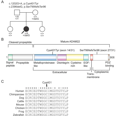
ADAM22segregated in an autosomal recessive manner in the family (figure 2A and figure e-2). The patient was compound heterozygous for a missense mutation c.1202G.A encoding the p.Cys401Tyr substitution (mutation nomenclature based onADAM22variant 1, RefSeq ID NM_021723) and a 1-bp frameshift deletion c.2396delG (p.Ser799IlefsTer96). The missense mutation is present in the heterozygous state in 2 individuals of European ancestry in the Exome Aggregation Consortium (ExAC) database
(http://exac.broadinstitute.org/), yielding an allele frequency of 1.65831025. The deletion mutation is novel. The missense substitution, p.Cys401Tyr, occurs in a highly conserved cysteine residue that forms a disulfide bond with Cys394 in the metalloproteinase domain25 (figure 2, B and C and figure e-3). The p.Cys401Tyr substitution is predicted to be deleterious by all 4 in silico methods used. The frameshift deletion is predicted to alter the last ;100 residues of ADAM22, thus abolishing the PDZ-binding motif (figure 2B). The deletion occurs in exon 27 of the longest isoform of
ADAM22(RefSeq ID NM_021723), which encodes
Figure 2 Compound heterozygous mutations inADAM22
residues in the cytoplasmic tail and is subjected to alternative splicing26,27(figures e-4 and e-5, see also Discussion).
Eight variants passed the filtering under the“de novo”hypothesis, where we looked for novel hetero-zygous variants (figure e-1, table e-1). Because the parents were not exome- sequenced, we analyzed seg-regation of these variants by capillary sequencing. All 8 variants were inherited from an unaffected parent and thus were not considered pathogenic. Variants in mitochondrial DNA were known polymorphisms (data not shown).
Analysis of CNVs in the proband-parent trio did not yield likely pathogenic variants (data not shown).
ADAM22 mutants show aberrant binding to LGI1.
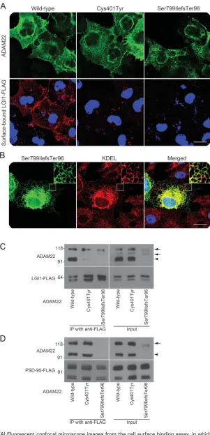
Using a cell surface binding assay, we first examined whether LGI1 interacts with the ADAM22 mutants. When LGI1 was cotransfected with wild-type ADAM22, LGI1 efficiently bound to ADAM22 on the cell surface as previously reported1 (figure 3A). In contrast, LGI1 did not bind to the Cys401Tyr and Ser799IlefsTer96 mutants on the cell surface. Although the localization of the Cys401Tyr mutant was similar to that of the wild type, most of the Ser799IlefsTer96 mutant protein was localized in the endoplasmic reticulum (ER), labeled by anti-KDEL antibody (figure 3, A and B).
We next analyzed the heterologous expression of
ADAM22mutants in HEK293T cells by Western blot-ting. The expression level of Cys401Tyr was comparable to that of the wild type, whereas Ser799IlefsTer96 was less expressed in HEK293T cells and remained as an immature form (propeptide not cleaved, see inputs in figure 3, C and D). When LGI1 was cotransfected with wild-type ADAM22, ADAM22 was efficiently coimmu-noprecipitated with LGI1 (figure 3C). In contrast, nei-ther the Cys401Tyr nor the Ser799IlefsTer96 mutant was coimmunoprecipitated with LGI1 (figure 3C, arrowheads). We also found that the wild-type ADAM22 and Cys401Tyr mutant, which have a PDZ-binding motif at their C-terminus, bound to the postsynaptic scaffolding protein PSD-95, as shown previously for wild-type ADAM22,1 but Ser799Ilef-sTer96 lacking the motif did not (figure 3D).
Figure 3 ADAM22 mutant proteins do not bind to LGI1
(A) Fluorescent confocal microscope images from the cell surface binding assay, in which indicatedADAM22complementary DNAs were cotransfected with wild-type FLAG-tagged LGI1into COS7 cells. Surface-bound FLAG-tagged LGI1 was labeled before cell permeabi-lization (red) and then ADAM22 was stained (green). Both ADAM22 mutants fail to bind to LGI1. Bar: 20mm. (B) Ser799IlefsTer96 mutant (green) is localized in the endoplasmic retic-ulum labeled by the anti-KDEL antibody (red). Regions outlined with white squares are
DISCUSSION We identified compound heterozygous loss-of-function mutations in ADAM22 in a patient with rapidly progressing severe encephalopathy with intractable seizures and profound intellectual disability. After initially normal CT scans, a remarkable feature of her disease was the rapidly progressing cerebral atrophy (within only a few weeks) with subdural effusions that became apparent approximately 2 months after seizure onset. The brain imaging changes were very similar to those seen in Menkes syndrome,28,29 and the patient’s symptoms also resemble those seen in Alpers syndrome.30 The disease course of the patient was also unusual. The progression was very rapid in infancy after the onset of intractable seizures at 3 months of age, following the apparently normal first months of life. Thereafter the condition stabilized, leaving the patient nonambulatory with intractable seizures, and she is still alive at the age of 26 years. A rapidly progressing disease course in infancy may be a characteristic feature forADAM22-associated disease, related to the role of ADAM22 in postnatal brain development.
We searched for pathogenic recessive and de novo variants in the exome and CNV data. The only variants passing the filtering and having a compatible segregation pattern in the family were compound heterozygous mu-tations inADAM22(figure e-1). The ability to dramat-ically reduce the number of candidate variants when only the proband was exome-sequenced was possible by filtering against comprehensive variant databases including population-matched individuals, in our case Finns in the ExAC database.
Our functional assays indicated that both mutations cause a nonfunctional ADAM22 protein. The Cys401-Tyr mutant did not bind to LGI1 in cell surface binding and immunoprecipitation assays. The p.Cys401Tyr substitution is predicted to break a disulfide bond in the metalloproteinase-like (M) domain. The crystal structure of the extracellular domain of ADAM22 has been recently determined25(figure e-3). Based on this structure, the authors suggested a modular movement model of ligand binding to ADAM22, where the ligand, LGI1, competes with the M domain in binding with the cysteine-rich domain (C), making the M domain move away from the C domain.25Because the p.Cys401Tyr substitution likely affects the tertiary structure of the M domain, it may hinder the conformational change that is necessary for ADAM22 in implementing its protein– protein adhesion function.
Because the C-terminal PDZ-binding motif is intact in the Cys401Tyr mutant, its binding to PSD-95 appeared normal in the immunoprecipitation assay. On the contrary, the frameshift mutation is predicted to alter the;100 amino acids in the cytoplasmic part of the protein, including residues binding to cytoplas-mic PSD-95 (figure 2B) and 14-3-3 proteins.1,5,31Our
binding assays showed that the frameshift mutation leads to a nonfunctional protein that is localized mainly in the ER and does not bind to LGI1 or to PSD-95.
The highly conserved exon 27 containing the frameshift deletion is subjected to alternative splicing (figure e-4), as are several other exons encoding parts of the cytoplasmic domain of ADAM22. Exon 27 is expressed in several regions of the human and mouse brain13,26,27,32and has a progressive temporal expres-sion pattern that differs from that of ADAM22
exons that are not subjected to alternative splicing (figure e-5).33 Accounting for all exons, ADAM22 expression is highest in postnatal stages (reference 1 and figure e-5). In embryonic stages, exon 27 is expressed in lower levels than other exons; the pro-portion of splice variants expressing exon 27 rapidly increases toward the end of the embryonic develop-ment and reaches a plateau at the first postnatal months. Exon 27 does not contain known peptide motifs, but its temporal expression pattern suggests that it contains residues playing a role in the CNS function in postnatal stages in particular. Reflecting the impor-tance of LGI1–ADAM22/23 complexes in postnatal brain development, the Lgi12/2, Adam222/2, and
Adam232/2 mice start developing seizures around 10 postnatal days.2,11–14 The expression pattern of
ADAM22 and data from knockout mice are in line with the development of symptoms in our patient, who was apparently asymptomatic at birth, with the first symptoms appearing at approximately 2 months of age and the disease progressing rapidly thereafter.
Because not allADAM22transcripts express exon 27, the frameshift mutation is likely not a total null mutation but rather a hypomorphic one, its effect increasing temporally. This may also explain why the compound heterozygous mutations in our patient were not lethal in early life, in contrast toLgi12/2,
Adam222/2, andAdam232/2mice, which die at 2–3 postnatal weeks.2,11–14Furthermore, exon 27 is skip-ped from ADAM22 in the PNS,8,13,32 which may explain why sural biopsy shows normally myelinated nerves in the proband, whereas the knockout mice present with marked hypomyelination of the periph-eral nerves.13
been shown for a monoallelic LGI1 mutant.4 Reduc-tion of synaptic AMPARs as a consequence of the disruption of LGI1-ADAM22 interaction has also been suggested34to be the underlying epileptogenic pathomechanism in autoimmune limbic encephali-tis, which manifests with amnesia and seizures and is caused by autoantibodies against LGI1.35,36 It re-mains unknown whether seizure activity contributes to the rapidly progressive degeneration of cortical regions in our patient or whether ADAM22 dys-function has a direct negative effect on the develop-ment of neuronal connections, increasing seizure susceptibility.
Our data suggest that loss of ADAM22 function in the CNS underlies the severe rapidly progressing infantile-onset encephalopathy with epilepsy in our patient. Given that heterozygous LGI1 mutations underlie ADLTE, previous studies have attempted to identify dominantly inherited mutations in
ADAM22in ADLTE families.37,38However, the fact that the parents of the proband—who are heterozy-gous carriers of loss-of-function mutations—do not have epilepsy indicates that heterozygous deleterious mutations are tolerated in ADAM22 and that
ADAM22is primarily a recessive disease gene. This hypothesis is supported by the presence of 9 truncat-ing variants of the canonicalADAM22 transcript in the ExAC database, whereas there are none forLGI1. Lack of rare biallelic mutations in ADAM22 in other exome data sets (appendix e-1) indicates that
ADAM22-associated encephalopathy is rare, which may partially be because complete knockout muta-tions ofADAM22 are not tolerated and only hypo-morphic mutations cause a phenotype similar to that in our study. Future studies are needed to confirm this genetic association and define the phenotypic presentations of ADAM22 dysfunction.
AUTHOR CONTRIBUTIONS
M. Muona: analysis and interpretation of genetic data, drafting and revis-ing the manuscript. Y. Fukata: functional analysis and interpretation, drafting and revising the manuscript. A.-K. Anttonen: patient evaluation, study concept and design, drafting and revising the manuscript. A. Laari: analysis and interpretation of genetic data, revising the manuscript. A. Palotie: study concept and design, revising the manuscript. H. Pihko: patient evaluation, revising the manuscript. T. Lönnqvist: patient evalu-ation, revising the manuscript. L. Valanne: patient evaluevalu-ation, revising the manuscript. M. Somer: patient evaluation, revising the manuscript. M. Fukata: functional analysis and interpretation, drafting and revising the manuscript. A.-E. Lehesjoki: study concept and design, interpretation of data, drafting and revising the manuscript.
ACKNOWLEDGMENT
The authors thank the family for participation in the study, Dr. Toshitaka Kawarai (Tokushima University Graduate School) for complementary DNA of humanADAM22, Carol Scott and Jillian Durham (Wellcome Trust Sanger Institute) for exome sequence processing, Sinikka Lindh for management of patient samples, Dr. Kaisa Kettunen (Institute for Molec-ular Medicine Finland) for whole-genome genotyping data generation, and CSC–IT Center for Science, Ltd., for the allocation of computational
resources. Exome Aggregation Consortium, EuroEPINOMICS RES con-sortium, and The Deciphering Developmental Disorders study are acknowledged for data sharing.
STUDY FUNDING
This study was supported by the Folkhälsan Research Foundation (A.-E.L.), Wellcome Trust grants 089062 and 098051 (A.P.), the Euro-pean Commission FP7 project no. 242167 SynSys (A.P.), Health-2010 project no. 261433 BioSHare (A.P.), the Academy of Finland (grants no. 251704 and 263401) (A.P.), Emil Aaltonen Foundation (M.M. and A.-K.A.), Epilepsy Research Foundation (M.M.), University of Helsinki Funds (M.M.), Doctoral Programme in Biomedicine (M.M.), Arvo and Lea Ylppö Foundation (M.M. and A.-K.A.), Finnish Brain tion (M.M.), Paulo Foundation (M.M.), Biomedicum Helsinki Founda-tion (M.M.), Helsinki University Central Hospital Research Fund (A.-K. A.), and the Ministry of Education, Culture, Sports, Science, and Tech-nology (15H04279 and 15H01299) (Y.F.) and (15H01570) (M.F.).
DISCLOSURE
Mr. Muona has received research support from the Emil Aaltonen Foun-dation, the Epilepsy Research FounFoun-dation, University of Helsinki Funds, the Doctoral Programme in Biomedicine University of Helsinki, the Arvo and Lea Ylppö Foundation, the Finnish Brain Foundation, the Paolo Foundation, and the Biomedicum Helsinki Foundation. Dr. Y. Fukata has received research support from the Ministry of Education, Culture, Sports, Science, and Technology; the Astellas Foundation for Research on Metabolic Disorders; the Takeda Science Foundation; and the Mochida Memorial Foundation for Medical and Pharmaceutical Research. Dr. Anttonen has received research support from the Arvo and Lea Ylppö Foundation and the Helsinki University Central Hospital Research Fund. Ms. Laari reports no disclosures. Dr. Palotie is a member of the Pfizer Genetics Scientific Advisory Panel, has received travel expenses and/or honoraria for lectures or educational activities not funded by industry, and has received research support from the Finnish Academy, the Euro-pean Union NIH, National Institute of Neurological Disorders and Stroke, the Juselius Foundation, and the Finnish Foundation for Cardi-ovascular Research. Dr. Pihko receives publishing royalties forTextbook of Child Neurology, Duodecim 2014. Dr. Lönnqvist, Dr. Valanne, and Dr. Somer report no disclosures. Dr. M. Fukata has served on the editorial board of theEuropean Journal of Neuroscienceand has received research support from The Ministry of Education, Culture, Sports, Science, and Technology. Dr. Lehesjoki has received research support from the Academy of Finland, the Sigrid Juselius Foundation, the Liv och Hälsa Foundation, and the Folkhälsan Research Foundation. Go to Neurology.org/ng for full disclosure forms.
Received August 26, 2015. Accepted in final form November 20, 2015.
REFERENCES
1. Fukata Y, Adesnik H, Iwanaga T, Bredt DS, Nicoll RA, Fukata M. Epilepsy-related ligand/receptor complex LGI1 and ADAM22 regulate synaptic transmission. Science 2006;313:1792–1795.
2. Fukata Y, Lovero KL, Iwanaga T, et al. Disruption of LGI1-linked synaptic complex causes abnormal synaptic transmission and epilepsy. Proc Natl Acad Sci U S A 2010;107:3799–3804.
3. Duffy M, Mullooly M, O’Donovan N, et al. The ADAMs family of proteases: new biomarkers and therapeutic tar-gets for cancer? Clin Proteomics 2011;8:9.
4. Zhou YD, Lee S, Jin Z, Wright M, Smith SEP, Anderson MP. Arrested maturation of excitatory synapses in autosomal dominant lateral temporal lobe epilepsy. Nat Med 2009;15:1208–1214.
6. Benarroch EE. ADAM proteins, their ligands, and clinical implications. Neurology 2012;78:914–920.
7. Ogawa Y, Oses-Prieto J, Kim MY, et al. ADAM22, a Kv1 channel-interacting protein, recruits membrane-associated guanylate kinases to juxtaparanodes of myelinated axons. J Neurosci 2010;30:1038–1048.
8. Özkaynak E, Abello G, Jaegle M, et al. Adam22 is a major neuronal receptor for Lgi4-mediated Schwann cell signal-ing. J Neurosci 2010;30:3857–3864.
9. Yokoi N, Fukata Y, Kase D, et al. Chemical corrector treat-ment ameliorates increased seizure susceptibility in a mouse model of familial epilepsy. Nat Med 2015;21:19–26. 10. Kalachikov S, Evgrafov O, Ross B, et al. Mutations in
LGI1 cause autosomal-dominant partial epilepsy with auditory features. Nat Genet 2002;30:335–341. 11. Yu YE, Wen L, Silva J, et al. Lgi1 null mutant mice exhibit
myoclonic seizures and CA1 neuronal hyperexcitability. Hum Mol Genet 2010;19:1702–1711.
12. Chabrol E, Navarro V, Provenzano G, et al. Electroclinical characterization of epileptic seizures in leucine-rich, glioma-inactivated 1-deficient mice. Brain 2010;133: 2749–2762.
13. Sagane K, Hayakawa K, Kai J, et al. Ataxia and peripheral nerve hypomyelination in ADAM22-deficient mice. BMC Neurosci 2005;6:33.
14. Owuor K, Harel NY, Englot DJ, Hisama F, Blumenfeld H, Strittmatter SM. LGI1-associated epilepsy through altered ADAM23-dependent neuronal morphol-ogy. Mol Cell Neurosci 2009;42:448–457.
15. Koskinen LL, Seppala EH, Belanger JM, et al. Identifica-tion of a common risk haplotype for canine idiopathic epilepsy in the ADAM23 gene. BMC Genomics 2015; 16:465.
16. Epi4K Consortium; Epilepsy Phenome/Genome Project, Allen AS, Berkovic SF, Cossette P, et al. De novo muta-tions in epileptic encephalopathies. Nature 2013;501: 217–221.
17. Boycott KM, Vanstone MR, Bulman DE, MacKenzie AE. Rare-disease genetics in the era of next-generation sequencing: discovery to translation. Nat Rev Genet 2013;14:681–691.
18. EuroEPINOMICS-RES Consortium; Epilepsy Phenome/-Genome Project; Epi4K Consortium. De novo mutations in synaptic transmission genes including DNM1 cause epileptic encephalopathies. Am J Hum Genet 2014;95: 360–370.
19. Muona M, Berkovic SF, Dibbens LM, et al. A recurrent de novo mutation in KCNC1 causes progressive myoclo-nus epilepsy. Nat Genet 2015;47:39–46.
20. DePristo MA, Banks E, Poplin R, et al. A framework for variation discovery and genotyping using next-generation DNA sequencing data. Nat Genet 2011;43:491–498. 21. Van der Auwera GA, Carneiro MO, Hartl C, et al. From
FastQ data to high confidence variant calls: the Genome Analysis Toolkit best practices pipeline. Curr Protoc Bio-inform 2013;11:11.10.1–11.10.33.
22. McKenna A, Hanna M, Banks E, et al. The Genome Analysis Toolkit: a MapReduce framework for analyzing next-generation DNA sequencing data. Genome Res 2010;20:1297–1303.
23. Wang K, Li M, Hadley D, et al. PennCNV: an integrated hidden Markov model designed for high-resolution copy number variation detection in whole-genome SNP geno-typing data. Genome Res 2007;17:1665–1674. 24. Fukata M, Fukata Y, Adesnik H, Nicoll RA, Bredt DS.
Identification of PSD-95 palmitoylating enzymes. Neuron 2004;44:987–996.
25. Liu H, Shim AHR, He X. Structural characterization of the ectodomain of a disintegrin and metalloproteinase-22 (ADAM22), a neural adhesion receptor instead of metal-loproteinase: insights on ADAM function. J Biol Chem 2009;284:29077–29086.
26. Harada T, Nishie A, Torigoe K, et al. The specific expres-sion of three novel splice variant forms of human metalloprotease-like disintegrin-like cysteine-rich protein 2 gene in brain tissues and gliomas. Jpn J Cancer Res 2000;91:1001–1006.
27. Gödde NJ, D’Abaco GM, Paradiso L, Novak U. Differ-ential coding potDiffer-ential of ADAM22 mRNAs. Gene 2007; 403:80–88.
28. Barkovich AJ, Good WV, Koch TK, Berg BO. Mitochon-drial disorders: analysis of their clinical and imaging char-acteristics. AJNR Am J Neuroradiol 1993;14:1119–1137. 29. Jacobs DS, Smith AS, Finelli DA, Lanzieri CF, Wiznitzer M. Menkes kinky hair disease: characteristic MR angiographic findings. AJNR Am J Neuroradiol 1993;14:1160–1163.
30. Harding BN. Progressive neuronal degeneration of child-hood with liver disease (Alpers-Huttenlocher syndrome): a personal review. J Child Neurol 1990;5:273–287. 31. Zhu P, Sang Y, Xu H, et al. ADAM22 plays an important
role in cell adhesion and spreading with the assistance of 14-3-3. Biochem Biophys Res Commun 2005;331:938–946. 32. Lonsdale J, Thomas J, Salvatore M, et al; GTEx
Consor-tium. The Genotype-Tissue Expression (GTEx) project. Nat Genet 2013;45:580–585.
33. Kang HJ, Kawasawa YI, Cheng F, et al. Spatio-temporal transcriptome of the human brain. Nature 2011;478: 483–489.
34. Ohkawa T, Fukata Y, Yamasaki M, et al. Autoantibodies to epilepsy-related LGI1 in limbic encephalitis neutralize LGI1-ADAM22 interaction and reduce synaptic AMPA receptors. J Neurosci 2013;33:18161–18174.
35. Irani SR, Alexander S, Waters P, et al. Antibodies to Kv1 potassium channel-complex proteins leucine-rich, glioma inactivated 1 protein and contactin-associated protein-2 in limbic encephalitis, Morvan’s syndrome and acquired neu-romyotonia. Brain 2010;133:2734–2748.
36. Lai M, Huijbers MGM, Lancaster E, et al. Investigation of LGI1 as the antigen in limbic encephalitis previously attributed to potassium channels: a case series. Lancet Neurol 2010;9:776–785.
37. Chabrol E, Gourfinkel-An I, Scheffer IE, et al. Absence of mutations in the LGI1 receptor ADAM22 gene in auto-somal dominant lateral temporal epilepsy. Epilepsy Res 2007;76:41–48.
DOI 10.1212/NXG.0000000000000046
2016;2;
Neurol Genet
Mikko Muona, Yuko Fukata, Anna-Kaisa Anttonen, et al.
atrophy and epilepsy
Dysfunctional ADAM22 implicated in progressive encephalopathy with cortical
This information is current as of January 21, 2016
Neurology. All rights reserved. Online ISSN: 2376-7839.
an open-access, online-only, continuous publication journal. Copyright © 2016 American Academy of is an official journal of the American Academy of Neurology. Published since April 2015, it is
Services
Updated Information &
http://ng.neurology.org/content/2/1/e46.full.html including high resolution figures, can be found at:
Supplementary Material
http://ng.neurology.org/content/suppl/2016/01/21/2.1.e46.DC1 Supplementary material can be found at:
References
http://ng.neurology.org/content/2/1/e46.full.html##ref-list-1 This article cites 38 articles, 11 of which you can access for free at:
Citations
http://ng.neurology.org/content/2/1/e46.full.html##otherarticles This article has been cited by 4 HighWire-hosted articles:
Subspecialty Collections
http://ng.neurology.org//cgi/collection/mri
MRI
http://ng.neurology.org//cgi/collection/mental_retardation
Mental retardation
http://ng.neurology.org//cgi/collection/developmental_disorders
Developmental disorders
http://ng.neurology.org//cgi/collection/all_genetics
All Genetics
following collection(s):
This article, along with others on similar topics, appears in the
Permissions & Licensing
http://ng.neurology.org/misc/about.xhtml#permissions its entirety can be found online at:
Information about reproducing this article in parts (figures,tables) or in
Reprints
http://ng.neurology.org/misc/addir.xhtml#reprintsus Information about ordering reprints can be found online:
Neurology. All rights reserved. Online ISSN: 2376-7839.
an open-access, online-only, continuous publication journal. Copyright © 2016 American Academy of is an official journal of the American Academy of Neurology. Published since April 2015, it is