1696 | P a g e
‘Triphala -An Indian Ayurvedic Herbal Formulation’: A
comparative study of some biological properties of triphala,
its ingredients, & triphala silver nanoparticles
Pratiksha Chandorkar
1, Jyoti Mantri
2Microbiology Department of Sophia Collage, University of Mumbai, (India)
ABSTRACT
India has an age old heritage of traditional herbal medicine. Triphala is a well-known powdered preparation which consists of equal proportion of Terminellia chebula, Teminellia bellerica and Embellica officinalis, as described in the ‘Sushrut Samhitas’.
This powder preparation is believed to promote health, immunity, and detoxify the whole body. Triphala contains several compounds such as gallic acid, chebulic acid, ellagic acid, chebulinic acid and polyphenol, which responsible for its claimed health benefits.
One of the major limitations of Triphala is that it is effective only in high doses. The present study is undertaken to overcome this limitation, by preparing Triphala-silver nanoparticles (TSNPs), and to study if it is effective in low doses.
Characterization of the prepared silver nanoparticles and Triphala silver nanoparticles was done using UV-Visible spectroscopy. A comparative study of the therapeutic effects of the prepared TSNPs with Triphala showed more pronounced anti-bacterial, anti-oxidant, anti-inflammatory and anti-coagulant properties for TSNPs.
Keywords: Antibacterial, Anti-Coagulant, Anti-Inflammatory, Antioxidant, Triphala-Silver
Nanoparticles
I. INTRODUCTION
Herbs have been used as food and medicinal purposes for centuries. Triphala is a popular herbal formulation consisting of the dried and powdered fruits of three plants, Terminalia bellerica (family Combretaceae), Terminalia chebula (family Combretaceae) and Emblica officinalis (family Phyllanthaceae) [1-3]. This powder preparation is believed to promote health, immunity, clean the gastro-intestinal tract and detoxify the whole body. Triphala is rich in gallic acid, Vitamin C, ellagic acid, chebulic acid, bellericanin, β-sitosterol and flavonoids and phenolic
compound such as flavonoids, tannins and syringic acid [4-5]. Because of these compounds triphala is claimed to have antibacterial [6], antifungal [7], antiviral [8], anti-oxidant [9], anti-inflammatory [6], and anticancer activity [10-11].
1697 | P a g e
further limits the utility of the Triphala. These limitations can be addressed by using nanoparticles as carriers for delivery of Triphala [12].Among the other noble metal nanoparticle, silver nanoparticles have gained boundless interest in the medical field, because of their unique properties of chemical stability, good conductivity and most important antibacterial, antiviral, antifungal and anti-inflammatory [13].
The aim of this research work was to carry out green synthesis of Triphala silver nanoparticles and to compare the efficacy of Triphala-silver nanoparticles with Triphala powder and its individual constituent for their antibacterial, anti-inflammatory, anti-oxidant and anti-coagulant properties. This research work demonstrates that the use of silver nanoparticles as a carrier of Triphala improved its therapeutic properties by overcoming its shortcomings.
II. MATERIALS & METHODS
2.1 Green synthesis of Triphala-Silver Nanoparticles (TSNPs)
10.0g of fine Triphala powder was suspended in 100 ml of 1mM silver nitrate (AgNO3) solution in 1:10 ratio (w/v). The flask (black paper covered) was kept on a shaker in dark for 24 hours. A change in color indicating the formation of silver nanoparticles was observed. The mixture was centrifuged at 6000 rpm for 30 minutes, double filtered and the supernatant was used for further experiments [14]. These nanoparticles are further characterized by scanning electron microscopy (SEM) and UV-Visible spectroscopy.
2.1.1 Characterization of silver nanoparticles by UV-Visible spectroscopy
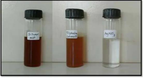
The addition of Triphala powder to silver nitrate solution resulted in colour change from light brown to dark brown due to production of silver nanoparticles. The bioreduction of silver ions in aqueous solution was monitored by UV-VIS spectra of the solution between 300 nm – 600 nm.
2.1.2 Characterization of Triphala silver nanoparticles by Scanning Electron Microscopy (SEM)
A drop of Triphala-silver nanoparticles solution was put on a piece of copper plate and was allowed to dry and was then used for SEM analysis which was done by using ZEISS ultra plus scanning electron microscope.
2.2. Preparation of Aqueous extract of Triphala and its ingredients
1698 | P a g e
2.3. Determination of antibacterial activity of Triphala, its ingredients and TSNPs
Antibacterial activity was determined by agar well diffusion method against 24 hours old cultures (0.1 optical densities at 570 nm)ofStreptococcus pyogenes, Escherichia coli, Shigella species, Salmonella typhi, Salmonella paratyphi A, Salmonella paratyphi B. etc. Nutrient agar plates were seeded with these different pathogens. Wells of 6mm were prepared using cork borer and the wells were charged with 100µl of samples (extract of Triphala, its ingredients and Triphala-silver nanoparticles). Zones of inhibition were measured after 24 hours incubation period.
2.4. Determination of antioxidant activity of Triphala, its ingredient and TSNPs
6 ml of the reagent solution (2ml of 0.6M sulfuric acid, 2 ml of 28 mM sodium dihydrogen phosphate and 2ml of 4 mM ammonium molybdate) was combined with 0.6 ml of different concentrations of ascorbic acid (standard solutions) and samples. The tubes was capped and incubated at 95°C for 90 min. After cooling the tubes to room temperature, absorbance of the standards and test solutions was measured at 670 nm using colorimeter. The anti-oxidant capacity of each sample is expressed as ascorbic acid equivalent in µg/ml. Higher the concentration higher is the total anti-oxidant activity [9].
2.5. Determination of anti-inflammatory activity of Triphala, its ingredients and TSNPs
The RBC membrane is analogous to the lysosomal membrane and its stabilization implies that the test sample may as well stabilize lysosomal membranes. This stabilization plays an important role in limiting inflammatory responses, by preventing the release of lysosomal constituents consisting of bactericidal enzymes and proteases. Stabilization of HRBC by hypotonicity induced membrane lysis can be taken as an in vitro measure of anti-inflammatory activity of the drugs or plant extract. [15]
For this assay fresh blood was collected and mixed with the Alsever solution. The mixture was centrifuged at 3000 rpm for 10min. The cells were washed with isosaline. The volume of blood was recorded and reconstituted as 10% v/v suspension with isosaline. The assay mixture was prepared by adding 1ml of 0.2M phosphate buffer and 1ml of 0.36% hyposaline to 1ml of the sample and standards (Diclofinac solution of various concentrations). For control, hyposaline was replaced with distilled water to give 100% haemolysis. The tubes were capped and incubated at 37°C for 30 min, centrifuged and the absorbance was measured at 560nm using a colorimeter. The result was recorded as the % of the membrane stabilization of HRBC.
(1
) The percentage of hemolysis of HRBC membrane was calculated as follows-Percentage of hemolysis = optical density of test/optical density of control x 100 (2) The percentage of HRBC membrane stabilization was calculated as follows-
1699 | P a g e
2.6. Proteinase inhibitory activity of Triphala, its ingredients and TSNPs
Neutrophils carry serine proteinases in their lysosomal granules and these have been implicated in arthritic reactions. It has been reported that proteinases from leukocyte play an important role in the development of tissue damage during an inflammatory reactions and significant level of protection was provided by proteinase inhibitors [15].
Trypsin can hydrolyse casein (a protein in milk) as it possesses proteinase activity. Thus trypsin can be used to assay proteinase inhibitory activity of samples by investigating inhibition of breakdown of casein. The reaction mixture contained 1 mL of test sample, 1.0 mL of 20 mM TrisHCl buffer (pH 7.4) containing 0.06 mg trypsin, and the
mixture was incubated at 37°C for 5 min. Then 1 mL of 0.8% (w/v) casein was added. The mixture was incubated for an additional 20 min. 2 ml of 70% perchloric acid was added to terminate the reaction. Cloudy suspension was centrifuged and the absorbance of the supernatant was read at 340 nm against buffer as blank. Diclofinac sodium was used as a standard (1000mcg/ml).
Percentage of proteinase inhibitory activity was calculated as follows- Percentage of proteinase inhibitory = optical density of test/optical density of control x 100
2.7. Determination of anticoagulant activity of Triphala, its ingredient and TSNPs
Anticoagulant activity was determined by prothrombin time determination assay [16]. The prothrombin time is a one stage test based upon the time required for a fibrin clot to form after the addition of tissue factor (TF) (historically known as tissue thromboplastin), phospholipid and calcium to decalcified, platelet poor plasma. ‘Thromboplastin’ is a plasma protein, aiding blood coagulation through catalyzing the conversion of prothrombin to
thrombin. This reagent activates the extrinsic coagulation pathway. Thromboplastin contains significant amounts of tissue factors (TF) and phospholipid [16].
1700 | P a g e
III. RESULTS AND DISCUSSION
3.1 Synthesis & Characterization of triphala-silver nanoparticles
It is well known that silver nanoparticles exhibit yellowish brown color in aqueous solution due to excitation of surface Plasmon vibrations. As the Triphala extract was mixed in the aqueous solution of the silver ion complex, it started to change the color from light brown to dark brown due to reduction of silver ion which indicated formation of silver nanoparticles. The nanoparticles were characterized by UV-Visible spectroscopy. Absorption spectra of TSNPs formed in the reaction mixture showed an absorbance peak at 420 nm, and broadening of peak indicated that the particles are polydispersed. The size of the Triphala silver nanoparticles was found to be around 200nm.
Figure 1: Color change indicating synthesis of Nanoparticles
1701 | P a g e
Figure 3: UV-VIS analysis of obtained triphala-silver Nanoparticles (TSNPs)3.2 Antibacterial activity
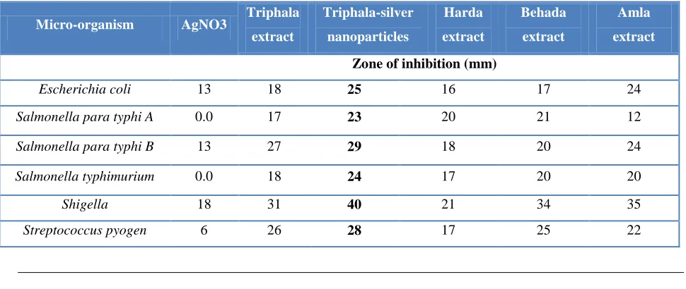
It was seen that all the samples showed clear zone of inhibition against all the tested micro-organisms. TSNPs displayed maximum antibacterial activity followed by aqueous extract of triphala, individual ingredients of triphala and lastly silver nanoparticles. Triphala contains 3 different medicinal fruits; T.bellerica, T.chebula, E.officinalis in equal proportions. The enhanced antibacterial activity of triphala could be due to the additive or synergistic effects of the three fruits together. Use of nanoformulations of triphala leads to increase in bioactivity of Triphala by reducing the size of particles, resulting in improved solubility and enhanced chemical reactivity. This could be the reason behind the maximum activity of TSNPs as compared to the other samples.
Table 1: Results of antibacterial activity by agar well diffusion method
Micro-organism AgNO3 Triphala
extract
Triphala-silver
nanoparticles
Harda
extract
Behada
extract
Amla
extract
Zone of inhibition (mm)
Escherichia coli 13 18 25 16 17 24
Salmonella para typhi A 0.0 17 23 20 21 12
Salmonella para typhi B 13 27 29 18 20 24
Salmonella typhimurium 0.0 18 24 17 20 20
Shigella 18 31 40 21 34 35
1702 | P a g e
3.3 Antioxidant activity
The total antioxidant capacity of Triphala, its individual ingredients and Triphala-silver nanoparticles was
determined by using the phosphomolybdenum method. This assay is based on the reduction of Mo(V) to Mo(V) in the presence of antioxidant compound results in the formation of colored complex at acidic pH, resulting that Amla powder has the highest antioxidant activity followed by Harda, TNSPs, Triphala and Behada. The overall antioxidant activity is attributed to its polyphenolic and other phytochemical constituents. The improved antioxidant activity of TSNPs as compared to triphala could be due to the enhanced chemical reactivity of the nanoformulation.
Figure: 4:Results of antioxidant activity by phosphomolybdenum assay. Color change after incubation at
95°C for 90 min
Figure 5: Comparison of antioxiadant activity of extract of Triphala, its individual ingredients and
1703 | P a g e
3.4 Anti-Inflammatory Activity
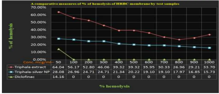
Both Triphala aqueous extract and Triphala-silver nanoparticle exhibited strong anti-inflammatory response. The anti inflammatory activity of TSNPs and triphala was concentration dependent. As showed in figure 7 at 1000 and 50 concentration TSNPs showed a protection of 84.27% and 71.92% respectively whereas triphala showed a protection of 73.04% and 35.96% respectively.
Thus both Triphala aqueous extract and TSNPs exhibit capacity of membrane stabilization of HRBC and hence are anti-inflammatory. However TSNPs had more anti-inflammatory activity than Triphala extract. Though the exact mechanism of the membrane stabilization by the extract and nanoparticle is not yet known; the increase in aqueous stability and permeability through biological membrane due to the reduction in the size of the TSNPs may be responsible for its enhanced anti-inflammatory activity [15].
Figure 6: Hypotonicity induced HRBC membrane stabilization test. A comparative measures of % of hemolysis of HRBC
Figure 7: A comparative measures of % protection of HRBC membrane given by extract of Triphala,
1704 | P a g e
3.5 Proteinase inhibitory activity
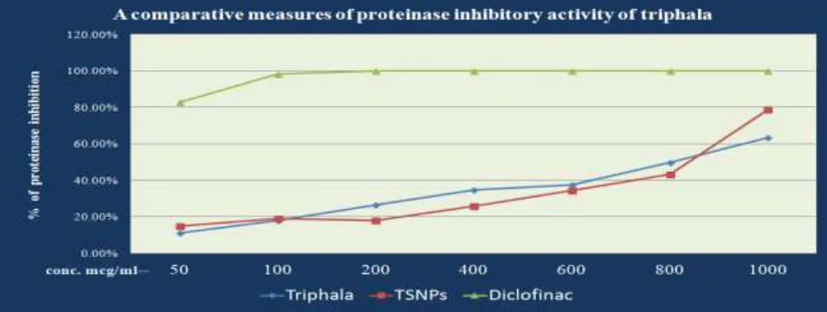
The aqueous extract of Triphala and TSNPs exhibited proteinase inhibitory activity. At 1000μg/m and 50μg/ml concentration the Triphala extract showed an inhibition of 63.49% and 11.11% respectively whereas TSNPs showed an inhibition of 78.83% and 14.81% respectively. Standard diclofinac sodium shows a 98.41% inhibitory activity at 100μg/ml concentration. Thus this increased proteinase inhibitory activity of the TSNPs may be preventing the
release of potent serine proteases from the lysosomal granules thus aiding in its anti-inflammatory activity.
Figure 8: Graphical representation of proteinase inhibitory action of Triphala extract, Triphala-silver
nanoparticles and standard diclofinac
3.4 Anticoagulant activity
The prothrombin time for all samples was determined. It was found that after addition of uniplastin reagent in all test samples the clot formation was delayed upto more than 23 seconds (control) which is average time for human plasma clotting process. Triphala and TSNPs showed comparable delay in clot formation followed by Harda, Behada and Amla. Thus, results indicate that both Triphala and TSNPs can help in preventing platelet aggregation, thereby thinning consistency of blood.
Table 2: Results of anticoagulant activity
Sample Plasma
(ml)
Sample (µl)
Thromboplastin reagent
(ml)
Time taken for clot formation
(seconds)
Control 0.5ml - 0.2 ml 23
Amla 0.5ml 50 µl 0.2 ml 38
Harda 0.5ml 50 µl 0.2 ml 82
Behada 0.5ml 50µl 0.2 ml 63
Triphala extract 0.5ml 50 µl 0.2 ml 110
Triphala-silver
1705 | P a g e
Figure 9: Results of anticoagulant activityIV. CONCLUSION
Green synthesis of Triphala-silver nanoparticles was done and its antioxidant, anti-inflammatory, anticoagulant and antibacterial activity was compared with apueous extract of Triphala and its individual ingredients. It was found that Triphala-silver nanoparticles (TSNPs) had higher antioxidant, anti-inflammatory and antibacterial activity as compare to aqueous extract of Triphala and its individual ingredients. Anticoagulant activity of TSNPs and Triphala aqueous extract were comparable.
These results indicate that the nanoformulation of Triphala improved the therapeutic efficacy of Triphala. This improved efficacy could be due to a decrease in size of particles from the micron to the nm range resulting in increased surface area to volume ratio. Such nanoparticles are chemically more reactive and have amplified dissolution rate thus addressing issues associated with poor solubility. Thus nanoparticle formulation strategies provide a means to develop a new drug delivery system with improved therapeutic outcome and addressing unmet medical needs.
BIBLIOGRAPHY
1. The Ayurvedic Formulary of India, Part –II, Department of Indian System of Medicine and Homeopathy, New Delhi; 2002.
1706 | P a g e
3. Jeganathan NS, Kannan K. HPTLC method for the estimation of ellagic and gallic acid in triphala churanum formulations. Research journal of phytochemistry 2008; 2:1‐9.
4. Jagetia GC, Baliga MS, Malagi KJ, Settukumar KM. The evaluation of the radioprotective effect of Triphala (an Ayurvedic rejuvenating drug) in the mice exposed to gamma-radiation. Phytomedicine, 9, 2002, 99-108.
5. Singh DP, Govindarajan R, Rawat AKS. High Performance Liquid Chromatography as a tool for the Chemical Standardisation of Triphala – an Ayurvedic Formulation, Phytochemical Analysis, 19, 2008, 164-168.
6. D.V. Gowda, Ganesh Muguli, Rangesh P.R, Rohan D. Deshpande. Phytochemical and pharmacological actions of triphala: ayurvedic formulation - a review Int. J. Pharm. Sci. Rev. Res., 15(2), 2012; nᵒ 12, 61-65 issn 0976 – 044x
7. V. Eugin amala, M. Jeyaraj. Determination of antibacterial, antifungal, bioactive constituents of triphala by FT-IR and GC-MS analysis. International journal of pharmacy and pharmaceutical sciences issn- 0975-1491 vol 6, issue 8, 2014
8. Hozumi T, Oyama H. Crude drugs for treating AIDS, Japan KokaiTokkyo Koho JP, 09, 1997, 87-185. 9. Deepa Babu, Prema Gurumurthy, Sai Krishna Borra and K. M. Cherian. Antioxidant and free radical
scavenging activity of triphala determined by using different in vitro models. Journal of Medicinal Plant Research Vol. 7(39), pp. 2898-2905, 17 October, 2013 DOI 10.5897/JMPR2013. 5124 ISSN 1996-0875 © 2013 Academic Journals
10. Sandhya T, Lathika KM, Pandey BN, Mishra KP. Potential of traditional Ayurvedic Formulation, Triphala as a novel anticancer drug, Cancer Letters, 231, 2006, 206-214.
11. Kaur S, Arora S, Kaur K, Kumar S. The in vitro antimutagenic activity of Triphala- an Indian Herbal Drug.
Food and Chemical Toxicology, 40(4), 2002, 527-534.
12. C.S. Shreedhara, Sowmya S, Dhirendra B. Sanghai1, Kranti P. Musmade, K.B. Koteshwara. Preparation of nanoformulation of triphala extract and evaluation of it’s in vitro antioxidant and cytotoxic potential.
International Journal of Pharmaceutical and Phytopharmacological Research (eIJPPR)
13. Shakeel Ahmed, Mudasir Ahmad, Babu Lal Swami, Saiqa Ikram. (2016) A review on plants extract mediated synthesis of silver nanoparticles for antimicrobial applications: A green expertise Department of Chemistry, Faculty of Natural Sciences, Jamia Millia Islamia, New Delhi 110025, India Journal of Advanced Research (2016) 7, 17–28
14. Asmita J. Gavhane1, P. Padmanabhan, Suresh P. Kamble and Suresh N. Jangle (2012). Synthesis of silver nanoparticles using extract of neem leaf and triphala and evaluation of their antimicrobial activities.
1707 | P a g e
15. Chandrappa Cp, Govindappa M, Anil Kumar NV and Channabasava R in vitro anti-inflammatory activityof carmona Retusa (vahl.). World journal of pharmacy and pharmaceutical sciences Volume 2, issue 5, 3991-3997. Research article issn 2278 – 4357
16. Website- Prothrombin Time [PT] A Practical Guide To Laboratory Haemostasis www.practical-haemostasis.com/screening%20tests/pt.htm